Case Based Learning Teaching Methodology in
Undergraduate Health Sciences
Kaitlyn BROWN*
1, Mary COMMANDANT
1, Adi KARTOLO
1, Casey ROWED
1, Agatha
STANEK
1, Heebah SULTAN
1, Kabir TOOR
1, Victoria WININGER
11 Student, University of Ottawa, Canada
* Auteur(e) correspondant | Corresponding author: N/A
Résumé :
(traduction)
L’apprentissage par cas (APC) est une méthode d’enseignement interactive fai- sant intervenir de petits groupes de discussion afin de déterminer un éventail de solutions à un cas clinique donné. Vu la réussite de cette approche dans de nom-breux programmes professionnels et de premier cycle, des étudiants seniors en sciences de la santé ont présenté, en 2009, un projet pilote dans lequel ils jouaient le rôle de facilitateurs de l’APC dans les cours de premier cycle de l’École interdisciplinaire des sciences de la santé (ÉISS) de l’Université d’Ottawa. En collaboration avec des professeurs de la faculté, ces facilitateurs ont élaboré des séances d’APC composées d’études de cas reflétant les objectifs de base des cours de sciences de la santé. Au total, 144 étudiants de premier cycle de trois cours de l’ÉISS ont participé à ces séances et ont été évalués en fonction du ni- veau de leur participation et de leurs réponses à un questionnaire composé de cinq questions évaluant leur maîtrise des notions abordées lors des séances d’APC. Ces étudiants ont également évalué le projet pilote. Sur une échelle de 1 à 5, les étudiants ont obtenu une note moyenne de 4,13 sur 5,00 (SD 1.48) au questionnaire. Dans leur évaluation du projet, les étudiants ont noté 3,82 sur 4 la valeur globale de l’apprentissage. Ils ont donc perçu ce programme comme ayant une certaine valeur d’apprentissage. Et les résultats du questionnaire ont confirmé que l’APC facilite l’application à des cas pratiques des connaissances théoriques acquises. Ces premières conclusions suggèrent que l’implantation de l’APC à l’ÉISS améliorerait l’expérience académique des étudiants. Des évalua- tions plus rigoureuses avant et après les séances, conduiraient à une améliora-tion des séances basées sur ce modèle.
Mots-clés :
Apprentissage basé sur les cas;; méthodologie d’enseignement;; éducation de pre- mier cycle; projets pilotesAbstract:
Case-based learning (CBL) is an interactive teaching approach involving small-group discussion to determine a range of solutions for a presented pa-tient case. In light of the success that the approach has achieved in numerous professional and undergraduate programs, a pilot project was introduced in 2009 by senior health sciences students, who acted as CBL facilitators, at the University of Ottawa for undergraduate courses in the Interdisciplinary School of Health Sciences (ISHS). In collaboration with faculty professors, the facilitators developed CBL sessions consisting of patient cases that were reflective of the core objectives of health sciences courses. A total of 144 un-dergraduate students from three ISHS courses took part in these sessions; they were evaluated based on the calibre of their participation and a quiz. The quiz consisted of 5 questions that evaluated the students’ mastery of the con- cepts covered in the CBL session. The students also completed an evaluation of the pilot project. On a nominal scale of one to five, the students on average scored 4.13 out of a possible 5.00 (SD 1.48) marks on the quiz. In the evalua-tion, the students rated the project as having an overall learning benefit of 3.82 on a nominal scale of one to four. The evaluation indicates that the stu-dents perceived the program as having significant learning value and the quiz marks confirmed that CBL promoted the application of lecture content to practical scenarios. These preliminary findings suggest that implementing CBL in ISHS would enhance students’ academic experience. Further sessions based on this model would improve from more rigorous pre- and post-session assessments.Keywords:
Case-based learning, teaching methodology, undergraduate education, pilot projectsIntroduction
Case-based learning (CBL) is an interactive teaching ap-proach involving small-group discussion to determine a range of solutions for a presented patient case. Similar to the problem-based learning (PBL) approach used at medi-cal schools, CBL promotes application of course-based knowledge to applied and practical situations. An experi-enced facilitator encourages students towards the solution, which they determine through discussion, critical thinking, deductive reasoning and group consensus. Since it was in-troduced at McMaster University’s medical school in the 1970s, CBL has been lauded by experts in pedagogy, in-cluding Dr. John Cavanaugh, professor and associate chair and Wayne State University, as a superior learning tech-nique that results in improved knowledge retention, heightened critical thinking, better collaboration amongst colleagues, and increased opportunity for interactions be-tween teaching staff and students. A systematic review of the literature by Koh, Khoo, and Wong (2008) highlight the positive impact of PBL on the cognitive and social skills of medical students and physicians. Recently this self-learning approach has been expanded to several academic domains including medicine, science, business, and law. An emerging development is the implementation of CBL/ PBL in undergraduate programs, where opportunities to learn interactively are valuable. The multitude of literature (cf. Bibliography) supporting the CBL approach, as well as its inclusion in the McMaster Health Sciences Program, has led to the initiative to incorporate CBL in the Universi-ty of Ottawa’s Interdisciplinary School of Health Scienc- es’ (ISHS) undergraduate curriculum. The CBL initiative began in 2009 as a pilot project, spearheaded by senior health sciences students at the University of Ottawa. In conjunction with the program director, Dr. Linda Garcia, these students created a curriculum and evaluation frame-work, approved by the university’s Faculty of Medicine that could be smoothly integrated into second and third year health science courses. The goals of the pilot project were to develop students’ problem-solving skills and ability to apply course knowledge into practical scenarios and to cul-tivate collaborative skills and ability to work in groups. These skills are essential and invaluable to future health care professionals. The CBL curriculum structure was de-rived from the University of Ottawa Faculty of Medicine’s CBL program. Facilitators were selected and trained on a set of standards and criteria developed and approved by Dr. Bell from the Faculty of Medicine, University of Otta-wa. Session anatomy was based directly on the Faculty of
tators for CBL sessions, the senior Health Sciences stu-dents designed numerous patient cases, which were then reviewed at weekly meetings. In the fall of 2009, the facili-tators began running CBL sessions within French and Eng-lish sections of Biological Basis of Disease, a third year health sciences course. In response to the overwhelmingly positive feedback these sessions received, additional stu-dents were recruited as facilitators and CBL was included in two more third year courses, Introduction to Pharmacol-ogy and Health Problems. The objective of this paper is to review the implementation of the 2009-2010 CBL pilot project by analysing the program’s success, based on quiz- zes and student evaluations, and to provide recommenda-tions for future CBL integration in the ISHS faculty and in other undergraduate programs across Canada.
Methodology
Development and implementation of the CBL pilot project included the following stages: selection and training of fa-cilitators, creation of patient cases, the CBL session itself, and evaluation of the students.
Stages of Case Creation
The process of developing patient cases began with a thor-ough review of course curriculum to determine which cepts would be conducive to the CBL approach. After con-sulting with ISHS faculty and medical education experts, the facilitators conducted extensive research around the pertinent pathophysiological, clinical, and psychosocial aspects of the patient case. Subsequently, a PowerPoint presentation of the case was created which included an overview of the CBL project, the case presentation, detailed patient history, test result interpretation, diagnosis, treat-ment, and prognosis. A tutor guide was also designed to ensure standardised facilitation of the CBL sessions. Fol-lowing case design, detailed tutor guides were prepared to ensure the standardisation of session delivery by facilita-tors (see Appendix 1 for an example from Health Prob-lems).
Preparatory Material
contained an overview of basic physiology and pathophysi-ology of the system of interest in the case. A sample SLM, created by facilitators, can be found in Appendix 2. Materi-als found in the SLM were taken directly from relevant ma-terial found in the courses selected for the pilot project.
Session Structure
The two hour long sessions began with taking attendance and familiarising the students with the CBL approach. As students reviewed the patient’s symptoms as presented in the PowerPoint presentation, facilitators guided discussion and posed thought-provoking questions. At various points in the case, students were guided through the process of differential diagnosis (encouraged to make a list of poten-tial diagnoses) with the help of research materials such as: text books, lecture notes, and internet sources. The pa-tient’s medical, social, and occupational histories were also considered. As the session progressed, students modified their differential diagnosis and interpreted test results with the facilitator’s guidance. Once the diagnosis had been es- tablished, prognosis and treatment for the patient’s condi- tion were briefly discussed.
Marking Scheme
In all three courses, CBL participation counted towards five percent of the students’ final marks. Students obtained a pass or fail participation grade, as described in Appendix 3. This participation portion of the grade promoted active presentation during the case. In addition to a participation mark, students also completed an end-session quiz, an ex-ample of which is found in Appendix 4. The final mark was out of a possible 5 points and was divided equally between the quiz and participation (as agreed upon by all
partici-pating faculty). After completing the quiz, students were encouraged to complete an evaluation of the CBL session.
Results
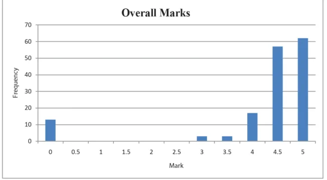
A total of 144 students participated in the CBL pilot study and received marks. Students wrote the quiz designed by the CBL team and completed an evaluation. There were five versions of the quiz per class, ensuring that all topics were evaluated equally over the entire student population and to limit the opportunity for copied answers. The data presented does not have a large distribution as the quiz mark is out of 2.5 (based out of five questions). The first of the three classes had an average mark of 2.10 with a stand-ard deviation of 0.65. The second class, which had the larg-est population of the three, had an average of 2.20 with a standard deviation of 0.37. The final class had an average of 2.30 with a standard deviation of 0.38. Overall the aver-age for the entire program was 2.21 with a standard devia-tion of 0.46. The overall mark was comprised of the quiz result and participation (participation criteria found in Appendix 3). The overall average, out of 5, was 4.13 with a standard deviation of 1.48 (see Figure 1).
The data presented suggests that CBL is an effective teach-ing method at this level of education and professional de-velopment. All the averages presented include participants who were not present and received a mark of 0. The aver-age participation mark was 2.33 with a standard deviation of 0.40. The average difference between participation mark and quiz mark was 0.13 (SD of 0.47). This suggests an as-sociation between superior performance participation and performance on the quiz. No correlation study was con-ducted.
The second set of data collected for CBL was the evalua-tion. This included 11 questions (of which two were binary)
Average Mark (Standard Deviation)
Class 1 2.10 (0.65)
Class 2 2.20 (0.37)
Class 3 2.30 (0.38)
Overall 2.21 (0.46)
Table 1
Frequency of marks for all three classes: Marks are a combination of participation mark and question mark (2.5 marks each) making the entire mark out of five.based on the students’ opinions of the session. Out of 11 questions, nine were evaluated on a nominal scale of one to four. Of the eleven questions in the Appendix 5, three will be looked at in depth as they provide the most relevant da-ta for the present subject of interest.
The first question asked was whether or not the SLM was read by the students. This question was extremely im-portant to this study because the students would be learn-ing new material and readlearn-ing the SLM prior to the sessions would enable them to participate in the discussion with a greater knowledge set and understanding of the relevant material. It is crucial for students to have this background information to be able to engage in the case based learning sessions effectively. Of the two classes that answered this question, 54.7% of the students in the first class answered that they completed the reading compared to 91.3% in the second class. Professor involvement is a major factor in maintaining high compliance. Without proper mastery of the SLM, students put themselves at a major disadvantage for the session.
The second question to be analysed in this paper is ques-tion six, which asked if this type of integrated teaching was a beneficial learning tool. This is asking the perceived ben-efit of the program. It is useful to know if this type of teach-ing aided the students’ understandteach-ing of core material and whether it encouraged conceptual learning and integration, as this was the ultimate goal of the project. The average for
this question was 3.82 with a standard deviation of 0.41. The third and final question is question 8, which asked the students’ overall satisfaction with the CBL session. This was to determine if the students enjoyed the CBL sessions or not. This is where CBL attempts to set itself apart from traditional lecture methodology. With the students engag-ing each other they learn in a way that might be perceived as more interactive and enjoyable than the typical universi-ty teaching suniversi-tyle. It was the hope of this study that this fact may make CBL a more attractive and novel teaching ap-proach and that it would encourage its adoption within the faculty. The average response for this question was 3.80 with a standard deviation of 0.41.
The above data is subject to a number of biases and also relied on categorical data. Nonetheless, the data strongly supports the popularity and the use of the program. The SLM is the lowest scoring portion of the program. Out of the courses assessed for completion of the SLM, the course with the lower completion rate is also the higher level course of those examined. The marks for the quiz were above averages for the respective courses showing the ef-fectiveness of CBL. The overall mark indicates the same findings. An analysis of the opinions of the students shows that participating students found the CBL to be an effective and enjoyable teaching tool.
Figure 1
Frequency of marks for all three classes. Marks are a combination of participation mark and quiz mark (2.5 marks each) making the entire mark out of five.Discussion
Feasibility of CBL implementation
The viability of implementing CBL sessions in health sci-ence courses is measured by the following parameters: (1) quiz marks;; (2) students’ preference;; (3) students’ evalua- tion of the facilitators and the program; and (4) financial resources of the Interdisciplinary School of Health Scienc-es.
The majority of students scored well on both discussion and quiz sections. The average grade out of 2.5 was 2.21 with a standard deviation of 0.46. The average of the three participating classes is relatively high and the standard deviation indicates minimum spread. The data suggests that CBL may be a viable teaching methodology that allows students to apply their knowledge with colleagues. Results of the questionnaires suggest that most health sciences students prefer to have CBL as a supplement to traditional lectures and course work. Interest is crucial to students’ subjects of study; they may be motivated to learn and un-derstand the material they feel is relevant, resulting in a better integration and conceptualisation of knowledge, CBL also prepares students for the problem-based nature of a career in health care and it encourages them to devel-op interprofessional skills (such as codevel-operation and com-munication) early on in their professional development. The material utilised in all aspects of the CBL was taken directly from the course material that makes up each course curriculum. As such, the CBL was directly relevant to the student’s studies.
In addition, students gave positive evaluations to the facili-tators’ skills and the program as a whole. Facilifacili-tators’ skills are an important part of the evaluation because they en-courage students to think critically and steer the discussion in the appropriate direction. Thus, CBL may not only offer a style of learning which motivates students, the program also offers a necessary environment to conduct such learn-ing experience. As the program operates on a volunteer basis, it requires minimal work on the part of the profes-sors and low costs associated with printing quizzes and reserving facilities. The use of third year health sciences students allows the faculty to conduct many small class size sessions which promotes the opportunity for student dis-cussion and engagement with the topics in disdis-cussion. Moreover, CBL provides senior health sciences students with an opportunity to contribute to the program by facili-tating future sessions for first and second year students.
For the two classes with available data regarding the use of the SLM, the lower level course demonstrated a much higher rate of completion. This may suggest that students at a higher academic level were not willing to complete the SLM reading when it was not required or tested upon. There is room for bias in this result as the question evalu-ating the use of SLM was self-reported. Results are high with an average of 3.82 out of 4 (with a standard deviation of 0.41). This question was used to assess students’ percep- tion of the CBL program and its integration in the health science program. The CBL facilitators were in the room at the time of evaluation which may have influenced partici-pants’ evaluations of the facilitators, thus suggesting an observational bias. Overall, the students’ opinion of the program is favourable, as mentioned in the results. This indicates the students’ enthusiasm for the CBL program as it compliments these and other courses in the Interdiscipli-nary School of Health Sciences.
Limitations
As a pilot project, this study contains several limitations that need to be addressed. This study lacks a control group. As such, confidence intervals could not be calculated. In addition, there were a significant number of students who chose not to read the SLM prior to attending their CBL ses-sions. The SLM is a crucial component to the CBL program as it provides students with the basic required knowledge to participate in the discussion. Failure to complete this assigned reading may have prevented optimal participa-tion. Results of the quizzes may be negatively skewed, giv-en that the quizzes giv-entailed only a small number of ques-tions in comparison to the amount of material conveyed. The study results, although rather persuasive, cannot be generalised to courses outside of the ones included in the pilot program. There were only six participating courses in both French and English out of a total of 48 courses availa-ble in the ISHS. Resources, both financial and material, were limited. For instance, the availability of rooms to con-duct CBL sessions was restricted to evening reservations at a location not on main campus. This led to some transpor-tation difficulties and a lack of punctuality. In fact, the most widely criticised issue of the program according to the evaluation was the location of the CBL sessions.
The results indicate that health sciences students partici-pating in the CBL sessions benefitted from and enjoyed taking part in CBL to complement their studies. The post-session assessments had all participating students averag-ing above 80%, indicataverag-ing an understandaverag-ing of course ma-terial in an environment that promoted application and critical thinking. The evaluations suggest that the students felt that they learned and enjoyed the sessions overall. Sev-eral limitations have been considered. Financial resources and authority within the faculty would help overcome a majority of these limitations. Future programs based on this model would also benefit from more rigorous pre- and post-session assessments. Attendance could be improved by holding future sessions on the main campus. Further research would benefit from the use of control groups and more rigorous testing methods.
Acknowledgements
We wish to acknowledge the following individuals for their time and effort during the course of the CBL program at the University of Ottawa: Dr. Linda Garcia, Dr. Karen Phil-lips, Dr. Robert Bell, Dr. Ron Saulnier, Dr. Eric Lavigne, Dr. Anne Konkle, Dr. Frédérique Tesson, Dr. Raywat De-onandan.
We would also like to acknowledge the founding members of this pilot program and facilitators who were not authors: Valerie Audette, Derek McLellan, Michael Cecchini, Premal Patel, Armin Yazdani, Maria Blahoianu, Mirhad Loncar, Sarah Mansour.
References
Carder, L., Willingham, P., & Bibb, D. (2001). Case-based, problem-based learning: Information literacy for the real world. Research Strategies, 18(3), 181-190. Retrieved from
http://dx.doi.org/10.1016/S0734-3310(02)00087-3 Chan, W. P., Hsu, C. Y., & Hong, C. Y. (2008, November). Innovative “case-based integrated teaching” in an under- graduate medical curriculum: Development and teachers’ and students’ responses. Annals Academy of Medicine, 37 (11), 952-956.
Dutch, B. J., Groh, S. E., & Allen, D. E. (Eds.). (2001). The Power of Problem-Based Learning. Sterling: Stylus Pub-lishing.
Ferguson, K. J., & Kreiter, C. D. (2007, September). As-sessing the relationship between peer and facilitator evalu-ations in case-based learning. Medical Education, 41(9), 906-908. doi: 10.1111/j.1365-2923.2007.02824.x
Koh, G. C.-H., Khoo, H. E., & Wong, M. L. (2008, January 1). The effects of problem-based learning during medical school on physician competency: A systematic
re-view. Canadian Medical Association Journal, 178(1), 34-41. doi: 10.1503/cmaj.070565
Appendix 1
Introduction: Offer a Brief Description of CBL
Offer a brief description of CBL Explain format and marking. Answer any questions regarding marking scheme.
Some people may be worried about participation marks; explain that good participation does not involve incessant talking – it is about meaningful contribu-tions and good listening skills
Learning Objectives
Case Presentation
What is important on this slide?
What is the significance of the fictional patient’s age – what issues are age-specific?
What does productive cough mean?
Patient has a “wet cough” (brings up mucus or other fluids)
What is dyspnea?
Shortness of breath.
If the patient tires easily, what could this be a sign of? What is the difference between mucoid and purulent? What does each one mean and why is the change from mucoid to purulent significant?
Mucoid cough is a cough that yields mucus. Purulent cough yields pus.
The transition could indicate an infection. Occasional tension in chest is a symptom for a
variety of diagnoses. What are some of them? Considering the other signs and symptoms ex-hibited by this patient, what ones are likely?
Possible Causes
Encourage group discussion
information, on the board. Each person can contribute to one item.
Medical History
What is the significance of the long-lasting symptoms? Prolonged wheezing and dyspnea are major
cri-teria for lung diseases.
Indicate that the patient is not suffering from a seasonal bronchitis or cold.
What are some of the symptoms and disease associated with smoking? What does being a smoker increase his risk for?
Emphysema, lung cancer, chronic bronchitis, asthma, coughing, dyspnea, wheezing, etc. What are some of the reasons for which the patient
might wake up frequently in the night? Polyuria (frequent urination).
Polyphagia and polydipsia (excessive eating and drinking).
Chronic pain. Sleep apnea.
Note that in this case sleep apnea is the reason and that the first three are signs of diabetes. Why might this be the first time reporting to a
physi-cian? Why is this significant and what might have driv-en him to seek medical help?
Can explore sociology of health – limited access to physicians, reasons why someone may be un-willing to visit the doctor, etc.
Patient likely sought medical help because his symptoms became significantly worse/
interfered with daily routines or because a fami-ly member urged him to.
Since patient hasn’t seen doctor, symptoms haven’t been monitored or treated, so the condi- tion has likely developed and worsened.
Social and Occupational History
Why is the fact that he lived in urban locations rele-vant? What does this put him at greater risk for?
Exposure to carcinogens and pollution may put him at a higher risk for cancers and lung disease. Given that his spouse smokes, what risks can we say
he’s been exposed to?
Thirty years of secondhand smoke, which is ade-quate to cause emphysema.
Is there anything significant in his occupational histo-ry?
Welding is a risk factor because particulate mat-ter might be inhaled that could irritate lungs and increase risk of disease.
What other occupations may increase risk of respirato-ry disorders?
Physical Examination: Vital Signs
Is temperature normal? What is normal value for tem-perature? Why might the patient have a fever?
Normal temperature is 37.2 degrees Celsius. Therefore, this patient has a slight fever – sign
of infection.
What is BMI and what are the various categories? Which one does our patient fall under? Given his BMI, is he at a greater risk for any diseases?
Less than 20 = underweight. 20 – 25 = normal range. 25 – 30 = overweight. 30—35 = obese.
35 and over = morbidly obese. Patient is slightly overweight.
What is normal blood pressure? Is this blood pressure anything to be concerned about?
Normal value is 120/80.
The patient is slightly hypertensive, but that is normal in a male patient this age and probably is not too concerning (but still worth noting). What is the normal rate of respiration? What can we
say about our patient’s respiration?
Normal values are 12-20 breaths per minute. This patient is tachypneic.
What are normal heart rates? Is the patient’s heart rate a concern?
Normal values are 70-75 bpm, but this can vary considerably based on a number of factors. This patient is slightly tachycardic.
What is the fifth vital sign? In many hospitals, a sixth is also used – any idea what it might be?
Fifth vital sign is pain.
The sixth vital sign varies but often it is blood glucose or pupil size.
What are rales? What might cause them and what might they indicate?
Rales are clicking, rattling, or clacking noises that can be heard in the lung using a stetho-scope.
They can be coarse or fine (fine ones are higher pitched, less intense, and shorter).
The sound is made when fluid (mucus) collects in the peripheral portions of the lung, the alveoli collapse, and their walls stick together. When the patient attempts to inhale, the alveolar walls are forced to pop open and a crackling sound is heard during inspiration.
Can indicate pneumonia, bronchitis, collapsed lung tissue, etc.
What is implicated by a longer expiration time? Provoke questions about what would make it
The longer expiration time is because fluid in lungs (from chronic bronchitis) narrows the bronchi, therefore less air can be breathed out at once.
What is meant by barrel chest? Why would someone have one?
A barrel chest is a broad, deep chest that is ex-panded outwards, as shown in the picture. It is common in patients with emphysema. It
happens because of air pocket accumulation in-side the thoracic cavity and increased intratho-racic pressure that allows wall to naturally ex-pand outward.
The person is having more trouble breathing so they must use all their accessory muscles. What does the coughing up of thick mucus indicate?
How is it related to the rales?
Indicates inflammation and explains the rales (fluid in the lung causes the cracking sound). What other organs should be tested and why?
Answers might include the heart (complications of lung problems can include heart problems; also, some of the early symptoms included those of a heart attack), liver (fairly standard)
What tests might you want to do next?
Lead to discussion of a sputum sputum test. They would want to do this because they should suspect some kind of an infection, along with more serious problems. This is done because it is suspected there is an infection, along with more serious problems.
Sputum Test
How is a sputum test done
Patient coughs deeply and expels material that comes up from the lungs into a sterile cup. The sample is placed in a medium where
organ-isms can grow; a positive culture can identify organisms to help diagnose bronchitis,
tubercu- Mixed with animal cells, observe characteristic changes to the cel in order to identify the virus. What does Streptococcus pneumoniae cause? Is it
gram positive or gram negative?
Gram positive (has a thicker layer of peptidogly-can membrane. Can cause a variety of infectious diseases including pneumonia and acute sinusi-tis.
So the patient has an infection…but does this explain all the symptoms?
No!
Differential Diagnosis
What is a differential diagnosis?
Systematic list of all possible diagnoses elimi-nate until you get the final diagnosis.
Have them write their differential diagnosis on the board and explain each possible diagnosis.
Ask for ideas about what further tests they might want to do or what information they will need to narrow it down.
ABG
What is an ABG test and what is it used to measure? Arterial Blood Gas test; uses blood from an
ar-tery.
Most common puncture site is the radial artery at the wrist.
It is used to determine the pH of the blood, the partial pressure of carbon dioxide and oxygen, and the bicarbonate levels.
Mainly used in pulmonology to determine gas exchange levels in the blood (related to lung function).
Results for ABG
What do you notice about the pH of the blood? What is meant by acidosis and alkalosis?
pH higher than 7.45 = alkalosis pH lower than 7.35 = acidosis
Our patient’s pH is significantly lower than nor- mal levels, which means he has acidosis.
How is pH linked bicarbonate ion and carbon dioxide? How do acidosis and alkalosis occur?
The formula represents the blood buffer system, which is responsible for regulating blood pH. Acidosis results from a build-up of carbon
diox-ide in the blood – hence the elevated carbon di-oxide levels.
Alkalosis results from a loss of carbon dioxide (often caused by hyperventilation).
To compensate for the increase or decrease in carbon dioxide, there are metabolic mecha-nisms; the reverse happens. Think about Le Chatelier’s principle and use the formula. What is the difference between respiratory and
meta-bolic acidosis? Which does our patient probably have? Respiratory acidosis or alkalosis is caused by
various problems in the lungs, while metabolic is caused by metabolic disorders that result in an imbalanced pH.
Given that he has lung disease, our patient prob-ably has respiratory acidosis.
One key way to determine whether the acidosis is respiratory or metabolic is that in respiratory acidosis, the CO2 is increased while bicarbonate is either unchanged or increased. This matches our patient’s ABG results, so he has respiratory acidosis.
Why would he have respiratory acidosis?
The build-up of carbon dioxide makes sense if he has lung disease….air is trapped in lungs, can’t exit as quickly, etc. so it has a high propor- tion of CO2 in it.
Imaging Techniques
What is an X-ray and when is it used?
Due to differences in composition, the lungs and the bones of the chest can be distinctly visual-ised
White shadows on X-ray represent more dense or solid tissues such as bone.
Darker shadows represent air-filled tissues, such as lungs.
X-ray radiation is absorbed by solid tissues (such as bone) and therefore white shadows are obtained whereas the lung absorbs very little so the X-ray beams pass through and make darker shadows.
Note: For COPD (Chronic Obstructive Pulmo-nary Disease), should find hyper-inflated lungs with flattened diaphragm, hyperlucent lungs (greater than normal blackening), central pul-monary artery enlargement, bullae (areas of de-stroyed lung tissue create large, dilated air sacs). What is PET (Positron Emission Technology) scan and
when is it used?
Uses short-lived radioactive substances to pro-duce a 3-D coloured image of those substances in the body
Studies metabolic activity or body function. Used mainly in cardiology, neurology, and
on-cology.
Patient receives a short half-lived radiopharma-ceutical (therefore patient doesn’t receive a high amount of radiation…it is about the same as two chest x-rays)
Radiopharmaceuticals discharge positrons and as they encounter electrons within the body, a reaction producing gamma rays occurs. Radiopharmaceuticals contain a chemical
com-monly used by the body so one can view meta-bolic processes (ex: glucose with radioisoptope to see glucose consumption in a tumor).
is it used?
An x-ray procedure that combines many x-ray images using a computer to generate cross-sectional views of internal organs and structures of body.
Used to define normal and abnormal structures in the body and/or assist in procedures by help-ing to accurately guide the placement of instru-ments or treatinstru-ments.
Note: CT scan could be used to assess COPD, but it is not cost-effective and is not usually done, because x-ray and spirometry are adequate. What is microscopy and why would it be used?
Examine microbes under a microscope. In this case, use it to test for the infection What is spirometry and what is it used for?
Used to test the air capacity of lungs.
Breathe into a mouthpiece that is connected to a spirometer.
Patient breathes and then exhales into sensor as hard as possible for as long as possible.
Test is usually repeated at least three times to ensure accuracy and reproducibility.
Can only be used on people who are able to un-derstand and follow the instructions (aka can’t use on young children or severely mentally handicapped).
Which of these techniques should we use for the pa-tient?
X-ray, spirometry, and microscopy (the sputum test is completed at that stage, and there may be no need for).
Spirometry Graph
What does the graph show us?
and normal lungs.
What is obstructive lung disease versus restrictive dis-ease? What might cause each one?
Obstructive lung disease is when the bronchial tubes become blocked or narrower and conse-quently, expiration is difficult. Narrowing is of-ten due to infections or pathologies that cause fluid build-up in the lung.
Causes of obstructive lung disease include COPD, chronic bronchitis, asthma, pneumonia Restrictive lung disease is when lungs cannot
expand completely and are stiff (scar tissue and air pockets).
Causes of restrictive lung disease include scar-ring of lungs and emphysema.
What are FEV1, FVC, and FEV1/FVC?
FEV1 = Forced expiratory volume in first second (how much air can they blow out in one second) FVC = Forced vital capacity (how much air can
they blow out in total).
FEV1/FVC = Ratio of the two; in normal people it is 75 – 85% depending on age, height, weight, health status, ethnicity, etc.
The first two are measured in litres, third is measured as a decimal or percentage. What do you observe on this graph?
How do the different pathologies explain the trends seen on the graph?
OBSTRUCTIVE: The forced vital capacity does not change; the person can breathe out the same amount of air as before, but it takes them much longer to do it because the bronchial tubes are blocked. This accounts for the lower FEV1 and the resulting lower FEV1/ FVC%.
RESTRICTIVE: The lungs are stiff and there is air trapped in them that cannot be expired. Therefore, the forced vital capacity is lower. There is nothing blocking the bronchial tubes
a lower absolute value than normal, because there is less air to blow out). The lower FVC and relatively normal FEV1 cause the FEV1/FVC ra-tio to be relatively high.
Spirometry Results
Based on the values given in the previous slide, what type of lung disease does our patient have?
Mainly obstructive, but note that the FVC is low-er than normal, so he has some restrictive lung disease as well. This finding is a major “key” to the fact that it is COPD.
What is ventolin? If the patient’s values do not change when he is given this, what does this indicate?
Ventolin is frequently given to asthmatics as a sympathomimetic and in an asthmatic, it would improve their FEV1/FVC percentage. However, in our patient it does not, so we can rule out asthma.
Note: COPD damage is irreversible, so generally ventolin (or similar drugs) will not work. In a small percentage of COPD patients, these drugs may increase FEV1 slightly (suggesting that they also have some asthma?).
Based on these findings, can you rule out any diagno-ses or lean towards others?
Imaging Results
What differences do you notice between the X-rays? In patient, there is a flattened diaphragm,
hy-perinflation, increased AP diameter, thick mu-cus in lungs, and thickened central pulmonary artery.
Think about the implications…is this purely obstruc- tive or purely restrictive?
No. There are elements of both.
Pink Puffer versus Blue Bloater
Which one is associated with restrictive and with which obstructive?
Pink puffer is associated with restrictive lung
disease; the lungs are stiffer (from the air pock-ets present in walls) and it is harder for them to expand and contract. Consequently, the person must work much harder to breathe (use accesso-ry muscles, pursed-lip breathing, leaning for-ward). They are thin and more muscular because of this constant exertion. Cough is absent be-cause unlike obstructive, they do not have mu-cus in lungs.
Blue bloater is associated with obstructive lung disease; there is mucus in the lungs and bron-chial tubes are clogged. This inflammation is associated with the excess body fluids and the cough (trying to bring up mucus). It is difficult to breathe because of blockages and narrowing, so there is dyspnea on exertion.
What are some diseases associated with each? Emphysema, scarring of lung tissue associated
with restrictive.
Asthma, pneumonia, bronchitis associated with obstructive.
COPD is primarily obstructive, but also restric-tive (depends on relarestric-tive severity of the emphy-sema and chronic bronchitis).
Final Diagnosis
Students discuss possibilities and come to conclusion that it is COPD. Bring up key points that point towards this diagnosis.
Final Diagnosis: COPD with chest infection
How was this conclusion arrived at? How were otherdiagnoses ruled out?
Students should be able to link the symptoms and test results to COPD. Some key points are detailed briefly below.
Epidemiological points: COPD is more common in males but only because more males smoke (historically), Occupational history and smoking
are critical indicators that he is at risk for COPD. Symptoms related to chest infection: sputum
becoming purulent, fever (although COPD is associated with transient fever), the positive sputum test.
Long-term development of symptoms; dyspnea, barrel chest, etc.
Talk about restrictive versus obstructive disease; how the spirometry and pink puffer/ blue bloat-er indicated that our patient was obstructive and slightly restrictive, pointing towards COPD. For ABG, respiratory acidosis (and not alkalosis)
because air trapped in the lungs, poor ability to expire, etc would cause increased CO2. It would-n’t be very plausible for a COPD patient to have alkalosis…hyperventilation isn’t likely for some- one with obstructive lung disease.
Pathological Findings
What differences do you notice about emphysema and bronchitis? Which can be considered restrictive and which is obstructive?
Ensure students understand what each disease is and that the combination of the two is COPD.
Possible Complications
What are some likely complications?
What is meant by each of the complications? How might these happen?
Cor pulmonale = enlargement/weakness of the right ventricle caused by a respiratory disorder. Chronic cor pulmonale tends to cause hypertro-phy of right ventricle (muscle cells grow larger to push the blood against increased resistance), whereas acute cases often cause dilation (ventricle stretched out to accommodate pres-sure changes) instead. Both are caused by in-creased pressure in right ventricle. Heart failure can result if untreated. Treat underlying cause.
Secondary polycythemia = increase in the pro-portion of RBCs in the blood. Caused by in-creased RBC production (absolute polycythe-mia) or decreased plasma levels (relative polycy-themia). Secondary polycythemia caused by in-creases in production of erythropoietin
(hormone controlling RBC production), leading to an increased production of erythrocytes). Normal adaptation at high altitudes or for dis-eases like COPD.
Bullous lung disease = formation of bullae (air-filled wall tissue in lungs) so lung can’t expand or contract fully. Leads to shortness of breath and infection. Treat with surgery or steroids. Pulmonary hypertension = increase in blood
pressure in pulmonary artery, pulmonary vein, or capillaries leading to dyspnea, fainting, and dizziness. Exacerbated by exertion. Caused by vasoconstriction of blood vessels connected to lungs, making it harder for heart to pump blood through lungs.
Malnutrition – dyspnea makes it difficult to eat and COPD patients often lack an appetite but require 10 times the calories of a healthy person. Consequence of malnourishment is lack of ener-gy and worsened dyspnea.
Pneumothorax = air enters pleural cavity. COPD patients have weaker lungs that are more vul-nerable to tears/holes.
General Treatment Measures
What are some important treatment measures that should be taken? What is the single most important one?
Smoking cessation is the most important. Reversible bronchospasms should be treated. Explain what each treatment measure involves? Why is each important?
Adequate hydration helps to keep mucous thin and easier to cough up
Pulmonary rehabilitation is physiotherapy and education that helps patients develop stronger accessory muscles to assist in breathing, clear mucus, etc.
Vaccinations for seasonal influenza (flu worsens symptoms of COPD), pneumovax (to prevent pneumonia, which COPD patients are at higher risk for).
What is pulmonary hygiene?
A set of methods used to clean mucous and se-cretions from the airways to prevent respiratory secretions (suctioning airway, analgesics, cough-ing, percussion, proper positioncough-ing, etc).
General Treatment Measures Continued
When would surgical intervention be necessary? Only considered if the usual treatments don’t
relieve symptoms and quality of life is poor. High risks associated with lung surgeries. What is lung volume reduction surgery? When would a
lung transplant be done?
Lung volume reduction = 20-30% of the top por-tion of lung is removed (this is usually the most damaged area) so air can move more freely through the lung and decrease COPD symptoms. Controversial procedure.
Lung transplant is only recommended for ad-vance stage patients who will die without it and are no longer smokers.
What diet considerations are important for a COPD patient?
Low sodium (otherwise, body becomes dehy-drated and breathing is difficult), avoid alcohol (suppresses breathing), high protein (improves appetite and is more substantial for people who have trouble eating), high caloric intake (to com-pensate for calories lost in use of accessory mus-cles to breathe).
What are corticosteroids used for?
To treat inflammatory conditions that ma affect joints, skin, digestive tract, respiratory system, eyes, and ears. In this case, they are given orally. Work by blocking production of substances such as prostaglandins which trigger inflammation in the body; also partially suppress certain compo-nents of immune system.
Why would there be inflammation?
Inhaled irritants cause inflammation as the re-sult of neutrophil activity, T lymphocyte, and macrophage activity. Hyperplasia and hypertro-phy of glandular tissue occurs and there is in-creased mucus production which plugs the bron-chi.
What role do the corticosteroids play in the treatment of COPD?
Liquefy mucus and reduce swelling in breathing tubes.
Note that not all patients respond to them. What does theophylline do and when should it be
used?
Relaxes muscles surrounding breathing tubes and widens them to ease breathing.
However, it is toxic and therefore used only as a last resort.
What are sympathomimetics?
Andrenergic agonists which produce symptoms of flight-or-fight response and stimulate sympa-thetic nervous system. Result is bronchodilation. Some adverse effects though.
Other Recommendations
What recommendations would you give the patient about his environment?
Avoid cold, dusty, or damp environments. What would rehabilitation programs entail?
Breathing exercises to improve function of dia-phragm, strengthen muscles, improve gas ex-change, conserve energy, and relax breathing. What types of exercise would you recommend to the
patient?
Aerobic exercise programs, such as walking to improve exercise tolerance and ultimately re-duce dyspnea.
Why avoid traveling at high altitudes?
Lower level of oxygen would cause extreme hypoxemia and more profound dyspnea. Anything else?
Oxygen therapy.
Prognosis and Follow-Up
What are some important indicators of prognosis? Age, smoking status, FEV1 values, poor response
to bronchodilator therapy, severe hypoxemia, development of complications.
Why is a follow-up necessary? Think about the nature of the disease (irreversible!)
If very progressive, more advanced therapy will be needed. Otherwise, should be seen monthly if severe or biannually when stable.
Why would a patient require nocturnal oxygen? If he has severe dyspnea.
Evaluation
Individual completion of the quiz and evaluations.
Appendix 2
Health Problems (Winter 2010) CBL- Self-Learning Mod-ule
Basic Physiology of the Respiratory System
The respiratory system obtains oxygen from the external environment and delivers it to the blood for distribution throughout the body.
The passageway from external to internal is as follows: na-sal/oral cavity -> pharynx -> trachea -> Larynx -> bronchi -> bronchioles -> alveoli.
The branching structure, which includes the bronchi and bronchioles, is known (quite literally) as the bronchial tree. The terminal end of the resp. system, the alveoli, is where gas exchange occurs.
Alveoli are thin-walled sacs surrounded by blood capillar-ies and are the site of gas exchange. Air that is inspired dif-fuses into the blood and the hemoglobin portion of eryth-rocytes (red blood cells) becomes saturated with oxygen. Carbon dioxide diffuses in the opposing direction from the blood to the alveoli in order to be expired. This is the basis of the cellular respiration.
Pathophysiology of the Respiratory System
Asthma: Inflammation of the small airways that carry air in an out of the lungs, which can cause wheezing, coughing and difficulty in breathing.
COPD: A disease in which the airways and tiny air sacs in-side the lungs are partially obstructed or destroyed. The result is labored breathing. This disease may occur when a person breathes in lung irritants of some kind.
Emphysema: An irreversible chronic lung disease in which the alveoli (small air sacs in the lung) become damaged; smoking is the most common cause of emphysema. Bronchitis: An inflammation of the mucous membranes of the bronchial tubes, causing a persistent cough that pro-duces considerable quantities of sputum.
Pleurisy: Inflammation of the pleura (membrane lining of the throat between the lungs and abdomen) characterized by fever, painful and difficult breathing, and a dry cough. Pneumonia: An inflammation of one or both lungs that is frequently but not always due to infection. The infection
may be bacterial, viral, fungal or parasitic. Symptoms may include fever, chills, cough with sputum production, chest pain, and shortness of breath.
Tuberculosis: An infection transmitted by inhalation or ingestion of tubercle bacilli that is manifested in fever and small lesions called tubercles (usually in the lungs but in various other parts of the body in acute stages).
Terminology and Definitions
FVC – Forced Vital Capacity – after the patient has taken in the deepest possible breath, this is the volume of air, which can be forcibly and maximally exhaled out of the lungs until no more can be expired. FVC is usually ex-pressed in units called liters. This PFT value is critically important in the diagnosis of obstructive and restrictive diseases.
FEV1 – Forced Expiratory Volume in One Second – this is the volume of air, which can be forcibly exhaled from the lungs in the first second of a forced expiratory maneuver. It is expressed as liters. This PFT value is critically important in the diagnosis of obstructive and restrictive diseases. FEV1/FVC – FEV1 Percent (FEV1%) – This number is the ratio of FEV1 to FVC – it indicates what percentage of the total FVC was expelled from the lungs during the first sec-ond of forced exhalation – this number is called FEV1%, % FEV1 or FEV1/FVC ratio. This PFT value is critically im-portant in the diagnosis of obstructive and restrictive dis-eases.
FEV3 – Forced Expiratory Volume in Three Seconds – this is the volume of air, which can be forcibly exhaled in three seconds – measured in Liters – this volume usually is fairly close to the FVC since, in the normal individual, most of the air in the lungs can be forcibly exhaled in three sec-onds.
FEV3/FVC – FEV3% – This number is the ratio of FEV3 to the FVC – it indicates what percentage of the total FVC was expelled during the first three seconds of forced exhalation. This is called %FEV3 or FEV3%.
PEFR – Peak Expiratory Flow Rate – this is maximum flow rate achieved by the patient during the forced vital capacity maneuver beginning after full inspiration and starting and ending with maximal
FEF – Forced Expiratory Flow – Forced expiratory Flow is a measure of how much air can be expired from the lungs.
It is a flow rate measurement. It is measured as liters/ second or liters/minute. The FVC expiratory curve is divid-ed into quartiles and therefore there is a FEF that exists for each quartile. The quartiles are expressed as FEF25%, FEF50%, and FEF75% of FVC.
MVV – Maximal Voluntary Ventilation – this value is de-termined by having the patient breathe in and out as rapid-ly and fulrapid-ly as possible for 12 -15 seconds – the total vol-ume of air moved during the test can be expressed as L/sec or L/min – this test parameter reflects the status of the respiratory muscles, compliance of the thorax-lung com-plex, and airway resistance. Surgeons like this test value because it is a quick and easy way to assess the strength of the patient’s pulmonary musculature prior to surgery – a poor performance on this test suggests that the patient may have pulmonary problems postoperatively due to muscle weakness. MVV can therefore be viewed as a meas-ure of respiratory muscle strength. One major cautionary note is that this test is effort dependent and therefore can be a poor predictor of true pulmonary strength and compli-ance.
Appendix 3
Pass/Fail Evaluation of Case Based Learning
Passing Criteria
The student shows up on time and well prepared to the case based learning session.
The student shows respect and regard to peers and the facilitator throughout the session. Offensive language will not be tolerated and will result in the removal of the student from the session and a consequent failing grade.
The students shows cooperation and commitment to the group’s overall goals.
The student actively searches for the answers to the problems.
The student participates throughout the discussion and contributes to the problem solving process. The student meets all the learning objectives designed
The student follows the case based learning process which starts from the introduction and ends in conclu-sion and ending questions.
The student completes the session evaluation of the facilitator
The student stays for the entire CBL session.
The Student Identifies the core issues in a timely man-ner.
The student asks for appropriate help from the facilita-tor or Professor in the event of a problem.
Failing Criteria
Absence of greater than 5 minutes results in a fail mark Offensive language will not be tolerated and will result
in the removal of the student from the session and a consequent fail.
The Student does not participate in the group discus-sion.
The student is distracting and does not cooperate with the group.
The student is unorganized and did not prepare for the session.
The Student does not follow the case based learning process which starts from the introduction and ends in conclusion and ending questions.
The student does not meet the learning objectives. The Student is constantly talking and does not allow
others to comment or participate in the discussion. The Student does not Identify the core issues and is
constantly distracted.
Appendix 4:
Health Problems CBL Quiz
1. What FEV1/FVC% value would be consistent with severe COPD?
A) 20% B) 60% C) 75% D) 100%
5. Where is COPD most prevalent? A) Women
B) Men
C) Men and Women equally D) Children
10. Which one of these is not one of the possible complica-tions of COPD?
A) Respiratory Infection B) Pneumothorax C) Cor Pulmonale D) Pleurodesis
11. What is the normal respiratory rate range? A) 10 – 15 breaths/minute
B) 12 – 20 breaths/minute C) 15 – 30 breaths/minute D) 20 – 25 breaths/minute
21. What conditions are associated with COPD? A) Bronchitis
B) Emphysema C) Asthma D) A and B
Appendix 5:
Seminar Facilitation Evaluation
Student Facilitators:
1. ______________ 2. ______________
Rating: Poor (1) Satisfactory (2) Very Good (3) Superior (4) Have you read the self-learning module? Yes No
1. Facilitation Skills: (1) (2) (3) (4)
Facilitators ask questions and use strategies that draw out knowledge of theory/experience; facilitators are knowl-edgeable and offer correction and guidance when neces-sary.
2. Organisation: (1) (2) (3) (4)
Seminar is structured in a clear and logical sequence.
3. Originality: (1) (2) (3) (4)
Visual and written aids are interesting, innovative/ creative and helpful.
4. Engagement: (1) (2) (3) (4)
Facilitators generate a high degree of student interest; re-spectful and inclusive; all students are encouraged to par-ticipate.
5. Discussion: (1) (2) (3) (4)
Discussion is focused, relevant and engaging; theory (readings) related to experience; application and implica-tions clear and accurate.
6. Experience: (1) (2) (3) (4)
Participation in this exercise increased understanding of
knowledge; complement the theoretical portion on the class.
7. Self-learning module: (1) (2) (3) (4)
The self-learning module was helpful in enhancing ability to participate actively in discussion during the session.
Seminar Improvements
1. Overall Satisfaction: (1) (2) (3) (4)Rate the level of satisfaction with the seminar in combining theoretical concepts and practical problem solving.
2. Effectiveness: (1) (2) (3) (4)
Rate the effectiveness of the seminar to complement theo-retical concepts acquired in the class.
3. How would you improve the CBL experience?
4. Do you think that this seminar should be offered as a component of fourth year courses, in order to integrate all previous knowledge?
5. Would you recommend this CBL seminar to be offered in other Health Sciences courses?
6. Is the seminar helpful only to someone who wants to become a medical doctor? Can you think of other Health Science-oriented careers where CBL seminars are useful?