The developmental genetics of Hirschsprung’s disease
Karl-F. Bergeron1, David W. Silversides2 and Nicolas Pilon1
1 Molecular Genetics of Development Laboratory, Department of Biological Sciences and BioMed
Research Center, University of Quebec at Montreal (UQAM), Quebec, Canada.
2Department of Veterinary Biomedicine, Centre de recherche en reproduction animale (CRRA), Faculty of Veterinary Medicine, University of Montreal, Quebec, Canada.
Corresponding Author: Nicolas Pilon, Department of Biological Sciences and BioMed Research Center, Faculty of Sciences, University of Quebec at Montreal (UQAM), 141 President-Kennedy Ave, Montreal, PQ, Canada, H2X 3Y7; Tel.: 514-987-3000 x3342; Fax: 514-987-4647; Email:
pilon.nicolas@uqam.ca
Key words: Intestinal Motility, Enteric Nervous System, Hirschsprung’s disease, Aganglionic
Megacolon, Neural Crest Cells, Neurocristopathy.
Abstract: Hirschsprung’s disease, also known as aganglionic megacolon, derives from a congenital
malformation of the enteric nervous system (ENS). It displays an incidence of 1 in 5000 live births with a 4:1 male to female sex ratio. Clinical signs include severe constipation and distended bowel due to a non-motile colon. If left untreated, aganglionic megacolon is lethal. This severe congenital condition is caused by the absence of colonic neural ganglia and thus lack of intrinsic innervation of the colon due in turn to improper colonization of the developing intestines by ENS progenitor cells. These progenitor cells are derived from a transient stem cell population called neural crest cells (NCC). The genetics of Hirschsprung’s disease is complex and can involve mutations in multiple genes. However, it is estimated that mutations in known genes account for less than half of the cases of Hirschsprung’s disease observed clinically. The male sex bias is currently unexplained. The objective of this review is to provide an overview of the pathophysiology and genetics of Hirschsprung’s disease, within the context of our current knowledge of NCC development, sex chromosome genetics and laboratory models.
Intestinal motility disorders (e.g. gastroesophageal reflux, irritable bowel syndrome and chronic constipation) are very frequent in the North American population. For instance, the prevalence of irritable bowel syndrome and chronic constipation ranges between 20-25% and 12-19%, respectively (1-3). Gastrointestinal motility is controlled by the enteric nervous system (ENS), which operates largely independently of the central nervous system. The ENS is the largest and most complex part of the peripheral nervous system, being found throughout the length of the gastrointestinal tract. The ENS notably coordinates mixing and peristaltic bowel contractions by interacting with enteric smooth muscle cells. Thus, anything that impairs this interaction can lead to a gut motility disorder.
Hirschsprung’s disease (HSCR) (4) is one of the most severe forms of organic constipation. This review focuses on the tissue architecture underlying gut motility, the embryonic origin of HSCR, as well as the genetic mutations known to cause different forms of this severe congenital disease. We also discuss the currently unexplained male-sex bias seen in HSCR, and present new mouse models to further our understanding of HSCR pathogenesis.
Structure and function of the enteric nervous system
The enteric nervous system (ENS) is the nervous system intrinsic to the digestive system. It consists of thousands of interconnected small ganglia embedded in the walls of the entire digestive tract (Fig.1A). Each ganglion is composed of neurons and associated glial cells. As summarized below, the extensive neural circuits of the ENS are capable of local autonomous function (5). In addition, the ENS works in concert with the central nervous system (CNS) through widespread two-way connections, to control the digestive system in the context of local and whole body physiological demands (5, 6). These connections are provided by both vagal and sympathetic fibers along mesenteric vessels (Fig.1B)
The gut wall is composed of a mucosal layer, a smooth muscle layer, and a serosal layer. The muscle layer consists of at least two sheets of parallel muscle fibers typically arrayed at right angles to one another: a circular layer wrapping around the gut and a longitudinal layer running along it. Most neurons of the ENS are found in the mucosal and muscle layers, forming two main networks of nerve ganglia extending from oesophagus to anus, and known as the submucosal plexus and the myenteric plexus, respectively (Fig.1A). The myenteric plexus lies between the longitudinal and circular muscle sheets of the gut wall and exerts control primarily over digestive tract motility. The submucosal plexus, as its name implies, is buried underneath the mucosa. Its principal role is in sensing the environment within the lumen, regulating gastrointestinal blood flow and controlling epithelial cell function.
Enteric neurons can be classified into three broad classes according to their function: intrinsic primary afferent neurons (also referred to as sensory neurons), interneurons and motor neurons. Sensory neurons receive information from receptors in the mucosa and muscle responding to mechanical, thermal, osmotic and chemical stimuli (7). Mucosal chemoreceptors sensitive to acids, glucose and amino acids allow sampling of luminal contents whereas sensory receptors in muscle respond to stretch and tension. Interneurons integrate information from sensory neurons and provide it to motor neurons. Collectively, motor neurons directly control gastrointestinal motility, blood flow and secretion through direct interaction with numerous effector cells, including smooth muscle cells.
Myenteric motor neuron subtypes include excitatory and inhibitory neurons interacting with both the longitudinal and circular muscle layers. Excitatory circular muscle motor neurons contain cholinergic markers and tachykinins such as substance P, while inhibitory motor neurons contain nitric oxide synthase (NOS), as well as vasoactive intestinal peptide (VIP) (8). Enteric glial cells provide support for neurons. However, unlike individual Schwann cells, enteric glial cells ensheath axons in groups. Enteric glial cells differ markedly from other glial cells in the peripheral nervous system and are similar to CNS astrocytes with respect to S-100 protein and glial fibrillary acidic protein (GFAP) expression (9). These observations have led some researchers to classify the ENS as a distinct nervous system, outside of the peripheral nervous system, more closely resembling a second CNS.
Neural crest cells: the multipotent precursors of the ENS
Cells forming the ENS are derived from a transient–and vertebrate specific−population of multipotent cells called neural crest cells (NCC). These cells are initially specified at the border of the non-neural ectoderm (the future epidermis) and the neural plate (the future central nervous system) (10). During neurulation, bending of the neural plate places NCC at the dorsal edge of the forming neural tube. NCC subsequently undergo an epithelial-mesenchymal transition, leading to an anteroposterior wave of delamination and migration from the neural tube. This results in the eventual colonization of numerous embryonic structures (Fig.2A). NCC can differentiate into several different cell types including melanocytes (pigment cells), peripheral neurons and glia, craniofacial chondrocytes and some cardiac cells, to name a few. However, the NCC population is heterogenous and NCC are not fully equivalent with respect to their differentiation potential (11). As such, NCC are subdivided into several subpopulations in accordance with their origin along the anterior-posterior axis: cranial, cardiac, vagal, trunk and sacral. NCC differentiation is controlled by a combination of their origin along this axis with their exposure to regionally distinct environmental cues. For instance, cranial NCC contribute extensively to the cranial nerves (neurons and glia), the head mesenchyme (cartilage, bone and connective tissue) and the inner ear (sensory hair cells and melanocytes of the stria vascularis) in addition to generating skin melanocytes.
Much of the ENS is derived from vagal NCC, which are formed adjacent to somites one to seven. These vagal NCC initially migrate in a dorsal to ventral direction to colonize the foregut (future oesophagus and stomach), where they are now named enteric (e) NCC. As illustrated in Fig.2B, vagal NCC colonize the foregut via two distinct migratory patterns, the most posterior vagal NCC contributing only a small subset of foregut eNCC (12). Foregut colonization from anterior vagal NCC is followed by a rostral to caudal migration within the mesenchyme to invade the midgut (future small intestine) and hindgut (future colon and rectum) (13, 14) (Fig.2B-C). Gut colonization by eNCC occurs during a relatively long period of time (from embryonic day 9 to 15 in mice and from week 4 to week 7 in humans) and is performed in parallel with gut tissue growth and rearrangement. Such timing appears important for proper colonization of the hindgut since a subpopulation of eNCC crosses from the prospective small intestine to the future colon via the mesentery during a developmental time period in which these gut regions are transiently juxtaposed. These trans-mesenteric eNCC constitute a large part of the hindgut ENS (15). Finally, a small subset of the ENS is also derived from sacral NCC (formed adjacent to somite 28 and beyond). This minor contingent of NCC colonizes the hindgut up to the
cecum in a caudal to rostral direction (Fig.2B,C) but is unable to compensate for a lack of vagal NCC colonization (16). During and following migration, eNCC will undergo extensive proliferation and differentiation up to one month after birth in order to form the mature ENS (17).
An introduction to HSCR
HSCR is caused by the absence of myenteric neural ganglia in the terminal regions of the gut resulting in tonic contraction of the affected segment, which in turn leads to obstruction and massive distention anterior to this point. The particular migration pattern of eNCC is believed to be the main contributing factor to the pathogenesis of HSCR. Accordingly, the colon, found furthest from the original vagal source of eNCC, is the last segment of bowel to be colonized and is the most susceptible to aganglionosis. However, animal models have shown that deficient cell migration is not the sole cause of aganglionosis. Indeed, deficient cell proliferation, survival or differentiation of eNCC can also lead to bowel aganglionosis affecting the rectum up to a varying anterior limit (18, 19).
The incidence of HSCR is approximately one in 5000 live births (20). Males are much more frequently affected than females with an overall ratio of 4:1. In 12% of cases, HSCR is associated with chromosomal anomalies such as trisomy 21, and in 18% of cases, it is part of a syndrome in recurrent combination with other defects (20).
Infants affected by HSCR frequently present in the first two months of life with symptoms of impaired intestinal motility such as failure to pass meconium (within the first 48 hours of life), constipation, and abdominal pain or distention. However, HSCR can also be diagnosed later in life (in childhood or even in adulthood) in individuals with lifelong severe constipation. The aganglionic segment includes the distal rectum and a variable length of contiguous proximal gut (21). Accordingly, HSCR is classified in four sub-categories:
- In 80% of individuals, aganglionosis is restricted to the rectosigmoid colon (short-segment disease). The incidence of short-segment disease is around five times greater in males than in females.
- In approximately 15%-20%, the aganglionosis extends proximal to the sigmoid colon (long-segment disease). The male sex bias is less than 2:1 for long-segment HSCR.
- In approximately 5% of individuals, aganglionosis affects the entire large intestine (total colonic aganglionosis).
- Rarely, the aganglionosis extends into the small bowel or even more proximally to encompass the entire bowel (total intestinal aganglionosis).
Modern treatment for HSCR, performed as early as 1948 (22), involves surgical resection of the aganglionic segment and reconnection of the proximal bowel to the anus (a procedure nicknamed "pull-through"). Alternatively, individuals with extensive intestinal aganglionosis and irreversible intestinal failure may be candidates for intestinal transplantation (23). However, these surgical procedures essentially convert the lethal defect into a chronic medical ailment that frequently involves constipation, fecal soiling and enteritis (24). Thus, there is a need for novel therapeutic strategies to better address this condition and stem cell therapy approaches are a very promising avenue (25). Indeed, mammalian (including human) neural crest progenitors derived from foetal or neonatal guts are
able to form distinct neurogenic and gliogenic structures in culture and, when transplanted into rodent aganglionic guts, reconstruct ganglionic function (26-28). Enteric glial cells are a particularly interesting candidate for transplantation into affected gut segments, as they possess neurogenic potential, even in adult mice (29, 30).
Gene mutations linked to HSCR
The genetics of HSCR is complex: whereas a mendelian mode of inheritance has been described for syndromic HSCR, isolated HSCR (>70% of patients) stands as a model for genetic disorders with complex patterns of inheritance. Mutations in several genes have been implicated in HSCR and tend to display incomplete penetrance. Collectively, known mutations account for slightly more than 50% of the familial (mostly long-segment HSCR) and around 20% of the sporadic cases (mostly short-segment HSCR), thus contributing to only a small proportion of the genetic heritability of HSCR (31). HSCR-associated genes and susceptibility loci as well as animal models have been extensively reviewed previously (19, 20, 32). A comprehensive and updated list (including relevant references) is presented in Table 1 but will not be discussed in detail here.
Most of known HSCR-associated genes encode players of two main signaling pathways: the RET (proto-oncogene REarranged during Transfection) and EDNRB (endothelin receptor, type B) pathways (Table 1). Both of these pathways regulate survival, differentiation, migration, and proliferation of neural crest-derived cells (33-36). Furthermore, cross talk between RET and EDNRB pathways in the coordination of ENS development has been demonstrated both in vitro and in vivo (33, 37, 38). RET is the main gene associated with HSCR: the majority of patients harbor either heterozygous mutations in the coding region or a hypomorphic allele in a conserved enhancer sequence in its first intron. Mutations within RET account for about 15% of patients with sporadic HSCR and 50% of familial cases (20).
In 18% of cases, HSCR is found as a feature of a larger developmental syndrome for which a mendelian inheritance pattern is typically observed (20). Most of these HSCR-associated syndromes are neurocristopathies, i.e. NCC developmental defects leading to disease (39). Given the extensive NCC contribution to the developing embryo, such associations are not surprising. A classical example is Waardenburg-Shah syndrome which features aganglionic megacolon in combination with sensorineural hearing loss (and balance problems in some patients), as well as pigmentary abnormalities of the hair, skin and eyes. Three genes have been implicated in WSS: SOX10 as well as two members of the EDNRB signaling pathway (EDNRB and ET3) (40).
The male sex bias of HSCR
The 4:1 male sex bias seen clinically for HSCR is currently poorly understood at the molecular level, in part because it is poorly replicated in animal models. A weak male sex bias (around or less than 2:1) is only occasionally detected in single and double mutant animal models of short-segment aganglionosis involving hypomorphic Ret and Ednrb alleles. Interestingly, these modelswhich depend
on gene dosage as well as genetic backgroundhave shown that gender correlated with extent of aganglionosis (38, 41, 42).
It has been previously proposed that a X-linked gene may be responsible for the HSCR sex bias. In this regard, it is noteworthy that a small subset of patients suffering from hydrocephalus associated with mutations of the X-linked gene L1CAM, also display HSCR (43). However, the estimated incidence of HSCR among patients with mutations in the L1CAM gene is low (~3%) and no mutation in L1CAM or any other X-linked gene have been identified through genome-wide association studies thus far (20, 44). Interestingly, in mouse models, L1cam has been shown to act as a modifier gene for aganglionosis-causing mutations of Sox10 but not for mutations of the major HSCR-associated genes
Ret and Gdnf (45). These observations suggest that the genetic basis of the male-sex bias of HSCR is
likely to be complex and we now propose the novel hypothesis that a Y-linked gene may be involved. Among the very few genes on the Y chromosome that are not involved with aspects of spermatogenesis, SRY is such a candidate.
SRY (Sex determining region Y) is expressed in the male genital ridge where it functions as the
key genetic switch for sex determination in mammals (46). SRY was the first SOX gene identified (47), and conferred its name to the family (SOX: SRY-related HMG bOX). Although not well appreciated, extra-genital ridge expression of SRY has been reported in a variety of cell types including developing and mature neurons (48-51). At the functional level, it has been suggested that the SRY protein might compete with other SOX proteins via their well conserved HMG-box domain, which is involved in DNA binding as well as protein-protein interactions (52). Accordingly, it is postulated that SRY could be involved in the etiology of multiple male-biased neurological disorders such as Parkinson’s disease, autism and schizophrenia (53). Endogenous SRY expression has not been reported in the normal developing ENS. However, it is noteworthy that the SRY promoter of several species (including human) contains sequences that can define a NCC expression pattern (including eNCC) in transgenic mice (54). Taken together, these observations suggest that, for some individuals, SRY gene expression might be inappropriately turned on in ENS progenitors where it could interfere with the critical function of at least one other SOX member, SOX10. This possibility is strongly supported by very recent data indicating that the SRY protein is detected in HSCR tissues and can repress the RET promoter by preventing the formation of a SOX10-containing transcriptional complex (55). Therefore, among the sex-specific genes that may contribute to the male-sex bias seen in HSCR, the candidacy of SRY should now be seriously considered.
New mouse models for HSCR and associated syndromes
Coding sequence mutations of most known HSCR-associated genes result in long-segment as well as syndromic HSCR but fail to explain the transmission of the more frequent short-segment form (56). This suggests the importance of non-coding variations and implies that additional genes involved in ENS development still await discovery. In order to identify new loci involved in NCC development and ENS formation, we generated new mouse models through a random transgene insertional mutation approach. To this end, we used mice from the inbred FVB/n strain which are albino due to a homozygous mutation in their Tyrosinase gene. A minigene including functional Tyrosinase sequences
was injected as a transgene (57). Animals were screened for rescue of pigmentation, with concurrent pigmentation abnormalities indicative of NCC development defects. This approach yielded multiple lines with pigmentation flaws (to be described elsewhere) from which two have been confirmed to have disruption of a novel locus regulating NCC development and exhibit a phenotype resembling human HSCR:
- TashT mice display uniform spotted pigmentation in all individuals, irrespective of zygosity. Interestingly, aganglionic megacolon develops in a fraction of homozygotes with a strong male sex bias; female are very rarely affected. Moreover, the penetrance of this defect can be increased by exogenous stress. The TashT transgene insertion was localised to mouse chromosome 10 in a large gene-free region near the cytoband B2-B3 boundary. This region is syntenic with human chromosome 6q16.3 which is devoid of known HSCR-associated loci.
- Spot mice possess a few spots of faint pigmentation. A fraction of second generation individuals display increased pigmentation defects and aganglionic megacolon concomitant with hyperactive circling behavior, a symptom of balance problems. This new mouse line is thus a model for Waardenburg-Shah syndrome (WSS), combining HSCR with hypopigmentation and inner ear abnormalities. The Spot transgene insertion was localised to mouse chromosome 10 cytoband D2. This region is syntenic to human chromosome 12q15 which is devoid of any known HSCR- or WSS-associated genes.
Conclusions
Clinical expression of HSCR suggests it is a multifactorial disease, involving at least one major gene (i.e. RET) and genetic structural variants (modifier loci) conferring risk to HSCR. Intriguingly, in some mouse models of short-segment HSCR typically involving gene-dosage dependence, there is no direct correlation between the extent of aganglionosis and severity of the megacolon phenotype. A fraction of animals possessing a substantial aganglionic colon segment even fail to present with signs of intestinal dysmotility or abdominal distension, especially within females (38) (unpublished observations). Moreover, non-genetic exogenous factors like oxidative stress (58, 59) and vitamin A deficiency (60) compromise NCC development and can contribute to the etiology of HSCR, at the very least in animal laboratory models. Altogether, these observations suggest that our understanding of HSCR pathophysiology is still incomplete and that further studies are required to identify additional genetic and non-genetic factors. In this regard, it is anticipated that our work with the mouse models described above will lead to a new understanding of ENS formation in health and disease, with the objective of converting basic research discoveries into the improved clinical management (screening, diagnosis and treatment) of HSCR as well as other gut motility disorders.
Acknowledgements
The authors thank Dr Christophe Faure for critical review of the manuscript. Research in the Pilon lab is supported by the Canadian Institutes for Health Research (CIHR), the Natural Sciences and Engineering Research Council of Canada (NSERC), the Fonds de la Recherche du Québec-Santé (FRQS) and the Fonds de la Recherche du Québec-Nature et Technologie (FRQNT).
Table 1: Human and mouse loci associated with intestinal aganglionosis or hypoganglionosis
Human Locus Gene Details Human etiology Mouse synteny Mouse models†
10q11.21 RET ret proto-oncogene, HSCR1 non-syndromic HSCR, multiple
endocrine neoplasia type 2 6F1 Ret
-, Ret51, RetS697A,
RetC620R, RetY1062F
5p13.2 GDNF glial cell line-derived neurotropic factor, HSCR3
non-syndromic HSCR 15A1 Gdnf
-19p13.3 NRTN neurturin, NTN non-syndromic HSCR 17D no ENS phenotype reported
10q25.3 GFRα1 GDNF family receptor alpha 1 ** 19D2 Gfra1
-8p21.3 GFRα2 GDNF family receptor alpha 2 ** 14D2 Gfra2
-12p13.31 NTF3 neurotrophin 3, NT3 ** 6F3 NT3
-15q25.3 NTRK3 neurotrophic tyrosine kinase
receptor type 3, TRKC ** 7D2 TrkC
-13q22.3 EDNRB endothelin-B receptor, HSCR2 WSS and non-syndromic HSCR 14E2.3 ppiebald lethal (sl), Ednrb
-20q13.32 ET3 endothelin 3, EDN3, HSCR4 WSS and non-syndromic HSCR 2H4 lethal spotted (ls), Et3
-1p36.12 ECE1 endothelin converting enzyme 1 non-syndromic HSCR 4D3 Ece
-2q36.1 PAX3 paired box 3 ** 1C4 Pax3Sp
22q13.1 SOX10 sex determining region Y-box 10 WSS and non-syndromic HSCR 15E1 Sox10Dom, Sox10LacZ
2q22.3 ZEB2 zinc finger E-box binding
homeobox 2, SIP1, ZFHX1B Mowat-Wilson syndrome with HSCR 2B Zfhx1b
-4p13 PHOX2b paired-like homeobox 2b Haddad syndrome with HSCR 5C3.1 Phox2b
-12q23.2 ASCL1 achaete-scute complex homolog 1,
MASH1 Haddad syndrome with HSCR 10C1 Ascl1
-18q21.2 TCF4 transcription factor 4 Pitt-Hopkins syndrome
with HSCR 18E2 no ENS phenotype reported 10q22.1 KBP kinesin family member 1 (KIF1)
binding protein, KIAA1279 Goldberg-Shprintzen syndrome 10B4 * Xq28 L1CAM L1 cell adhesion molecule X-linked hydrocephalus
with HSCR XA7.3 L1cam
-(Sox10 modifier locus)
8p12 NRG1 neuregulin 1 susceptibility locus 8A3 no ENS phenotype
reported
9q31 * HSCR5 susceptibility locus 4B1-3 *
3p21 * HSCR6 susceptibility locus 5G1.3; 9F1-4; 14A3-B *
19q12 * HSCR7 susceptibility locus 7B2-3;
13A3.2; 14A2
*
16q23 * HSCR8 susceptibility locus 8D3-E1 *
4q31.3-32.3 * HSCR9 susceptibility locus 3E3-F1;
8B3.1-3.3
*
2q35 IHH indian hedgehog ** 1C4 Ihh
-20q13.2 SALL4 sal-like 4 ** 2H3 Sall4
-1q41 HLX H2.0-like homeobox ** 1H5 Hlx
-20q11.21 POFUT1 protein O-fucosyltransferase 1 ** 2H1 Pofut1
-10p11.22 ITGB1 integrin beta 1 ** 8E2 Itgb1
-4q34.1 HAND2 heart and neural crest derivatives
expressed transcript 2 ** 8B2 Hand2
-1p35.3 PHACTR4 phosphatase and actin regulator 4 ** 4D2.3 Phactr4humdy * not determined/ not available ** no known causative mutation in humans
† see Guillemot et al., 1993 (61) ; Heanue and Pachnis, 2007 (19); Amiel et al., 2008 (20); Okamura et al., 2008 (62) ; Wallace and Anderson, 2011 (32); Lei et al., 2011 (63) ; Zhang et al., 2012 (64).
REFERENCES
1. Chang L. Review article: epidemiology and quality of life in functional gastrointestinal disorders. Aliment Pharmacol Ther 2004: 20 Suppl 7: 31-39.
2. Lacy BE, Weiser K. Gastrointestinal motility disorders: an update. Dig Dis 2006: 24: 228-242. 3. Parkman HP. Training in gastrointestinal motility. Dig Dis 2006: 24: 221-227.
4. Hirschsprung H. Stuhlträgheit Neugeborener in Folge von Dilatation und Hypertrophie des Colons. Jahrbuch für Kinderheilkunde und physische Erziehung (Berlin) 1888: 27: 1-7.
5. Furness JB. The enteric nervous system and neurogastroenterology. Nat Rev Gastroenterol Hepatol 2012: 9: 286-294. 6. Mayer EA. Gut feelings: the emerging biology of gut-brain communication. Nat Rev Neurosci 2011: 12: 453-466. 7. Furness JB, Jones C, Nurgali K et al. Intrinsic primary afferent neurons and nerve circuits within the intestine. Prog
Neurobiol 2004: 72: 143-164.
8. Hao MM, Young HM. Development of enteric neuron diversity. J Cell Mol Med 2009: 13: 1193-1210. 9. Gershon MD, Rothman TP. Enteric glia. Glia 1991: 4: 195-204.
10. Stuhlmiller TJ, Garcia-Castro MI. Current perspectives of the signaling pathways directing neural crest induction. Cell Mol Life Sci 2012.
11. Le Douarin NM, Creuzet S, Couly G et al. Neural crest cell plasticity and its limits. Development 2004: 131: 4637-4650.
12. Durbec PL, Larsson-Blomberg LB, Schuchardt A et al. Common origin and developmental dependence on c-ret of subsets of enteric and sympathetic neuroblasts. Development 1996: 122: 349-358.
13. Anderson RB, Newgreen DF, Young HM. Neural crest and the development of the enteric nervous system. Adv Exp Med Biol 2006: 589: 181-196.
14. Wallace AS, Burns AJ. Development of the enteric nervous system, smooth muscle and interstitial cells of Cajal in the human gastrointestinal tract. Cell Tissue Res 2005: 319: 367-382.
15. Nishiyama C, Uesaka T, Manabe T et al. Trans-mesenteric neural crest cells are the principal source of the colonic enteric nervous system. Nat Neurosci 2012: 15: 1211-1218.
16. Burns AJ, Champeval D, Le Douarin NM. Sacral neural crest cells colonise aganglionic hindgut in vivo but fail to compensate for lack of enteric ganglia. Dev Biol 2000: 219: 30-43.
17. Liu MT, Kuan YH, Wang J et al. 5-HT4 receptor-mediated neuroprotection and neurogenesis in the enteric nervous system of adult mice. J Neurosci 2009: 29: 9683-9699.
18. Chalazonitis A, Gershon MD, Greene LA. Cell death and the developing enteric nervous system. Neurochem Int 2012. 19. Heanue TA, Pachnis V. Enteric nervous system development and Hirschsprung's disease: advances in genetic and
stem cell studies. Nat Rev Neurosci 2007: 8: 466-479.
20. Amiel J, Sproat-Emison E, Garcia-Barcelo M et al. Hirschsprung disease, associated syndromes and genetics: a review. J Med Genet 2008: 45: 1-14.
21. Badner JA, Sieber WK, Garver KL et al. A genetic study of Hirschsprung disease. Am J Hum Genet 1990: 46: 568-580.
22. Swenson O. Early history of the therapy of Hirschsprung's disease: facts and personal observations over 50 years. J Pediatr Surg 1996: 31: 1003-1008.
23. Bond GJ, Reyes JD. Intestinal transplantation for total/near-total aganglionosis and intestinal pseudo-obstruction. Semin Pediatr Surg 2004: 13: 286-292.
24. Gershon MD. Transplanting the enteric nervous system: a step closer to treatment for aganglionosis. Gut 2007: 56: 459-461.
25. Hotta R, Thapar N. Advances in enteric neurobiology: how close are we to clinical use? J Pediatr Gastroenterol Nutr 2011: 53 Suppl 2: S43-45.
26. Bondurand N, Natarajan D, Thapar N et al. Neuron and glia generating progenitors of the mammalian enteric nervous system isolated from foetal and postnatal gut cultures. Development 2003: 130: 6387-6400.
27. Pan WK, Zheng BJ, Gao Y et al. Transplantation of neonatal gut neural crest progenitors reconstructs ganglionic function in benzalkonium chloride-treated homogenic rat colon. J Surg Res 2011: 167: e221-230.
28. Almond S, Lindley RM, Kenny SE et al. Characterisation and transplantation of enteric nervous system progenitor cells. Gut 2007: 56: 489-496.
29. Joseph NM, He S, Quintana E et al. Enteric glia are multipotent in culture but primarily form glia in the adult rodent gut. J Clin Invest 2011: 121: 3398-3411.
30. Laranjeira C, Sandgren K, Kessaris N et al. Glial cells in the mouse enteric nervous system can undergo neurogenesis in response to injury. J Clin Invest 2011: 121: 3412-3424.
31. Emison ES, Garcia-Barcelo M, Grice EA et al. Differential contributions of rare and common, coding and noncoding Ret mutations to multifactorial Hirschsprung disease liability. Am J Hum Genet 2010: 87: 60-74.
32. Wallace AS, Anderson RB. Genetic interactions and modifier genes in Hirschsprung's disease. World J Gastroenterol 2011: 17: 4937-4944.
33. Barlow A, de Graaff E, Pachnis V. Enteric nervous system progenitors are coordinately controlled by the G protein-coupled receptor EDNRB and the receptor tyrosine kinase RET. Neuron 2003: 40: 905-916.
34. Lee HO, Levorse JM, Shin MK. The endothelin receptor-B is required for the migration of neural crest-derived melanocyte and enteric neuron precursors. Dev Biol 2003: 259: 162-175.
35. Nagy N, Goldstein AM. Endothelin-3 regulates neural crest cell proliferation and differentiation in the hindgut enteric nervous system. Dev Biol 2006: 293: 203-217.
36. Young HM, Hearn CJ, Farlie PG et al. GDNF is a chemoattractant for enteric neural cells. Dev Biol 2001: 229: 503-516.
37. Carrasquillo MM, McCallion AS, Puffenberger EG et al. Genome-wide association study and mouse model identify interaction between RET and EDNRB pathways in Hirschsprung disease. Nat Genet 2002: 32: 237-244.
38. McCallion AS, Stames E, Conlon RA et al. Phenotype variation in two-locus mouse models of Hirschsprung disease: tissue-specific interaction between Ret and Ednrb. Proc Natl Acad Sci U S A 2003: 100: 1826-1831.
39. Bolande RP. Neurocristopathy: its growth and development in 20 years. Pediatr Pathol Lab Med 1997: 17: 1-25. 40. Pingault V, Ente D, Dastot-Le Moal F et al. Review and update of mutations causing Waardenburg syndrome. Hum
Mutat 2010: 31: 391-406.
41. Dang R, Torigoe D, Suzuki S et al. Genetic background strongly modifies the severity of symptoms of Hirschsprung disease, but not hearing loss in rats carrying Ednrb(sl) mutations. PLoS ONE 2011: 6: e24086.
42. Uesaka T, Nagashimada M, Yonemura S et al. Diminished Ret expression compromises neuronal survival in the colon and causes intestinal aganglionosis in mice. J Clin Invest 2008: 118: 1890-1898.
43. Parisi MA, Kapur RP, Neilson I et al. Hydrocephalus and intestinal aganglionosis: is L1CAM a modifier gene in Hirschsprung disease? Am J Med Genet 2002: 108: 51-56.
44. Okamoto N, Del Maestro R, Valero R et al. Hydrocephalus and Hirschsprung's disease with a mutation of L1CAM. J Hum Genet 2004: 49: 334-337.
45. Wallace AS, Tan MX, Schachner M et al. L1cam acts as a modifier gene for members of the endothelin signalling pathway during enteric nervous system development. Neurogastroenterol Motil 2011: 23: e510-522.
46. Sekido R, Lovell-Badge R. Genetic Control of Testis Development. Sex Dev 2012.
47. Sinclair AH, Berta P, Palmer MS et al. A gene from the human sex-determining region encodes a protein with homology to a conserved DNA-binding motif. Nature 1990: 346: 240-244.
48. Clepet C, Schafer AJ, Sinclair AH et al. The human SRY transcript. Hum Mol Genet 1993: 2: 2007-2012.
49. Dewing P, Chiang CW, Sinchak K et al. Direct regulation of adult brain function by the male-specific factor SRY. Curr Biol 2006: 16: 415-420.
50. Mayer A, Lahr G, Swaab DF et al. The Y-chromosomal genes SRY and ZFY are transcribed in adult human brain. Neurogenetics 1998: 1: 281-288.
51. Mayer A, Mosler G, Just W et al. Developmental profile of Sry transcripts in mouse brain. Neurogenetics 2000: 3: 25-30.
52. Silversides DW, Raiwet DL, Souchkova O et al. Transgenic mouse analysis of Sry expression during the pre- and peri-implantation stage. Dev Dyn 2012: 241: 1192-1204.
53. Lee J, Harley VR. The male fight-flight response: a result of SRY regulation of catecholamines? Bioessays 2012: 34: 454-457.
54. Boyer A, Pilon N, Raiwet DL et al. Human and pig SRY 5' flanking sequences can direct reporter transgene expression to the genital ridge and to migrating neural crest cells. Dev Dyn 2006.
55. Li Y, Tabatabai ZL, Garcia-Barcelo MM et al. SRY regulation of the RET gene suggests a potential role of the Y-chromosome gene in sexual dimorphism in Hirschsprung disease. Abstracts of the American Society of Human Genetics Annual Meeting 2012: 408.
56. Pusch CM, Sasiadek MM, Blin N. Hirschsprung, RET-SOX and beyond: the challenge of examining non-mendelian traits (Review). Int J Mol Med 2002: 10: 367-370.
57. Methot D, Reudelhuber TL, Silversides DW. Evaluation of tyrosinase minigene co-injection as a marker for genetic manipulations in transgenic mice. Nucleic Acids Res 1995: 23: 4551-4556.
58. Barlow AJ, Dixon J, Dixon MJ et al. Balancing neural crest cell intrinsic processes with those of the microenvironment in Tcof1 haploinsufficient mice enables complete enteric nervous system formation. Hum Mol Genet 2012: 21: 1782-1793.
59. Morgan SC, Relaix F, Sandell LL et al. Oxidative stress during diabetic pregnancy disrupts cardiac neural crest migration and causes outflow tract defects. Birth Defects Res A Clin Mol Teratol 2008: 82: 453-463.
60. Fu M, Sato Y, Lyons-Warren A et al. Vitamin A facilitates enteric nervous system precursor migration by reducing Pten accumulation. Development 2010: 137: 631-640.
61. Guillemot F, Lo LC, Johnson JE et al. Mammalian achaete-scute homolog 1 is required for the early development of olfactory and autonomic neurons. Cell 1993: 75: 463-476.
62. Okamura Y, Saga Y. Notch signaling is required for the maintenance of enteric neural crest progenitors. Development 2008: 135: 3555-3565.
63. Lei J, Howard MJ. Targeted deletion of Hand2 in enteric neural precursor cells affects its functions in neurogenesis, neurotransmitter specification and gangliogenesis, causing functional aganglionosis. Development 2011: 138: 4789-4800.
64. Zhang Y, Kim TH, Niswander L. Phactr4 regulates directional migration of enteric neural crest through PP1, integrin signaling, and cofilin activity. Genes Dev 2012: 26: 69-81.
65. Boyer A, Pilon N, Raiwet DL et al. Human and pig SRY 5' flanking sequences can direct reporter transgene expression to the genital ridge and to migrating neural crest cells. Dev Dyn 2006: 235: 623-632.
66. Pilon N, Raiwet D, Viger RS et al. Novel pre- and post-gastrulation expression of Gata4 within cells of the inner cell mass and migratory neural crest cells. Dev Dyn 2008: 237: 1133-1143.
67. Farlie PG, McKeown SJ, Newgreen DF. The neural crest: basic biology and clinical relationships in the craniofacial and enteric nervous systems. Birth Defects Res C Embryo Today 2004: 72: 173-189.
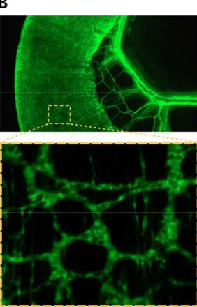
Figure 1. Structure of the enteric nervous system.
A) Simplified cross-section of the gut showing the relative positions of the different muscle, neural and mucosal layers. Adapted from Heanue and Pachnis, 2007 (19).
B) Side view of a SRYp[1.4kb]-YFP (65) transgenic mature mouse intestine displaying intrinsic as well as extrinsic (along mesenteric vessels) in vivo innervations. The magnified section shows the fluorescently-labeled neurons of the muscle layers and reveals the typical lattice-like organization of the myenteric plexus.
Figure 2. Neural crest cell migration.
A) Wnt1-Cre::R26R-YFP (66) double transgenic mouse embryo at embryonic day (e) 9.5. The dashed line indicates the level at which the transverse section shown in the inset was made. Fluorescently labeled NCC originate from the dorsal neural tube (NT in the inset) and migrate extensively through the embryo to colonize various structures (white arrows in the inset). Note that the ENS is entirely derived from NCC of the vagal and sacral levels (red and blue arrows).
B) Vagal (red arrows) and sacral (blue arrows) NCC initially reach gut tissue from the neural tube via a dorsal to ventral migratory route. Colonization is then completed by migration within the embryonic gut mesenchyme. Note that, during a relatively short window of time, a subset of enteric NCC of vagal origin also takes a shortcut from the midgut to the hindgut via the mesentery. Adapted from Farlie et al., 2004 (67). C) Gata4p[5kb]-GFP (66) transgenic mouse colons at different stages of development showing the migration
of enteric NCC to form the colonic ENS between e11.5 and e15.5. The distal-most part of the colon is outlined in red to facilitate visualization of the whole colon tissue. White arrows indicate the directional migration of enteric NCC of vagal origin. Note that colonization of the hindgut by sacral NCC is preceded by an accumulation of these NCC bilateral to the caudal hindgut at e13.5 (middle panel).