Up-regulation of Vascular Endothelial Growth Factor-A by Active
Membrane-type 1 Matrix Metalloproteinase through Activation of
Src-Tyrosine Kinases
iNor Eddine Sounni‡, Christian Roghi§, Vincent Chabottaux‡ii, Mathias Janssen‡, Carine Munaut‡iii, Erik Maquoi‡, Beatriz G. Galvez**, Christine Gilles‡, Francis Frankenne‡, Gillian Murphy§, Jean-Michel Foidart‡, and Agnes Noel‡
‡Laboratory of Tumor and Development Biology University of Liège, Sart Tilman, 4000 Sart-Tilman, B-4000 Liège, Belgium
§Department of Oncology, Cambridge Institute for Medical Research, University of Cambridge, CB2 2XY Hills Road Cambridge, United Kingdom
**Departamento de Inmunologìa, Hospital de la Princesa, 28006 Madrid, Spain
Abstract
Membrane-type 1 matrix metalloproteinase (MT1-MMP) and vascular endothelial growth factor (VEGF) are two key molecules involved in pericellular proteolysis and cell proliferation during tumor growth and an-giogenesis. Our previous data showed that MT1-MMP overexpression in human breast carcinoma MCF7 cells induced an up-regulation of VEGF expression. This effect was associated in vivo with accelerated tumor growth and angiogenesis. We now provide evidence that MT1-MMP overexpression specifically affected VEGF-A production and failed to influence that of other VEGF family members (VEGF, B, C, D, or P1GF) or their receptors. The up-regulation of VEGF-A by MT1-MMP was related to an increased transcriptional activation rather than to a modification of mRNA stability. It was blocked by synthetic MMP inhibitors, TIMP2, but not TIMP-1 and abolished by a partial deletion of the catalytic domain or the cytoplasmic tail of MT1-MMP. Analysis of the signal transduction mechanisms demonstrated that MT1-MMP acts through a signaling pathway involving Src tyrosine kinases. Thus, our results provide new insight into the mechanisms of action of MT1-MMP during angiogenesis and suggest that the full enzymatic activity of MT1-MT1-MMP is required for a specific up-regulation of VEGF-A through an activation of Src tyrosine kinase pathways.
The abbreviations used are: VEGF, vascular endothelial growth factor; P1GF, placental-like growth factor; NRP-1, neuropilin-1; MT-MMP, membrane-type matrix metalloproteinase; TIMP-1, tissue inhibitor of
metalloproteinase; rhTIMP, human recombinant TIMP; PP2, 4-amino-5-(4-chlorophenyl)-7-(t-butyl)pyrazol[3,4-d]pyrimidine; EGFP, enhanced green fluorescent protein; DMEM, Dulbecco's modified Eagle's medium; RT, reverse transcriptase; MAPK, mitogen-activated protein kinase; ERK, extracellular signal-regulated kinase.
Angiogenesis, the formation of new blood vessels from preexisting ones, is a key event in tumor progression controlled by a balance between positive and negative regulators (1, 2). From this observation has emerged the concept of the "angiogenic switch" in which endothelial activation status is determined by the induction of angiogenic factors and/or the loss of inhibitors (3). Positive regulators include at least vascular endothelial growth factor family (VEGF-A, -B, -C, -D),1 fibroblast growth factors, placental-like growth factor (PlGF), angiopoietins, their tyrosine kinase receptors (VEGF-R1, -R2; Tie1 and Tie2) and neuropilin-1 (NRP-1), a co-receptor for VEGF (1, 4). An increasing number of negative regulators have been identified such as inhibitors of proteinases, thrombospondins, interferons, chemokines (IP-10 and PF-4), and bioactive fragments of the extracellular matrix (2, 4). Once a tumor has acquired an angiogenic phenotype, tumor cell migration, invasion, and vessel sprouting require the dynamic and coordinated action of many cell surface molecules, including proteinases and inte-grins mediating cell-matrix interactions.
Among the cell-associated proteinases, matrix metallopro-teinases (MMPs) anchored to the plasma membrane, called membrane-type MMPs (MT-MMPs) play a pivotal role in pericellular proteolysis. Of the six MT-MMPs that have been identified to date, four (MT1-, MT2-, MT3-, and MT5-MMP) contain a transmembrane domain
i This work was supported in part by grants from the Communauté française de Belgique (Actions de Recherches Concertées), the
Commission of European Communities (QLK3-CT02- 02136 and FP6), the Fonds National de la Recherche Scientifique (FNRS, Belgium), the Federation Beige Contre le Cancer, the Fonds spéciaux de la Recherche (University of Liège), the Centre Anticancéreux près l'Université de Liège, the FB Assurances, the Fondation Leon Frédéricq (University of Liège), the Direction Generale des Technologies, de la Recherche et de l'Energie from the Région Wallonne, and the Interuniversity Attraction Poles Programme-Belgian Science Policy from (Brussels, Belgium). The costs of publication of this article were defrayed in part by the payment of page charges. This article must therefore be hereby marked "advertisement" in accordance with 18 U.S.C. Section 1734 solely to indicate this fact.
ii Recipient of a grant from FNRS-Télévie iii Research Associates from the FNRS.
followed by a cytoplasmic tail and two (MT4- and MT6-MMP) are tethered to the cell surface via a
glycosylphosphatidylinositol link (for review, see Refs. 5-8). It now appears that all MT-MMPs may have the ability to activate pro-MMP2 (9-13). Although, each of the four physiological tissue inhibitors of MMPs (TIMPs) can non selectively bind to all active MMPs, TIMP1 is unable to inhibit effectively MT1, 2, 3, and 5-MMPs (14). TIMP-2 has dual functions, inhibiting the activity of each MT-MMP and participating in the activation of MMP2 through the formation of a ternary complex formed of MT1-MMP, TIMP-2, and pro-MMP2 (for review, see Refs. 7, 15, and 16). pro-MMP2 activation by MT1-MMP could be promoted by interaction with integrin (17) and oligomerization of MT1-MMP (18, 19).
MT1-MMP exerts pleiotropic effects on cell behavior by cleaving matrix components (20), as well as by controlling expression and functional status of cell surface receptors, including integrins (21), transglutaminase (22), and CD44 (23). Numerous studies have demonstrated that it is up-regulated in aggressive tumors of diverse cell/tissue origin (7, 16). MT1-MMP has been implicated in the neovascularization process, because it can act as a potent collagenase and fibrinolysin promoting tubulogenesis in collagen or fibrin gels (24-26). The pro-angiogenic activity of MT1-MMP is further supported by the effects of MT1-MMP overexpression on tumorigenicity, growth, and vascularization of human melanoma (27), breast adenocarcinoma (28), or glioma (29) cells transfected with MT1-MMP cDNA. We have previously provided evidence that this MT1-MMP pro-angiogenic effect was mediated at least in part by an up-regulation of VEGF at both mRNA and protein levels (28). Such a link between the expression of VEGF and MT1-MMP was further confirmed in authentic tumors, by im-munohistochemical and RT-PCR analysis of human glioma tissue samples (30). However, the molecular mechanisms underlying this transcriptional regulation of an angiogenic factor by a membrane-type MMP is currently unknown. The objectives of this study were to determine whether MT1-MMP regulates the expression of other angiogenic factors (VEGF-B, -C,-D, PlGF, and ANG-1 and -2) and their receptors (VEGF-Rl,-R2, NRP-1, and Tie-1 and -2), as well as to provide mechanistic insights into VEGF regulation by MT1-MMP.
TABLE I: Sequence of primers used for RT-PCR studies
Forward primer (5' → 3') Reverse primer (5' → 3') Expected size bp No. of
cycles sRNA mRNA
MT1-MMP 5'-GGATACCCAATGCCCATTGGCCA-3' 5'-CCATTGGGCATCCAGAAGAGAGC-3' 269 221 25
TIMP-2 5'-CTCGCTGGACGTTGGAGGAAAGAA-3' 5'-AGCCCATCTGGTACCTGTGGTTCA-3' 269 155 25
VEGF-A 5'-CCTGGTGGACATCTTCCAGGAGTA-3' 5'-CTCACCGCCTCGGCTTGTCACA-3' 311 275, 407,
and 479 29
VEGF-R1 5'-TCCCTTATGATGCCAGCAAGT-3' 5'-CCAAAAGCCCCTCTTCCAA-3' 78 35
VEGF-R2 5'-CTTCGAAGCATCAGCATAAGAAACT-3' 5'-TGGTCATCAGCCCACTGGAT-3' 155 35
VEGF-B 5'-CAGAGGAAAGTGGTGTCATGGA-3' 5'-ACCGGATCATGAGGATCTGCA-3' 223 35
VEGF-C 5'-CTCTCAAGGCCCCAAACCA-3' 5'-AGGTCTTGTTCGCTGCCTGA-3' 151 35
VEGF-D 5'-TCTCGCTCAGCATCCCATC-3' 5'-CACCTCCACGCACGTTTCT-3' 152 33
P1GF 5'-CCTACGTGGAGCTGACGTTCT-3' 5'-TCCTTTCCGGCTTCATCTTCT-3' 132 35
NRP-1 5'-CACAGTGGAACAGGTGATGACTTC-3' 5'-AACCATATGTTGGAAACTCTGATTGT-3' 111 29
Tie-2 5'-CAAAACGTGAGGGAAGAACCA-3' 5'-AAATTGCCCTCCCCAATCA-3' 148 35
Ang-1 5'-GGAACCGAGCCTATTCACAGTA-3' 5'-CAAGCATCAAACCACCATCCT-3' 211 35
FIG.1.Influence of MT1-MMP over-expression on the expression of putative angiogenic factors and their
receptors.
MT1-MMP-overexpressing clones (MT clones: MT/1 and MT/3) and control clones (C10 and C11) were used to profile by RT-PCR, the mRNA expression of VEGFs (A, B, C, D), their receptors (VEGF-R1 and VEGF-R2), PlGF, neuropilin-1 (NRP-1), angiopoi-etin-1 (Ang-1), Tie2, and TIMP-2. In some assays, RNAs isolated from glioblastoma (G) or placenta (P) have been added as positive controls. In some cases, for quantification purpose, synthetic RNAs (CTR1, 2, or 5) were added in each sample to control the efficiency of RT-PCR for 28S (CTR1) and MT1-MMP (CTR1), TIMP-2 (CTR2) and VEGF-A (CTR5) as described under "Experimental Procedures." 28 S ribosomal RNA is shown as a loading control.
FIG.2.VEGF mRNA stability in MT clones and control cells.
MT1-MMP overexpressing clones (MT clones: MT/1 and MT/3) or control clones (C10 and C11) were incubated in serum-free medium with actinomycin D (10 µg/ml) for 0, 1, 2, 3, 4, 8, and 12 h. After RNA extraction, VEGF and MT1-MMP mRNA levels were analyzed by RT-PCR using specific primer and internal synthetic control RNAs: CTR5 for VEGF, CTR1 for MT1-MMP, and 28 S as described under "Experimental Procedures." The experiment presented is representative of three independent assays. 28 S ribosomal RNA is shown as a loading control.
EXPERIMENTAL PROCEDURES Reagents and Chemicals
The synthetic MMP inhibitors hydroxamic acid-based (BB94) (31) or the pyrimidine-2,4,6-trione-type inhibitor (RO 28-2653) (32) were prepared as 10 mM stock solutions in Me2SO and used at final concentration of 10 µM in 0.1% Me2SO. Human recombinant TIMP-1 (rhTIMP-1) and TIMP-2 (rhTIMP-2) were obtained as described previously (33). Actinomycin D, genistein, and herbimycin A were from Sigma (St. Louis, MO), PD98059 and SB 203580 were from Alexis Biochemicals (Lausanne, Suisse), wortmannin and 4-amino-5-(4-chlorophenyl)-7-(t-butyl)pyrazol[3,4-d]pyrimidine (PP2) were from Cal-biochem (La Jolla, CA). Aprotinin, leupeptin, and pepstatin A were obtained from Sigma, and Pefabloc SC (4-(2-aminoethyl)benzenesulfo-nyl fluoride hydrochloride) was form Roche Diagnostics. Neutralizing anti-human MT1-MMP monoclonal antibody LEM2/15.8 was generous gift from Dr. Alicia Arroyo (Madrid, Spain).
cDNA Constructions
Full-length wild type human MT1-MMP cDNA (huMT1-MMP) without both 5'- and 3'-untranslated regions was subcloned between the HindIII and EcoRI sites of pCDNA3.1 Zeo + mammalian expression vector (Invitrogen Ltd., Paisley, UK). Point mutations (E240A, T567A, C574S, and S577A) were generated by site-directed
mutagenesis using Pfu DNA polymerase (Stratagene, Amsterdam, The Netherlands). Cytotail truncated MT1-MMP construct (∆577) was prepared from full-length MT1-MT1-MMP by inserting a stop codon after Arg-576 by site-directed mutagenesis.
Oligonucleotides coding for huMT1-MMP signal sequence (amino acids 1-30) were annealed, and the resulting product was cloned upstream of EGFP in the NheI-AgeI sites of the pEGFP-C1 vector to generate the
huMT1SSC1 recombinant vector. In the resulting construct the huMT1-MMP signal sequence is linked by the short linker PVAT to EGFP. Residues 283-582 of huMTl-MMP containing the hinge, hemopexin,
transmembrane, and cytoplasmic tail were amplified by PCR using the primers 5'- AAAAAAAGATCTTATGGGGGTGAGT-CAGGGTTCCCCACC-3' (called CHR087) and 5'-AAAAGAATTCTCA-GACCTTGTCCAGCAGGGA-3' (called CHR057). The resulting PCR product was cloned into the BgIΠ-EcoRI sites of huMTlSSCl in-frame with EGFP. All constructs were verified by sequencing using Big Dye Terminator kit (Applied Biosystems, Warrington, UK).
Cell Culture and Treatments
MCF7 clones stably expressing MT1-MMP or control clones were generated by transfection of adenocarcinoma MCF7 with full-length MT1-MMP cDNA or empty vector as previously described (28). Two clones
overexpressing MT1-MMP (MT clones: MT/1 and MT/3) and two control clones (C10 and C11) were routinely grown in Dulbecco's modified Eagle's medium (DMEM) supplemented with 10% (v/v) fetal calf serum
(Invitrogen, Merelbeke, Belgium), penicillin (100 IU/ml), streptomycin (100 µg/ml), 2 mM glutamine, 10 mM Hepes buffer, and G418 (400 µg/ml). For treatments with MMP inhibitors or other reagents, cells were seeded in 12-well plates (Falcon) to a density of 2 x 10⁵ cells/well and incubated in serum-containing medium for 24 h. Cells were then washed twice with phosphate-buffered saline and subsequently supplemented with 2 ml of serum-free DMEM (untreated control), BB94, or RO 28-2653 (final concentration 1 µM); rhTIMP-1 (10 µg/ml) or rhTIMP-2 (0.1-10 µg/ml); leupeptin (1 µM), aprotinin (0.3 µM), pepstatin A (1 µM), or Pefabloc SC (1 µM); blocking anti-MT1-MMP LEM2 (10 µg/ml); genistein (5-50 µM), herbimycin A (0.03-1 µM), PD98059 (50 µM), SB 203580 (20 µM), wortmannin (250 n M),or PP2 (1 µM). After 24 h of incubation, the resulting cell
monolayers were collected for RNA or proteins extraction. To block transcription, actinomycin D (10 µg/ml) was added after a 12-h incubation in serum-free medium. Cells were harvested before (0 h) or after several time points (1, 2, 3, 4, 8, or 12 h) following the addition of actinomycin D, and RT-PCR analysis was performed for VEGF mRNA and MT1-MMP mRNA expression.
Cell Transfection with Wild Type or Mutated MT1-MMP cDNA
Stable transfection of MCF7 cells with the different constructs were performed using the non-liposomal formulation FuGENE-6 transfection reagent (Roche Applied Science, Indianapolis, IN), according to the manufacturer's instructions. Two different vectors containing wild type MT1-MMP cDNA were used: pc3MT1800s with neomycin (28) or huMT1-MMP with zeocin (see above) as selected markers, respectively. Transfectants were selected with Zeocin (100 µg/ml) (Invitrogen) or G418 (400 µg/ml) according to the vector used.
Reverse Transcriptase-PCR Analysis
After 24 h of treatment in serum-free medium, total RNA was extracted from cells by using the High Pure RNA Isolation Kit (Roche Applied Science). RT-PCR amplification was performed using the GeneAmp Thermostable rTth reverse transcriptase RNA PCR kit (Applied Biosystems, Branchburg, NJ), specific pairs of primers (5 pmol each) (see Table I), 10 ng of total RNA and, when available, a known copy number of an internal
standard (multiplex synthetic standard RNA, see below) per 25 µl of reaction mixture (final volume). Reverse transcription was performed at 70 °C for 15 min. For 28 S, MT1-MMP, and TIMP-2 transcripts, PCR
amplification was run as follows: 15 s at 94 °C, 20 s at 68 °C, and 10 s at 72 °C (number of cycles as indicated in Table I), followed by a final 2-min extension step at 72 °C. Other cycling conditions are the followings: 20 s at 94 °C, 20 s at 66 °C, and 30 s at 72 °C for VEGF-A; 20 s at 94 °C, 20 s at 60 °C, and 30 s at 72 °C for VEGF-B; 15 s at 94 °C, 20 s at 55 °C, and 15 s at 72 °C for VEGF-C and VEGF-D. For VEGF-R1, VEGF-R2, and neuropilin-1: 15 s at 94 °C, 30 s at 60 °C, and 30 s at 72 °C. For P1GF, Tie-2, and ang-1: 15 s at 94 °C, 30 s at 60 °C, and 15 s at 72 °C. RT-PCR products were resolved on 10% polyacrylamide gels and analyzed using a Fluor-S Multilmager (Bio-Rad, Hercules, CA) after staining with Gelstar dye (FMC BioProducts, Rockland, ME). The selected primer pairs for each human mRNA are described in Table I; their selectivity and efficiency were tested by RT-PCR using 10 ng of total RNA extracted from human placenta or human dermal fibroblasts or 50 ng of human DNA as negative control. For RT-PCR quantification of VEGF-B, -C, -D, P1GF, VEGFR1,
VEGFR2, neuropilin, angiopoietin-1, and Tie2, their expression was normalized to 28 S rRNA. For a more precise quantification of MT1-MMP, TIMP-2, VEGF mRNA, and 28 S rRNA, synthetic standard RNAs were generated as previously described (28, 34, 35). These synthetic RNAs have two main characteristics. First, they can be amplified by primers used for RT-PCR amplification of the endogenous mRNA of interest. Second, the size of their amplification product is different from the size of the RT-PCR product amplified from the mRNA, permitting their discrimination after electrophoresis. The ratio between the signals for a specific mRNA and the standard RNA was shown to increase linearly with the amount of cellular RNA.
FIG.3. Effect of MMP inhibitors on VEGF-A production.
MT1-MMP-overexpressing clones (MT clones: MT/1 and MT/3) or control clones (C10 and C11) were incubated for 24 h in serum-free medium supplemented with synthetic MMP inhibitors: BB94 or RO 28-2653 (1 µM). A, total RNA of MT clones and control clones were extracted and analyzed by quantitative RT-PCR using standard RNA (CTR1 and CTR5). The histograms on the right correspond to the scanning quantification of the three different VEGF-A isoforms considered together ("VEGF"). Results are expressed as the ratio of (VEGF-A mRN(VEGF-As/synthetic VEGF mRN(VEGF-A (CTR5)) divided by the ratio of (28 S mRN(VEGF-A/synthetic 28 S mRN(VEGF-A (CTR1)). They correspond to the mean ± S.D. of quantifications performed on three independent assays. B, cell lysates were analyzed by immunoblotting using an anti-VEGF polyclonal antibody. The recombinant human VEGF used as positive control corresponds to a species of 30 kDa.
FIG.4.Effect of protease inhibitors, recombinant TIMP-1, TIMP-2, and blocking anti-MT1-MMP on the
VEGF-A expression.
A clone overex-pressing MT1-MMP (MT/1) and a control clone (C10) were incubated for 24 h in serum-free medium supplemented with leupeptin (1 µM), aprotinin (0.3 µM), pep-statin A (1 µM), or Pefabloc SC (1 µM) (A), recombinant TIMP-1 (10 µg/ml), or different concentrations of recombinant TIMP-2 (0.1, 1, and 10 µg/ml) (B), or with blocking anti-MTl-MMP LEM2 (10 µg/ ml) (C). The histograms correspond to the quantification (mean ± S.D.) of RT-PCR analysis of VEGF-A performed on two assays (see legend of Fig. 3).
FIG.5.Analysis of signaling pathways involved in VEGF up-regulation by MT1-MMP.
MT1-MMP-overexpressing clones (MT clones: MT/1 and MT/3) and control clones (C10 and C11) were treated with various
pharmacological inhibitors specific to different signaling pathways. A, one MT clone (MT/3) and one control clone (C10) were treated with PD98059 (50 µM), wortmannin (250 µM),or herbimycin A. B, one MT clone (MT/1) and one control clone (C12) were treated with PD98059 (50 µM), herbimycin A (250 nil), or SB203580 (20 µM). C, two MT clones and two control clones were treated with genistein (10 µM). After 24 h of treatment without serum, the expression of VEGF mRNA was measured by RT-PCR. The experiments presented are representative of two different assays performed in triplicate. 28 S ribosomal RNA is shown as a loading control.
Western Blotting Analysis
Total cell extracts were prepared by incubating cells in radioimmune precipitation assay buffer (50 ml Tris-HC1, pH 7.4; 150 ml NaCl; 1% Nonidet P40; 1% Triton X-100; 1% sodium deoxycholate; 0.1% SDS; 5 mM
iodoacetamide; 2 mM phenyl-methylsulfonyl fluoride). After centrifugation at 15,000 rpm for 30 min, at 4 °C, the supernatant was stored frozen at -20 °C. Protein concentration was determined by using the DC protein Assay Kit (Bio-Rad Laboratories, Hercules, CA) and adjusted to 2 mg/ml. Samples of total cell extracts were resolved by SDS-PAGE under reducing conditions, and proteins were transferred to nitrocellulose membranes (Hybond-ECL membrane, Amersham Biosciences, Arlington Heights, IL). The membranes were exposed to primary mouse monoclonal antibody 2D-7 raised against the hemopexin-like domain of MT1-MMP (kindly provided by M.-C. Rio, Instit de Genetique et de Biologie Moleculaire et Cellulaire, Illkirch, France) (28) or rabbit polyclonal antibody against human VEGF165, -189, and -121 amino acid splice variants at dilution of 1/500 ((A-20):sc-152 Sanver Tech). After extensive washings, the membranes were incubated with a secondary horseradish peroxidase-conjugated goat anti-mouse (for MT1-MMP) or mouse anti-rabbit antibody (for VEGF) at 1/1000 (DAKO, Glostrup, Denmark). Signals were detected using an enhanced chemiluminescence (ECL) kit (PerkinElmer Life Sciences, Boston), according to the manufacturer's instructions.
Gelatin Zymography
Conditioned media were prepared by incubating subconfluent cells (2 x 10⁵) in 24 well-plates (Falcon, BD Biosciences) for 24 h, in serum-free DMEM, in the presence of an exogenous source of MMP-2 (medium conditioned by Chinese hamster ovary cells transfected with full-length MMP-2 cDNA) (36). Conditioned media were harvested, clarified by centrifugation, and stored frozen at -20 °C. Aliquots (5-10 µl) of conditioned media standardized for cell DNA content were mixed with equal volume of non-reducing sample buffer (62.5 mM Tris-HCl, pH 6.8; 2% SDS; 10% glycerol; 0.1% brom-phenol blue) and were resolved on 10% gels SDS-PAGE containing 0.1% gelatin (w/v) according to the procedure previously described (27).
Densitometric Analysis
Gels were analyzed with Quantity One software (version 4.2.2., Bio-Rad Laboratories, Hercules, CA) after densitometry scanning of the gels using a Fluor-S Multilmager (Bio-Rad).
RESULTS
MT1-MMP Overexpression Is Associated with a Specific Up-regulation of VEGF-A
We first examined the possibility that several angiogenic factors and their receptors could be a target of the up-regulatory effect of MT1-MMP. We have used human breast adenocarcinoma MCF7 clones stably
transfected with MT1-MMP cDNA (MT/1 and MT/3 clones, designed "MT clones") and control MCF7 cells stably transfected with control vector (C10 and C11 clones called "control clones") (28). We examined by RT-PCR analysis the expression of different forms of VEGF A, -B, -C, -D), P1GF, their receptors (VEGF-R1, VEGF-R2), neuropilin-1 (NRP-1), angiopoietin-1, its receptor Tie2 and TIMP-2. In accordance with our previous data (28), the mRNA levels of three isoforms of VEGF-A (VEGF121, VEGF165 and VEGF189) were 2- to 3-fold increased in MT clones as compared with control clones (Fig. 1). In sharp contrast, no modulation of the expression of VEGF-C, VEGF-D, VEGF-R2, NRP-1, Ang-1, Tie2, or TIMP-2 was induced by MT1-MMP expression. In our conditions, P1GF, VEGF-B, and VEGF-R1 were never detected in the clones analyzed. These findings emphasize the specific effect of MT1-MMP on the mRNA expression of the three VEGF-A isoforms (VEGF121, VEGF165, and VEGF189) without affecting the expression of other angiogenic factors.
MT1-MMP Overexpression Does Not Affect VEGF mRNA Stability
To determine whether MT1-MMP overexpression affects VEGF mRNA stability, we evaluated the effect of actinomycin D, an inhibitor of RNA transcription, on the evolution of VEGF mRNA levels in the different clones expressing or not MT1-MMP. In the absence of actinomycin D, the VEGF mRNA expression was increased 3-fold in MT clones as compared with control clones (Fig. 2). In the presence of actinomycin D, VEGF mRNA levels decreased progressively with time and in a similar manner in both MT clones. Almost no VEGF mRNA remained after 3 h of actinomycin D treatment, whereas MT1-MMP mRNAs persisted until 8 h and was not anymore detected after 12 h (Fig. 2). These results demonstrate that the VEGF up-regulation by MT1-MMP could not result from an enhancement of mRNA stability but to a transcriptional activation.
VEGF Up-regulation by MT1-MMP Is Blocked by MMP Inhibitors
To determine whether MT1-MMP activity is required for VEGF regulation, the effect of synthetic (BB94 and RO 28-2653) or recombinant physiological inhibitors (rTIMP-1 and rTIMP-2) on VEGF expression was examined in MT and control clones. Although the treatment of cells with BB-94 or RO 28-2653 (1 µM) inhibitors reduced VEGF mRNA levels in MT clones, it did not affect the basal VEGF mRNA levels in control clones (Fig. 3A). Quantitative RT-PCR analysis using synthetic standard mRNAs revealed that treatment of MT clones with both inhibitors down-regulated VEGF mRNA production to the levels found in control clones (Fig. 3A). The inhibition of VEGF production in cells treated with BB94 was confirmed by measuring VEGF at a protein level by Western blot analysis (Fig. 3B). To assess the effect of physiological MMP inhibitors on VEGF regulation, cells were incubated for 24 h with rhTIMP-1 (10 µg/ml), which is unable to inhibit MT1-MMP activity or with increasing concentrations of rhTIMP-2 (0.1, 1, and 10 µg/ml), which binds MT1-MT1-MMP (Fig. 4B). TIMP-2 decreased significantly the level of VEGF mRNAs in MT clones when used at 10 µg/ml but not at lower concentrations (0.1 and 1 µg/ml). In sharp contrast, TIMP-1 did not affect VEGF expression. These findings suggest the importance of the enzymatic activity of MT1-MMP in the transcriptional regulation of VEGF. The specificity of MT1-MMP as compared with other proteases, was assessed by using different protease inhibitors. Inhibitors of serine proteases (aprotinin and Pefabloc SC), cystein and serine proteases (leupeptin), or cathepsin and aspartic proteases (pepstatin A) failed to modulate the VEGF expression (Fig. 4A). In sharp contrast, specific anti-MT1-MMP blocking antibody (LEM2) reduced VEGF mRNA levels to that observed in control clones (Fig. 4C).
VEGF Up-regulation by MT1 -MMP Involves Nonreceptor Src Tyrosine Kinases
To provide further insights into the mechanisms underlying the up-regulating effect of MT1-MMP on VEGF, cells were cultured in the presence of several inhibitors of different signaling pathways, and the levels of VEGF were examined by RT-PCR analysis. As illustrated in Fig. 5 (A and B), neither the phosphatidylinositol 3-kinase pathway inhibitor (wortmannin), the MAPK/ERK kinase pathway inhibitor (PD98059), nor the p38 MAPK pathway inhibitor (SB203580) affected MT1-MMP-mediated VEGF induction. In contrast, herbimycin-A, an inhibitor of tyrosine kinase, strongly inhibited the up-regulating effect of MT1-MMP. Similarly to herbimycin-A, genistein, another tyrosine kinase inhibitor markedly suppressed the VEGF mRNA induction in MT clones without affecting the VEGF expression in control clones (Fig. 5C).
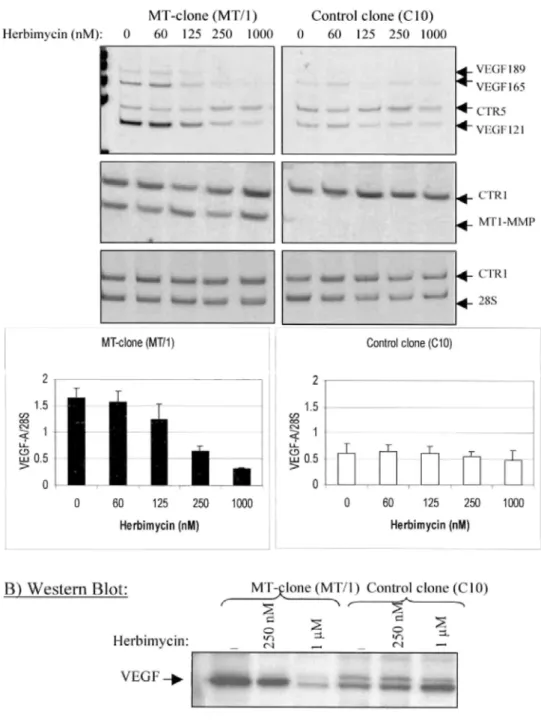
The VEGF mRNA expression in MT clones was not significantly affected by herbimycin-A used at doses ranging from 60 to 125 nM, but was reduced to a basal level by herbimycin-A at 250 nM (Fig. 6A). Incubation of MT clones with increasing concentration of genistein (5-50 µM) decreased VEGF expression in a
dose-dependent manner (Fig. 7A). Interestingly, MT1-MMP expression levels were not affected by the different concentrations of herbimycin or genistein used (Figs. 6A and 7A). In addition, VEGF expression in control clones was never modulated by herbimycin-A or genistein at the different concentrations used (Figs. 6A and 7A). In accordance to RT-PCR data, the inhibition of VEGF up-regulation in MT clones by herbimycin-A and genistein was confirmed at protein level by Western blotting (Figs. 6B and 7B).
To further investigate the tyrosine kinase pathway, we tested a specific inhibitor of Src protein, 4-amino-5-(4-chloro-phenyl)-7-(t-butyl)pyrazol[3,4-d]pyrimidine (PP2, 1 µM) (37). Similarly to herbimycin and genistein, PP2 abolished the VEGF up-regulation without affecting MT1-MMP expression (Fig. 8). These results indicated that VEGF up-regulation in MT clones was depend on nonreceptor Src tyrosine kinases.
FIG. 6. Effect of herbimycin A on VEGF up-regulation by MT1-MMP.
One MT1-MMP-overexpressing clone (MT clone: MT/1) and one control clone (C10) were incubated with increasing concentrations of herbimycin A (0-1000 nM). A, VEGF and MT1-MMP mRNA levels were analyzed by RT-PCR. The histograms correspond to the
quantification of VEGF mRNA in MT1-MMP clones treated or not with herbimycin A (mean ± S.D. of three different experiments) (see legend of Fig. 3). B, Western blot analysis of VEGF production in MT clones treated or not with 250 nM or 1 µM of herbimycin A, using an anti-VEGF polyclonal antibody. Similar results were obtained with the other MT clone (MT/3) and control clone (C11) (data not shown).
FIG.7. Effect of genistein on VEGF up-regulation by MT1-MMP.
One MT1-MMP-overexpressing clone (MT clone: MT/1) and one control clone (C10) were incubated with increasing concentrations of genistein (0-50 µM). A, VEGF and MT1-MMP mRNA levels were analyzed by RT-PCR (see legend of Fig. 3). The histograms correspond to the quantification of VEGF mRNA (mean ± S.D. of three independent assays) (see legend of Fig. 3). B, Western blot analysis of VEGF production in MT clones treated or not with 5 µM or 10 µM of genistein, using an anti-VEGF polyclonal antibody. Similar results were obtained with the other MT clone (MT/3) and control clone (C11) (data not shown).
FIG.8. Effects of tyrosine kinases and Src-type tyrosine kinases on VEGF and MT1-MMP expression.
Representative inhibition of VEGF-A up-regulation in one MT1-MMP overexpressing clone (MT clone: MT/1) and one control clone (C10) incubated with herbimy-cin (1 µM), genistein (10 µM) or PP2 (1 µM). MT1-MMP expression of the MT clone was not affected by cell treatment. The mRNA levels of VEGF, MT1-MMP, and 28 S were analyzed by RT-PCR as described under "Experimental Procedures."
VEGF Up-regulation Requires MT1-MMP Catalytic and Cytoplasmic Domain
Because catalytic and cytoplasmic domains of MT1-MMP have been shown to be important for pro-MMP2 activation, we investigated their respective requirement for VEGF regulation. Therefore, we generated different MT1-MMP constructs encoding proteins with deletions in the catalytic domain (∆cd) or the cytoplasmic domain (∆577) (Fig. 9A). In addition, we designed four MT1-MMP mutants with a substitution in the active site (E240A) or in the cytoplasmic domain (S577A, C574S, or T567A) (Fig. 9A). These constructs and wild type MT1-MMP cDNA were transfected in MCF7 cells and stable cell populations were selected by antibiotic treatment. MT1-MMP production was assessed both at protein and mRNA levels by Western blotting (data not shown) and RT-PCR analysis (Fig. 9C). The capacity of WT and mutated forms of MT1-MMP expressed by these cells to process pro-MMP2 was evaluated by zymographic analysis performed on medium conditioned by transfected cells incubated with exogenous pro-MMP2 (Fig. 9B). As expected, MT1-MMP forms with the catalytic domain deleted (∆cd) as well as inactive MT1-MMP (MT1-E240A), were both unable to activate exogenous pro-MMP2. Pro-MMP2-activating capacity was not affected by deletion or two point mutations (S577A or T567A) in the cytoplasmic tail. However, mutation at position 574 (MT1-C574S) led to a reduction of pro-MMP2 activation (Fig. 9C). RT-PCR analysis indicated a 2- to 3-fold increased levels of VEGF mRNA in cells transfected with WT MT1-MMP cDNA as compared with cells transfected with control vector (Fig. 9D). Transfection with two mutated forms (S577A or T567A) led to a similar VEGF up-regulation. In sharp contrast, no up-regulation of VEGF expression was detected in cells expressing deleted forms of MT1-MMP (MT1-∆cd and MT1-∆577), or MT1-MMP mutated at position 574 (MT1-C574S). Similarly, cell transfection with enzymatically inactive MT1-MMP cDNA (E240A) failed to induce VEGF mRNA expression.
DISCUSSION
In addition to its direct role on pro-MMP activation and extracellular matrix degradation, emerging functions of MT1-MMP include at least the activation and/or shedding of cell surface molecules (21-23). We and others provided evidence that MT1-MMP and VEGF can be functionally linked in tumoral angiogenesis (7). In support of this functional link is the fact that hypoxia-induced up-regulation of MT1-MMP in murine bone marrow-derived stromal cells correlates with a stimulation of VEGF (38). However, the mechanisms underlying VEGF production in response to MT1-MMP overexpression remain to be elucidated. In this work, we demonstrate a specific effect of MT1-MMP on VEGF-A expression without affecting other members of VEGF family or their receptors. This MT1-MMP effect is dependent on the presence of functional catalytic and cytoplasmic domains of the enzyme and involves the activity of Src tyrosine kinases.
By RT-PCR analysis, we demonstrate that MT1-MMP over-expression specifically up-regulated VEGF-A, whereas the mRNA levels of other VEGF family members (VEGF-C and -D) and their receptors (VEGF-R2, NRP-1, Ang-1, and Tie-2) were not affected. The mRNA for VEGF-B, P1GF, and VEGF-R1 were not expressed either in MCF7 parental cells or in MT clones. This specific correlation between MT1-MMP and VEGF-A is in agreement with in vivo observation in glioma tissue samples (30, 39). In the present study, we provide evidence that this specific regulation of VEGF-A production by MT1-MMP occurred at a transcriptional level without a concomitant increase in VEGF mRNA stability. The specific effect of MT1-MMP was assessed by using a blocking MT1-MMP antibody.
The localization of MT1-MMP at the surface of some cells in clathrin-coated pits (40) and caveolae (41-43) raised the possibility that MT1-MMP activates intracellular signaling events (8). By using a series of specific inhibitors for different signaling pathways putatively involved in VEGF regulation, we found that the activation of MAPK/ERK kinase, p38 MAPK, or phosphatidylinositol 3-kinase pathways is not required for VEGF up-regulation in our experimental conditions. Interestingly, herbimycin A or genistein, both selective inhibitors of protein tyrosine kinases, suppressed VEGF up-regulation in MT clones, without affecting VEGF mRNA levels in control clones. In addition, PP2, a specific inhibitor of Src tyrosine kinases, also strongly abolished the VEGF up-regulation in MT clones. The possibility of a nonspecific toxic effect of the inhibitors was excluded, because mRNA levels for MT1-MMP were not altered in the presence of inhibitors. The finding that an Src tyrosineinase inhibitor is effective in blocking VEGF transcription agrees with reports indicating that these nonreceptor kinases participate in VEGF induction by hypoxia (44, 45), adrenergic stimulation (37), or prostaglandin PGE2 (46). Our data indicate that the pathway through which MT1-MMP regulates VEGF is distinct to that implicated in the induction of cell migration which involves extracellular signal-regulated protein kinase (ERK) (47). In addition, MT1-MMP has been shown to control integrin maturation by processing the αv integrin subunit, leading to tyrosine phosphorylation of focal adhesion kinase and thereby to cell migration (48). The involvement of such an αv convertase activity by MT1-MMP and an indirect activation of focal adhesion kinase pathway is unlikely in our model, because MCF7 cells failed to produce αvβ3 (7). Altogether these data indicate that MT1-MMP can contribute to the activation of different intracellular pathways leading to distinct biological responses.
Deletion of the six C-terminal amino acids of the cytoplasmic domain or mutation of Cys-574 (C574S)
completely abolished VEGF up-regulation suggesting that the integrity of specific sites of the cytoplasmic tail of MT1-MMP is essential for proper VEGF stimulation. In sharp contrast, S577A and T567A mutants retained their ability to increase VEGF expression. These results highlight the importance of cysteine residue at position 574 in the MT1-MMP cytoplasmic tail. This amino acid residue has been reported to be essential for the formation of a disulfide bridge linking two MT1-MMP molecules (49). Such homophilic complex formation is thought to be involved in pro-MMP-2 activation (18, 49). Accordingly, we observed by zymographic analysis that the substitution of Cys-574 residue by Ser in MT1-MMP led to a reduction of exogenous pro-MMP2 activation. However, because MCF7 cells do not express pro-MMP2 (7), it is likely that this enzyme is not involved in VEGF up-regulation. Furthermore, no correlation could be established between the capacity to activate pro-MMP2 and the up-regulation of VEGF. Indeed, although the deletion of the six C-terminal amino acids of MT1-MMP failed to affect pro-MT1-MMP2 activation, it inhibited VEGF up-regulation.
Whether the regulation of gene expression by MT1-MMP occurs directly or indirectly by regulating other cell surface receptors remains to be demonstrated. Recently, Kajita et al. (23) reported that the shedding of the hyaluronate receptor (CD44) ectodomain by MT1-MMP resulted in enhanced cell migration. On the other hand, Okamoto et al. (50) showed that CD44 cell surface shedding could be followed by an intracellular cleavage resulting in the release of CD44 intracellular domain translocating to the nucleus and promoting gene
transcription. A putative link between MT1-MMP-induced shedding of CD44 and gene expression remains to be established. However, these findings raised the possibility that processing of a cell surface receptor by
MT1-MMP might induce a subsequent intracellular signaling.
FIG.9. Effect of MT1-MMP mutations on VEGF-A expression. A, schematic representation of different mutated forms of MT1-MMP cDNA constructs. Full-length MT1-MMP (wild-type (wt)) contains a signal peptide, a prodomain (Pro), a furin cleavage site ("Furin"), a catalytic domain ("Catalytic"), a hinge region, a hemopexin-like domain ("Hemopexin"), a transmembrane domain (TM), and a cytoplasmic tail (CT). The ∆577 form corresponds to the deletion of six amino acids in the cytoplasmic tail. The ∆cd form is characterized by replacement of the prodomain and the entire catalytic domain by EGFP. MT1-MMP forms with point mutations are: S577A (serine 577 substituted by alanine); C574S (cysteine 574 replaced by serine); T567A (threonine 567 substituted by alanine); and inactive E240A (glutamic acid 240 replaced by alanine in the catalytic domain). MCF7 cells were stably transfected with vectors containing either neomycin ("neo") or zeocin ("Zeo") for selection. Cell populations were obtained after transfection with empty vector ("CTR/neo+" or "CTR/Zeo+"), vector containing full-length MT1-MMP cDNA ("WT/neo+" or "WT/Zeo+"), or mutated MT1-MMP cDNA as described under "Experimental Procedures." B, zymographic analysis of medium conditioned by transfected cells incubated in the presence of an exogenous source of pro-MMP2. The medium conditioned by MT/1 clone was used as positive control. C, RT-PCR analysis of MT1-MMP and VEGF-A in selected populations. The 28 S ribosomal RNA is shown as a loading control. CTR1 corresponds to synthetic internal control RNA for 28 S rRNA. Histograms correspond to the quantification of VEGF and MT1-MMP mRNA (see Fig. 3 legend).
The catalytic activity of MT1-MMP appears to be important for VEGF mRNA up-regulation, because this effect was markedly reduced by synthetic (BB94 and RO 28-2653) and physiological (TIMP-2) MMP inhibitors, as well as by deletion of or point mutation (E240A) within the catalytic domain of the enzyme. The lack of inhibitory effect of TIMP-1 even at high concentration is in favor of a specific role of MT1-MMP without involvement of other MMPs. Accordingly, in vitro tubular formation of endothelial cells within fibrin gels was
inhibited in the presence of high concentration of TIMP-2 (10 µg/ml) but not of TIMP-1 (26). In addition, the present data confirm our previous demonstration that the in vivo anti-angiogenic effect of TIMP-2 in murine mammary carcinoma cells was associated with a strong reduction of VEGF expression (35). Accordingly, synthetic broad spectrum MMP inhibitors also down-regulated in vivo VEGF expression in a T cell lymphoma model (51). Therefore, our data demonstrate that, for VEGF up-regulation, both the catalytic and cytoplasmic domains of MT1-MMP are essential. This observation is in accordance with the role of both domains of this enzyme in signal transduction through ERK activation pathway (47).
Altogether, our data support the recently emerging concept of a functional link between TIMPs, MMPs, and VEGF. Although TIMP1 has been shown to up-regulate VEGF expression in some cells (52), TIMP-3 can inhibit angiogenesis by blocking VEGF binding to VEGFR-2 (53). The control of VEGF bioactivity by MMPs is supported by the findings that MMP-9 induces the release of VEGF from the extracellular matrix (54). In addition, MMP-1, -3, -7, and -13 selectively degrade connective tissue growth factor bound to VEGF165 and thereby promote the angiogenic activity of VEGF165 (55).
In conclusion, our work provides new insights into the mechanisms of MT1-MMP action during tumoral angiogenesis. MT1-MMP controls VEGF expression at a transcriptional level through the activation of Src-tyrosine kinase pathway. This effect depends on both the presence of the catalytic and cytoplasmic domains. These results strengthen the emerging view that the role of MT1-MMP in tumor progression is not strictly restrained to its classical role in activation of pro-MMPs and matrix destruction but also to the control of gene expression. Such findings have direct impact in cancer therapy by providing a possibility to trigger
simultaneously VEGF production and tissue remodeling associated with tumoral angiogenesis by inhibiting MT1-MMP.
Acknowledgment
We thank Carlos Lopez-Otin for his comments.
REFERENCES
1. Carmeliet, P., and Jain, R. K. (2000) Nature 407, 249-257
2. Liekens, S., De Clercq, E., and Neyts, J. (2001) Biochem. Pharmacol. 61, 253-270 3. Hanahan, D., and Folkman, J. (1996) Cell 86, 353-364
4. Yancopoulos, G. D., Davis, S., Gale, N. W., Rudge, J. S., Wiegand, S. J., and Holash, J. (2000) Nature 407, 242-248 5. Egeblad, M., and Werb, Z. (2002) Nat. Rev. Cancer 2, 161-174
6. Overall, C. M., and Lopez-Otin, C. (2002) Nat. Rev. Cancer 2, 657-672
7. Sounni, N. E., Janssen, M., Foidart, J. M., and Noel, A. (2003) Matrix Biol. 22, 55-61 8. Zucker, S., Pei, D., Cao, J., and Lopez-Otin, C. (2003) Curr. Top. Dev. Biol. 54, 1-74 9. Takino, T., Sato, H., Shinagawa, A., and Seiki, M. (1995) J. Biol. Chem. 270, 23013-23020 10. Wang, Y., Johnson, A R., Ye, Q. Z., and Dyer, R. D. (1999) J. Biol. Chem. 274, 33043-33049
11. Llano, E., Pendas, A. M., Freije, J. P., Nakano, A., Knauper, V., Murphy, G., and Lopez-Otin, C. (1999) Cancer Res. 59, 2570-2576 12. Velasco, G., Cal, S., Merlos-Suarez, A., Ferrando, A. A., Alvarez, S., Nakano, A, Arribas, J., and Lopez-Otin, C. (2000) Cancer Res. 60, 877-882
13. Morrison, C. J., Butler, G. S., Bigg, H. F., Roberts, C. R., Soloway, P. D., and Overall, C. M. (2001) J. Biol. Chem. 276, 47402-47410 14. Baker, A. H., Edwards, D. R., and Murphy, G. (2002) J. Cell Sci. 115, 3719-3727
16. Yana, I., and Seiki, M. (2002) Clin. Exp. Metastasis 19, 209-215
17. Deryugina, E. I., Ratnikov, B., Monosov, E., Postnova, T. I., DiScipio, R.. Smith, J. W., and Strongin, A. Y. (2001) Exp. Cell Res. 263, 209-223
18. Itoh, Y., Takamura, A., Ito, N, Maru, Y., Sato, H., Suenaga, N, Aoki, T., and Seiki, M. (2001) EMBO J. 20, 4782-4793 19. Lehti, K., Lohi, J., Juntunen, M. M., Pei, D., and Keski-Oja, J. (2002) J. Biol.Chem. 277, 8440-8448
20. d'Ortho, M. P., Will, H., Atkinson, S., Butler, G., Messent, A., Gavrilovic, J., Smith, B., Timpl, R., Zardi, L., and Murphy, G. (1997)
Eur. J. Biochem. 250, 751-757
21. Deryugina, E. I., Bourdon, M. A., Jungwirth, K., Smith, J. W., and Strongin, A Y. (2000) Int. J. Cancer 86, 15-23
22. Belkin, A M., Akimov, S. S., Zaritskaya, L. S., Ratnikov, B. I., Deryugina, E. I., and Strongin, A Y. (2001) J. Biol. Chem. 276, 18415-18422
23. Kajita, M., Itoh, Y., Chiba, T., Mori, H., Okada, A., Kinoh, H., and Seiki, M. (2001) J. Cell Biol. 153, 893-904 24. Hiraoka, N, Allen, E., Apel, I. J., Gyetko, M. R., and Weiss, S. J. (1998) Cell 95, 365-377
25. Hotary, K., Allen, E., Punturieri, A., Yana, I., and Weiss, S. J. (2000) J. Cell. Biol. 149, 1309-1323
26. Lafleur, M. A., Handsley, M. M., Knauper, V., Murphy, G., and Edwards, D. R. (2002) J. Cell Sci. 115, 3427-3438
27. Sounni, N. E., Baramova, E. N, Munaut, C, Maquoi, E., Frankenne, F., Foidart, J. M., and Noel, A. (2002) Int. J. Cancer 98, 23-28 28. Sounni, N. E., Devy, L., Hajitou, A., Frankenne, F., Munaut, C, Gilles, C, Deroanne, C, Thompson, E. W., Foidart, J. M., and Noel, A. (2002) EASEB J. 16, 555-564
29. Deryugina, E. I., Soroceanu, L., and Strongin, A. Y. (2002) Cancer Res. 62, 580-588
30. Munaut, C, Noel, A., Hougrand, O., Foidart, J. M., Boniver, J., and Deprez, M. (2003) Int. J. Cancer 106, 848-855 31. Eccles, S. A., Box, G. M., Court WJ, Bone, E. A, Thomas, W., and Brown, P. D. (1996) Cancer Res. 56, 2815-2822
32. Grams, F., Brandstetter, H., D'Alo, S., Geppert, D., Krell, H. W., Leinert, H., Livi, V-, Menta, E., Oliva, A., Zimmermann, G., Gram, F., Brandstetter, H.. DAlo, S., Geppert, D., Krell, H. W., Leinert, H., Livi, V. E., Oliva, A., and Zimmermann, G. (2001) Biol. Chem. 382, 1277-1285
33. Baramova, E. N, Bajou, K, Remade, A, L'Hoir, C, Krell, H. W., Weidle, U. H., Noel, A., and Foidart, J. M. (1997) EEBS Lett. 405, 157-162
34. Goffin, F., Munaut, C, Frankenne, F., Perrier, D. S., Beliard, A., Fridman, V., Nervo, P., Colige, A, and Foidart, J. M. (2003) Biol.
Reprod. 69, 976-984
35. Hajitou, A, Sounni, N. E., Devy, L., Grignet-Debrus, C, Lewalle, J. M., Li, H., Deroanne, C. F., Lu, H., Colige, A., Nusgens, B. V., Frankenne, F., Maron, A, Yeh, P., Perricaudet, M., Chang, Y., Soria, C, Calberg-Bacq, C. M., Foidart, J. M., and Noel, A. (2001) Cancer
Res. 61, 3450-3457
36. Maquoi, E., Frankenne, F., Baramova, E., Munaut, C, Sounni, N. E., Remade, A, Noel, A., Murphy, G., and Foidart, J. M. (2000) J. Biol.
Chem. 275, 11368-11378
37. Fredriksson, J. M., Lindquist, J. M., Bronnikov, G. E., and Nedergaard, J. (2000) J. Biol. Chem. 275, 13802-13811
38. Annabi, B., Lee, Y. T., Turcotte, S., Naud, E., Desrosiers, R. R., Champagne, M., Eliopoulos, N, Galipeau, J., and Beliveau, R. (2003)
Stem Cells 21, 337-347
39. Nuttall, R. K, Pennington, C. J., Taplin, J., Wheal, A., Yong, V. W., Forsyth, P. A., and Edwards, D. R. (2003) Mol. Cancer Res. 1, 333-345
40. Jiang, A., Lehti, K, Wang, X., Weiss, S. J., Keski-Oja, J., and Pei, D. (2001) Proc. Natl. Acad. Sci. U. S. A. 98, 13693-13698 41. Puyraimond, A., Fridman, R., Lemesle, M., Arbeille, B., and Menashi, S. (2001) Exp. Cell Res. 262, 28-36
43. Remade, A., Murphy, G., and Roghi, C. (2003). J. Cell Sci. 116, 3905-3916 44. Mukhopadhyay, D., Tsiokas, L., Zhou, X. M., Foster, D., Brugge, J. S., and Sukhatme, V. P. (1995) Nature 375, 577-581
45. Ellis, L. M., Staley, C. A, Liu, W., Fleming, R. Y., Parikh, N. U., Bucana, C. D., and Gallick, G. E. (1998) J. Biol. Chem. 273, 1052-1057
46. Fukuda, R., Kelly, B., and Semenza, G. L. (2003) Cancer Res. 63, 2330-2334
47. Gingras, D., Bousquet-Gagnon, N, Langlois, S., Lachambre, M. P., Annabi, B. and Beliveau, R. (2001) FEBS Lett. 507, 231-236 48. Deryugina, E. I., Ratnikov, B. I., Postnova, T. I., Rozanov, D. V., and Strongin. A Y. (2002) J. Biol. Chem. 277, 9749-9756
49. Rozanov, D. V., Deryugina, E. I., Ratnikov, B. I., Monosov, E. Z., Marchenko. G. N, Quigley, J. P., and Strongin, A. Y. (2001) J. Biol.
Chem. 276, 25705-25714
50. Okamoto, I., Kawano, Y., Murakami, D., Sasayama, T., Araki, N, Miki, T., Wong, A. J., and Saya, H. (2001) J. Cell Biol. 155, 755-762 51. Arlt, M., Kopitz, C, Pennington, C, Watson, K. L., Krell, H. W., Bode, W., Gansbacher, B., Khokha, R., Edwards, D. R., and Kruger, A. (2002) Cancer Res. 62, 5543-5550
52. Yoshiji, H., Harris, S. R., Raso, E., Gomez, D. E., Lindsay, C. K., Shibuya, M.. Sinha, C. C, and Thorgeirsson, U. P. (1998) Int. J.
Cancer 75, 81-87
53. Qi, J. H., Ebrahem, Q., Moore, N, Murphy, G., Claesson-Welsh, L., Bond, M. Baker, A., and Anand-Apte, B. (2003) Nat. Med. 9, 407-415
54. Bergers, G., Brekken, R., McMahon, G., Vu, T. H, Itoh, T., Tamaki, K, Tanzawa, K, Thorpe, P., Itohara, S., Werb, Z., and Hanahan, D. (2000) Nat. Cell Biol. 2, 737-744