THE ROLE OF SURFACE LAYER PROTEINS IN CLOSTRIDIUM DIFFICILE BACTERIOPHAGE INFECTION
Par
Maicol Ospina Bedoya
Département de microbiologie et infectiologie
Mémoire présenté à la Faculté de médecine et des sciences de la santé en vue de l’obtention du grade de maitre ès sciences (M. Sc.)
en microbiologie
Sherbrooke, Québec, Canada Juin, 2017
Membres du jury d’évaluation
Dr. Louis-Charles Fortier, Microbiologie et infectiologie. Directeur de recherche Dr. Alfredo Menendez, Microbiologie et infectiologie. Membre de jury interne
Dr. Vincent Burrus, Biologie. Membre de jury externe du programme
to complete this research and write this document.
I want to thank all my laboratory colleagues: Julian Garneau, Ognjen Sekulovic, François Kirouac, Mathieu Larocque, Auréliane Michaud and Émilie St-Pierre.
I want to give thanks equally to the professors and other members of the Faculty of Medicine and the Department of Microbiology. Specially to Alfredo Menendez, Hervé Vennin-Rendos, Vilcy Reyes, Gisela Marrero, Edith Alvarado, Alexandre Cloutier, Nancy Laterreur, Emmanuel Bajon, Nancy Martinez and Rebeca Martinez
To all my friends that are not in the mountains anymore and understand the struggles and rewards of searching another sun. David Loaiza, Andrea Perez, Cristiam Santa, Sergio Muñoz, Andrea Manrique, Juan Pablo Narvaez, David Vasquez, Henry Arenas, Camilo Rodriguez and Steve Roldan.
I also want to express a special feeling of gratitude to the Ospina family: Hugo, Elsy, Brian, Andrés, Isabella, Ana Sofia, Viviana, Luisa, Elkin y Ligia.
I must acknowledge as well the philosophical group “La Murgandad”.
My thanks and appreciation to the music bands that provided me comfort along this process: We Are Scientist, Robi Draco Rosa, Arctic Monkeys, The Vines, OutKast, The Strokes, Foo Fighters, Queen of the Stone Age, The Beatles, Bon Iver and The White Stripes.
Finally, I want to thank the last note of this song. The whirl and the wind that brought direction to my life. Mélissa Charbonneau-Bisier muchas gracias por volar a mi lado.
Todavía no habré descendido la primera nube. Mas, la delicia está en curvar el arco
y en suponer la flecha donde la clava el ojo.
Yo, señor, soy acontista»
Le rôle des protéines de surface dans l’infection des bactériophages de Clostridium difficile.
Maicol Ospina Bedoya. Programme de microbiologie. Mémoire présentée à la Faculté de médecine et des sciences de la santé en vue de l’obtention du diplôme de maitre ès sciences
(M.Sc.) en microbiologie, Faculté de médecine et des sciences de la santé, Université de Sherbrooke, Sherbrooke, Québec, Canada, J1H 5N4
Les phages sont des parasites bactériens présents dans tous les types d’écosystèmes et ont un effet important sur le cycle de vie des cellules procaryotes. Malgré l’importance des bactériophages dans la biologie bactérienne, leur fonction dans le cycle de vie de Clostridium difficile n’a pas encore été étudié de manière exhaustive. C. difficile est un pathogène bactérien préoccupant qui cause des infections intestinales sévères chez les humains et les animaux. Dans ce travail, nous investiguons le rôle de deux protéines de surface, CwpV et SlpA, dans l’infection de C. difficile par des bactériophages. La fonction de la protéine SlpA n’est pas entièrement connue. Un possible rôle dans l’infection par des bactériophages a déjà été suggéré sans toutefois avoir d’évidences expérimentales. C. difficile est susceptible à l’infection par des bactériophages, les récepteurs utilisés par ceux-ci sont toutefois inconnus. CwpV est la plus grosse protéine de la famille des protéines CWP chez C. difficile. CwpV possède une région variable en C-terminal qui est constituée de séquences répétées dont la séquence et le nombre varient selon la souche de C. difficile étudiée. Comme premier objectif, nous utilisons l’hôte hétérologue Lactococcus lactis afin de transférer l’effet de la protéine CwpV comme mécanisme de défense contre l’infection par des bactériophages. Un effet protecteur de la protéine CwpV contre le bactériophage p2 chez L. lactis NZ9000 (EOP = 4,4x10⁻²) a été noté. De plus, des essais de survie bactérienne ont montré une réduction de la susceptibilité au bactériophage p2 des souches de L. lactis exprimant la protéine CwpV (environ 60%). L’expression de cette protéine n’empêche toutefois pas l’adsorption du bactériophage p2 sur la bactérie (92,8 ± 1,0 % pour le contrôle et 91,5 ± 2,9 % pour la souche test), ce qui suggère que CwpV n’interfère pas lors de l’interaction entre le récepteur primaire et le bactériophage. Dans la deuxième partie de cette étude, nous montrons par l’utilisation d’un mutant slpA- issus de la souche épidémique R20291 que l’absence de la protéine SlpA à la surface de la cellule rend la bactérie insensible à l’infection par trois bactériophages appartenant à la famille des Siphoviridae: ϕCD38-2, ϕCD111 et ϕCD146. La complémentation du mutant slpA- avec l’allèle de type sauvage rétabli la susceptibilité à l’infections par ces bactériophages. La réintroduction de cinq allèles provenant de différentes souches de C. difficile chez le mutant slpA- confère aussi une susceptibilité à d’autres bactériophages de la famille des Myoviridae qui n’infectent pas normalement la souche R20291. Finalement, la co-expression de deux allèles de la protéine SlpA dans la souche sauvage R20291 (types 4 et 12) confère une double sensibilité aux bactériophages des deux différentes familles. L’objectif de recherche de ce travail est d’approfondir et de comprendre à plus large échelle la relation hôte-bactériophage du point de vue des protéines de surface de C. difficile.
Mots clés: Clostridium difficile, Lactococcus lactis, bactériophage, CwpV, système d’antiphage, SlpA, récepteur
SUMMARY
The role of surface layer proteins in Clostridium difficile bacteriophage infection Maicol Ospina Bedoya. Microbiology Program. Thesis presented at the Faculty of medicine
and health sciences for the obtention of Master degree diploma (M.Sc.) in Microbiology, Faculty of medicine and health sciences, Université de Sherbrooke, Sherbrooke, Québec,
Canada, J1H 5N4
Phages are bacterial parasites that are present in virtually all ecosystems and have a massive effect on the life cycle of bacterial cells. Despite the importance of bacteriophages in bacterial biology, their function in the biology of Clostridium difficile has not been extensively studied. C. difficile is an important bacterial pathogen that causes severe intestinal infections in humans and animals. With this work, we seek to understand the role of two closely related surface layer proteins, CwpV and SlpA in C. difficile bacteriophage infection. The function of SlpA is still not completely understood. A possible role in bacteriophage infection has been suggested, although experimental evidence is lacking. C. difficile is prone to infection by bacteriophages, and the bacterial receptors used by these bacteriophages are unknown. CwpV is the largest protein of the C. difficile Cwp family. The variable region of CwpV is located toward the C-terminal end, and it is composed of a serine-glycine enriched flexible linker which is followed by repetitive sequences, whose sequence and number change depending on the C. difficile strain. The N-terminal domain possesses the cell-wall anchoring activity. Like SlpA, the CwpV protein undergoes maturation into two subunits that are re-associated in a non-covalent manner that forms a heterodimeric complex. Based on previous data from our lab on CwpV, the first objective of this study involved the use of the heterologous host Lactococcus lactis to transfer the antiphage functionality of CwpV against a new bacteriophage. We observed that the expression of CwpV conferred antiphage protection against bacteriophage p2 in L. lactis NZ9000 (EOP= 4.4x10⁻²). Additionally, bacterial survival assays showed a reduced susceptibility to p2 bacteriophage infection in L. lactis expressing CwpV (around 60 %). Also, the adsorption of bacteriophage p2 is not prevented in cells expressing cwpV, suggesting that CwpV does not block a primary bacteriophage receptor (92.8 ± 1.0 % for the control and 91.5 ± 2.9 % for the test strain). Using a slpA- mutant derived from the epidemic strain R20291, in the second part of our study, we show that the absence of SlpA from the cell surface renders the bacterium completely insensitive to infection by three related bacteriophages of the Siphoviridae family: ϕCD38-2, ϕCD111, and ϕCD146. Complementation of the mutant with a wild-type slpA allele restored susceptibility to bacteriophage infection. The reintroduction of five alleles conferred susceptibility to other bacteriophages of the Myoviridae family that typically do not infect the R20291 strain. Finally, co-expression of the slpA types 4 and12 in R20291 confers double susceptibility to bacteriophages of two different viral families. The main objective of this work was to better understand and examine on a broader level bacteriophage-host interactions through the surface layer proteins of C. difficile.
Keywords: Clostridium difficile, Lactococcus lactis, bacteriophage, CwpV, antiphage system, SlpA, phage receptor
Résumé ... iv
Summary ... vi
Table of content ... vii
List of figures ... ix
List of tables ... xi
List of abbreviations ... xii
CHAPTER I: INTRODUCTION ... 1
1.1 Brief insights into Clostridium difficile history ... 1
1.1.1 Importance of Clostridium difficile research ... 1
1.1.2 Epidemics and evolution of Clostridium difficile ... 3
1.1.3 Pathogenesis of Clostridium difficile infections... 4
1.2 Clostridium difficile virulence factors ... 5
1.3 Clostridium difficile cell surface architecture ... 6
1.3.1 SlpA protein ... 6
1.3.2 Polysaccharides ... 9
1.3.3 Flagella ... 10
1.3.4 Fimbriae and pilli ... 10
1.3.5 Clostridium difficile Adhesins ... 11
1.3.6 Cwp proteins ... 11 1.3.6.1 Cwp84 cystein protease ... 13 1.3.6.2 Cwp66 adhesin ... 13 1.3.6.3 CwpV protein ... 13 1.4 Bacteriophages ... 15 1.5 Bacteriophage taxonomy ... 16
1.6 Bacteriophage replication cycle ... 18
1.6.1 Host receptors and bacteriophage adsorption... 18
1.6.2 DNA injection ... 19
1.6.3 Replication cycle ... 20
1.6.3.1 Lytic cycle ... 20
1.6.3.2 Lysogenic cycle ... 21
1.7 Bacteriophages of Clostridium difficile ... 22
1.8 Lactococcus lactis and its bacteriophages ... 23
1.9 Antiphage systems: Infinite bacterial war ... 26
1.9.1 Restriction-modification systems ... 26
1.9.2 The CRISPR-cas system ... 26
1.9.3 Blocking of bacteriophage adsorption... 27
1.9.3.1 Blockage of receptors ... 28
1.9.3.2 Extracellular matrix production ... 29
1.9.4.2 Gram-negative bacteria Sie systems ... 30
1.9.4.3 Gram-positive bacteria Sie systems ... 31
1.9.5 CwpV protects against bacteriophage infection in C. difficile ... 32
Objectives of the project ... 35
Objectives ... 35
CHAPTER II: MATERIALS AND METHODS ... 36
2.1 Bacterial strains, plasmids, and bacteriophages ... 36
2.2 Bacteriophage amplification ... 40
2.3 Bacteriophage titer determination, spot test and efficiency of plaquing (EOP) by top agar ... 40
2.4 Cloning of CwpV in Lactococcus lactis ... 41
2.5 Bacterial transformation ... 44
2.5.1. Induction of cwpV expression in Lactococcus lactis ... 45
2.5.2. Induction of slpA expression in Clostridium difficile... 45
2.6 Bacterial survival assays ... 47
2.7 Bacteriophage adsorption assays ... 48
CHAPTER III: IMPACT OF CWPV ON BACTERIOPHAGE INFECTION LACTOCOCCUS LACTIS ... 49
3.1 CwpV protects Lactococcus lactis from bacteriophage infection ... 49
3.2 Like with Clostridium difficile myophages, expressed in Lactococcus lactis, CwpV protects against the siphophage p2 ... 55
3.3 Adsorption of bacteriophage p2 is not prevented in cells expressing cwpV .... 55
3.4 Expression of CwpV in L. lactis suggests an increase in cellular aggregation . 56 CHAPTER IV: ROLE OF SLPA IN CLOSTRIDIUM DIFFICILE BACTERIOPHAGE INFECTION ... 58
4.1 Inactivation of slpA in R20291 leads to complete bacteriophage resistance, and its complementation restores bacteriophage sensitivity ... 58
4.2 The absence of SlpA leads to a reduction in the bacteriophage adsorption of ϕCD38-2 and ϕCD146, but not ϕCD111... 60
4.3 Complementation of the R20291 slpA- mutant with different slpA alleles changes the susceptibility to bacteriophage infection ... 61
4.4 SlpA co-expression and host range prediction. ... 63
CHAPTER V: DISCUSSION AND CONCLUSION ... 65
5.1 Discussion ... 65
5.2 Conclusion ... 72
REFERENCES ... 74
Figure 1. C. difficile transmission cycle ………..5
Figure 2. The C. difficile cell wall architecture is mainly composed of a single protein, SlpA.7 Figure 3. Phylogeny of whole genomes and slpA………9
Figure 4. Genetic organization around the cwp cluster in the strain CD630………12
Figure 5. Visual representation of natural CwpV types………14
Figure 6. Illustration of the lytic and lysogenic life cycles of phage λ ………16
Figure 7. Diversity in bacteriophage families: Morphologies, genome natures, and comparative genome sizes……….18
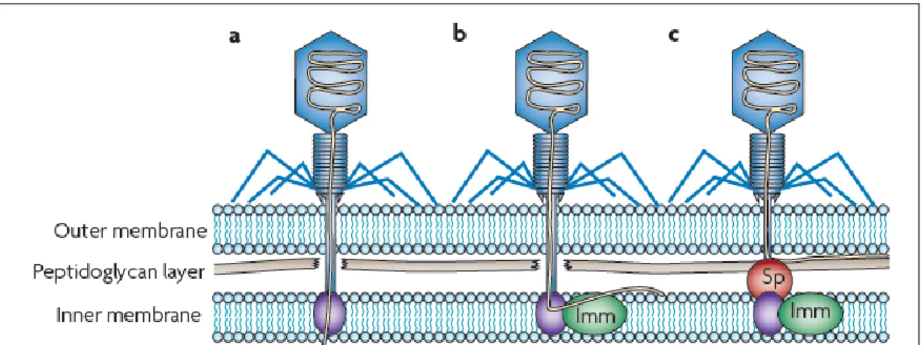
Figure 8. Strategies used by bacteria to block bacteriophage adsorption ………28
Figure 9. Blocking bacteriophage DNA translocation into the bacterial cell by a bacteriophage-encoded Superinfection exclusion (Sie) system………31
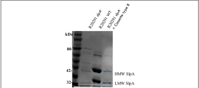
Figure 10. SDS-PAGE of SlpA extractions………..47
Figure 11. The susceptibility of Lactococcus lactis to bacteriophage infection in spot-test assays……….50
Figure 12. Immunofluorescence detection of CwpV expression………52
Figure 13. Susceptibility of L. lactis to bacteriophage infection in spot-test assays…...53
Figure 14. Bacterial survival assay following infection with lactococcal bacteriophage p2..54
Figure 15. Bacteriophage adsorption assay of p2 in Lactococcus lactis expressing or not CwpV type II……….56
Figure 16. Inactivation of slpA in R20291 leads to complete bacteriophage resistance ……59
Figure 17. Complemented slpA mutant restores bacteriophage sensitivity ……….59
Figure 18. The absence of SlpA leads to a reduction in the bacteriophage adsorption of ϕCD38-2 and ϕCD146, but not ϕCD111 ………..61
Figure 20. Proposed generalist mechanism of infection by a bacteriophage under a bacterial host expressing SlpA or CwpV proteins.………...…68 Supplementary figure 1………..……90
Table 1. C. difficile bacteriophage morphology, host, and origin of the viral particles used in
this study………25
Table 2. List of strains, plasmids, and phages utilized in this study………..36
Table 3. List of primers utilized in this study……….42
Table 4. General PCR conditions………..……44
Table 5. Complementation of the R20291 slpA- mutant with different slpA alleles changes the susceptibility to bacteriophage infection……….62
ATc DNA RNA BHI BLAST CDAD CRISPR CDI CWB2 Cwp CwpV dNTP ECM EDTA EOP GC gDNA GM17 HMW-SLP ICTV M MLST mM MOI NCBI LMW-SLP LTA LPS Anhydrotetracycline Deoxyribonucleic acid Ribonucleic acid Brain Heart Infusion
Basic Local Alignment Search Tool Clostridium difficile Associated Diarrhea
Clustered regularly interspaced short palindromic repeats Clostridium difficile infection
Cell_wall_binding_2 Cell Wall Protein
Cell Wall Protein Variable Deoxyribonucleotides
Extracellular Matrix Proteins Ethylenediaminetetraacetic acid Efficiency of Plaquing
Guanine- Cytosine genomic DNA Glucose-M17
High Molecular Weight Surface layer Protein International Committee on Taxonomy of Viruses Molar
Multilocus sequence typing Millimolar
Multiplicity of Infection
National Center for Biotechnology Information Low Molecular Weight Surface layer Protein Lipoteichoic acid
LB OD600nm PaLoc PBS PCR PFU/mL pH PMC PSI RBP R–M system Sie System SDS SLP TAE buffer TEM TMP TY Luria-Bertani
Optical density at 600nm of wavelength Pathogenicity Locus
Phosphate-Buffered Saline Polymerase Chain Reaction
Plaque-Forming Units per Milliliter Potential of Hydrogen
Pseudomembranous Colitis Polysaccharide I
Receptor Binding Proteins
Restriction–modification systems Superinfection exclusion system Sodium Dodecyl Sulfate
Surface Layer Protein
Tris base, acetic acid, EDTA buffer Transmission Electron Microscopy Tape Measure Protein
INTRODUCTION 1.1. Brief insights into Clostridium difficile history
Although humans are not the sole organisms fighting against bacteria, in recent years science has been working on different flanks to find new weapons that can be used in the battle against the oldest inhabitants of the planet Earth. Obviously, regarding human health, the microorganisms that can cause diseases are the ones who receive the most attention. However, it is important to clarify that most of the microbes that abide in our body cannot cause disease. On the contrary, several can protect us against other microorganisms that can be harmful or cause an alteration in the health state of an individual. A pathogen is an infectious organism that can cause disease in a susceptible host. A particular case is depicted when some of these microorganisms only reach their quality as pathogens when its host suffers an alteration of the immune system or a modification of other protective factors that maintain it in a stable health state. This kind of organism is called opportunistic pathogen (NHI, 2007).
Clostridium difficile is an opportunistic pathogen in healthcare-associated infections that was discovered in 1935. However, it was only associated with a particular disease in humans in 1979. After that and during the next three decades, extensive clinical research completely associated C. difficile as the primary origin of CDAD (Clostridium difficile associated diarrhea) and posterior PMC (pseudomembranous colitis). Additionally, Clostridium difficile infection (CDI) was linked to antibiotic exposure in patients in Western countries. In the past, CDIs were never considered as a real menace to public health. Usually, epidemics were small and mortality and economic burden were never significant (Rupnik & Mastrantonio, 2016). Two prominent outbreaks in Canada and the USA in 2003 by the strain named C. difficile BI/ NAP1/027 delimited a new epidemic age in which bacterial infections caused by C. difficile would play a significant role (Rupnik & Mastrantonio, 2016).
The reemergence of new C. difficile strains brought economic problems to the health care systems, and in parallel, high virulence, escalation in incidence, mortality, and severity was progressively detected. Previous findings have evidenced C. difficile as one of the most important nosocomial-associated pathogens (along with Escherichia coli, Staphylococcus aureus, and Pseudomonas aeruginosa) and equally CDI as one of the major public health concerns around the world (Cartman, Heap, Kuehne, Cockayne, & Minton, 2010).
Preoccupation about the future of C. difficile epidemics is rising due to new reports showing modification of the infection pattern from an opportunistic microorganism usually affecting elderly people and hospitalized patients under antibiotherapy, to a pathogen affecting patients with underlying clinical conditions, younger patients, and individuals without previous antibiotic exposure. Equally alarming, C. difficile infections are proliferating to the animal population, primarily animals utilized for food production (pigs being the most affected due to antibiotic overuse). Reports of contaminated vegetables, livestock and meats have worried scientist and health administration entities regarding the possibility of CDI as a food transmitted zoonosis (Rupnik & Mastrantonio, 2016).
C. difficile is a Gram-positive, bacillus-shaped, spore-forming strictly anaerobic bacterium. When the bacterial cells are under stress, they produce spores that are capable of resisting extreme conditions that metabolically active bacteria cannot tolerate. The spores can remain inactive, but once established in the human intestine, the spores can germinate and cause disease. The disease range from mild clinical effects to severe diarrhea and ultimately can degenerate into PMC (Rupnik, Wilcox, & Gerding, 2009).
A meta-analysis using PUBMED and EMBASE databases shows that around 8% of patients admitted to hospitals are carriers of toxinogenic C. difficile strains with almost six times higher risk of illness compared with patients that are admitted but not colonized (Zacharioudakis, Zervou, Pliakos, Ziakas, & Mylonakis, 2015). Contaminated hands, clinical instruments, and asymptomatic adult patients are not the sole reservoirs for the transmission
asymptomatic presence and act as reservoirs as well (Rousseau et al., 2012). In the last decade, it has been demonstrated that the gut microbiota (the network of microorganisms that abide in the gastrointestinal tissue) plays a major role in the physiological stability of the host, contributing to the gut health. C. difficile population dynamics is limited and controlled by the presence of other significant anaerobic bacteria. Recently, it has been shown that the capability of C. difficile to infect and colonize a host depends mostly on the inability of the normal gut population to retain C. difficile expansion through the intestinal cavity (Britton & Young, 2014; Buffie et al., 2014). Colonization is a vital part of the development of the disease and can happen because of the exposure to broad-spectrum antibiotics. It is well understood from various studies that older people, peripartum women, and children, immunocompromised patients and individuals with recent surgeries tend to have higher CDI susceptibility (Block, 2001; Cózar-Llistó, Ramos-Martinez, & Cobo, 2016).
Even with the upcoming of antibiotic cocktails and the development of better targeted antibiotic therapies (through the amelioration of patient diagnosis), one of the main characteristics of CDIs is the high recurrence rate (i.e., failure in the CDI contention by the first antibiotic treatment). These recurrences can result from relapses of the first infecting bacteria or reinfections from other C. difficile strains (Bien, Palagani, & Bozko, 2013; Rupnik et al., 2009).
1.1.2 Epidemics and evolution of Clostridium difficile
C. difficile epidemic behavior has changed in the last millennium. Over the history, CDI cases were underrepresented, and when they were occurring they were related to various types of strains. After the year 2000, numerous cases and clinical isolates have been evaluated (equally as the strain distribution on the continents) indicating the reemergence of specific strains belonging to the hypervirulent BI/NAP1/027 group. This lineage has been detected and monitored across Europe and North America (it has spread in several western countries). The strains belonging to this cluster are characterized by an overproduction of toxins A and B, production of a binary toxins and resistance to fluoroquinolone antibiotics. These changes led to an increase in the morbidity and mortality (McDonald et al., 2005; O’Connor, Johnson, & Gerding, 2009; Warny et al., 2005). However, in the last years, there has been a debate
regarding the mode of hypervirulence in the BI/NAP1/027 group. Other factors such as motility, antibiotic resistance, adherence, and sporulation can be added to the emergence of epidemic strains from this group (M. Merrigan et al., 2010; Stabler et al., 2009).
Across Canada, numerous studies have followed the historical development, morbidity, and mortality of CDI, where interestingly the province of Quebec takes a local relevance. Retrospective research of CDI in a period starting in 1991 and finishing in 2003 showed an increase in the incidence from 35.6 cases per 100,000 residents to 156.3 cases per 100,000 residents, respectively. With a similar pattern in the same period, the rate of complications during infection treatment increased from 7.1% to 18% (Pépin et al., 2004). A later study in hospitals mainly from Sherbrooke and Montreal metropolitan areas reported an incidence of 22.5 per 1,000 admissions and a 30-day attributable mortality rate of 6.8%. For the studied cases, a predominant strain from the group BI/NAP1/027 (82.2 percent of the cases) resistant to fluoroquinolones was found (Loo et al., 2005).
One of the latest studies performed in the country assessing the incidence, medical cost, and loss of productivity resulting from CDI, estimated that for the year 2012 there were around 37,932 cases. From there, Quebec had one of the highest numbers of bed days attributable to CDIs (5,560,668 bed days). Of those patients, around 73% were newly infected people, and 27% were recurrence cases. Sixty-one percent were cases with mild to moderate symptoms, and 54% of the patients were 75 years old or more. Quebec presents the highest estimated number of CDI of all Canadian provinces and territories with 16,562 cases (numbers far superior to equally developed provinces like Ontario, British Columbia, and Alberta) (Freeman et al., 2010; Levy et al., 2015).
Presently, the epidemic group BI/NAP1/027 is diminishing in proportion, and CDI has been caused by different strains (Poxton, 2013). This phenomenon also includes new strains from the 078 PCR-ribotype groups which both can be found in human patients and animals for meat production (Goorhuis et al., 2008; Knetsch et al., 2014).
As we previously stated, resident gut microbiota alteration permits spore germination and colonization of the host intestinal cavity. This process starts with the vegetative cell of C. difficile that, after metabolic activation, evades the host immune system and multiplies. The production of toxins and virulence factors start finally causing clinical manifestation (Figure 1). In that order of ideas, it is important to understand the virulence factors and main structural features that are critical in colonization and other steps of the disease progression.
1.2. Clostridium difficile virulence factors
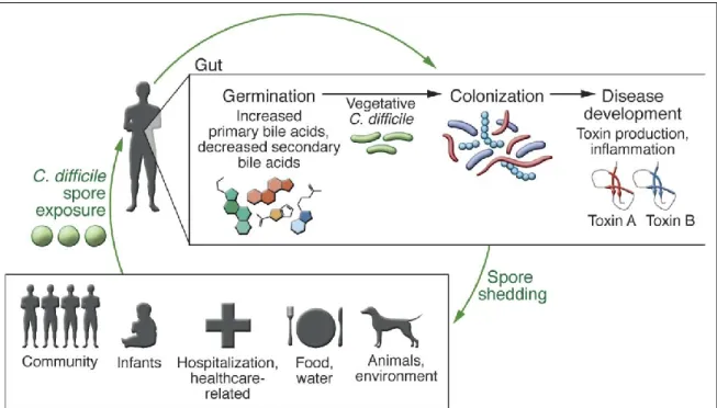
Figure 1. C. difficile transmission cycle. Original spore exposure from various sources does not inevitably result in disease in a healthy human. A healthy microbiota is capable of blocking C. difficile spore germination and vegetative growth. However, if the microbial ecology of the gut has been disturbed, spore germination and toxin production will emerge. The start of the life cycle activation can result in epithelial damage, inflammation, and apparent disease as the toxin production takes place. C. difficile sporulation, spore release into the environment, and transmission to new individuals continues the infectious cycle. Image taken from (Seekatz & Young, 2014).
After metabolic activation and establishment of the infection, one of the major virulence factors are produced, the exotoxins TcdA and TcdB. Both genes are located in a region of the bacterial chromosome called pathogenicity locus or PaLoc (Monot et al., 2015). The initial step of the intoxication process starts when the C-terminal receptor domain of the protein interacts with the cellular receptor (it is known that both toxins can activate a clathrin-mediated endocytosis. Also, it has been shown that the poliovirus receptor-like 3 (PVRL3) functions as a cellular factor essential for TcdB-mediated cytotoxicity). This interaction promotes the endocytosis of the toxins and posterior trafficking via endosomes. Acidification of the endosomal compartment and autocatalytic cleavage leads to the liberation of the active N-terminus into the cytosol. These domains inactivate numerous members of the Rho GTPases family which affect critical downstream cellular processes resulting in a loss of cytoskeleton integrity (Awad, Johanesen, Carter, Rose, & Lyras, 2014). Regarding the phenotypical manifestation of the cellular intoxication, the toxic effect could be compared to the outcome caused by cell apoptosis (Just et al., 1995). The PaLoc also encodes three accessory proteins named TcdR, TcdC, and TcdE. TcdE has been speculated to be involved in the release of TcdA and TcdB through permeabilization of the cell-wall. TcdR is an alternative sigma factor and TcdC may function as a negative regulator of toxin production. However, the functions of these accessory proteins remain in part controversial and are still investigated (Awad et al., 2014; Voth & Ballard, 2005).
In addition to the toxins, there are other factors like the endospore, flagella, pili, surface layer proteins, cell-wall proteins and adhesins that are equally implicated in the C. difficile virulence (Awad et al., 2014; Janoir, 2016b). Even if TcdA and TcdB are the primary virulence factors of C. difficile, other putative virulence factors may play a role in important steps of colonization, the establishment of the disease and the adherence to epithelial cells. However, these factors are also an integral part of the cellular structure and architecture of the C. difficile cell-wall, interaction with the environment and displacement. Some of them will be discussed in the following section.
SlpA protein (for Surface-Layer Protein A) is composed of two subunits, the LMW-SLP (low-molecular-weight S-layer protein) and the HMW-SLP (high-molecular weight S-layer protein), both are a major component of the para-crystalline S-layer of C. difficile (E Calabi et al., 2001). The two subunits are produced from the cleavage of the protein precursor SlpA by the cysteine protease Cwp84 (described below in section 1.3.6.1) (Bruxelle et al., 2016; Dang et al., 2010). Both subunits are noncovalently re-associated after the protease action (R.P. Fagan, Albesa-Jove, Qazi, Brown K.A., & Fairweather, 2009) (Figure 2).
It has been proposed that SlpA presence has a structural role in the integrity of the bacterial shape. Different experiments have also demonstrated that both, natural SlpA proteins and recombinant SlpA, can dock to the cells from the cell line Hep-2 and also to human gastrointestinal tissues taken from healthy patients (Emanuela Calabi, Calabi, Phillips, & Fairweather, 2002). In addition, it has been proven that SlpA proteins are essential in bacterial
Figure 2. The Clostridium difficile cell wall architecture is mainly composed by a single protein, SlpA. This protein and other cell wall proteins have a dedicated secretion
path (SecA2). SlpA undergoes a cleavage event, mediated by a cell wall cysteine protease Cwp84, forming the HMW and LMW SLPs which form the assembled S-layer. Image modified from (Reynolds et al., 2011a)
adherence to the colonic cell line Caco-2. Pre-incubation of bacteria with antibodies against LMW-SLP or HMW-SLP reduced adherence to Caco2 cells (M. M. Merrigan et al., 2013). Additionally, HMW-SLP presents an in vitro capability to attach to proteins such as type I collagen, vitronectin and thrombospondin (Emanuela Calabi et al., 2002). LMW-SLP and HMW-SLP domains can activate the host innate immune system, through the recognition of Toll-Like Receptors 4 (TLR-4). This activation stimulates signaling pathways that lead to maturation of dendritic cells and macrophage activation. However, the immunomodulatory kinetics of the SlpA proteins does not happen to correlate with the strain virulence considering that no particular immunomodulatory pattern has been discovered for the epidemic strain BI/ NAP1/027 when compared to more classical strains (Bianco et al., 2011; Poxton, 2013; Vohra & Poxton, 2012).
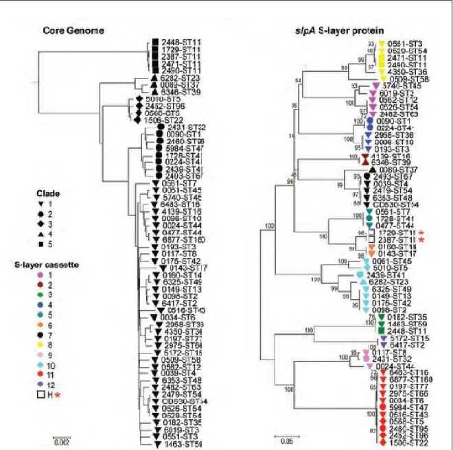
Studies of the phylogeny of 58 C. difficile isolates based on the core genome (genes common to all samples) showed the historical five clades of the population structure as determined using MLST data (K. E. Dingle et al., 2013). This pattern was also conserved in genes neighboring the cwp cluster (this cluster is explained in the section 1.3.6). However, in regions surrounding the genes slpA, cwp66, and secA2, an entirely dissimilar strain relationship appeared. A neighbor-joining phylogenetic tree of the slpA gene of the 58 isolates identified 12 distinct clusters (Figure 3), different from the usual five clades. Equally, neighbor-joining trees for the cwp66 adhesin and secA2 translocase grouped these 58 isolates identically to slpA. These results indicate that slpA, cwp66, and secA2 coevolve as an independent 10-kb gene cassette, which contains at least 12 genetically divergent variants (K. E. Dingle et al., 2013). Our understanding of the role of SlpA in CDI (and in other fields like bacteriophage infection and virus susceptibility) has been limited by the failure to obtain a mutant, despite several attempts by different research groups. Nevertheless, using genetically modified contractile R-type bacteriocins (phage-like particles) from C. difficile strain CD4 to kill BI/NAP1/027-type strains (Gebhart et al., 2015), a group from AvidBiotics Corp. isolated the first mutant for the slpA gene in the epidemic strain R20291 (unpublished data at the time of writing these lines). This mutant has an adenine insertion early in the sequence of the gene (nucleotide position 98) that leads to a premature TAA stop codon,
creating a truncated protein (personal communication from Gregory Govoni, AvidBiotics Corp., South San Francisco, California, USA).
1.3.2 Polysaccharides
Figure 3. Phylogeny of whole genomes and slpA. Phylogeny created from the whole
genomes: Black forms indicate the clades. Phylogeny constructed from the slpA gene: Shapes specify the clade, and colors designate the S-layer variants. Hybrids are indicated by a H and a star. Numbers indicate bootstrap support. Image taken from (Dingle et al., 2013).
In metabolically active C. difficile cells, it has been described three types of polysaccharides, PSI, PSII, and PSIII, including relevant ribotypes like 027, which has been found to contain more PSII than the other two kinds of molecules (Monteiro et al., 2013). Importantly, the presence of these molecules has stimulated the production of vaccines and technics for their use in diagnosis. Vaccines containing PSII have been tested in different animal models (Awad et al., 2014). The same molecule has been used in saccharide microarrays to detect IgG antibodies in the sera of CDI patients (Martin et al., 2013). Interestingly, new works in a mouse model and a PSII glycoprotein conjugated to TcdA and TcdB fragments indicated that antibodies targeting PSII and toxins were produced by mice, this result indicates that a combination of sugars and proteins could be a promising topic for vaccine development (Romano et al., 2014).
1.3.3. Flagella
C. difficile is motile due to the action of the flagellar machinery. A plethora of diseases-related bacteria relies on flagella as an important virulence factor (helping in tissue invasion and host colonization). Genes that regulate the assembly of the C. difficile flagella are ordered into three operons (Awad et al., 2014; Janoir, 2016a). The F3 locus comprises just early stage genes and contains the FliA sigma factor (also named SigD). FliA regulates the expression of late-stage flagellar genes from the F1 locus, such as genes coding for the flagellin FliC and the cap protein FliD. The F2 regulon is responsible for post-translational modification of flagella proteins that have been shown indispensable for efficient flagellar assembly and motility of C. difficile. The three loci show variability among lineages of C. difficile. Interestingly, the F3 locus is not present in the emerging virulent 078 clades, signifying in some manner that flagella and therefore motility are unessential for C. difficile virulence (Janoir, 2016a). One study has even shown that fliD mutants were more virulent than the wild-type strain (Baban et al., 2013). More data is necessary to understand the precise role of the flagellar machinery in the virulence of C. difficile.
Fimbriae are not exclusive to C. difficile strains. In other bacteria, such as E. coli, Bordetella pertussis, Staphylococcus and Streptococcus species, the expression of fimbriae is correlated with the ability of bacteria to attach to the host and cause disease (Connell et al., 1996). However, different studies showed no correlation between serious human or animal epidemic strains, expression of fimbriae and their ability to cause disease (Borriello et al., 1988). These results suggest that at least in C. difficile, the expression is not necessarily prominent in the development of the disease. On the other hand, bioinformatic investigation on the phylogenetic class Clostridia shows that in the genome sequences of the microorganism belonging to this taxon there are several putative type IV pilus genes (Melville & Craig, 2013). Specifically, in the epidemic strains 630 and R20291 at least nine pilli or putative pilus-like genes were identified (Maldarelli, De Masi, von Rosenvinge, Carter, & Donnenberg, 2014). Recently, it has been demonstrated in historical epidemic strains 630 and R20291 that the type IV pilus contributes to biofilm formation, especially in the strain R20291. Additionally, it was also reported that this strain is capable of motility in a type IV pilus-depended manner (Purcell, McKee, Bordeleau, Burrus, & Tamayo, 2015).
1.3.5. Clostridium difficile adhesins
C. difficile has been shown to be able to adhere to various cell lines. For example, HT-29-MTX cells, enterocyte-like Caco-2 cells, Hep-2 cells, Vero and HeLa cells (T Karjalainen et al., 1994; Naaber, Lehto, Salminen, & Mikelsaar, 1996; O’farrely, Baird, Drudy, Fenelon, & O’Donoghue, 2001). Equally, C. difficile attaches to a plethora of extracellular matrix proteins (ECM) in vitro: fibronectin, fibrinogen, vitronectin, and type -I, -III, -IV and -V collagens (Cerquetti, Serafino, Sebastianelli, & Mastrantonio, 2002). The broad picture of C. difficile adhesion shows that the host-bacterium attachment requires adhesins from a bacterial origin that in general terms are proteins present on the cell surface. A plethora of adhesins has been characterized: fibronectin binding proteins, the heat shock protein GroEL, cell wall proteins (Cwp), flagella, fimbriae, etc. However, their function in the pathogenesis of CDI is still not fully understood. In the next sections, we will focus on the Cwp protein family.
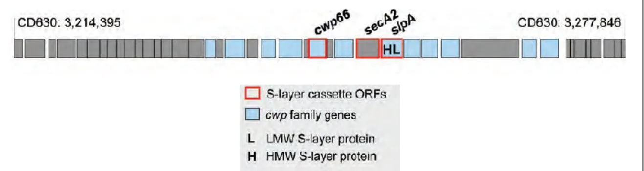
The Cwp proteins are a group of closely related paralogous surface-associated proteins that are composed of 29 members characterized by the presence of a conserved binding domain (Pfam 04122) that attaches the proteins to the bacterial cell-wall by PSII molecules (R. P. Fagan et al., 2011; Willing et al., 2015). Almost half of the Cwp proteins are found within the “clostridial wall (cwp) gene cluster.” This cluster has 18 open reading frames (ORFs) (Figure 4). The rest of the Cwp proteins are scattered throughout the bacterial chromosome. Different studies have shown that some of these proteins (like the protein CwpV) are secreted by the dedicated secretion system, SecA2 (K. E. Dingle et al., 2013). It is possible that other members of the Cwp family can use the same secretion system to be transported to the cell-wall. The cwp genes that are located inside the cwp cluster, like the cysteine protease Cwp84, presents high variability among C. difficile strains (Tuomo Karjalainen, Saumier, Barc, Delmée, & Collignon, 2002; Savariau-Lacomme, Lebarbier, Karjalainen, Collignon, & Janoir, 2003) This variability is also true for proteins like SlpA, Cwp66, and SecA2 where their coding sequences are shown to somehow be variable (regions prone to genetic exchanges by homologous recombination (K. E. Dingle et al., 2013)). In this order of ideas, the genetic variability present in Cwp surface proteins emerges from the immunological pressure from the host and contributes to its avoidance. Several of the Cwp proteins are immunogenic in patients (Drudy et al., 2004; Pechine, 2005; Pechine, Janoir, & Collignon, 2005; Wright, Drudy, Kyne, Brown, & Fairweather, 2008). Therefore, they could play an essential function in the colonization process and pathogenesis of the disease.
Figure 4. Genetic organization around the cwp cluster in the strain CD630. The C.
difficile S-layer is encoded by the slpA gene, positioned within the 36.6-kb cell-wall protein (cwp) gene cluster. Image modified from (Dingle et al., 2013)
1.3.6.1. Cwp84 cysteine protease
The cysteine protease Cwp84 coding sequence is present in the cwp cluster and is transcribed using its promoter (Savariau-Lacomme et al., 2003). Cwp84 is found localized embedded in the bacterial S-layer and requires the processing of its pro-peptide signal by another cell-wall associated cysteine protease, Cwp13 (which is also present in the cwp gene cluster and can be found incorporated into the S-layer) (de la Riva, Willing, Tate, & Fairweather, 2011). Once Cwp84 is fully processed, its complete conformation is responsible for the proteolytic cleavage of the SlpA precursor. Mutants of the Cwp84 proteins present retention of proteins of the cell-wall. This mutation results in a colony phenotype in which the colony formation is not cohesive and formless (de la Riva et al., 2011). Other studies also indicate that possibly the function of Cwp84 in the disease pathogenesis is not essential due to results showing that a mutant for the protease is as virulent as the wild-type strain in a hamster model (Kirby et al., 2009).
1.3.6.2. Cwp66 adhesin
Cwp66 was the first adhesin to be identified in the class Clostridia (Waligora et al., 2001). The cwp66 gene is found inside the cwp cluster and synthesizes a protein overproduced at the surface of the cell just after a heat-shock. It was proven to mediate adhesion to the cell line Vero (Waligora et al., 2001). As other proteins from the same cluster, Cwp66 contains two domains, one of which is highly variable. Both domains can be detected by antibodies in bacterial surface extracts. The function of Cwp66 in cell adherence was demonstrated by utilizing the same antibodies in attaching inhibition studies with the purified domains. However, no mutant for the protein has been evaluated yet and its role in the pathogenesis of the disease is unknown.
1.3.6.3. CwpV protein
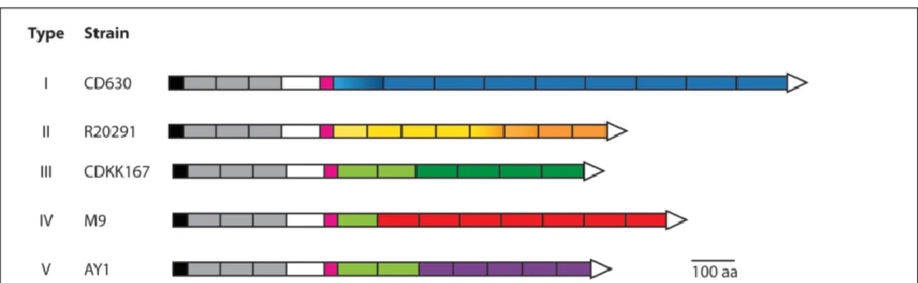
Of all the members of the Cwp family, CwpV is the largest protein. Its coding sequence is not present in the cwp cluster. The variable region of CwpV is located toward the C-terminal end; this domain starts with a serine-glycine enriched flexible linker which is followed by
several repetitions, whose sequence and number change depending on the C. difficile strain (Figure 5). The N-terminal domain possesses the cell-wall anchoring activity via three CWB2 motifs that recognize PSII molecules embedded in the bacterial cell-wall (Willing et al., 2015). Like SlpA, CwpV protein undergoes maturation into two subunits that are re-associated in a non-covalent manner to form a heterodimeric complex that attaches to the bacterial cell-wall (Robert P Fagan & Fairweather, 2014; Reynolds, Emerson, de la Riva, Fagan, & Fairweather, 2011a). Interestingly, and different from SlpA, CwpV post-translational cleavage is mediated by intramolecular auto-proteolysis (this phenomenon is also present in Cwp13) (Dembek, Reynolds, & Fairweather, 2012). CwpV is exported to the cell-wall through the secA2-secretion system (Robert P Fagan & Fairweather, 2011). CwpV is a major component of C. difficile cell-wall, representing about 13% of the whole surface layer proteins (Reynolds et al., 2011a).
In a controlled laboratory environment, only 5% of the viable cells of the entire bacterial population express the protein. The phase variation of CwpV is regulated by DNA inversion, a mechanism of transcriptional termination by the creation of a stem-loop structure followed by a poly-U tract, which promotes transcriptional termination, in the OFF (non-transcriptional) position (Emerson et al., 2009). The C-terminal domain of CwpV presents auto-aggregative characteristics (Reynolds et al., 2011a), but its link with biofilm formation (and possible role in gut colonization) is still unknown. Future analyses are necessary to assess the biological role of CwpV. More information about the role of CwpV in bacteriophage infection can be found in the section 1.9.5.
1.4. Bacteriophages
Bacteriophages (phages) are parasites that infect bacteria and are presently viewed as the most abundant and diverse biological entities on Earth. Their vastness is not only quantitative (they surpass bacterial organisms by a ratio of 1:10), their ecology, morphology, infection strategies and other qualitative characteristics also represent relevant and exciting study fields (Brüssow & Hendrix, 2002). For example, it has been stated that only in the seas phages infect bacteria sufficiently to produce more than 1011 kilograms of carbon per day from natural populations, thus affecting the ecology of the planet directly (Suttle, 2007).
Phages have the capacity of killing their bacterial host. But also their genomes can transfer to the bacterial genome toxins and complementary virulence factors that can modify the host and affect human bacterial pathogens (Fortier & Sekulovic, 2013). In an identical manner, phage diversity contributes to bacterial diversity directly. Phages can function as genetic vehicles for transduction of various genes, for example, antibiotics resistance genes among different populations (Figure 6) (Grose & Casjens, 2014). In all ecosystems on the planet, a large array of phages and hosts are tangled in an endless co-evolution cycle, in which emerging phage resistant hosts continuously preserve bacterial taxa, but on the other hand,
Figure 5. Visual representation of natural CwpV types. The CwpV type II from the
strain R20291 was used in this study. Color code: Black, signal peptide; grey, cell-wall anchoring domain (PF04122); white, non-described function; pink, serine-glycine-rich region; blue, type I repetition; orange, type II repetition, green, type III repetition; red, type IV repetition; purple, type V repetitions. Image modified from (Sekulovic et al., 2015a).
counter-resistant phages attack these new bacterial groups. Widely speaking, phages and phage resistance mechanism have an important function in the control of bacteria in most habitats on the planet.
1.5. Bacteriophage taxonomy
At present day, bacterial viruses include at least 14 officially accepted families, and approximately 5500 phages have been isolated and investigated by electron microscopy (H.-W. Ackermann, 2007; Hans-(H.-W. Ackermann, 2009). The categorization of these phages has been essential in understanding the biodiversity and their ecological role. In the same manner,
Figure 6. Illustration of the lytic and lysogenic life cycles of phage λ. Taken from
viruses are polyphyletic and do not possess conserved genes among them (for example ribosomal RNA genes) that are used in phylogenetic analyses for classification purposes. In this order of ideas, phage taxonomy can be considered as a quite complicated task, also adding up the fact that phages have the capability of acquiring, transmitting, and mixing genetic material in a fast way. The International Committee on Taxonomy of Viruses (ICTV) has proposed a classification system in which it is taking into account the nature of the nucleic acids, nucleotide sequences and the viral structure of the phages (van Regenmortel, Mayo, Fauquet, & Maniloff, 2000).
From the current taxonomy, we know that most phages belong to the order Caudovirales and this group is divided among three main families; Myoviridae, Podoviridae, and Siphoviridae. From these families, Podoviridae contains around 60% of all known phages (IUMS, 2016). In a similar manner, the different phages have different morphologic features. However, they can conserve certain common structures. In fact, all phages contain their genetic material (DNA or RNA) inside a capsid that is made of protein or lipoproteins. Nevertheless, the main structural characteristic of the phage from the order Caudovirales is the presence of a “tail” that helps in the recognition, attachment, and injection of the DNA into the cytosolic space. It is important to remark that there are differences among the tails of the three families. The Myoviridae tail is relatively short but rigid and contractile, whereas the Siphoviridae tail is long and non-contractile. The phages of the Podoviridae family have a very short tail but non-contractile (H.-W. Ackermann, 2007; Hans-W. Ackermann, 2009; Grath & Sinderen, 2007; Hyman & Abedon, 2012). We show examples of the family structures in Figure 7.
1.6. Bacteriophage replication cycle
Despite the exceptional biodiversity that phages can bear, the mechanisms responsible for viral replication are conserved among the different families. Phages are forced to overtake the cellular machinery and metabolic processes to complete their infection cycle and achieve their replication (Grath & Sinderen, 2007).
1.6.1. Host receptors and bacteriophage adsorption
Adsorption is the first step of the infection process; the attachment of the phage to the cell surface. This initial recognition is made at first in a reversible manner by the interaction of the virion with a receptor on the surface of the bacterium. In phages with tail structures, this reversible interaction is made by the utilization of proteins localized in the distal part of the
Figure 7. Diversity in bacteriophage families: Morphologies, genome natures, and comparative genome sizes. Bacteriophages are arranged in order of decreasing genome
sizes. Blue coloration indicates capsids, red indicates tails, and yellow refers to lipids. Tailed bacteriophages are members of viral order Caudovirales. Image taken from (Hyman & Abedon, 2012).
interaction between the receptor binding proteins (RBP) of the phage and the bacterial receptors on the surface (Silva, Storms, & Sauvageau, 2016; Weinbauer, 2004). Different surface molecules are recognized to play the role of a receptor in the adsorption process, for example, the polysaccharides, lipopolysaccharides (LPS), teichoic acids, lipoteichoic acids (LTAs), and glycoproteins function as entry ports to the cell (Grath & Sinderen, 2007; Silva et al., 2016). In particular cases, phages can recognize the capsule, conjugation pilus, and flagella as means of attachment to the bacterial surface (Geoffrey William Hanlon, 2007). This natural diversity in receptors and structures of both sides reflects the evolutionary relationship between phages and bacteria.
However, just one receptor may not be involved in the attachment and adsorption; it is possible to find in nature a two-host receptor entry mechanism. Even the proteins and molecules involved in the reversible part of the adsorption could be different than the proteins and receptor involved in the irreversible part of the adsorption. For example, the phage T4 contains long tail fibers that interact with E. coli membrane, and this interaction is responsible for the first reversible binding. The short fibers of the virions interact with the heptose fraction of the LPS of the host and account for its irreversible binding (Silva et al., 2016). In the same manner for the Gram-positive bacterium Bacillus subtilis, its phage SPP1 utilizes the glycosylated cell-wall lipoteichoic acids (LTA) for a primarily reversible binding, followed by the interaction between the phage protein gp21 and the cell membrane protein YueB that determines the irreversible adsorption. It is hypothesized that the ability of a phage to engage an irreversible adsorption to molecules that are more expressed or commonly found on the cell-wall can increase the probability of finding the cellular receptor that determines its irreversible adsorption (Chatterjee & Rothenberg, 2012).
1.6.2. DNA injection
Right after the adsorption, the downstream step of phage infection is the injection of the viral DNA into the host cytoplasm. This process can be done in different ways, depending on the structural morphology of the phage that is infecting. For example, the phages from the Myoviridae family contract a rigid protein sheath that is capable of piercing the bacterial membrane and force the viral DNA to be released from the capsid and be translocated to the
cytosol of the bacterium. In some cases, hydrolytic enzymes can be present in the virion and degrade the cell wall polymers and facilitate viral DNA entry (González-Huici, Salas, & Hermoso, 2004; Geoffrey William Hanlon, 2007). However, although phage adsorption and DNA injection have been studied since Hershey–Chase experiments in 1952, these particular steps of the phage replication cycle are still poorly understood.
In general terms, for DNA liberation from the capsid, a signal activation must take place from the host receptor to the phage virion structure, which triggers the capsid opening and future DNA injection. Still, each phage has its unique mechanism and strategy for DNA injection. For the coliphage λ which infects E. coli, it is hypothesized that the viral DNA enters just by diffusion (Filali Maltouf & Labedan, 1985; González-Huici et al., 2004). Phage T4 pierces the cell membrane using its contractile tail and a cell-puncturing apparatus in the baseplate of the virions (Hans-W. Ackermann, 2009). For phage T5, the injection process has two steps: 8% of the viral DNA is translocated to the cytoplasm. From there, the proteins A1 and A2 are synthesized from the region that was transferred; these two proteins are necessary for the translocation of the rest of the viral genomes. It is thought that DNA binding domains in the proteins A1 and A2 pull the DNA into the cell (Letellier, Boulanger, de Frutos, & Jacquot, 2003). In the Gram-positive bacterium, B. subtilis, the phage Ø29, that has a non-contractile tail, also presents a two-step process. In the first step, DNA is injected into the cytoplasm possibly by the differential of pressure created from the virion to the cell. Thus, synthesis of early operons is activated. The second step is initiated when one of the early viral proteins, p17, participates in the molecular complex that initiates the translocation of the rest of the genome inside of the bacterium (González-Huici et al., 2004).
1.6.3. Replication cycle
In most known phages, there are two main ways of replication: the lytic cycle and the lysogenic cycle. Phages that use the lytic cycle are called virulent or strictly lytic, whereas phages that have the capability of changing their replication from the lytic to the lysogenic cycle are called temperate phages.
During the lytic cycle, phages replicate their genome producing a plethora of significant changes in the host bacterium. Ultimately, these changes cause the bacterium to stop most its cellular processes and all the cell machinery is deviated to assure the production of new viral particles. Just after the injection of the viral DNA into the cytosol of the bacterium, the bacterial RNA polymerase is recruited to transcribe early genes that are implicated in the transcription, gene regulation, DNA replication, and recombination. Afterward, late genes are transcribed, allowing the synthesis of structural proteins (capsid, tail, RBP, and so forth). In parallel, the bacterial DNA polymerase is recruited, permitting the replication of the viral genome. Altogether, viral structural proteins and DNA are assembled, forming new infective virions. The last step of the process is the liberation of the viral offspring from the intracellular space to the environment. This cellular lysis is possible by the activity of two main enzymes, the endolysin, and the holin. On one hand, the endolysins are hydrolytic enzymes that degrade the peptidoglycan cleaving different types of bonds at the interior of the polymeric structure. On the other hand, the holins are hydrophobic proteins that have the function of inserting themselves in the cytoplasmic membrane (at a precise moment of the lytic cycle) and produce pores that ultimately will allow the endolysins to reach the peptidoglycan and cleave it. After the cellular lysis, virions are released into the environment ready to find a new cell and start again the lytic cycle (Borysowski, Weber-Dabrowska, & Górski, 2006; Kutter & Sulakvelidze, 2005; Weinbauer, 2004).
1.6.3.2. Lysogenic cycle
The particularity of temperate phages is their capability to alternate between the lytic and lysogenic replication cycles. After the infection and the translocation of the viral DNA into the host cytosol, a molecular decision is taken toward one of the replication cycles. The lytic cycle was described above. On the contrary, if the phage “decides” to take the lysogenic cycle, the necessary genes for the lytic cycle are repressed, and the phage genome is integrated into the bacterial chromosome (Hans-W. Ackermann, 2009; Grath & Sinderen, 2007). Already part of the host genome, every single division of the bacterium signifies the replication of the bacterial chromosome along with the replication of the viral genome. Bacteria with an inserted phage genome are called lysogens, and the phage takes the name
of prophage. The decision regarding the infection cycle is made early after the infection, and this one is firmly affected by the condition of the intracellular environment inside the bacterium. In fact, DNA damage and unfavorable environmental conditions are known to bias the phage to take the lytic cycle (Kutter & Sulakvelidze, 2005). In that way, when a phage opts for the lysogenic cycle, some repressors are transcribed and translated to block the expression of the genes implicated in the lytic cycle (for example, CI, CII and CIII in the coliphage λ) (Anderson & Yang, 2008; Babic & Little, 2007). However, the decision of taking the lysogenic cycle is complex, and many of the mechanisms are still not understood. The enzyme integrase is also expressed and is responsible for the viral genome integration into the bacterial chromosome. Phages are remarkably stable once inserted into their host genome. However, under the right condition, it is possible to reintroduce the phage to the lytic cycle and release themselves from the host (Babic & Little, 2007; Little, 1999). Certain intracellular conditions of the host can also cause the induction of the prophage and the initiation of the lytic cycle. Stress factors like U.V light, temperature changes, the presence of different compounds (H2O2, antibiotics) are well known for triggering an SOS response of the lysogen. In E. coli, when the bacterial protein RecA is activated, its activity causes the autocleavage of the repressor of the SOS genes, LexA. The inactivation of this repressor permits then the transcription and future translation of the SOS genes, those implicated in DNA damage reparation. Equally, activation of the RecA protein leads to the proteolytic cleavage of the CI repressor, that maintains the phage in his prophage state and regulates another repressor of the lysogenic cycle. The relaxation of the expression of the repressors CI, CII, and CIII, promotes the augmentation of the expression of the protein Cro, which can activate the transcription of some genes of the lytic cycle and repress at the same time the genes from the lysogenic cycle (Court, Oppenheim, & Adhya, 2007).
1.7. Bacteriophages of Clostridium difficile
To date, all members of the sequenced C. difficile phages that have been described in the scientific literature are temperate phages of the Caudovirales order (phages with tail) and are classified into the Myoviridae or Siphoviridae families (where the majority of the diversity
myoviruses can be sorted into three discrete morphological clades based on the capsid diameter and tail lengths. Small Myoviridae phages with capsid diameters of 40–60 nm and tail lengths of 105–110 nm. Intermediate sized with capsid diameters between ~60–70 nm and tail lengths of 110–130 nm. And finally, long-tailed with capsid diameter between ~60 to ~70 nm and tail lengths between 150–260 nm. All known C. difficile phages follow a lysogenic life cycle, their genome is characterized by the presence of predicted integrase genes, and their genome sizes range from ~31 to ~131 kbps with a GC content of 28.4% to 30.8%. The C. difficile phages utilized in this study are presented in Table 1.
1.8. Lactococcus lactis and its bacteriophages
Lactococcus lactis is a microorganism that has been tamed and utilized for millennia in dairy fermentations. L. lactis abides in niches close to animal and plant surfaces, and is also found in the gastric cavities. However, L. lactis industrial strains used in cheese buttermilk, and sour cream fermentation have the ability to grow faster and rapidly produce lactic acid in milk. Although it has evident industrial interest, L. lactis has become an important research model among other Gram-positive bacteria for subjects such as metabolism, physiology, genetics, and molecular biology. However, at the same time, the investment in fundamental research has stimulated potential new applications such as heterologous metabolite/protein synthesis, massive culture protection, and oral vaccines (Bolotin et al., 2001).
Lactococcal phages are ubiquitous in nature where the host is present, but also in the dairy environment. The virulent nature of all lactococcal phages, their biodiversity and its adverse effect in large-scale fermentations, promoted their study around the world. Hence, several lactococcal phages have been isolated and characterized. In fact, only coliphages have been more studied than phages from the dairy industry (H. W. Ackermann, 2001). All phages from L. lactis known to date present a double-stranded DNA and noncontractile tail structure and fall within the Caudovirales order. Lactococcal phages are primarily classified in the Siphoviridae family and a few members are in the Podoviridae family. Historically, lactococcal viruses were classified using virion morphology and DNA homology index. This classification sorted dairy pages in three different main groups: the 936, c2, and P335 phages. As research techniques advanced, investigators continued with this three-group classification
but adapting multiplex PCR to rapidly assign new phages to those three groups (Deveau, Labrie, Chopin, & Moineau, 2006; S. Labrie & Moineau, 2000). The fast growth of L. lactis cultures permits the easy amplification and replication of phages in solid and liquid broth. The well-known secretion system of L. lactis also allows the intracellular expression and secretion of heterologous proteins, making this bacterium a useful model microorganism. Equally, the absence of a lysogenic cycle among its phages greatly simplifies the study of antiphage systems.
study. Virions were observed by TEM after negative staining with uranyl acetate. Phages are not displayed on the same scale. The black bars represent 100 nm. Phages Φ38-2 and ΦCD52 are not shown. When available, the Genbank accession number is indicated. NS, no-sequence. Pictures are taken from (Meessen-Pinard, 2010) and (Sekulovic et al., 2014).
ΦCD111 ΦCD146 ΦCD481-1 ΦCD481-2 Natural host: CD111 Origin: Human Family: Siphoviridae LN681535 Natural host: CD146 Origin: Human Family: Siphoviridae LN681536 Natural host:CD481 Origin: Horse Family: Myoviridae LN681538 Natural host:CD481 Origin: Horse Family: Myoviridae NS ΦCD505 ΦCD506 ΦCD508 Natural host:CD505 Origin: Dog Family: Myoviridae LN681539 Natural host: CD506 Origin: Dog Family: Myoviridae LN681540 Natural host:CD508 Origin: Dog Family: Myoviridae NS ΦMMP01 ΦMMP02 ΦMMP03 ΦMMP04 Natural host: CD19 Origin: Human Family: Myoviridae LN681541 Natural host: CD343 Origin: Human Family: Myoviridae NC_019421 Natural host:CD368 Origin: Human Family: Myoviridae LN681542 Natural host: CD380 Origin: Human Family: Myoviridae NC_019422
1.9. Antiphage systems: Infinite bacterial war
Bacteria and phages are tangled in a constant war for survival, and these interactions lead to advantageous or damaging effects in the species that are interacting. Both cannot be described and studied in isolation. As traditional ecological models show, a predatory-prey interaction (parasite-host) stimulates the continuous adaptation of the involved species to keep their fitness constant. Leigh Van Vales proposed in his “Red Queen” hypothesis that between two species, an advantageous adaptation of one species declines the fitness of the other. This fundamental evolutionary existence leads to a constant cycle of adaptation and counter-adaptation that could act as a strong force in evolution. As preys of viruses, bacteria have evolved a plethora of antiphage mechanisms to survive the constant pressure for survival. Phage resistance is a decisive phenomenon present in almost all ecological niches which can take different kinds of molecular strategies at various levels of phage infection. Below, I describe the most common antiphage systems in bacteria and how phages can overturn these mechanisms.
1.9.1. Restriction–modification systems
Restriction–modification (R–M) systems are widespread among different taxa of archaea and bacteria. Their functions are mediated by numerous heterogeneous proteins that have been sorted into at least four different groups (type I–type IV) (S. J. Labrie, Samson, & Moineau, 2010; Samson, Magadán, Sabri, & Moineau, 2013). The R-M systems are vital components of the prokaryotic arsenal against genetic parasites. When unmethylated, phage DNA enters the cytoplasm of a cell bearing an R–M system, the restriction endonuclease cleaves the foreign DNA at specific places of its sequence (restriction sites) (Vasu & Nagaraja, 2013). To evade the restriction activity on its own DNA, the host uses a methyltransferase. The host blocks the endonuclease by inserting methyl groups on the restriction sites of its DNA. Phage DNA can be also methylated. When the methylation occurs on viral DNA, the new viral particles become resistant to the cognate restriction enzyme and easily infect adjacent bacteria containing the same R–M system (Samson et al., 2013; Vasu & Nagaraja, 2013)
One of the most important anti-phage systems regarding biotechnological applications is the CRISPR–Cas system. The function of clustered regularly interspaced short palindromic repeats (CRISPRs) and the CRISPR-associated (cas) genes on phage infection and replication have been extensively described in the last decade (Boudry et al., 2015; Rath, Amlinger, Rath, & Lundgren, 2015). Phylogenetic analysis has shown that they are present in several bacterial, archaeal, and even viral genomes (S. J. Labrie et al., 2010). The locus is a composition of short repeated sequences separated by non-repetitive spacers. CRISPRs can be found in chromosomal DNA, plasmids, and phage DNA. The spacers are adapted from exogenous sequences from viruses and plasmids, giving the idea that the system could have emerged as a bacterial immunity system (Rath et al., 2015). The acquisition of news spacers renders the organism resistant to viruses containing the target sequences. When a phage-sensitive strain carrying a functional CRISPR-Cas system is infected by a virulent phage, natural phage-resistant mutants will eventually emerge. These mutants are known to have acquired at least one novel repeat-spacer unit at the 5′ end of the repeat-spacer region of a CRISPR locus (Rath et al., 2015). The newly added spacer is close or identical to a sequence called the proto-spacer which is found in the genome of the infecting virulent phage or plasmid. CRISPR activation involves a set of cas genes that are normally located near the CRISPR sequences. The cas genes encode proteins that are necessary for the recognition of invading DNA. For further details on the CRISPR–Cas system in C. difficile see Boudry et al., 2015 (Boudry et al., 2015).
1.9.3. Blocking of bacteriophage adsorption
Adsorption to bacterial receptors is the first step of phage infection and possibly one of the most complex events, as phages must sense a particular host cellular factor. The ability to infect a host is lost when the phages cannot interact with the receptors. In normal conditions, phages encounter an enormous biodiversity in terms of the chemical composition of host membranes and cell-walls. Moreover, bacteria have developed a plethora of mechanisms to block phage adsorption. Adsorption resistance can be classified into three different