Université du Québec
Institut National de la Recherche Scientifique,
Centre Énergie Matériaux Télécommunications
"LAMP: AN
APPROPRIA TE TECHNOLOGY FOR POINT
-OF-CARE (POC) APPLICATION IN MEDICAL DIAGNOSTICS"
BY
SHARIFUN NAHAR
Thesis
Submitted in partial fulfillment for the requirements of the degree of
Master of Science (M. Sc.)
August, 2014
Evaluated by a jury composed of
President of jury and internaI examiner InternaI examiner
External examiner Director of research
Prof. Ana Tavares, INRS- EMT
Prof. Marc-André Gauthier, INRS- EMT Prof. Joanne Turnbull, Concordia University Montreal, Quebec, Canada
Dedication
This thesis is dedicated to my father Md. Nurul Islam and mother Mrs. Nazma Begum, who encouraged me to learn and taught me to be curious; to my husband Dr. Nazmul Alam, who inspired and supported me to pursue my research career; and my beloved daughters Sadia Naoshin and Nabeeha Nawal for their emotional support.
Abstract
In the field of medical diagnostics, point-of-care (POC) applications are simple to use, portable, easily disposable, and stable under different operating conditions. A high-throughput, automated robotic processing instrument is generally not affordable or feasible in low-resource settings that lack the necessary laboratory infrastructure. Though many methods are already established for medical diagnostics, not aIl of them are suitable for point-of-care diagnostics. Nucleic acid amplification methods are very sensitive and specific due to target amplification and base-pairing interactions. Over polymerase chain reaction (PCR), the isothermal amplification of DNA/RNA has recently drawn interest since it does not require a large thermal cycler. LAMP (100p mediated isothermal amplification) is an isothermal amplification technique and considered as a robust method in terms of sensitivity, tolerance to inhibitory substances present in the real sample, and easy naked eye detection. Therefore, it is a simpler and more energy efficient approach, making it an excellent choice for POC applications. l developed a low cost plastic pouch using a plastic bag (e.g. simple re-sealable zipper storage bag) for the detection of Herpes Simplex Viruses (HSV). The LAMP method was easily incorporated into this plastic pouch and allowed the detection of 6.08 x 101 copies/ill of HSV -1 DNA and 0.598 copies/ill of HSV-2 DNA within 45 minutes. Since the LAMP method is less sensitive to inhibitory substances present in the real sample, we also could detect viral DNA without purifying it. The result was easily evaluated -colorimetrically using the naked eye via the addition of hydroxynaphthol blue (HNB) dye in the reaction mix. Therefore, colorimetric detection by the naked eye makes for easy result analysis. -The lack of need for expensive instruments and its low cost and portability make this invension a perfect candidate for point-of-care (POC) diagnosis both in the laboratory and in low-resource countries.
Keywords: Point-of-care (POC), LAMP, Herpes Simplex Virus-l&2, HNB dye, colorimetric
detection, plastic pouch.
Résumé
Dans le domaine du diagnostic médical, les applications points de soins (PDS) sont simples à utiliser, portables, disponibles, et stables dans différentes conditions de fonctionnement. Les instruments automatiques robotisés à haut-débit de traitement ne sont généralement pas accessibles ou réalisables dans les milieux à faibles ressources qui manquent d'infrastructures dans les laboratoires. Bien que de nombreuses méthodes sont déjà établis pour le diagnostic médical, elles ne sont pas toutes adaptées pour le diagnostic aux points de soins. Les méthodes d'amplification d'acides nucléiques sont très sensibles et spécifiques en raison de l'amplification de la cible et des interactions d'appariement de bases. Au cours de la réaction en chaîne polymérase (PCR) , l'amplification isotherme de l'ADN / ARN a récemment suscité de l'intérêt, car elle ne nécessite pas un thermocycleur. LAMP (Loop-mediated isothermal amplification) est une technique d'amplification isotherme, considérée comme une méthode robuste en termes de sensibilité, de tolérance avec des substances inhibitrices présentes dans l'échantillon réel, et qui permet la détection du résultat avec l'œil nu. Par conséquent, il s'agit d'une approche plus efficace et plus simple, ce qui en fait un excellent choix pour les applications de points de soins (PDS). J'ai développé un étui en plastique à faible coût en utilisant des sacs en plastique (par exemple, les sacs à fermeture zip simples) pour la détection du virus d'herpès simplex (HSV). J'ai incorporé la méthode LAMP dans cette pochette de plastique et j'été capable de détecter 6,08 x 101 copies /
III
de HSV-I ADN et 0.598 copies / III de HSV-2 ADN dans 45 minutes.Mots-clés: point de servIce (PDS), LAMP, virus d'herpès simplex (HSV), HNB colorant, détection colorimétrique, le sachet en plastique.
Preface
This thesis includes three chapters. The first chapter is the introductory chapter where l briefly discuss point-of-care (POC), target analytes for disease diagnosis, and methods used for POC diagnostics. Though many methods are already established for medical diagnosis, not all of them are suitable for POC applications. Nucleic acid amplification methods are very sensitive and specific due to target amplification and base-pairing interactions. The isothermal amplification of
DNAIRNA has recently drawn considerable interest since in contrast to the polymerase chain reaction, it does not require a large thermal cycler. l focused on the LAMP (100p mediated isothermal amplification) technology because it is an isothermal amplification method and considered as a robust technique in terms of sensitivity, tolerance with inhibitory substances present in the real sample and facile detection with the naked eye. LAMP technology is a simpler and more energy efficient approach, making it an excellent choice for POC applications. Accordingly, l was motivated to use LAMP in my thesis work. The second chapter encompasses a discussion of the LAMP method and importantly describes my first experimental results. l developed a low cost plastic pouch for the detection of Herpes Simplex Viruses using the LAMP method. The amplified product of LAMP could be detected in various ways, for example, colorimetrically, electrochemically, or even by the naked eye. In my work, l used visual detection facilitated by a dye binding to the product. In the third and finalchapter l described an example of an electrochemical detection of LAMP product, which was a collaborative effort work with members of our laboratory at INRS. This work has been published in the journal
Analyst in 2013 and is entitled: "Real-time electrochemical detection of pathogen DNA using electrostatic interaction of a redox probe".
Acknowledgements
l would like to express my special appreciation and thanks to my supervisor Professor Fiorenzo Vetrone, INRS-EMT (Institut National de la Recherche Scientifique- Énergie Matériaux Télécommunications) for his guidance, support and time invested to complete this assignment. l would also like to thank Dr. Mohammed Zourob, my previous supervisor, who gave me the ide a and much of the laboratory support to do my work. In my experiments, l had to work with the Herpes Simplex Virus necessarily specifie biosafety facilities. This facility was not available at the INRS-EMT campus. Professor Angela Pearson, INRS-IAF (Institut National de la Recherche Scientifique-Institut Armand-Frappier), who was my former co-supervisor, allowed me to use her laboratory facilities. Without this l could not have finished my experiments. l would like to thank her for this support. Dr. Minhaz Uddin Ahmed, who was a post-doctoral fellow of INRS-EMT, taught and guided me throughout my work. l want to express my special gratitude to Prof. Marc A. Gauthier, INRS-EMT (Institut National de la Recherche Scientifique- Énergie Matériaux Télécommunications) for his guidance and support to complete my thesis writing. l also want to give special thanks to Prof. Federico Rosei, Director of INRS-EMT and Prof. Tsuneyuki Ozaki, Director of the programme Material Science for their support in persuading me to complete my degree. l would like to give special thanks to Ms. Mouna Moumene, a PhD. student of INRS-EMT, who helped me to translate the thesis summary into French. FinaIly, l
would like to thank aIl the members of Biosensors, BioMEMS and
Bionanotechnology laboratory (BBBL), INRS-EMT, and my family members for their support and encouragement.
Table of -contents
Abstract . . . .. . . . .. .. IV
Résumé . . . V
Preface ... vi
Acknowledgements ... . . . .. ... . .... vii
Chapter 1: Technologies for point-of-care testing (POCT) ... 1
1.1 Introduction. . . . .. .. 1 1.2 Point-of-care diagnostics. . . .. 2 1.3 Types of analytes ... 2 1.3.1 Protein ... 2 1.3.2 Cells ... 3 1.3.3 Nucleic acids ... 3 1.3.4 Small molecules . . . .. 4
1.4 Technologies used for pathogen detection ... 4
1.4.1 Nucleic -acid -amplification ... 8
1.4.2 PCR ... 9 1.4.3 Isothermal amplification. . . . .. . . 10 1.4.3.1 NASBA . . . .. 11 1.4.3.2 RCA ... 12 1.4.3.3 RDA ... 13 1.4.3.4 LAMP ... 14 1.5 Conclusion ... 15
Chapter 2: Loop mediated isothermal amplification (LAMP) technology for point-of-care diagnostics . . . 16
2.1 Objective ofmythesis ... 16
2.2 Introduction ... 16
2.3 LAMP primers . . . .. . ... 18
2.4 Principle ofLAMP ... 19
2.5 Materials and methods ... 22
2.5.1 Cells and Viruses ... 22
2.5.2 LAMP amplification ... .23
2.5.3 Plastic device fabrication and operation ... 23
2.5.4 LAMP in real samples ... 25
2.6 Results . . . .. '" ... 26
2.6.1 Optimization ofLAMP in tubes ... 26
2.6.2 Sensitivityand specificity of the LAMP ... 27
2.6.3 LAMP in real samples . . . .. . . .28
2.7 Discussion ... 30
2.8 Conclusion ... 32
Chapter 3: Real time detection ofLAMP product ... 33
3.1 Introduction. . . . .. 34
3.2 Materials and methods ... 37
3.2.1 Reagents and chemical. ... 37
3.2.2 Bacteria preparation and DNA extraction ... 37
3.2.3 LAMP reaction ... 37
3.2.4 Electrochemical detection ... 38
3.3 Results and discussion ... 39
3.3.1 Dose-response curve and chronocoulometric test ... 40
3.3.2 End-point LAMP amplicon detection ... .41
3.3.3 Real-time LAMP measurements ... 42
3.3.4 Real-time quantitative detection ... .44
3.4 Conclusion ... 46
References . ... 47
List of Figures
l.1 Flow chart of target and their detection technologies. . . . .. 3
1.2 ELISA Reader . . . .. 5
1.3 Cell culture laboratory setup . . . .. 6
1.4 Nucleic acid testing formats ... 8
1.5 Image of a conventional PCR machine. . . .. . ... 9
l.6 Schematic outline ofPCR steps ... 10
l.7 Schematic diagram ofNASBA ... 12
l.8 Schematic diagram ofRCA ... 13
l.9 HDA Technology ... 14
2.1 Schematic diagram of the position of LAMP primers in target ... 18
2.2 Steps (1-11) of LAMP method to produce amplified product ... 20
2.3 Analysis of the LAMP product by gel electrophoresis ... 22
2.4 Schematic diagram fabrication steps of the pouch made by plastic a bag ... 24
2.5 A photograph of the pouch made by plastic a bag ... 25
2.6 Gel analysis oftime optimization ofHSV DNA ... 26
2.7 Sensitivity of HSV DNA using the LAMP assay . . . .. 27
2.8 LAMP with real samples ... 28
2.9 Detection limit ofHSV DNA in real samples ... 29
3.1 Scheme ofSWV based electrochemical detection ofDNA and LAMP products ... 36
3.2 (A) Cyclic voltammograms of RuHex; (B) Plots of cathodic peak ... 40
3.3 Dose--response curve for the determination of salmon dsDNA ... 41
3.4 SWV behaviour of 15 /lM RuHex with type A electrode for end point detection ... 43
3.5 Real-time quantitative monitoring ofLAMP amplicon by the ratio ofpeak height for different concentrations of S. aureus . . . .. . ... 44
3.6 Real-time quantitative monitoring ofLAMP amplicon by the ratio ofpeak height for different concentrations of E. coli. . . .. . ... 45
List of Tables
1 . 1 C l a s s e s o f a s s a y s f o r P O C t e s t i n g . . . 4 2 . 1 P r i m e r s u s e d f o r L A M P a n d t h e i r l o c a t i o n i n t h e g e n e . . . . 2 3 2.2 LAMPoptimization:time,temperatureandsensitivity... ... ..30 3.1 Primer sequences, target regions and individual target genes of S. aureus and E. coli . .38
Chapter 1
Technologies
for point-of-care
testing (POCT) for medical
diagnostics
1.1 Introduction
Infectious diseases cause 9.5 million deaths per year, almost all in developing countries. (Holzscheiter, 2010) This rate is significantly high compared to developed countries, due to the delay of diagnosis and treatment in limited resource settings. Early diagnosis of disease and treatment can have an important role in preventing the development of long-term complications or in intemrpting transmission of the infectious agent. It also provides appropriate and timely care to patients, preventing nosocomial infections and providing crucial surveillance data for both emergency public health interventions and long-term public health strategies (Yager, et al. 2008). In rural areas, especially in the developing countries, laboratories cannot afford high throughput platforms. Conventional and low-sensitive technologies create delays or prevent diagnosis of the disease. Healthcare professionals, therefore, are seeking more affordable, smaller-scale, field-ready diagnostic technologies that can quickly and accurately identify pathogens for infectious diseases. To be useful, diagnostic methods must be accurate, simple and affordable for the population for which they are intended. Point-of-care (POC) diagnostics can meet these needs (Dwortzan, 2013)
In the area of medical diagnostics, POC applications are simple to use, portable, easily disposable, stable under different operating conditions (such as temperature, humidity especially in low resource area). Lab-on-a-chip (LOC) devices offer many advantages for pathogen detection such as miniaturization, small sample volume, portability, and short detection time for POC diasnosis.
1.2 Point-of-care (POC)
POC testing (POCT) is - defined as medical testing at or near the site of patient care. These are simple medical tests which can be performed at the bedside and include tests such as those found in typical medical examinations: urine dip stick tests, simple imaging such as with a portable ultrasound device, etc. (Kost, 2002).
The driving notion behind POCT is to bring the test conveniently and immediately to the patient. This increases the likelihood that the patient, physician, and care team will receive the results more quickly, which facilitates for immediate clinical management decisions to be made. POCT includes: blood glucose testing, blood gas and electrolytes analysis, rapid coagulation testing (PT/INR, Alere, Microvisk Ltd), rapid cardiac markers diagnostics (TRIAGE, Alere), screening of drug abuse, urine strip testing, pregnancy testing, fecal occult blood analysis, food pathogen screening, hemoglobin diagnostics, infectious disease testing, and cholesterol screening (TriMark Publications, 2013). Cheaper, smaller, faster, and smarter POCT devices have increased the use of POCT approaches by making testing cost-effective for many diseases.
1.3 Types of analytes
For medical diagnostics including POC diagnostics, different types of analyes are detected, for example, proteins, nucleic acids, cells, and small molecules (Chin, et at.2007).
1.3.1 Proteins
Proteins in clinical specimens are mainly found in body fluids, e.g. in whole blood, saliva, urine, and other intracellular substances. These are used for clinical diagnostics and monitoring disease states .POC for detecting proteins includes both immunoassays and enzymatic assays. Clinical tests for POC include viral infections (anti-HIV antibodies, antibodies against influenza A"/B virus, rotavirus antigens), bacterial infections (antibodies against Streptococcas A and B, Chlamydia trachomatis, Treponema pallidum), parasitic infections (histidine-rich protein 2 for Plasmodium. falcipantm, trichomonas antigens), and noncommunicable diseases (PSA for prostate cancer, C-reactive protein for inflammation, HbAlc for plasma glucose concentration) (Ahmed et aL.2007, Chin et aL.2007, Ahmed et al. 2009)
Culture,
Neutralization, Flow cwromertv,
Figl.1 : Flow chart showing target analytes and thefu detection technologies for disease diagnosis. ELISA : Enzyme-linked immunosorbent assay; PCR : Polymerase chain reaction; LAMP : Loop mediated isothermal amplification; NASBA : Nucleic acid sequence based amplification; RCA : Rolling cycle amplification; HDA : Helicase-dependent amplification.
1.3.2 Cells
Cells are mainly blood and tissue origin. Viruses are present in body fluid in low quantities. They are cultured in living cells in the presence of culture media and analysed neutralizing assays or under a microscope to determine cell morphology. For viruses that target blood cells, cell countprovide information in diagnosing and monitoring of diseases such as anemia and HIV/AIDS. For example, CD4 cell counting is used to monitor the progression of HIV/AIDS. Cell-based POC testing is often needed for disease diagnosis and hematological analysis.
1.3.3 Nucleic acids
Clinical diagnoses can be made based on the analysis of DNA or RNA sequences. Nucleic acid detection and analysis can identify the type of infection or pathogen and disease. It can be used in prenatal diagnosis ofinherited disorders, clinical disease diagnosis (genetic disease, infection, disease staging, drug resistance mutation, and pathogen presence/abundance), and forensic investigations Nucleic acid testing (NAT) offers detection that is highly sensitive (due to amplification) and specif,rc (due to specific base pairing of complementary nucleotides).
Currently available systems are primarily used in hospitals and centralized laboratories with complex operation steps and high-cost instruments (Safavieh et al.2012) . In order to achieve NAT for POC diagnosis, a fully integrated system is preferable; for instance, to avoid contamination issues, reduce worker steps, and deliver rapid results.
Table 1.1: Classes of assays for POC testing (adapted from: (Chin et aI. 2012)
1.3.4 Small molecules
Small molecules from body fluids are used to monitor health parameters in disease prevention. The ranges of electrolytes Q.{a*, K*, Cl-, C}\, general chemistries (pH, urea, glucose), blood gases (pCO2, pO2 ), and hematology (hematocrit) are analyzed (Chin et al. 2012). Curently, methods are based on electrochemical detection such as potentiometry, amperometry, optical sensing (Safavieh etal.2012) and conductance (Chin etal.2007).
1.4 Technologies
used for POCT
Many methods (Table 1.1) are already established for the identification of pathogens but not all of them are suitable for POC testing. For example, ELISA (Enzyme Linked Immuno-Sorbent Assay) is a standard assay for pathogen detection in the laboratory. However, it is a multi-stepped assay that is less sensitive than other assays. This method requires an ELISA reader (Fig. 1.2) to analyze the result, which is large as well as expensive. Additionally ELISA is not well suited for use outside the sophisticated climate-controlled laboratory manned with highly trained personnel.
Class of assays Ch"r*rt
Immunoassays Nucleic acid testing
Examples Glucose, HbAlc Troponin, PSA HfV viral load Method of detection Direct detection Signal amplification
Target amplifi cation followed by signal amplification
Availabilitv of POC products Qualitative Quantitative Wtd*p*"d V-/td*p*"d Widesoread Limited
Fig. 1.2: ELISA Reader
(Source: htB://www.moleculardevices.comÆroducts/Instruments/Microplate-Readers)
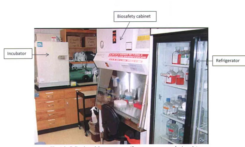
Cell culture is another standard method for the detection of bacterial or viral pathogens. To perform this method specific laboratory setup, bio safety cabinet, hepa frlter (Fig. 1.3), and trained personnel are needed. Although reliable this technique takes 3-7 days to obtain results. Additionally, since live cultures of pathogens are required, biosafety rules must be strictly maintained according to their safety level. For this reason, this method is not suitable for limited resource settings such as remote clinics as well as for POC application. Therefore, a real need exists for more rapid, sensitive and specific diagnostic technologies for infectious disease to replace the time-consuming and limited culture methods (Yageret al. 2008).
Fig. 1.3: Cell culture laboratory setup (Source: www.vetmed.wisc.edu)
A major class of POC diagnostic tests is the lateral flow test. In this test, a membrane or paper strip is used to indicate the presence of protein markers such as pathogen antigens or host antibodies. On a membrane, the addition of the sample induces a capillary action. The sample flows across the membrane, reacts with reagents that are pre-embedded in the membrane, and flows over an area that contains captured molecules. The labeled captured analyes fotm a visible band on the membrane which is detected by eye. (Chin et aI.2012). Lateral flow tests are used for the diagnosis of pregnancy as well as infections with streptococcus or flu or to diagnose HIV. Although the test is simple to perform, the single-flow action does not mimic the multi-step procedures of laboratory-based assays that are crucial for producing highly reproducible, quantitative, and sensitive results. Blood glucose analysis is another major class of successful POC tests. This test is also performed on membranes but is distinct from lateral flow immunoassays. It uses signal amplification by a redox enzyme, typically ending in an electrochemical leadout (Price et al. 2010).
Many POC test systems are devised as easy-to-use membrane-based test strips, often enclosed by a plastic test cassette. This concept is often realized in test systems for detecting pathogens.
Recently such test systems for rheumatology diagnostics have been developed (Zhang 2004). These tests require only a single drop of whole blood, urine or saliva, and they can be performed and interpreted by any general physician within minutes.
For nucleic acid amplification-based testing, polymerase chain reaction (PCR) was the first technology to be used in POC devices. However, PCR requires an expensive thermal cycler and relatively sensitive optics for real-time detection. The use of a thermal cycler and sensitive optics are not ideally suited for POC devices where the goal of POC is not only shorten the test time, but also to reduce the cost (Steel et al. 1999). Accordingly, isothermal techniques for DNA/RNA amplification represent a more promising option for nucleic acid amplification-based testing (Ho et al. 1987, Rodriguez and Bard 1990, Maruyama et al. 2002, Asiello and Baeumner 201I, Nagatani et al. 2011).
The commonly used isothermal technologies are NASBA, RCA, HDA, LAMP. Among these, LAMP has become a preferred technique for diagnosis of infectious diseases at the POC due to its rapidity, low equipment cost and robustness to inhibitors present in the clinical sample (Notomi et al. 2000, Defever et al.2011). The LAMP assay can also be monitored in real-time (Ho et al. 1987, Ahmad et aL.2011, Ahmed et al. 2013) for quantification, and can be used to differentiate single nucleotide polymorphisms (Ikeda et al. 2007). Human health related applications that would benefit from quantitative and multiplexed POC genetic testing include measuring viral load with HIV (Shen et al.2011), differentiation of point mutations for multiple drug resistance tuberculosis (Lee et al. 2010), or measurement of microRNA panels for diagnosing cancer (Li et al.2011). In general, genetic testing is aimed at detecting the presence or absence of genetic markers such as pathogen-specific virulence genes, antibiotic resistance genes, or disease-specific mutations. Sequencing, the most desirable genetic analysis, is not yet available using low-cost POC devices.
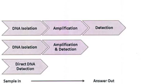
To incorporate nucleic acid (NA) testing into the POC device, several formats can be employed to effect sequence-specific detection as shown in Fig. 1.4.
DNA lsolation DNA lsolation Amplificatlon Amplification & Detection Detection Direct DNA Detection
Sample In Answer Out
Fig. 1.4: Nucleic acid testing formats (Craw and Balachandran 2012)
For the amplification of NAs, isolation or purifrcation of it from a clinical sample is an essential step. This is because, salt or sugar which present in clinical sample inhibit the amplification reaction. Firstly, NAs isolated from a clinical sample, then amplification followed by detection of the amplified product. For example, PCR, where NA (DNA/RNA) isolated from the clinical sample, then amplified in the thermo cycler, and then amplif,red product analyzed. Such methods have been well characterized, are extensively used and are widely applied across genetic determination and infectious diseases. ln other format, NAs are isolated and then amplification and detection steps are performed together. Even sometimes amplification and detection steps are carried out simultaneously without prior NAs isolation. These simplified tests therefore provide a robust format for the development of further NATs. With regard to POC applications, it is essential to simplify the assay procedure and avoid the multiple procedural steps for amplification and separate detection and thereby reduces the time-to-result (Craw and Balachandr an 2012) .
1.4.1 Nucleic acid testing (NAT)
NAT is performed to identify specific nucleic acid sequences from clinical samples. The presence of specific NA sequences indicates the presence of infection or genetic disease, progression and prognosis or, in the case of genomic medicine, suitability for a tailored therapy. In this section, several NAT methods, such as PCR, NASBA, RCA, HDA, LAMP are described.
1.4.2 Polymerase chain reaction (PCR)
PCR is a primer-mediated enzymatic amplification of specifically cloned or genomic DNA sequences. The main purpose of the PCR is to amplify template DNA using thermo stable DNA polymerase enzyme. This enzyme catalyzes the buffered reaction in which an excess of a template DNA (oligonucleotide primer pair) and four building blocks (deoxynucleoside triphosphates ordNTPs) are used to make millions of copies of the target sequence.
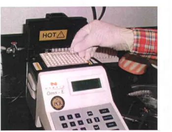
Fig. I .5: Image of a conventional PCR machine
(Source: http://biology.clc.uc.edr.r/fankhauser/labs/genetics/pcr/pcrlrotocol.htm) The PCR requires a repetitive series of the three fundamental steps that defines one PCR cycle. DNA amplification by the PCR is schematically outlined in Fig 1.6. There are three general steps to the process that are repeated for a number of cycles to exponentially increase the number of copies of a specific target region. The whole process is carried out in a thermo cycler (Fig. 1.5) which controls time and temperature according to the command. Genomic DNA is normally double-stranded (DS-DNA) (Kolmodin, Williams et al. 2000).
STEP I is to first unzip the DS-DNA, also denoted denaturation, into two complementary single strands of DNA by heating the reaction mix to 95 oC.
STEP 2 isolates the target region of the genomic DNA by the addition of two primers (Pl & P2), which exactly match two 20-30 unique base pair regions that flank the target region. This is known as annealing
STEP 3 is initiated once time is allowed for the primers to bind to the DNA and involves heating the mix to 72-75 oC at which point a special polymerase builds the DNA strand starting at the primers and continuing in the 5' direction. This is called extension. These three steps are repeated 25-40 times to produce millions of exact copies of the target region of DNA. During the second cycle of this process, extension can occur on both the original copy of genomic DNA and the newest pieces (the colored ones in the Fig. 1.6), thus subsequent extensions are quickly limited precisely to the target region (A).
Ds 3' s' DhJA 5, 3
oerarunrrron I
ssoc
v
3'rnr.ralltttttt:tttttt nrns Illtu AHNEALTT{G PI " -SOOC Fnrl --) EXTENSIOfl <-*^**^*t1,liæ z2ocFig. 1.6: Schematic outline of PCR steps (Source: www.flmnh.ufl.edu/cowries/amplify.html)
1.4.3 Isothermal amplifications
Novel developments in molecular biology of DNA synthesis in vivo demonstrate the possibility of amplifying DNA in isothermal temperature. Unlike PCR, isothermal amplification methods do not require a thermocycling machine to separate the two DNA strands and then to amplify the required fragment. DNA polymerase replicates DNA with various accessory proteins. Therefore, with identification of these proteins, we are able to develop new in vitro isothermal DNA amplification methods by mimicking these in vivo mechanisms. Though there are several
isothermal nucleic acid amplifications, in this section I will discuss the commonly used isothermal methods, such as NASBA, RCA, HDA and LAMP.
1.4.3.1 Nucleic acid sequence -amplifi cation (NASBA)
NASBA is a method in molecular biology which is used to amplify RNA sequences. The NASBA technique has been used to develop rapid diagnostic tests for several pathogenic viruses with single-stranded RNA genomes, e.g. influenza A (Collins et al. 2002), foot-and-mouth disease virus (Collins et al. 2002), severe acute respiratory syndrome (SARS)-associated coronavirus (Keightley et al. 2005), Human bocavirus (HBoV) (Bohmer et al. 2009) and also parasites llke Trypanosomo brucei (Mugasa et al. 2009). NASBA has been introduced into medical diagnostics, where it has been reported to provide more rapid results than PCR, and it can also be more sensitive.
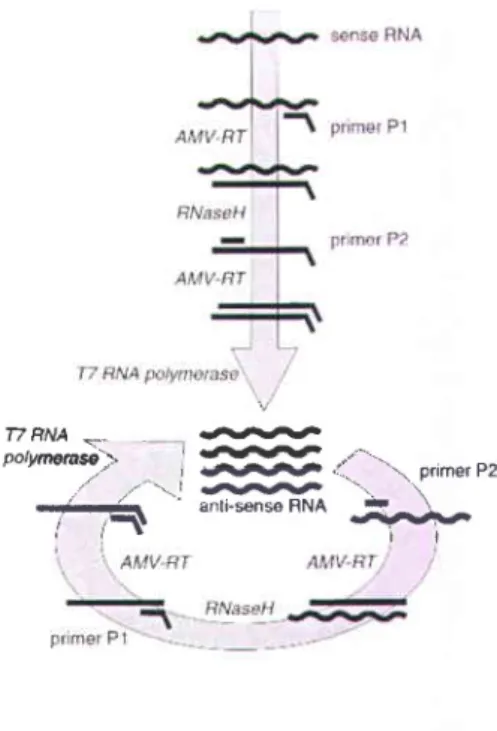
NASBA's main advantage is that it functions at isothermal conditions - usually at a constant temperature of 41 oC. NASBA technology is based on the concerted action of three enzymes (Fig. 1.7):
AMV Reverse Transcriptase: for cDNA synthesis
RNase H: for degradation of the RNA in the heteroduplex RNA-DNA T7 RNA polymerase: for synthesis of RNA from the T7 promotor
NASBA works as follows:
l. RNA template is added to the reaction mixture and then the first primer attaches to its complementary site at the 3'end of the template.
Reverse transcriptase synthesizes the opposite, complementary DNA strand.
RNAse H destroys the RNA template from the DNA-RNA compound (RNAse H only destroys RNA in RNA-DNA hybrids, but not single-stranded RNA).
The second primer attaches to the 5' end of the DNA strand.
Reverse transcriptase again synthesizes another DNA strand from the attached primer resulting in double stranded DNA.
r')
2 .
a J . 4. 5 . L1T7 RNA polymerase continuously produces complementary RNA strands of this template, which results in amplification. The amplicons (newly produced complementary RNA), however, are antisense to the original RNA template.
A cycle can now begin, starting with the RNA strands from the previous step, the second primer and reverse transcriptase and repeating the mechanisms of steps l-6.
Fig. 1.7: Schematic diagram of NASBA
(Source: htp ://nar. oxfordj ournals.org/content/3 0/ 6l e26 lF 2.large jp g)
1.4.3.2 Rolling circle amplifïcation (RCA)
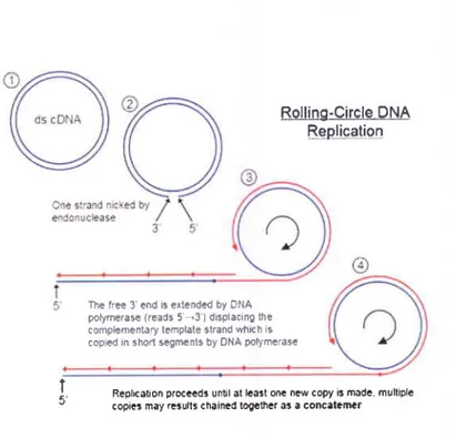
RCA is another isothermal, enzymatic process mediated by certain DNA polymerases in which long single-stranded (ss) DNA molecules are synthesized on a short circular ssDNA template by using a single DNA primer. This is a process of unidirectional nucleic acid replication that can rapidly synthesize multiple copies of circular molecules of DNA or RNA. This replication process involves continual synthesis of a polynucleotide which is 'rolled off of a circular template molecule (Fig. 1.8).
6 . T7 FINA * .ri/\ t potymorasà'1, /momse.:' 'æ..'^ , lJùJ-..'. '\ primerp2 éw T-É anli'6€ns€FNA L2
Rolling-Circle !M ts-epliee-tio,!
Replkôlon proceêd3 until ôl lcart onc ncr copy B môdê. multiple copies may ræulls chaimd logctfÉr as a concatcmcr
Fig. 1.8: Schematic diagram of RCA
(Source: http ://cronodon.com/images/Rolling-circle jpg)
The RCA method, traditionally used for ultrasensitive DNA detection in areas of genomics and diagnostics, has been used more recently to generate large-scale DNA templates for the creation of periodic nanoassemblies. Various RCA strategies have also been developed for the production of repetitive sequences of DNA aptamers and DNA enzymes as detection platforms for small molecules and proteins. Accordingly, RCA is rapidly becoming a highly versatile DNA amplification tool with wide-ranging applications in genomics, proteomics, diagnosis, biosensing, drug discovery, and nanotechnology (Zhao et al. 2008).
RCA uses specific DNA polymerases and primers to allow a circular piece of DNA to be replicated continuously until all reagents are exhausted. The overall reaction takes very little time compared to other methods and does not require NA purification or centrifugation. The results are easily analyzed, some cases, as little as 10 minutes (Alsmadi et al. 2003).
1.4.3.3 Helicase-dependent amplification (HDA)
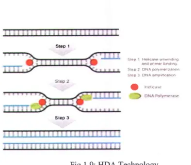
Helicase-Dependent Amplification (HDA) is an isothermal amplification method of nucleic acids. Like PCR, the HDA reaction selectively amplifies a target sequence defined by two primers. However, unlike PCR, HDA uses an enzyme called a helicase to separate DNA, rather than heat (Fig. 1.9). This allows DNA amplification without the need for thermo cycling. Thus,
1
6 'Th€ fr?e 3'cfld ir extendd by oilA polymerôta {rêads 5'-3 I displacing thè complcmenl,ary template strand ïvhÈh is copred ifl shotl spgtÏlcnts by ONA poF/metasc
two types of enzymes are required to complete the reaction: a helicase (to separate the ds-DNA strand) and a DNA polymerase (to precede the reaction). Like in PCR method, DNA polymerase can be is inhibited by elements present in the clinical sample, so the NA needs to be purified before the reaction. The HDA reaction can also be coupled with reverse transcription for RNA analysis. lirt9 I t{*æ ult6rlrÈ s d È ô e D 4 t 4 Ët6r3 tr?lIÂætæÊss : i E F 1 F a a @ l f f i I u.e*** srÊp A
Fig. 1.9: HDA Technology
(Source: https://www.neb.com/productslhOl I 0-isoamp-ii-universal-thda-kit) 1.4.3.4 Loop-mediated isothermal amplification (LAMP)
LAMP is a simple, rapid, specific and cost-effective isothermal nucleic acid (DNA or RNA) amplification technique developed by Notomi and colleagues over a decade ago (Notomi et al. 2000). In this technology, four different specifically designed primers are used to recognize six distinct regions on the target gene. Amplification and detection of the gene can be completed in a single step, by incubating the mixture of samples, primers, DNA polymerase with strand displacement activity and the reaction can proceed at a constant temperature. It provides high amplification efficiency 1l0e-10r0 times in 15-60 minutes). Thus, the presence of the amplified product can indicate the presence of the target gene. Thus this method may be of use in the future as a low cost alternative to detect diseases. The detailed of the LAMP method is described in the
Chapter 2.
5ûÈp r
1.5 Conclusion
POCT minimizes the gap between centralized laboratory diagnostics and rural healthcare service providers. Particularly in infectious diseases such as HW/AIDS and Tuberculosis (TB), where early detection is imperative to improve disease outcome, the development of an accurate test that is simple, rapid and robust can significantly alter the epidemiology and control of the disease. An effective POCT, however, can only serve its full potential when adopted in a comprehensive programmatic context linking patients to on-site case management. Immunochromatographic lateral flow devices for detection of antibody or antigen currently dominate available POCTs, and the development of such devices has relied on the discovery and optimization of definitive biomarkers suitable for such platforms. ln the future, however, there will be an increasing need to develop cost-effective POCTs that address biomarkers that are well established in laboratory settings but are not currently amenable to POCT, such as molecular tests for drug resistance in TB and viral load in HIV and viral hepatitis (Mohd Hanafiah et al. 2013).
There is no doubt that the need for POC diagnostics is crucial in developing countries and low-resource setting laboratories/hospitals in the developed countries. For example, a high-throughput, automated robotic processing instrument is generally not affordable or feasible in low-resource settings that lack the necessary laboratory infrastructure (Yager et al. 2008)
Chapter 2
Loop-mediated isothermal amplifÏcation (LAMP) for point-of-care
diagnostics
LAMP technology is a robust method to identify disease within very short time. The overall cost for this method is very low compared to other existing technologies. With this motivation I used the LAMP method in my MSc thesis work for the detection of Herpes Simplex Viruses.
2.1 Objective of my thesis
To develop a low-cost tool for the detection of Herpes Simplex Viruses suitable for low resource countries.
2.2lntroduction
Herpes simplex virus (HSV) infection is a major problem in both the industrialized and developing worlds. HSV has been characterized into two different serotypes: HSV t1,pe 1 (HSV-1, named as oral herpes) is generally associated with infections in the tongue, mouth, lips, pharynx, and eyes, whereas HSV type 2 (HSV-2, named as genital herpes) is primarily associated with genital and neonatal infections (Aurelian 1992). Both HSV-I and HSV-2 establish lifelong latency in human sensory neuronal ganglia, and subsequently reactivate. Following reactivation, each of these herpes viruses may cause significant clinical symptoms in the individual and may spread to uninfected persons. The virus can also be passed from mother to child during birth. Neonatal infection) can be very serious (Pinninti and Kimberlin 2013). Without treatment. 80% of infants with HSV infection die, and those who do survive are often carry physical damage throughout their life (Brown 2004).In one study in the United States of America (USA), four of nine infants born to women who acquired genital herpes shortly before labour developed neonatal infection, died (Brown et al. 1997). Thus the early diagnosis of the virus is important for the determination of clinical management and for an understanding of the clinical progress and prognosis.
There are many established methods for the identification of both HSV1 and HSV2 viruses. Among them, cell culture (Singh et al. 2005) and serological assays (Wald and Ashley-Morrow 2002) are standard methods of HSV diagnosis. These methods, however, require substantial time to obtain accurate final results. PCR is a highly sensitive and gold standard method for the detection of HSV DNA (Johnson et al. 2000, Gardella et al. 2010) compared to antigenic detection or cell culture methods (Koenig et al. 2001). Bedside monitoring of HSV infection, its progression could also be monitored by the real-time PCR quantitative analysis of viral DNA (Enomoto et al. 2005). This method however, has not yet become a cofitmon procedure in hospital laboratories and low-resource settings and is also not suitable for POC applications due to the requirement of specific expensive equipment (a thermal cycler), dedicated laboratory space, and specially trained technicians.
In 2000, Notomi and his colleagues reported a novel nucleic acid amplification method, he denoted as loop-mediated isothermal amplification (LAMP). In this method DNA is amplif,red under isothermal conditions (between 63 "C and 65 oC), which requires only simple and cost-effective equipment (e.g., a heating block) suitable for use in limited resort setting areas. The technique also exhibits both high specificity and high amplif,rcation efficiency. The LAMP method uses four primers which recognize six distinct target DNA sequences, yields extremely high specificity. This method does not need to denature double strand DNA to a single strand, which is a crucial step in PCR, because the Bsl DNA polymerase enzyme has strand displacement capacity. In contrast to PCR, there is no time lost due to temperature changes in each step. Therefore, the entire method can be conducted in a short time period (30-60 minutes). LAMP also exhibits extremely high amplification efficiency compared to PCR. As the reaction can be conducted at the optimal temperature for enzyme function, the inhibition reactions that often occur at later stages of typical PCR amplifications are less likely to occur (Enomotoet al. 2005). The LAMP method could potentially be a valuable tool for the rapid diagnosis of HSV (Reddy et al. 2011) as well as other infectious diseases in both commercial and hospital laboratories (Notomi et al. 2000).
Plastic/paper-based microfluidic devices possess many of the desired characteristics of a suitable POC viral DNA test (Fu et al. 2011, Pollock et aI. 2012). These diagnostic devices are inexpensive, portable, and simple to operate, making them appropriate for low-resource settings
(Martinez et al. 2010). We developed a simple plastic pouch for the detection of both HSV-I and HSV-2. In this device we could detect viral DNA within 45 minutes using the LAMP method. Since the LAMP reaction is less sensitive to inhibitory substances present in the clinical sample (Enomoto et al. 2005, Kaneko et al. 2005, Defever et al.2011), this allowed us to detect viral DNA without purifying it. The final result was evaluated by the naked eye via the addition of hydroxynaphthol blue GINB) dye in the reaction mix (Goto et aL.2009, Das et al. 2012). HNB is a metal indicator for magnesium and a colorimetric reagent for alkaline earth metal ions, which is usually purple color. With the LAMP reaction, magnesium pyrophosphate is produced as by-product, thus the amount of free magnesium ion (Mg*2) is reduced in the assay mixture. As the magnesium is reduced, due to the change of PH, the color of HNB dye changed from purple to light blue. Thus, positive reaction is indicated by a color change from purple to sky blue. The results of our colorimetric assay were further confirmed by 2% agarose gel electrophoresis.
2.3 LAMP primers:
There are four types of primers based on the six distinct regions of the target gene. The primers are denoted Forward Inner Primer (FIP), Forward Outer Primer (F3), Backward Inner Primer (BIP) and Backward Outer Primer (83)(Notomi et al. 2000). The positions of the primers are shown in the Fig. 2.1.
F.'lcFlcFlc Inrsrt0.\:l il 112 8.1 13 F2 Fl lllc ll2cl|lc j æ t , , n l . ' G " ' Bl ltl
Fig.2.1: Schematic diagram showing the position of LAMP primers in target DNA (Notomi et al. 2000)
t 2 c t l c
ç2 :--è
FIP consists of the Flc region at the 5' end, which is complementary to the Fl region, and the F2 region (at the 3' end) that is complementary to the F2c region of the target DNA.
F3 Primer consists of the F3 region that is complementary to the F3c region.
BIP consists of the 82 region at the 3' end that is complementary to the B2c region, and the Blc region that is complementary to the sequence of B1 region at the 5'end.
83 Primer consists of only 83 region that is complementary to the B3c region.
There are two other primers named Loop Primer Forward (LPF) and Loop Primer Backward (LPB) containing sequences complementary to the single-stranded loop region. These primers are used to increase the amplification rate.
2.4 Principle of LAMP
When the DNA template (the target gene) are incubated together with primers, DNA polymerase eîzyme and other reagents at a constant temperature, the reaction proceed as the following steps: (see Fig. 2.2)
Step 1z FIP primers anneal to the complimentary sequence of double stranded target DNA (light pink), and then initiates DNA synthesis using the Bst DNA polymerase with strand displacement activity which displaces and releases a single stranded DNA. With the LAMP method, in contrast to PCR, there is no need for heat denaturation of the double stranded DNA into a single strand (Figure 2.2).
Step 2: FIP is used to produce the complementary strand of the template DNA starting from the 3' end of the F2 region of the FIP.
Step 3: The F3 Primer anneals to the F3c region, outside of FIP on the target DNA and initiates strand displacement DNA synthesis by which the FlP-linked complementary strand is released. Step 4: A double strand is formed from the DNA strand synthesized from the F3 Primer and the template DNA strand.
Step 5: The FlP-linked complementary strand is released as a single strand because of the displacement by the DNA strand synthesized from the F3 Primer.
This released single strand forms a stem-loop structure at the 5' end because of the complementary of the Flc and Fl regions.
F.lc Flc Ftc Turxct Ir\.4 Bl Bl Bt FT T2 FI 8 l e B t t B I c Flc Fl Fl F l F ? c t l r B l c B h B J c : ! '
ût s2 B3
f 3 c F 2 c f t c . & t r t B t B l(?) T,
(E) ( t ) flc flc t'lc F3c Flc Flc Flc tlc ilc Br 81 lll Bt B2 n3nr Bl 83
f l c r.lt 5' r - t Ï ({} F t n 2 i l Blc BlcB.lc (5) ftc tl Fl F I Blc BlcB3c Blc B2cBlc Ble McBh 12 3'I
t
rl
f l i 0 l t BlcStep 6: In this step BIP initiates DNA synthesis, this newly synthesized DNA strands are separated by 83 primer. Starting from the 3' end, BIP anneals to the DNA strand produced in Step (5), so that synthesis of complementary DNA takes place. As DNA synthesis proceeds, the DNA reverts from a loop structure into a linear structure. The 83 Primer anneals to the outside of the BIP and then, through the action activity of the Bsl DNA polymerase and starting at the 3' end, the DNA synthesized from the BIP is displaced and released as a single strand before DNA synthesis from the 83 primer.
Step 7z B3-primed strand displacement DNA synthesis and the DNA reverts from a loop structue into a linear structure. Double stranded linear DNA is the product at the end of this step.
Step 8: At each end complementary sequences at the same strand (e.g. Fl, Flc and 81, Blc) forms a structure with stem-loops, which looks like a dumbbell structure
The Loop Primers containing sequences complementary to the single stranded loop region (either between the 81 and 82 regions, or between the Fl and F2 regions) on the 5' end of the dumbbell-like structure, provide an increased number of starting points for DNA synthesis for the LAMP method.
Step 9-11: After forming a dumbbell-like structure, the amplification cycle in the LAMP method begins. At this step, Loop Primer Forward (LPF) and Loop Primer Backward (LPB) bind with their complementary sequences (F2c and 82 region respectively) and primed the amplification. At each cycle of amplification, the number and the size of the existing products increased.Thus, at the end ofthe reaction the products can be resolved by agarose gel electrophoresis as a ladder like band instead of single band (Fig. 2.3)
Viewing the animation of the principle of LAMP will assist the understanding of the amplification procedure. (Animation link: http://loopamp.eiken.coip/e/lamp/anim.l-rtrnl)
Fig. 2.3: Analysis of the LAMP product by gel electrophoresis. The amplification reaction was performed at 65 "C for 40 minutes. The products were resolved on a2Yo agarose gel and visualized with ethidium bromide after the exposure to the UV light. Each lane shows a ladder-like band due to the combination of various sized dumbbell-shaped DNA molecule. M= 2KB marker, l: negative control (Water), 2-6: positive control.
2.5 Materials and methods
Before developing the amplification device, the LAMP protocol was first optimized using in conventional 0.2 ml PCR tubes to amplify HSV DNA. The final result was analyzed by the color change of HNB dye from purple to light blue and further confirmed by 2% agarose gel electrophoresis.
2.5.1 Cells and Viruses
African green monkey kidney (Vero) cells were maintained in DMEM, supplemented with 5% newbom calf serum, 0.5u/ml of penicillin and 0.5ug/ml of streptomycin at 37"C and 5% COz. The virus strain KOS (HSV-l) (Jacobson et al. 1998) and strain HG52 (HSV-2) were used. Vero cells were infected at multiplicity of infection (MOD of 0.01 with either HSV-I or HSV-2 and after 3 days virus were harvested and used or DNA extraction was performed with phenol/chloroform.
2.5.2 L ANI} amplifi cation
The amplification reaction was carried out using the previously described LAMP primers for Herpes Simplex Virus type 1 (Kaneko et al. 2005) and Herpes Simplex Virus type 2 (Enomoto et al. 2005). Details of LAMP primers used in this study are listed in Table 2.1. The LAMP reaction was carried out in a total 25 1tL reaction mixture containing 0.2 mM of each of the outer primers (F3/83), 0.8 mM of each of the loop primers (LF/LB) 1.6 mM of each of the inner primers (FIP/BIP). 0.4 mM dNTPs, 0.64 M Betaine (Sigma), 3 mM MgSOa, Bsr DNA polymerase, large Fragment, 1,600 units, (8,000U/ml) (New England Biolabs), lX ThermoPol reaction polymerase buffer (New England Biolabs Inc.) and 5 pL of double-stranded target DNA. Hydroxynapthanol blue 0.15 pllml- was also added to the reaction mixture to visualize the amplification of HSV DNA. Mineral oil (20 pL) was added to each tube to avoid evaporation. The mixture was incubated at 65oC for 45 minutes on a heat block.
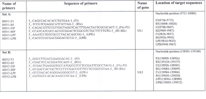
Table 2.1: Primers used for LAMP and their location in the gene
2.5.3 Plastic pouch fabrication and operation
For the development of the device, a conventional reseal-able plastic bag was used (Fig. 2.4). A small plastic chamber (1.5 x 0.2 cm) was fabricated by pressing using a plastic sealer. This small chamber can hold 25 1tL of reaction mixture including 5 prl- of sample. A small plastic spacer was placed into the bottom of the chamber, so that the liquid can easily full and retained by the
Name of primers
Sequence of primers Name
of gene
Location of target sequences
Set A: H S V I - F 3 H S V I - 8 3 H S V I - F I P H S V I . B I P H S V I - L P F H S V I - L P B 5_-CAGCCACACACCTGTGA A-l_(F3) 5_-TCCGTCGACGCATCGTTAG-3_ (B3c) 5 -CAGACGTTCCCTTGGTAGGTCACTTTGACTATTCGCGCACC-3-(Fl c-F2) s -CCATCATCGCCACGTCGGACTCGGCGTCTGCTTTTTGTG-3- (Bt-B2c) 5 -AAATCCTGTCGCCCTACACAGCGG-3_ (LPFc) 5 -CACCCCGCGACGGGACGCCC.3 (LPB) U L I Nucleotide position (972 I - 10080) F3(97s6-9773) 83( 10008- l 0026) F2(9788-9807) B2(9969-9987) F r (9836-e857) Bl(9926-994s) LPF(9810-9833) LPB(9948-9967) S e t B : HSV2-F3 HSV2-83 HSV2-FIP HSV2-BIP HSV2-LPF HSV2.LPB 5_-GGCCTTGACCGAGGACAC.3 (F3) 5_-CGACTCCACCGATCCAGT-3_ (B3c) 5 -TCGACTGAGGGTCCCATGGCCTCCTCCGATTCGCCTACG-3- (Flc-F2) 5 -GCAACCACTACTCCCCCCGACCGTTTCCTCCGGCGTAA-3- (Bl-B2c) s_-GCCGACACAGGGAGGGGCGT-3_ (LPFc) 5 -GATGCCCACACAAGCCGCAA-3 (LPB) UL5 Nucleotide position (138901 -139 140) F 3 ( 1 3 8 9 0 9 - 1 3 8 9 2 6 ) 8 3 ( 1 3 9 1 2 0 - l 3 9 I 3 7 ) F2(138926- 138945) 82( l 39082- l 39099) F l ( r 3 8 9 8 4 - l 3 9 0 0 4 ) B l ( 1 3 9 0 3 0 - 1 3 9 0 s 0 ) LPF(l 3896 r- l 38980) LPB( 139053- l 39072) 2 3
chamber. The reaction mixture and the sample were inserted using the long-edged loading tips and, after loading, the bag was sealed. The sealed bag was kept on a heat block at 65 oC for 45 minutes to amplify the DNA. Hydroxy naphthol blue (HNB), a metal indicator for magnesium and a colorimetric reagent for alkaline earth metal ions, was added to the reaction mixture. A positive reaction is indicated by a color change from purple to light blue. After completion of the reaction, the result was analysed visually without any instrument. The visual analysis was further confirmed by 2% agarose gel electrophoresis. The pouch was punched and the reaction product was collected and loaded to agarose gel
Step 1 Step4 t t U , {
step
1
t .'J
Step 5 0.2 cm Step 3 Step 6Fig. 2.4: Schematic diagram of the fabrication steps of the reaction pouch made by a plastic bag Step 1: A piece of double layered plastic bag cut in a rectangular shape; Step2: Pressed with sealer to make small chambers (L: 1.5 cm and W: 0.2 cm); Step 3: Small pieces of plastic were inserted at the bottom of each chamber which functions as spacers to ensure that the liquid inside the chamber; Step 4: Reagents and sample inserted with long-edge tips; Step 5: Pouch was sealed horizontally with sealer; Step 6: The pack was placed on the heat block for 45 min at 65'C; positive reactions turn to blue from purple.
The following figure (Fig. 2.5) is a photograph. The positive yielded a blue color after they were incubated on a heat block for 45 minutes at 65"C; negative samples purple color.
Purple = Negative
Bhre - Pnsifive
Fig.2.5: A photograph of the reaction in an Eppendorf tube (A) or plastic (B). The purple color changed to blue in a positive reaction.
This qualitative and colorimetric LAMP assay integrated in this very simple and low cost plastic pouch has potential usefulness for the rapid diagnostic of HSV in remote clinics, field site laboratories, hospital laboratories, and also in low-resource countries.
2.5.4 LAMP in real samples
Previous reports have indicated that the LAMP assay's sensitivity is less affected iby the presence of inhibitory substances in clinical samples than is PCR (Enomoto et al. 2005, Kaneko et al. 2005, Defever etaI.2011). To evaluate these advantages, we analyzed the tolerance of ILAMP against a real virus. Viral stocks were diluted in phosphate buffered saline (PBS), distilled water, and culture medium (Dulbecco's Modified Eagle's medium or DMEM). The concentration of viral stock was I .20 x 1 04 PFU/pL. We compared the results with purified DNA.
To assess the detection limit of the LAMP reaction in a real sample, virus stock was serially diluted 1O-fold with PBS. The original virus stock contained lxl04 PFU/FL of 1 and HSV-2 . The diluted viral fluids were used directly. Five microliters of each sample were added to the reaction mixture to a final volume of 25 pL, and the reaction was incubated at 65 oC for I h.
2.6 Results
2.6.1 Optimization of LAMP in tubes
The time, temperature, and dNTP concentrations were optimized for the LAMP assay. We performed the LAMP assay at three different temperatures (60 oC, 63 oC and 66 "C) for both HSV-I and HSV-2 DNA. We observed that HSV-I DNA was amplified at each of the three temperatures, whereas HSV-2 DNA was amplihed at 63-66"C (Table 2.2).
10 mins 20 mins 30 mins 40 mins t0 mins 20 mins 30 mins 40 mins
Fig. 2.6: Agarose gel analysis of time optimization of LAMP assay using HSV DNA. The assay was performed at four different times (10 mins, 20 mins, 30 mins and 40 mins).Water was used as negative control (NC) and l0 nglpl- of DNA used as positive control. (A) Assay using HSV-I DNA. The lowest detection limit was observed 10 fglpl of DNA for HSV-I when we performed the sensitivity test. M:2 kb Marker, 1: NC; 2= l0 nglpL DNA; 3: l0 fglpL DNA;4: NC; 5=
l0 nglprl- DNA; 6: 10 fglpL DNA; 7: NC; 8: l0 nglp'L DNA; 9: l0 fglpL DNA; 10: NC; ll: l0 ng/pl DNA; 12: l0 fglltL DNA. (B) Assay using HSV-2 DNA. The lowest detection limit was observed 1 fglpl of DNA for HSV-2 when we performed the sensitivity test. M:2 kb Marker, l: NC; 2: l0 nglp,L DNA; 3: I fglltL DNA; 4: NC; 5: l0 nglpL DNA; 6: I fglltL DNA; 7: NC; 8: 10 nglp,L DNA; 9: 1 fglpL DNA; l0: NC; ll: 10 ngltrù DNA; 12: I fglpL DNA.
To optimize the time, the LAMP assay was carried out for I0,20,30, and 40 (Fig 2.6). 10 ngl pL of HSV DNA served as positive control and water as
minutes at 65 oC negative control.
fglpL of DNA for HSV-I and 1 fglpl- of DNA for HSV-2) and this concentration we obtained when we performed the sensitivity test (Fig.2.7). An adequate signal for the positive control was observed at 20 minutes although a darker band was found at 30 minutes. However, the lower concentration of HSV-I DNA (10 fglpl-) was detected at 40 minutes. In contrast the HSV-2 positive control (10 nglpl.) was visible at 30 minutes. We observed a very faint band at 40 minutes for HSV-2 DNA (lfglpl-).
2.6.2 Sensitivity and specificity of the LAMP
The sensitivity of the LAMP assay was evaluated using a series of lO-fold diluted samples of HSV DNA (10 nglpl- to 1 fglpl-) and the assay was performed at 65oC for 45 minutes. We performed the test for 45 minutes to make sure that the DNA get adequate time to amplify. This test was performed in tubes and in the plastic pouch using 5 pL of DNA at each concentration.
M 1 2 3 4 5 6 7 8 9 1 0
I
Fig.2.7: Sensitivity of the LAMP assay forthe detection of HSV DNA.
The test was performed in both tube and plastic pouch (tube images are not shown) A: Agarose gel electrophoretic analysis of LAMP products of HSV- I DNA; B: LAMP reaction performed in plastic pouch with HSV-I DNA, the color is due to the HNB dye. Blue indicates a positive reaction where purple indicates a negative result; C: Agarose gel electrophoretic analysis of LAMP products of HSV-2 DNA; D: LAMP reaction was performed in plastic pouch with HSV-2 D N A . In a l l c a s e s M : 2 k b M a r k e r ; l : N C ( H z O ) ; 2 : N C ( l n g / p l E . c o l i D N A ) ; 3 : l 0 n g l p ' L DNA; 4 : lnglp"LDNA; 5 : 100 pglpLDNA; 6 : l0pg/pl DNA; 7 : 1 pg p/-DNA; 8 : 100 fglltL DNA; 9 : l0fg/pl DNA and 10 : I fglpL DNA. Total reaction volume was 25 pL including 5 pL of DNA
I 2 3 4 5 6 7 1 9 1 0 1 2 3 4 5 6 7 I I
^ n
Ei U
The LAMP products were detected by agarose gel electrophoresis and colorimetrically. The results are shown in Fig. 2.7 and summarized in Table 2.2. We observed that as little as l0 fglpl-of HSV-I DNA could be detected in both the tube and the plastic device (Fig 2.7A,2.78 and Table 2.2). By contrast, as low as I fglpl, of HSV-2 DNA was detected in both tube and plastic pouch. For negative controls both water and E. coli DNA were used. No band on the agarose gel (panels A and C, lanes I and 2) or color change (panels B and D, lanes I and 2) was observed in the negative controls.
2.6.3 LAMP in real samples
To verify the effect of inhibitory substances present in the real sample and to assess for the need for DNA extraction and purification, the viral stock was diluted in three different solvents: phosphate buffered saline (PBS), distilled water and culture medium (Dulbecco's Modified Eagle's medium or DMEM). The LAMP reaction was performed at 65oC for 45 min. Products were detected in eppendorf tube (Fig. 2.8 A) or on a 2%o agarose gel (Fig. 2.8 B) The concentration of viral stock was 1.20x10⁴ PFU/pL. We used HSV-I (l ng/prl) DNA as a positive control and water as negative control. A color change was observed in tubes 2-5 (panel A) and indicated that the DNA was amplified. The color did not change in tube 6 in which the virus was diluted using DMEM. This was due to the red color in DMEM. Detection using agarose gel electrophoresis (panel B) showed DNA in all solvents tested.
1 2 3 4 5 6
Fig. 2.8: LAMP with real samples.
LAMP products analyzed colorimetrically in tube (A) and by 2Yo agarose gel electrophoresis (B). M:2 kb Marker; 1 : NC (HrO); 2: PC (HSV I nglpl); 3 = HSV-I virus diluted in water; 4 : HSV-I virus diluted in water (lane 3 and 4 are duplicate samples); 5 : HSV-I virus diluted in PBS;6: HSV-I virus diluted in DMEM. The viral concentration used in tubes 3-6 was 1.2x10 PFU/pL.
Next we attempted to determine the detection limit of HSV DNA in real samples using the LAMP assay performed in the plastic pouch. Viral stocks of HSV-I (Fig 2.9A and 2.98) and HSV-2 (Fig2.9C and2.9D) were serially diluted lO-fold from 10a pftr/pl to 100 pfu/pl in PBS. Purified HSV DNA was used as positive control and water was used as negative control (lane 1). Cell-line (without virus) (Lane 7) was also used to see whether cellular DNA interferes in the amplification reaction. The products were visualized in the plastic pouch (panels A and C) and by agarose gel electrophoresis (panels B and D) Lane 2-6 indicated that we could detect as little as 100 pftr/pl of viral load in both cases.
Fig. 2.9: Detection limit of HSV DNA in a real sample using the LAMP assay carried out in a plastic pouch. A: LAMP product (1-7) of HSV-1. B: Agarose gel elechophoresis of A; C: LAMP product (l-7) of HSV-2. D: Agarose gel electrophoresis of C. M: 2 Kb Marker; 1 : NC (HzO); 2 : too pfu/pi;3: l0rpfr/pi; 4:702 pfu/pL;5: 103 p f u / p l ; 6 : l O a p f u / p l a n d T : c e l l s without virus.
Our findings in Figure 2.8 and 2.9 suggested that DNA extraction step could be omitted since the amplification results obtained with the positive control (purified HSV DNA) matched that obtained from unpurified DNA from viral stock. This will save time as well as cost.
D
Table 2.2:LAMP optimization: Time, temperature and sensitivity
Virus Primer Detection Time (Ampfified dtrO',20', 30',4O'l Temp (Amplified at 60 "C, 6 3 ' C , 6 6 ' C l Sensitivity (DNA was diluted from 10 nglUl-l felull
HSV-1 Set A (Table 1) Saturation time:
50 ng/reaction : 20' -40' 50 fglreaction: 40'
60-66 "C 50 fglreaction
HSV-2 Set B (Table 1) Saturation time:
50 nglreaction: 30'-40' 5 fglreaction: 40'
6 3 - 6 6 ' C 5 fglreaction
2.7 Discussion
To optimize the LAMP method, two different concentrations of dNTPs (0.4 mM and 1.4 mM) were used. The products of the LAMP reaction were aîalped by agarose gel electrophoresis and showed that both concentrations yielded the desired bands (data not shown). When 1.4 mM dNTP was used, the color changed from purple to blue before the addition of sample. One of the possible explanations is, when higher concentration of dNTP was added, the basic pH of the dNTP changed the color of the dye (HNB). Thus, a lower concentration of dNTP (0.4 mM) was used. The assay was carried out at 65 'C for 45 minutes at 0.4 mM dNTP concentration.
To examine see the cross reactivity of the primers used in these assays, we amplifred E. coli DNA and water as negative control. We did not observe any color change (plastic pouch) or band (agarose gel) in E. coli DNA or for the negative control (Fig 2.7). This proves the specificity of the primers used in the assay as well as the lack of interference of the plastic material in the reaction. The sensitivity of the assay towards viral DNA was evaluated by carrying out the LAMP reaction in tubes and in the plastic pouch. We observed the same detection limit in both the tube and the plastic pouch which was lOfg/pl (or 50 fglreaction since 5 pf of sample was used) for HSV-I (Fig2.7A,2.78) andl fglpL or 5 fglreaction for HSV-2 (Fig. 2.7D, Fig. 2.7D). These findings suggested that the tube and the plastic pouch function comparably. The detection limit of real virus was 100 pfuipl of viral concentration in both cases (Fig 2.9A aîd 2.98 for HSV-I and Fig ZSC and 2.9D for HSV-2). We noted that the assay
conducted with the cells lacking the virus (Lane 7) did not result in a color change, which prove that the cellular DNA does not interfere in amplif,rcation reaction.
The LAMP method has been incorporated in different kinds of POC devices to detect infectious diseases (Fang et al. 2010, Fang et al.2011). These devises need complex mechanical micro fabrication steps. Paper or plastic-based devices are comparatively cheap and easy to handle. Some other paper-plastic based devices are available (Rohrman and Richards-Kortum 2012),but they need sample processing and result analysis steps. Roskos and colleagues coupled isothermal amplification with NALF (Nucleic Acid Lateral Flow) (Roskos et al.2013). They developed a cartridge which consisted of a reaction pouch and a pump pouch. Several steps were required to fabricate the device, in addition to a sample processing step. The final result was analyzed by a lateral flow assay.
The plastic pouch reaction vessel is very simple and does not require any mechanical micro fabrication steps. The only laboratory instruments required to perform the assay are a micropipette, pipette tips, and a heating block. The attractive feature here is to incorporate the LAMP technique. Because the LAMP is less affected by the other elements present in the real sample, we do not need to process or purify the samples. The time consuming the sample processing steps, which typically take almost one hour, can be omitted.
Another advantage is the use of the HNB dye, which yields a color change after the reaction. HNB, a metal indicator for free magnesium and a colorimetric reagent for alkaline earth metal ions, was added to the reaction mixture which is usually purple color. When the LAMP reaction proceeds, magnesium pyrophosphate is produced, thus reduces the amount of free magnesium ion (Mg*2) in the reaction mixture. Accordingly, the color of the HNB dye changes from purple to light blue, indication of a positive reaction. Therefore, after completion of the reaction, the result can be analyzed visually without any instrument. We do not need to perform gel electrophoresis or to connect with other analyser which could save another hour. Our results can be obtained in only 45 minutes. With this plastic pouch, only qualitative analysis is possible which its limitation seems. The device is lightweight, small, and easy to make. Therefore, I believe all these together can make it a perfect candidate for POC diagnosis both in the laboratory and in low-resource settings.
Further improvement and modification is needed to use this plastic device in the field. We are planning to lyophilize the primers in paper then place inside the plastic bag, which will reduce reagent addition steps and also allow for storage at room temperature. By changing the pattern of sealing by the sealer, we can modify the design of this device. For example, to make a multiplexed device, we will make one sample loading hole, so the sample will move to different chambers where different lyophilized primer sets are already stored. In this manner, more than one virus could be detected in the same device. We are also planning to improve the material quality and design to make a perfect device for POC application.
2.8 Conclusion
We have developed a plastic device that allows the detection of 100 pfu/pl of herpes simplex virus 1 and 2 (HSV-I and -2) by loop-mediated isothermal amplification within 45 minutes. With this pouch there is no need for prior sample purification. In addition, colorimetric detection by eye makes analysis simple. This device is easy to handle and portable, without the need for expensive instruments. It is also low cost, which it a perfect candidate for point of care diagnosis both in the laboratory and in low-resource countries.
Chapter 3
Real-time detection of LAMP product
LAMP products can be detected by different techniques, for example, colorimetrically, by agarose gel electrophoresis, electrochemically, or even by the naked eye. In the previous chapter, the colorimetric detection of LAMP products was investigated. In this chapter, the electrochemical detection of LAMP product is described. The following work outlines the real-time monitoring of LAMP-amplified products using a redox probe as performed by our group at INRS. This work has been published in Analyst, 2013 where I am cited as a co-author. My contribution to the publication includes carrying out bacterial preparations, DNA purification and all of the LAMP reactions.
Analyst
RSC
Publishing
Cite this: Ar,.r/t/s t, 20'| 3. I 38. 907
Real-time
electrochemical
detection of pathogen DNA
using electrostatic
interaction of a redox probet
Minhaz Uddin Ahmed,*ob Sharifun Nahar,ô Mohammadali Safaviehu and Mohammed Zourob"o'
Abstract
"Electrostatic redox probes interaction has been widely rendered for DNA quantification. In this report our group has established a prooÊof-principle by using the ruthenium hexaamine molecule [Ru(NH3)6]'* u, u redox probe. We utilized real-time electrochemical monitoring of a loop-mediated isothermal amplification (LAMP) amplicon of target genes of Escherichia coli and Staphylococcus aureus by square wave voltammetry (SWV). Ruthenium hexaamine interaction with free DNAs in solution without immobilization onto the biochip surface enabled us to discard the time-consuming ovemight probe immobilization step in DNA quantifrcation. We have measured the changes in the cathodic current signals using screen printed low-cost biochips both in the presence and the absence of LAMP amplicons of target DNAs in the solution-phase. By using this novel probe, we successfully carried out the real-time isothermal amplification and