Validation of Adult Bone Marrow Stromal Cells in
Cellular Therapy Protocols, using a Mouse Model for
Parkinson’s Disease
Validation of Adult Bone Marrow Stromal Cells in
Cellular Therapy Protocols, using a Mouse Model for
Parkinson’s Disease
V. Neirinckx
1, E. Laudet
1, B. Rogister
1,2,3and
S. Wislet-Gendebien
1.
1GIGA Neurosciences, University of Liège, Belgium ;
2GIGA Development, stem cells and regenerative medicine, University of Liège, Belgium. 3Dept. of Neurology, C.H.U., Liège, Belgium;
Parkinson’s disease (PD) is the second most common neurodegenerative disorder after Alzheimer’s disease, with an estimated prevalence of 0,3% of the entire population in industrialised countries, reaching about 1% in people over 60 years of age. The main characteristic of PD relies in a progressive loss of dopaminergic neurons in the Substantia Nigra pars compacta (SNpc), resulting in a deficient dopamine release in the striatum and then promoting important defects in motility regulation. Unfortunately, motor symptoms are generally diagnosed once 80% of the neurons in the nigrostriatal pathway are already lost. The emergence of new neuroprotective and/or neurorestorative strategies is then raising hope, and a lot of researches now focus on cellular therapy protocols. Previous studies and clinical trials have already demonstrated significant improvement in motor symptoms and nigral lesions after grafting fetal dopaminergic midbrain neurons in PD patients’ brains. However, ethical and technical issues concerning the use of human fetuses leave no option but finding other ways to proceed. Adult bone marrow stromal stem cells (BMSCs) have already been suggested as good candidates for cell therapy in CNS lesions, as those cells are multipotent (including the ability to differentiate into functional neurons) and they can be easily harvested from the patient himself allowing autologous graft. These BMSCs are actually an heterogeneous population of cells at various stages of commitment, and deriving from different embryonic lineages. Indeed, it has recently been demonstrated that some BMSCs originate from the embryonic neural crest (NC), while the main part of bone marrow stroma arises from mesoderm (M). After characterizing the different properties of NC-BMSCs and M-BMSCs in terms of neural fate in vitro, our aim was to investigate the potential usefulness of both populations in the context of a neurological pathology. We have then validated a PD mouse model and started setting up a cell therapy experiment, using stereotaxic brain injection of neural crest-derived BMSCs and mesenchymal BMSCs.
Parkinson’s disease (PD) is the second most common neurodegenerative disorder after Alzheimer’s disease, with an estimated prevalence of 0,3% of the entire population in industrialised countries, reaching about 1% in people over 60 years of age. The main characteristic of PD relies in a progressive loss of dopaminergic neurons in the Substantia Nigra pars compacta (SNpc), resulting in a deficient dopamine release in the striatum and then promoting important defects in motility regulation. Unfortunately, motor symptoms are generally diagnosed once 80% of the neurons in the nigrostriatal pathway are already lost. The emergence of new neuroprotective and/or neurorestorative strategies is then raising hope, and a lot of researches now focus on cellular therapy protocols. Previous studies and clinical trials have already demonstrated significant improvement in motor symptoms and nigral lesions after grafting fetal dopaminergic midbrain neurons in PD patients’ brains. However, ethical and technical issues concerning the use of human fetuses leave no option but finding other ways to proceed. Adult bone marrow stromal stem cells (BMSCs) have already been suggested as good candidates for cell therapy in CNS lesions, as those cells are multipotent (including the ability to differentiate into functional neurons) and they can be easily harvested from the patient himself allowing autologous graft. These BMSCs are actually an heterogeneous population of cells at various stages of commitment, and deriving from different embryonic lineages. Indeed, it has recently been demonstrated that some BMSCs originate from the embryonic neural crest (NC), while the main part of bone marrow stroma arises from mesoderm (M). After characterizing the different properties of NC-BMSCs and M-BMSCs in terms of neural fate in vitro, our aim was to investigate the potential usefulness of both populations in the context of a neurological pathology. We have then validated a PD mouse model and started setting up a cell therapy experiment, using stereotaxic brain injection of neural crest-derived BMSCs and mesenchymal BMSCs.
Introductio
n
The experimental protocol is based on an acute i.p. administration of 1-methyl-4-phenyltetrahydropyridine (MPTP) (Jackson-Lewis, 1995). 14-week-old C57Bl/6J mice are injected with
20 mg/kg MPTP (Four times a day, at 2 hours interval). Saline solution is injected as control in sham
animals.
Using tyrosine-hydroxylase (TH) immunoreactivity, we confirmed that the number of dopaminergic cell bodies per plane in the SNpc of MPTP-treated mice was at least twice decreased (45,93 ± 4,86) compared to control (129,40 ± 10,50) (p < 0,001), and a loss of about 90% of axonal fibers was observed in the striatum of these animals (Figure 1). Ventral tegmental area (VTA neurons) are not significantly affected by MPTP, attesting for its specificity for nigral neurons.
G I G A
Université de Liège
1. Validation of a PD
mouse model
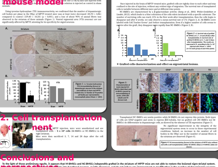
Survival rate of grafted cells.Once injected in the brain of MPTP-treated mice, grafted cells are tightly close to each other and stay confined to the site of injection, without any evident sign of migration. The survival rate of transplanted cells is variable between different mice and at different time points.
NC-BMSCs are characterized by a -galactosidase activity β (Jiang et al., 2002; Wislet-Gendebien et Laudet, 2012), which leads to a blue coloration of the cells when incubated with a specific substrate. The
number of surviving cells can reach 15% in the first week after transplantation, then the cells begin to disappear and after 4 weeks, we only observe a mean survival rate of 1% (Figure 2, A). M-BMSCs were labeled with Cell Tracker Green® just before transplantation. Even if a higher number of cells seems to
survive after the graft, they disappear more rapidly than NC-BMSCs (Figure 2, B).
j
Grafted cells characterization and effect on nigrostriatal lesions.
Transplanted NC-BMSCs are nestin-positive while M-BMSCs do not express this protein. Both types of cells are GFAP-negative and seem to express III-tubulin, but no grafted cell (NC-BMSCs nor M-β BMSCs) do differentiate in dopaminergic cells, as showed by the absence of TH expression (Figure 3).
Survival rate of grafted cells.
Once injected in the brain of MPTP-treated mice, grafted cells are tightly close to each other and stay confined to the site of injection, without any evident sign of migration. The survival rate of transplanted cells is variable between different mice and at different time points.
NC-BMSCs are characterized by a -galactosidase activity β (Jiang et al., 2002; Wislet-Gendebien et Laudet, 2012), which leads to a blue coloration of the cells when incubated with a specific substrate. The
number of surviving cells can reach 15% in the first week after transplantation, then the cells begin to disappear and after 4 weeks, we only observe a mean survival rate of 1% (Figure 2, A). M-BMSCs were labeled with Cell Tracker Green® just before transplantation. Even if a higher number of cells seems to
survive after the graft, they disappear more rapidly than NC-BMSCs (Figure 2, B).
j
Grafted cells characterization and effect on nigrostriatal lesions.
Transplanted NC-BMSCs are nestin-positive while M-BMSCs do not express this protein. Both types of cells are GFAP-negative and seem to express III-tubulin, but no grafted cell (NC-BMSCs nor M-β BMSCs) do differentiate in dopaminergic cells, as showed by the absence of TH expression (Figure 3).
To the light of those preliminary results, it appears that M-BMSCs and NC-BMSCs independtly grafted in the striatum of MPTP mice are not able to restore the lesioned nigro-striatal system. Indeed, transplanted cells don’t seem to differentiate into functional neurons, quickly disappear, and do not induce any improvement or modification in the number of dopaminergic neurons in the nigrostriatal pathway. Consequently, we tend to assume that those cells in such a “stem” state were not competent in those conditions. In accordance to several studies demonstrating a supportive effect of neuronal-primed MSCs in other models of neurological diseases (Fu et al. 2006, Hong et al. 2011, Xu et al. 2011), we hypothesize that maybe a pre-differentiation step would be required to trigger NC-BMSCs and M-BMSCs into a neuronal fate before grafting them in a MPTP-mouse brain, and that maybe one type of cells would exert better properties than the other one when used in those cellular therapy conditions. Therefore, we now prospect to test the ability of both types of BMSCs to differentiate in dopaminergic neurons in vitro ( Trzaska et al., 2007,2011), to finally find out the most valuable cell population and the most promising protocol to develop in terms of cellular therapy in PD patients.
To the light of those preliminary results, it appears that M-BMSCs and NC-BMSCs independtly grafted in the striatum of MPTP mice are not able to restore the lesioned nigro-striatal system. Indeed, transplanted cells don’t seem to differentiate into functional neurons, quickly disappear, and do not induce any improvement or modification in the number of dopaminergic neurons in the nigrostriatal pathway. Consequently, we tend to assume that those cells in such a “stem” state were not competent in those conditions. In accordance to several studies demonstrating a supportive effect of neuronal-primed MSCs in other models of neurological diseases (Fu et al. 2006, Hong et al. 2011, Xu et al. 2011), we hypothesize that maybe a pre-differentiation step would be required to trigger NC-BMSCs and M-BMSCs into a neuronal fate before grafting them in a MPTP-mouse brain, and that maybe one type of cells would exert better properties than the other one when used in those cellular therapy conditions. Therefore, we now prospect to test the ability of both types of BMSCs to differentiate in dopaminergic neurons in vitro ( Trzaska et al., 2007,2011), to finally find out the most valuable cell population and the most promising protocol to develop in terms of cellular therapy in PD patients.
Conclusions and
perspectives
Striatum Midbrain M P T P ( n = 5 ) Sh a m ( n = 5 ) A.Figure 1 : A. Tyrosine hydroxylase (TH) immunolabeling (brown) of dopaminergic neurons in the striatum
and midbrain of sham and MPTP-treated mice. B. Number of TH-positive
cell bodies per representative mesencephalic plane (Mean +/- SEM). SNpc SNpc VTA VTA Sham MPTP 0 20 40 60 80 10 0 12 0 14 0 Sham MPTP 0 20 40 60 80 100 120 * * * SNpc VTA B.
2. Cell transplantation
experiment
Five days after MPTP injection, mice were anesthetized and aretransplanted with ~ 5 x 10⁴ cells (M-BMSCs or NC-BMSCs) in the
right striatum.
Mice were then sacrificed 3, 7, 14 and 28 days after the cell transplantation.
3. Cell therapy results
N u m b e r o f TH + c e ll b o d ie s p e r p la n e N u m b e r o f T H + c e ll b o d ie s p e r p la n e 3 7 1 4 2 8 0 2 4 6 8 1 0 1 2 1 4 1 6 Days post-transplantation % o f su rv iv in g ce lls 3 7 1 4 0 2 4 6 8 1 0 1 2 1 4 1 6 Days post-transplantation % o f su rv iv in g ce lls
Figure 2 : A. Survival rate of grafted NC-BMSCs at 3, 7, 14 and 28 days after transplantation. B. Survival rate of grafted M-BMSCs at 3, 7, and 14 days after transplantation. The results are expressed in %, according to the 5 x 10⁴
injected cells (Mean +/- SEM) (n=3 in each group at each time point).
A. B. M P T P ( n = 5 ) S h a m ( n = 5 ) Figure 3 : Characterization of brain-injected NC-BMSCs (dark blue) and
M-BMSCs (green) by immunolabeling of nestin, GFAP,
BIII-tubulin and TH (brown/red).
No significant improvement in the integrity of the nigrostriatal system seem to occur in those experimental conditions. Indeed, no increase in the number of cell bodies in the SNpc nor in the number of axonal fibers in the striatum are observed (Figure 4).
Cell graft
Figure 4: TH immunostaining (brown) of the striatum of MPTP mice without and with a cell transplantation (either NC-BMSCs (blue) or M-BMSCs (data not
shown).
Nestin GFAP βIII-tubulin TH
M -B M S C s N C-B M SC s
We would like to thank the Fonds National pour la Recherche Scientifique (FNRS), University of Liège, and Fonds Léon Frédéricq. We also thank Alice Marquet for her helpful technical support.