Comparative Loss-of-Function Screens Reveal ABCE1 as an
Essential Cellular Host Factor for Efficient Translation of
Paramyxoviridae and Pneumoviridae
Danielle E. Anderson,
aKristin Pfeffermann,
bSo Young Kim,
c,dBevan Sawatsky,
bJames Pearson,
c,d* Mikhail Kovtun,
cDavid L. Corcoran,
cYvonne Krebs,
bKristmundur Sigmundsson,
eSharon F. Jamison,
cZhen Zhen Joanna Yeo,
a*
Linda J. Rennick,
fLin-Fa Wang,
aPierre J. Talbot,
gW. Paul Duprex,
f*
Mariano A. Garcia-Blanco,
a,c,d,hVeronika von Messling
a,b,gaProgramme in Emerging Infectious Diseases, Duke-NUS Medical School, Singapore, Singapore
bVeterinary Medicine Division, Paul-Ehrlich-Institute, Langen, Germany
cDepartment of Molecular Genetics and Microbiology, Duke University, Durham, North Carolina, USA
dDuke Center for RNA Biology, Duke University, Durham, North Carolina, USA
eProgramme in Cardiovascular and Metabolic Disorders, Duke-NUS Medical School, Singapore, Singapore
fNational Emerging Infectious Diseases Laboratories, Boston University, Boston, Massachusetts, USA
gINRS-Institut Armand-Frappier, University of Quebec, Laval, Canada
hDepartment of Biochemistry and Molecular Biology, University of Texas Medical Branch, Galveston, Texas,
USA
ABSTRACT
Paramyxoviruses and pneumoviruses have similar life cycles and share
the respiratory tract as a point of entry. In comparative genome-scale siRNA screens
with wild-type-derived measles, mumps, and respiratory syncytial viruses in A549
cells, a human lung adenocarcinoma cell line, we identified vesicular transport, RNA
processing pathways, and translation as the top pathways required by all three
vi-ruses. As the top hit in the translation pathway, ABCE1, a member of the
ATP-binding cassette transporters, was chosen for further study. We found that ABCE1
supports replication of all three viruses, confirming its importance for viruses of both
families. More detailed characterization revealed that ABCE1 is specifically required
for efficient viral but not general cellular protein synthesis, indicating that
paramyxo-viral and pneumoparamyxo-viral mRNAs exploit specific translation mechanisms. In addition to
providing a novel overview of cellular proteins and pathways that impact these
im-portant pathogens, this study highlights the role of ABCE1 as a host factor required
for efficient paramyxovirus and pneumovirus translation.
IMPORTANCE
The Paramyxoviridae and Pneumoviridae families include important
human and animal pathogens. To identify common host factors, we performed
genome-scale siRNA screens with wild-type-derived measles, mumps, and
respi-ratory syncytial viruses in the same cell line. A comparative bioinformatics
analy-sis yielded different members of the coatomer complex I, translation factors
ABCE1 and eIF3A, and several RNA binding proteins as cellular proteins with
pro-viral activity for all three viruses. A more detailed characterization of ABCE1
re-vealed its essential role for viral protein synthesis. Taken together, these data
sets provide new insight into the interactions between paramyxoviruses and
pneumoviruses and host cell proteins and constitute a starting point for the
de-velopment of broadly effective antivirals.
KEYWORDS
ABCE1, Paramyxoviridae, Pneumoviridae, RNAi screen, host factor,
respiratory syncytial virus
Citation Anderson DE, Pfeffermann K, Kim SY,
Sawatsky B, Pearson J, Kovtun M, Corcoran DL, Krebs Y, Sigmundsson K, Jamison SF, Yeo ZZJ, Rennick LJ, Wang L-F, Talbot PJ, Duprex WP, Garcia-Blanco MA, von Messling V. 2019. Comparative loss-of-function screens reveal ABCE1 as an essential cellular host factor for efficient translation of Paramyxoviridae and Pneumoviridae. mBio 10:e00826-19.https://doi .org/10.1128/mBio.00826-19.
Editor Diane E. Griffin, Johns Hopkins
Bloomberg School of Public Health
Copyright © 2019 Anderson et al. This is an
open-access article distributed under the terms of theCreative Commons Attribution 4.0 International license.
Address correspondence to Mariano A. Garcia-Blanco, [email protected], or Veronika von Messling,
[email protected]. * Present address: James Pearson, Bayer Crop Science LP, Research Triangle Park, North Carolina, USA; Zhen Zhen Joanna Yeo, Cancer Science Institute of Singapore, Singapore, Singapore; W. Paul Duprex, Center for Vaccine Research, University of Pittsburgh, Pittsburgh, Pennsylvania, USA.
D.E.A. and K.P. contributed equally to this work. This article is a direct contribution from a Fellow of the American Academy of Microbiology. Solicited external reviewers: Stacey Schultz-Cherry, St. Jude Children's Research Hospital; Michael Buchmeier, University of California, Irvine.
Received 4 April 2019 Accepted 5 April 2019 Published 14 May 2019
crossm
®
on August 20, 2019 at INRS-Institut Armand-Frappier
http://mbio.asm.org/
M
embers of the family Paramyxoviridae have recently been reclassified into two
families: Paramyxoviridae and Pneumoviridae (1). Both families include a broad
range of respiratory viruses with great relevance for human and animal health. These
viruses all have nonsegmented RNA genomes of negative polarity, and many of the
viral proteins share considerable structural and functional homology within a genus
and even between genera (2, 3). The negative-strand RNA genome by itself is not
infectious. Instead, the viruses rely on their own incoming polymerase complex for
transcription and replication (4, 5), resulting in relative independence from cellular
processes during these infection stages. Thus, cellular proteins involved in different
aspects of the Paramyxoviridae and Pneumoviridae life cycle are much less well
char-acterized than those of other virus families.
The availability of genome-scale loss-of-function screens has enabled an unbiased
investigation of the role of cellular proteins in diverse processes (6, 7). As obligate
intracellular parasites, viruses critically depend on the exploitation of the cellular
environment for their amplification, and genome-scale siRNA screens of infected cells
have yielded new insights in the host cell functional interactome of many virus families
(8, 9). For negative-strand RNA viruses, several proteins within the coatomer complex
I (COPI), cellular proteins essential for the secretory pathway, were originally identified
in a vesicular stomatitis virus (VSV) genome-scale siRNA screen (10) and then found to
also be required for replication of human parainfluenza virus 3 (HPIV3) (10), suggesting
a role for this pathway in the life cycle of several negative-strand RNA viruses. Screens
with human respiratory syncytial virus (HRSV) revealed actin-related protein 2 and the
calcium pump SPCA1 as host factors involved in different aspects of particle
matura-tion, egress, and spread, the last of which is also required for other RNA viruses (11, 12).
In addition, the large ribosomal subunit protein 40, one of the hits in the VSV screen,
has been shown to be specifically needed for the translation of negative-stranded RNA
virus mRNAs (13), but other host factors involved in viral mRNA translation remain to
be identified.
Measles virus (MeV), mumps virus (MuV), and HRSV are representatives of the
Paramyxoviridae and Pneumoviridae families with high clinical relevance. All three
viruses are transmitted by aerosol or large droplets as well as through contact and
fomites. MeV and MuV result in systemic infection, whereas HRSV remains restricted to
the respiratory tract (2). While the MeV and MuV vaccines are among the most
successful vaccines available today (14–18), an approved vaccine for HRSV remains an
unmet need. Aside from a prophylactic monoclonal antibody-based treatment against
HRSV, no approved therapeutics are available (19). Given the general propensity of
these viruses to infect the respiratory tract, identification of common cellular host
factors could lead to rapid symptom-based treatment options with rationally designed
broad-spectrum therapeutics. Towards this end, and to learn more about the biology
of these viruses, we performed comparative genome-scale siRNA screens of the three
viruses. To reproduce the common natural environment as closely as possible, the
human lung adenocarcinoma cell line A549, which shares many key features with the
respiratory epithelial cells (20), was selected for the screens. In addition to the
previ-ously identified secretory pathway (10), our screens identified translation, proteasomal
degradation, and RNA processing as important host cell pathways for the life cycles of
all three viruses.
After secondary screening of candidates and comparison with a screen for Hendra
host factors, we chose to focus our studies on the ATP-binding cassette (ABC)
trans-porter ABCE1. ABCE1 is a member of the superfamily of ABC transtrans-porters that contain
two nucleotide-binding domains and two N-terminal iron-sulfur clusters. Unlike most
ABC domain proteins, ABCE1 lacks a membrane-spanning domain and therefore is
unlikely to have a transporter function (21). ABCE1 plays an important role in translation
(22), as it is crucial for ribosome recycling in vivo and controls ribosome homeostasis
(23, 24). Additional roles have also been attributed to this protein (25–27). ABCE1 was
initially identified as an inhibitor of endoribonuclease L (RNase L) (28). More recently,
ABCE1 was demonstrated to act as an endogenous suppressor of RNA silencing (29),
May/June 2019 Volume 10 Issue 3 e00826-19 mbio.asm.org 2
on August 20, 2019 at INRS-Institut Armand-Frappier
http://mbio.asm.org/
and during HIV infection, it plays a critical role in particle formation by associating with
assembly-competent HIV Gag polypeptides (30–32). Here we show that ABCE1
knock-down strongly inhibits the translation of MeV mRNAs while only modestly affecting
global cellular translation, suggesting that these mRNAs, and likely mRNAs from all
Paramyxoviridae and Pneumoviridae, may be particularly dependent on the function of
this protein.
RESULTS
Genome-scale RNAi screens for host factors involved in the Paramyxoviridae
and Pneumoviridae life cycles. To identify host proteins important for Paramyxoviridae
and Pneumoviridae pathogenic to humans, independent genome-scale siRNA screens
were conducted with MeV and MuV, human pathogens of the Morbillivirus and
Rubu-lavirus genera, respectively, within the Paramyxoviridae family, and HRSV of the genus
Orthopneumovirus within the Pneumoviridae family (Fig. 1A). Recombinant
EGFP-expressing derivatives of wild-type MeV, MuV, and HRSV strains (Fig. 1B) were used to
facilitate quantification of infected cells by detection of green fluorescence (33–35). To
ensure comparable results, all screens were performed in the lung adenocarcinoma cell
line A549 engineered to stably express the human MeV receptor SLAM (A549-hSLAM).
We used a Qiagen genome siRNA library designed to target 21,705 human genes,
with at least four siRNAs per gene, formatted in two pools of two siRNAs each (see
Fig. S1A in the supplemental material). Target annotation for the library was updated
using the GRCh37 human assembly, resulting in a small minority of genes being
targeted with more or less than two pools. At 48 h after siRNA transfection, cells were
infected with the respective virus at an MOI determined to lead to infection of
approximately 50% of cells. When infected cells could be clearly identified and the
cytopathic effect was still limited, which corresponded to 33 h postinfection (hpi) for
the faster-growing MeV, and 72 hpi for MuV and HRSV, the percentage of infected cells
was analyzed using a Cellomics ArrayScan system (Fig. S1B). Infection levels in the
majority of negative-control wells ranged from 45 to 65% and revealed idiosyncratic
behavior for each virus, which is not surprising for individual siRNAs (Fig. 1C).
Impor-tantly, positive-control siRNAs against EGFP or COPA dramatically reduced the
percent-age of EGFP-positive cells for all viruses (Fig. 1C, red bars) nearly to the level of
uninfected cells (gray bars). For MeV and HRSV, the distribution of the siRNA-treated
wells was shifted toward lower infection levels, whereas the distribution in the MuV
screen was almost normal (Fig. 1C). For MeV and MuV, the distribution of the
siRNA-treated wells relative to that of the controls enabled the reliable identification of hits on
both ends of the phenotypic spectrum, while for HRSV, the behavior of the negative
controls suggested potentially less reliable identification of antiviral factors (see
Ta-ble S1).
The identification of the COPI complex, which is known to be required for
negative-strand RNA viruses (10), as a proviral factor for all three viruses (Fig. 2A) and the MeV
receptor SLAMF1 for only the MeV screen (Table S2) provided confidence that the
screening and analysis approaches were appropriately chosen to identify novel
paramyxovirus host factors. The complete data sets for all three screens are provided
in Table S2.
Analysis of individual screens reveals unique host factors for each virus. Each
of the genome-scale screens identified candidate proviral factors uniquely required for
MeV, MuV, or HRSV or shared by only two of these viruses (Fig. 2A). Interestingly, MeV
and MuV each shared more dependency factors with HRSV than they did with each
other, which is surprising given their phylogenetic relationship. Proteins involved in the
cellular translation machinery and control of the cell cycle and, surprisingly, the
activation of the innate immune response were also prominently represented among
the top proviral factors in each of the individual screens (Table S2). For MeV,
synap-totagmin 8, which is expressed in islet cells and involved in insulin expression (36), and
insulin itself were among the top hits, suggesting a hitherto-unknown role of glucose
metabolism in MeV infection. Furthermore, the calcium/calmodulin-dependent protein
on August 20, 2019 at INRS-Institut Armand-Frappier
http://mbio.asm.org/
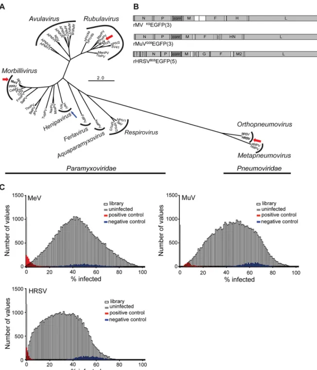
FIG 1 Selection of viruses and RNAi screen performance. (A) A total of 45 N protein sequences were downloaded from GenBank: measles virus (MeV),
rinderpest virus (RPV), canine distemper virus (CDV), pestes-des-petits ruminants virus (PPRV), phocine distemper virus (PDV), cetacean morbillivirus virus (CeMV), mumps virus (MuV), human parainfluenza viruses 1 to 4 (hPIV1 to -4), porcine rubulavirus (PorPV), Mapuera virus (MprPV), parainfluenza virus 5 (PIV5), Menangle virus (MenPV), Tioman virus (TioPV), Newcastle disease virus (NDV) avian paramyxoviruses 2 to 9 (APMV 2 to -9), Sendai virus (SeV), bovine parainfluenza virus 3 (bPIV3), Hendra virus (HeV), Nipah virus (NiV), J paramyxovirus (JPV), Beilong paramyxovirus (BeiPV), Salem virus (SalPV), Tupaia paramyxovirus (TupPV), Nariva virus (NarPV), Mossman virus (MosPV), fer-de-lance virus (FdlPV), Atlantic salmon paramyxovirus (AsaPV), bovine respiratory syncytial virus (BRSV), human respiratory syncytial virus (HRSV), avian metapneumovirus (aMPV), and human metapneu-movirus (hMPV). Amino acids were aligned using the MUSCLE plugin in Geneious 7.1.6. Alignments were visually inspected and manually curated.
The best protein evolution model was determined according to Akaike information criterion (AIC) scores using ProtTest. An LG⫹G⫹F maximum
likelihood analysis was run in PHYML using average likelihood ratio test (aLRT) statistics for branch support. MeV, MuV, and HRSV are boldfaced and highlighted with a thick red arrow to indicate that the viruses were screened, and HeV is boldfaced and highlighted with a thin blue arrow to indicate that data from a separate screen (43) were included in candidate host factor selection. (B) Schematic drawing of recombinant viruses. Gray and white boxes represent open reading frames and UTRs, respectively. The genes are indicated by their respective abbreviations. The inserted EGFP gene is shaded in dark gray, and the number in parentheses indicates the position of the gene in the viral genome. (C) Histogram of percent infection distribution for MeV, MuV, and HRSV. Distribution of percent infection values for each of the individual virus screens. For reference, distribution of positive (red), negative (blue), and uninfected (gray) controls is shown for each screen.
May/June 2019 Volume 10 Issue 3 e00826-19 mbio.asm.org 4
on August 20, 2019 at INRS-Institut Armand-Frappier
http://mbio.asm.org/
FIG 2 Identification and validation of common proviral hits. (A) Common Paramyxoviridae and Pneumoviridae host factors. Extent of
overlap among the genes required for the replication of MeV, MuV, and RSV. The number of genes inhibiting the replication of the
viruses in both A/B and C/D siRNA sets (robust Z score⬍ 0.8) is tabulated in the Venn diagram. (B) Extent of overlap among the genes
required for the replication of MeV, MuV, RSV, and HeV. The number of genes inhibiting the replication of the viruses is tabulated in the Venn diagram. Multiple siRNAs were used to target each of the six top-ranking genes identified by comparative analysis of the three primary screens and the previously published HeV screen (43). (C) MuV validation screen of 24 common hits identified by both Z score and KS analysis. Forty-eight hours after siRNA transfection, cells were infected with MuV at a multiplicity of infection of 1.0 and incubated for 48 h. The percentage of infected cells was calculated, and the mean percent infection was set at 1 for the NSC. The heat map shows the fold change compared to the NSC, and each square represents the average from three experiments for each individual siRNA. Functional categorization of genes is shown beside the heat map. The gene hits shown in red were then selected for MeV validation screening. (D) MeV validation screen of top proviral hits also identified in the HeV screen. Forty-eight hours after siRNA transfection, cells were infected with MeV at a multiplicity of infection of 0.05 and incubated for 48 h. The percentage of infected cells was calculated, and the mean percent infection was set at 1 for the NSC. The heat map shows the fold change compared to the NSC, and each square represents the average from five experiments for each individual siRNA.
on August 20, 2019 at INRS-Institut Armand-Frappier
http://mbio.asm.org/
kinase type II beta chain (CAMK2B), which was previously identified as a host factor
supporting influenza virus RNA transcription (37), also supports MeV replication. The
proapoptotic serine/threonine kinase 3 (STK3), one of the top hits in the MuV screen,
also acts as a proviral factor for influenza virus (38).
Actin-related protein 2, identified as a proviral host factor involved in virus egress
and dissemination in a similar screen using the tissue-culture adapted HRSV A2 strain
(12, 39), did not reach significance in our screen. However, cofilin 1, which associates
with the viral matrix (M) protein in HRSV particles and may modulate particle assembly
and release (40, 41), was among the top hits. Furthermore, dynamin 2 was also
identified as an important HRSV host factor. Dynamins are part of the endocytosis
pathway and were recently implicated in HRSV entry in airway epithelial cells (42). Thus,
our data also provide insights into the unique requirements for these viruses or their
genera.
Comparative meta-analysis identifies common candidate proviral factors. The
screens were first analyzed separately using a custom bioinformatics package that
calculates statistical significance for genes and pathways by considering the
distribu-tion of all wells that belong to a specific gene or pathway by Kolmogorov-Smirnov (KS)
testing. As a further step, we performed a meta-analysis of the three screens that
yielded 179 genes required for all three viruses (false discovery rate
⬍ 0.05), which are
thus candidate host proviral factors for Paramyxoviridae and Pneumoviridae
(high-lighted in Table S3). The list of candidate proviral factors was significantly enriched for
gene products involved in RNA processing, mRNA translation, and
proteasome-mediated degradation (Table S4).
To narrow down the list of the most promising pan-paramyxovirus host factor
genes, we performed a complementary meta-analysis of the three data sets using
robust Z score-based metrics. We identified putative host factors as genes with at least
two independent siRNA pools with Z scores of
⫺0.8 or below across all three screens
(Table S2). This second and very different analysis approach resulted in the
identifica-tion of only 42 proviral gene products required by all three viruses (Fig. 2A). Of these,
24 hits were also identified by the initial KS analysis (Table S5), and we consider these
24 to be the highest-confidence list of candidate proviral factors broadly required by
Paramyxoviridae and Pneumoviridae. Six of these hits (ABCE1, COPB2, FOXP4, HSD11B2,
PRR15, and RBM22) were also identified in a screen for Hendra virus (HeV) host factors
in HeLa cells using a different siRNA library (43) (Fig. 2B), and one (UXS1) was identified
in an HRSV screen in human haploid cells (11). It must be clear that these common hits,
which we address here, were not necessarily among the top hits for each individual
screen. To examine the lists of most potent proviral hits for each individual screen, we
refer the reader to Table S2.
Validation screens confirm membrane trafficking and translation as proviral
pathways. To assess the importance of these common proviral proteins in the
Paramyxoviridae life cycle, we next performed a validation screen of the 24 candidates
identified by both meta-analysis approaches using MuV, since an almost normal
distribution of infection in the siRNA-treated wells was observed in the initial siRNA
screen with this virus (Fig. 1C). Consistent with the fact that most of the 24 common hits
were not among the strongest hits for each of the individual screens, the overall extent
of inhibition of infection was moderate (Fig. 2C). A complementary validation screen
with MeV of the six genes that were also among the proviral factors identified in the
HeV screen (Fig. 2B) yielded similar results (Fig. 2D).
Among the top common proviral genes identified in our analysis were six
compo-nents of the coatomer complex I (ARCN1, COPA, COPG1, COPB1, COPB2, and COPZ1),
and pathway enrichment analysis highlighted multiple vesicle- and COPI-mediated
transport pathways (Tables S2, S4, and S5). The COPI complex is involved in retrograde
vesicular transport from the Golgi complex to the endoplasmic reticulum (44) and was
previously reported to be involved in the life cycle of several negative-stranded RNA
virus families, including paramyxoviruses (10, 43, 45). Our data also revealed important
May/June 2019 Volume 10 Issue 3 e00826-19 mbio.asm.org 6
on August 20, 2019 at INRS-Institut Armand-Frappier
http://mbio.asm.org/
roles for components of the translation machinery: ABCE1, EIF3A, and RPLP1 (Fig. 2C
and D and Tables S2, S4, and S5). While it was previously understood that general
translation components would be absolutely required, our analysis highlights specific
requirements for Paramyxoviridae and Pneumoviridae. For instance, we noted a
dispro-portionate dependence on proteins of the small ribosomal subunit (e.g., RPS9, which
appears in Fig. 2C and Table S5), something that was not observed in similar screens for
flaviviruses (46). Of interest is also the requirement for the ribosomal protein RPLP1, and
the likely requirement of its partner RPLP2, which did not satisfy the most stringent
cutoff criteria but was among the 179 gene products identified as common hits in the
KS analysis. These two components of the ancient ribosomal stalk, which are
dispens-able for bulk cellular translation, are exquisitely required for flavivirus translation (46,
47), suggesting a widespread role in the translation of viral proteins.
ABCE1 supports replication of MeV, MuV, and HRSV. We selected ABCE1 for
further study since it was found to be a common proviral factor for MeV, MuV, and HRSV
in our screens as well as for HeV (43). To reliably detect ABCE1, we generated a rabbit
antipeptide hyperimmune serum targeting the 19 C-terminal amino acid residues of
human ABCE1 and confirmed its specificity using a FLAG-tagged ABCE1 expression
plasmid (Fig. 3A). Since generation of stable ABCE1 knockout cells failed repeatedly,
and prolonged ABCE1 knockdown resulted in reduced cell proliferation, transient siRNA
knockdown was used for all experiments. We then validated the extent of ABCE1
protein knockdown using the most effective siRNAs, ABCE1_5 and ABCE1_6. For both
siRNAs, ABCE1 mRNA copy number was dramatically reduced, and protein expression
levels were decreased by more than 60% (Fig. 3B and C), illustrating specificity and
durability of the knockdown. Cells transfected with a plasmid encoding an
siRNA-resistant ABCE1 protein or C911 control siRNAs retained wild-type ABCE1 and MeV N
protein levels (Fig. 3D and E), demonstrating that the observed proviral effect is ABCE1
specific.
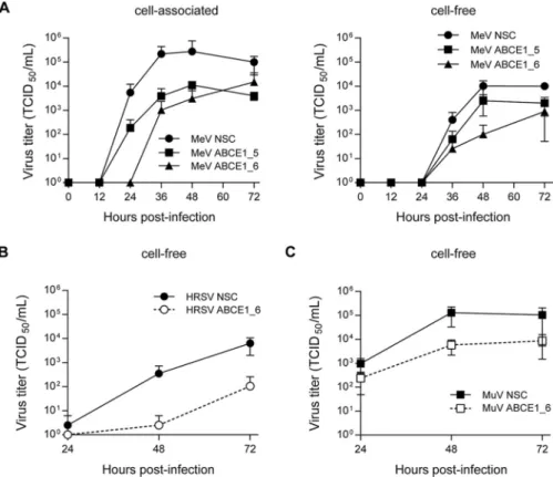
To evaluate the impact of ABCE1 on the viral life cycle, A549-hSLAM cells were
transfected with either nonsilencing control (NSC), ABCE1_5, or ABCE1_6 siRNAs. After
48 h of incubation, the cells were infected with MeV at an MOI of 0.01 for a multistep
growth curve, mimicking the screen conditions, and cell-associated and released virus
was quantified every 12 h for 72 h. In cells transfected with ABCE1_6, production of
cell-associated progeny virus was delayed by 12 h, and siRNA ABCE1_5 reduced virus
production more than 10-fold. At later time points, titers in cells with reduced ABCE1
levels remained around 100-fold lower than in NSC siRNA-treated cells (Fig. 4A, left
panel). While released virus was first detected after 36 h regardless of ABCE1 levels, a
similar reduction in virus titers was observed, with siRNA ABCE1_6 having the strongest
effect (Fig. 4A, right panel). HRSV displayed a similar dependency on ABCE1 (Fig. 4B),
whereas for MuV, only around a 10-fold reduction in viral titers was observed (Fig. 4C).
Taken together, these findings demonstrate that the reduction of cellular ABCE1 levels
correspondingly reduces MeV, MuV, and HRSV replication, confirming ABCE1 as a
biologically relevant host factor for Paramyxoviridae and Pneumoviridae.
ABCE1 acts at the level of viral protein accumulation. We then sought to
characterize the mechanism underlying the proviral effect of ABCE1. No redistribution
of ABCE1 in MeV-infected cells (Fig. S2A) and no colocalization of constituents of the
viral ribonucleoprotein (RNP) complex or envelope with ABCE1 was observed (Fig. S2B).
This suggests that ABCE1 does not interact with viral proteins during viral mRNA
transcription, genome replication, or virion packaging, all of which occur in close
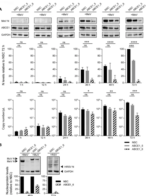
association with the viral RNP. To investigate how ABCE1 affects viral RNA and protein
synthesis, we followed MeV N protein and mRNA kinetics in NSC, ABCE1_5, or ABCE1_6
siRNA-transfected cells, which were infected after 48 h with an MOI of 0.01. Consistent
with the growth kinetics (Fig. 4A), N protein was first detected at 24 h after infection,
and expression levels consistently increased in control cells (Fig. 5A). Only minimal N
protein levels were detected in siRNA ABCE1_6-treated cells throughout the
experi-ment, whereas siRNA ABCE1_5 resulted in an intermediate phenotype (Fig. 5A). N
on August 20, 2019 at INRS-Institut Armand-Frappier
http://mbio.asm.org/
FIG 3 ABCE1 protein expression levels and knockdown efficiency. (A) Confirmation of rabbit anti-ABCE1 peptide antiserum
specificity. A549-hSLAM cells were transfected with an expression plasmid coding for human ABCE1 containing a FLAG tag (DYKDDDDK) at the N terminus or left untransfected. After 24 h, cells were lysed in RIPA buffer and clarified lysates were separated by SDS-PAGE and transferred to PVDF membranes. Western blot analyses were performed with either the rabbit anti-ABCE1 serum (right panel) or a monoclonal mouse anti-FLAG antibody (left panel); secondary antibodies against either
rabbit or mouse, respectively; and a mouse monoclonal antibody against-actin directly conjugated to HRP as an internal
control. (B) Reduction of ABCE1 mRNA levels following ABCE1_5 or ABCE1_6 siRNA transfection. ABCE1 mRNA copy numbers were quantified by real-time RT-PCR 48 h after siRNA transfection. ABCE1 mRNA copy numbers from three independent replicates were quantified. Error bars represent the standard deviation. (C) Continuous ABCE1 knockdown following ABCE1_5 or ABCE1_6 siRNA transfection. Reduction in cellular ABCE1 protein levels 48 h (left panel), 96 h (center panel), and 120 h (right panel) after siRNA transfection. ABCE1 bands from three independent replicates were quantified and normalized relative to an internal GAPDH control for each blot and are shown as percent reduction of ABCE1 expression relative to the NSC for each time point. Error bars represent the standard deviation. (D) Specificity of ABCE1_5 or ABCE1_6 siRNAs. A549-hSLAM cells were transfected with the FLAG-tagged ABCE1 expression plasmid or a derivative carrying 7 to 10 noncoding mutations in the binding sites of ABCE1_5 and ABCE1_6 siRNAs. After 48 h, the cells were transfected with the respective siRNAs, and ABCE1 protein levels were visualized by Western blotting using a FLAG-specific antibody 48 h later. (E) Validation of siRNA target specificity. Cells were transfected with the respective ABCE1 siRNAs or
(Continued on next page)
May/June 2019 Volume 10 Issue 3 e00826-19 mbio.asm.org 8
on August 20, 2019 at INRS-Institut Armand-Frappier
http://mbio.asm.org/
protein mRNA levels remained stable for the first 12 h after infection and then increased
around 100-fold within the next 12 h irrespective of ABCE1 levels, reflecting the first
round of replication (Fig. 5A). There was a gradual increase in mRNA levels as the
infection spread to new cells, which was less pronounced in ABCE1 siRNA-treated cells,
resulting in statistically significant differences starting at 48 h after infection. A similar
effect on viral proteins was observed with HRSV and MuV. Compared to the levels in
NSC-transfected cells, the HRSV N protein was also barely detectable in the absence of
ABCE1, and MuV N protein expression was reduced by more than 50% (Fig. 5B), which
correlated with the impact of ABCE1 knockdown on the replication efficiency of these
viruses (Fig. 4B and C), suggesting that ABCE1 plays an important role in the
accumu-lation of viral proteins during infection.
Viral mRNA transcription is ABCE1 independent. To determine if the observed
reduction in viral protein production is due to a direct role of ABCE1 in translation or
a consequence of a direct or indirect involvement in viral mRNA transcription, we first
compared viral and cellular mRNA levels in the presence and absence of cycloheximide
(CHX). Cells transfected with control or ABCE1_6 siRNA were infected 48 h later with
MeV at an MOI of 1 and then treated with CHX, which blocks de novo protein synthesis.
FIG 3 Legend (Continued)
their corresponding C911 control variants. After 48 h, cells were infected with MeV at an MOI of 0.01. ABCE1 levels in uninfected cells are shown in the left panel, and MeV N levels in MeV-infected cells are shown in the right panel. Protein levels from three independent replicates were quantified, and the NSC value was set to 100%. Error bars represent the
standard deviation.*, P⬍ 0.05; **, P ⬍ 0.01; ***, P ⬍ 0.001.
FIG 4 Validation of ABCE1 knockdown effect on MeV, MuV, and RSV replication. Reduction in virus
replication as a result of ABCE1 knockdown. (A) A549-hSLAM cells were transfected with control NSC, ABCE1_5, or ABCE1_6 siRNAs for 48 h. Cells were then infected with MeV at an MOI of 0.01. Cell lysates (left panel) and culture supernatants (right panel) were harvested at the indicated time points and titrated on A549-hSLAM cells. (B and C) A549-hSLAM cells were transfected with control NSC or ABCE1_6 siRNAs for 48 h. Cells were then infected with either RSV or MuV at an MOI of 0.01. Supernatant titers are shown for RSV (B) and MuV (C). Each data point represents at least three replicates, and error bars indicate the standard deviation.
on August 20, 2019 at INRS-Institut Armand-Frappier
http://mbio.asm.org/
FIG 5 Assessment of ABCE1 knockdown on viral protein and mRNA levels. (A) Kinetics of N protein (top and middle panels) and mRNA
(bottom panel) levels over the course of MeV infection at an MOI of 0.01. A549-hSLAM cells were transfected with control NSC, ABCE1_5, or ABCE1_6 siRNAs for 48 h. Cells were then infected, and samples were harvested at the indicated time points. N protein bands from three independent replicates were quantified and normalized relative to an internal GAPDH control for each blot and are shown as percent reduction of N protein expression relative to the NSC for each virus. Copy numbers of N gene mRNA were quantified from the same samples by real-time RT-PCR using a synthetized RNA standard. (B) Reduction of viral N protein expression in siRNA-transfected cells infected with either MeV or MuV (left panel) or RSV (right panel) at either 48 or 72 h postinfection, respectively. MeV, MuV, and RSV N protein bands from three independent replicates were each quantified and normalized relative to an internal GAPDH control for each blot and are shown as the percent reduction of viral N protein expression relative to the NSC for each virus.
Error bars represent the standard deviation. ns, Pⱖ 0.05; *, P ⬍ 0.05; **, P ⬍ 0.01; ***, P ⬍ 0.001; ****, P ⬍ 0.0001.
May/June 2019 Volume 10 Issue 3 e00826-19 mbio.asm.org 10
on August 20, 2019 at INRS-Institut Armand-Frappier
http://mbio.asm.org/
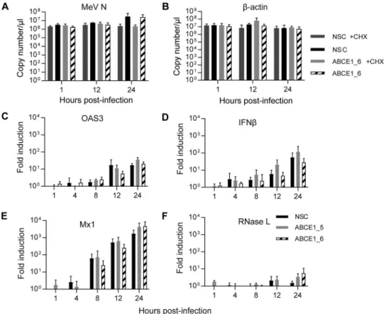
We observed a gradual increase but no differences between copy numbers of MeV N
mRNA in the presence or absence of cycloheximide for the first 12 h after infection
(Fig. 6A), which is expected for the initial phase of viral mRNA transcription in infected
cells driven by the viral polymerase complex provided in trans by incoming virions.
Consistent with the kinetics at an MOI of 0.01 (Fig. 5A), mRNA copy numbers increased
more than 10-fold in the absence of CHX at the 24-h time point, irrespective of cellular
ABCE1 levels (Fig. 6A), whereas CHX treatment had no effect on
-actin mRNA levels
(Fig. 6B). Taken together, this demonstrates that ABCE1 does not directly influence viral
mRNA synthesis.
Since ABCE1 was first identified as an RNase L inhibitor (28), we initially attempted
to investigate the effect of siRNA-mediated RNase L knockdown on the proviral effect
of ABCE1. However, the RNase L protein levels in A549-hSLAM cells were not reliably
detectable, leaving us unable to determine knockdown efficiency. We thus investigated
if RNase L indirectly affects viral replication either by enhancing MeV replication in the
presence of type I interferon (IFN) treatment or by inhibiting innate immune activation.
Type I IFN treatment had little effect on MeV N protein levels in the presence or absence
of ABCE1, indicating that virus replication is not affected (Fig. S3). Quantification of
relative mRNA induction over the first 24 h after infection with an MOI of 1 revealed a
gradual increase of 2=-5=-oligoadenylate synthase 3, the upstream activator of RNase L,
IFN-
, and the IFN-stimulated gene Mx1 (Fig. 6C to E), which mirrored the increase in
MeV N mRNA levels over the same period (Fig. 6A). While there was some variability,
FIG 6 Role of ABCE1 in mRNA transcription and innate immune activation. (A and B) Analysis of de novo mRNA
synthesis. A549-hSLAM cells were transfected with control NSC or ABCE1_6 siRNAs for 48 h, infected with MeV at
an MOI of 1, and treated with 100g/ml CHX or left untreated starting 30 min before infection. Copy numbers of
MeV N (A) and-actin mRNA levels (B) were quantified at different times after infection by real-time RT-PCR using
a synthetized RNA standard. (C to F) Evaluation of innate immune response activation. Induction of
interferon-related gene expression was assessed by quantifying relative mRNA levels of OAS3 (C), IFN- (D), Mx1 (E), and
RNase L (F). Relative changes in mRNA levels from three independent experiments were quantified by real-time
RT-PCR using the ΔΔCTmethod with GAPDH as standard and are shown relative to the NSC signal at 1 hpi for each
cytokine. Error bars indicate the standard deviation, and none of the differences seen reached statistical
signifi-cance (P⬎ 0.05).
on August 20, 2019 at INRS-Institut Armand-Frappier
http://mbio.asm.org/
there was no correlation with ABCE1 levels in the cells (Fig. 6C to E). RNase L mRNA
levels were also not affected and remained largely stable, with a slight increase at the
24-h time point (Fig. 6F). These results and the lack of an effect on viral RNA
accumu-lation indicate that ABCE1 does not support Paramyxoviridae and Pneumoviridae
rep-lication by modulating global innate immune activation.
ABCE1 is essential for efficient viral protein synthesis. Given the effects on viral
protein accumulation and the fact that ABCE1 is known to function in ribosome
recycling (23, 24), we evaluated the relative importance of ABCE1 in cellular and viral
protein translation, using catalyzed fluorescent labeling of proteins in cells treated with
O-propargyl-puromycin (OPP). We found that the siRNA-mediated knockdown of
ABCE1 resulted in a modest reduction of global cellular de novo protein synthesis in
noninfected and infected cells. In the context of infection, this reduction reached
statistical significance at longer labeling times (Fig. 7A), indicating a cumulative effect.
Knockdown of ABCE1 had no effect on protein synthesis of
-actin and pyruvate
dehydrogenase (PDH) E1
␣, two cellular proteins with moderate and high turnover
rates, respectively, whereas viral protein synthesis represented by the MeV M and
fusion (F) proteins was reduced by 30 to 50% compared to the expression levels
achieved in the presence of ABCE1 at the respective time points (Fig. 7B). Especially for
the MeV F protein, which is present at smaller quantities due to the transcriptional
gradient, the difference in the amount of newly synthetized protein increased over time
(Fig. 7B, far right panel). No significant difference in cellular or viral mRNA levels was
observed at the time point of the analysis (Fig. 7C), confirming that ABCE1 exerts its
effect at the translational level. Comparison of cellular and viral protein expression
levels in cells transfected with NSC or ABCE1_6 siRNA at the 30-min time point revealed
around a 60% reduction for MeV M and a 40% reduction for MeV F, which was
statistically significantly different to the cellular proteins (Fig. 7D). To assess the
importance of ABCE1 for Paramyxoviridae and Pneumoviridae protein translation, we
performed a CHX chase experiment. Cells were transfected with NSC and ABCE1_6
siRNA and then treated with 100
g/ml CHX starting 30 min before infection with MeV,
MuV, or HRSV at an MOI of 1. After 24 h, CHX was removed, and samples were collected
24 h later. Reduction of ABCE1 levels resulted in a significant inhibition of de novo
synthesis of viral proteins (Fig. 7E), demonstrating a specific role of ABCE1 in translation
of Paramyxoviridae and Pneumoviridae mRNAs.
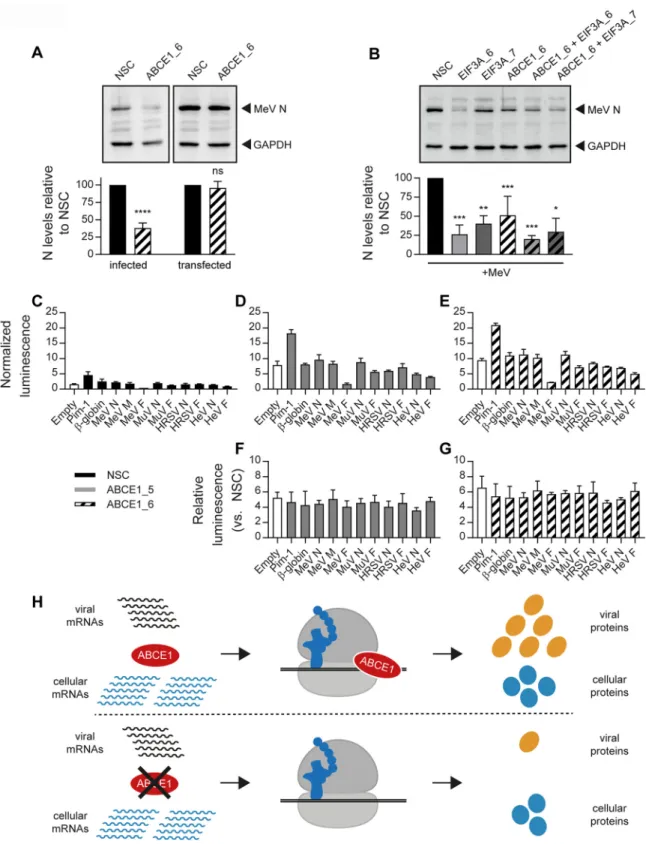
The ABCE1 proviral effect requires viral gene expression in the context of
infection. To further elucidate the virus-host interactions underlying the proviral
activity of ABCE1, we investigated if the effect could be reproduced if MeV N protein
was expressed from a cellular expression plasmid. In contrast to the reduction of MeV
N protein levels observed in the absence of ABCE1, there was no effect on MeV N
protein levels in cells transfected with a eukaryotic MeV N expression plasmid (Fig. 8A).
Comparison of the proviral activity of ABCE1 with eIF3A, another hit in our validation
screen and constituent of the cellular translation machinery, revealed a similar
reduc-tion in MeV N protein levels and no additive effect when the two genes were targeted
simultaneously (Fig. 8B), highlighting the exquisite dependence of Paramyxoviridae and
Pneumoviridae on specific translation factors.
Since the 5= untranslated regions (UTRs) of paramyxoviral and pneumoviral mRNAs
can vary in length depending on the gene and virus (3), sensitivity to ABCE1
knock-down was assessed. Toward this, the 5= UTRs of the MeV N, M, and F genes, as well as
those from the MuV, HRSV, and HeV N and F genes, were introduced upstream of a
firefly luciferase (ffLuc) reporter gene followed by a hepatitis C virus (HCV) internal
ribosomal entry site (IRES) that mediates translation of a Renilla luciferase (RenLuc)
reporter gene for normalization (48). While ABCE1 knockdown resulted in an overall
increase in ffLuc expression from all the tested plasmids independently of the 5= UTR
inserted (Fig. 8C to E), the relative expression levels remained stable, demonstrating
that viral 5= UTRs were not sufficient to confer sensitivity to ABCE1 knockdown (Fig. 8F
and G). Taken together, our results illustrate that Paramyxoviridae and Pneumoviridae
May/June 2019 Volume 10 Issue 3 e00826-19 mbio.asm.org 12
on August 20, 2019 at INRS-Institut Armand-Frappier
http://mbio.asm.org/
FIG 7 ABCE1 effect on de novo viral protein translation. (A) Quantification of total cellular protein synthesis by incorporation of
O-propargyl-puromycin (OPP). At 48 h after siRNA transfection, cells were infected with MeV, MuV, or HRSV for a further 48 h, after which
the cells were treated with OPP for the indicated times, and incorporated OPP was coupled to Alexa Fluor 594. The fluorescence signal was quantified by flow cytometry, and bars show the mean fluorescence intensity (MFI). Error bars represent the standard error of the
mean. Statistical significance was determined by one-tailed t tests. (B to D) Analysis of protein translation by35S pulse-labeling and
quantification of the corresponding mRNA levels. A549-hSLAM cells were transfected with control NSC or ABCE1_6 siRNAs for 48 h. Cells
were then infected with MeV at an MOI of 0.1 and labeled with [35S]Met-Cys 24 h later. (B) Immunoprecipitated cellular proteins-actin
and PDH E1␣ and MeV M and F proteins from infected cells were visualized by exposure to phosphor screens. Representative phosphor
screen images are shown. (C) Copy numbers of the respective mRNAs from three independent experiments corresponding to the35S
pulse-labeling were quantified by real-time RT-PCR using a synthetized RNA standard for each gene. (D) Quantification of35S pulse-label
incorporation. The expression levels in samples shown in panel B from ABCE1_6 siRNA-treated cells relative to NSC-treated cells were calculated for each protein. The average from three independent experiments for the 30-min time point is shown. (E) Analysis of de novo protein synthesis of MeV, MuV, and RSV. A549-hSLAM cells were transfected with control NSC or ABCE1_6 siRNAs for 48 h; infected with
(Continued on next page)
on August 20, 2019 at INRS-Institut Armand-Frappier
http://mbio.asm.org/
mRNAs are disproportionally sensitive to loss of ABCE1 function (Fig. 8H). Perturbations
in ABCE1 levels are tolerated by the cell but have a dramatic effect on the viral life cycle.
DISCUSSION
Even though nonsegmented negative-strand RNA viruses that replicate in the
cytoplasm exploit the host cell machinery for many aspects of their life cycles, the
cellular proteins and networks involved in these interactions remain largely unknown
(49). Here, we performed comparative whole-genome siRNA screens with wild-type
MeV, MuV, and HRSV strains in a human lung alveolar epithelial carcinoma cell line as
a representative target cell type. The meta-analysis of two independent comparative
analyses of the three siRNA screens highlighted 24 candidates that were validated
experimentally. Six of these genes, ABCE1, COPB2, RBM22, FOXP4, HSD11B2, and PRR15,
were also hits in a recently published HeV screen (43). Detailed characterization of
ABCE1, a multifunctional protein involved in inhibition of RNase L-mediated signaling,
HIV particle assembly, and ribosome recycling (50), revealed that it plays a critical role
in viral protein synthesis.
Previous screens using HeV and VSV in HeLa cells identified COPI and other proteins
involved in retrograde vesicular transport from the Golgi complex to the endoplasmic
reticulum as a common proviral pathway for all negative-stranded RNA viruses (10, 43).
While the function of this pathway in the biology of negative-stranded RNA viruses
remains to be characterized, it likely supports replication analogous to its role in the life
cycle of positive-stranded RNA viruses (51, 52). In addition to confirming the
impor-tance of this pathway, our screens also highlighted a prominent role for oxidative
phosphorylation. Mitochondria are at the crossroads of different cellular antiviral
defense mechanisms, and the importance of the pathway has been identified for
different virus families (53). For influenza virus, NOX4-mediated increase of reactive
oxygen species (ROS) is required for efficient replication by supporting the nuclear
export of RNPs and particle formation and budding (54). In HRSV, phosphorylated p38
mitogen-activated protein kinase was found to be sequestered in viral inclusion bodies
(55), and it is thus conceivable that such sequestration also occurs with other
paramyxoviruses.
ABCE1 is a member of the superfamily of ABC transporters that contain two
nucleotide-binding domains and two N-terminal iron-sulfur clusters. It is ubiquitously
expressed at high levels in the trachea, testis, and prostate (56). Unlike most ABC
domain proteins, members of the ABCE subfamily do not contain the
membrane-spanning domains and may thus not act as transporters (21). ABCE1 was initially
identified as a negative regulator of the interferon-induced 2-5A antiviral pathway,
where it functions by blocking RNase L (28). RNase L plays an important role in the
antiviral and antiproliferative activities of IFN and contributes to innate immunity and
cell metabolism (57, 58). ABCE1 in turn supports the replication of several RNA viruses
by inhibiting the IFN-induced activation of the 2-5A/RNase L pathway. During West Nile
virus infection, inhibition of ABCE1 results in cleavage of viral genomic RNA by RNase
L, indicating that RNase L plays a role in the cellular antiviral response to flaviviruses
(59). Furthermore, the potential of RNase L activator drugs to block paramyxovirus
infection was demonstrated by administration of a 2-5A homolog to reduce HRSV
replication in African green monkeys (60). Small-molecule activators of RNase L have
also been shown to have antiviral activity against Sendai virus and human
parainflu-enza virus 3 (61), highlighting the importance of this pathway. Additionally, OASL,
which was identified in our screens as a potential antiviral factor by the KS analysis (see
FIG 7 Legend (Continued)
MeV, MuV, or HRSV at an MOI of 1; and treated with 100g/ml CHX starting 30 min before infection. After 24 h, CHX was removed and
samples were harvested after an additional 24 h of incubation. MeV, MuV, and RSV N protein bands were quantified and normalized relative to an internal GAPDH control for each blot and are shown as the percent reduction of viral N protein expression relative to the NSC for each virus. Each graph shows the average from three independent experiments, and error bars represent the standard deviation.
Statistical significance in panels C to E is indicated as follows: ns, Pⱖ 0.05; ***, P ⬍ 0.001; ****, P ⬍ 0.0001.
May/June 2019 Volume 10 Issue 3 e00826-19 mbio.asm.org 14
on August 20, 2019 at INRS-Institut Armand-Frappier
http://mbio.asm.org/
FIG 8 Dependence of ABCE1 proviral effect on viral gene expression during infection. (A) Plasmid-expressed MeV N is not sensitive to
ABCE1 knockdown. A549-hSLAM cells were transfected with NSC or ABCE1_6 siRNAs. After 24 h, cells were transfected either with a control empty vector plasmid followed by infection with MeV at an MOI of 0.01 at 24 h posttransfection (“infected”) or with a plasmid expressing MeV N containing an N-terminal FLAG tag and left uninfected 24 posttransfection (“transfected”). The MeV N protein levels were quantified, and NSC was set to 100%. Data are representative of four replicates. Error bars represent standard deviations. Statistical
significance is indicated as follows: ns, Pⱖ 0.05; ****, P ⬍ 0.0001. (B) ABCE1 and eIF3A have similar effects on MeV replication. A549-hSLAM
cells were transfected with the respective siRNAs. After 48 h, cells were infected with MeV at an MOI of 0.01. Cell lysates were harvested 48 h after infection. MeV N protein levels were quantified, and NSC was set to 100%. Data are representative of three independent
replicates. ns, Pⱖ 0.05; *, P ⬍ 0.05; **, P ⬍ 0.01; ***, P ⬍ 0.001. (C to G) Effect of viral 5= untranslated regions (UTRs) on reporter gene
translation. Hep-G2 cells were transfected with either NSC or the ABCE1_5 and ABCE1_6 siRNAs in 96-well plates along with dual-luciferase
(Continued on next page)
on August 20, 2019 at INRS-Institut Armand-Frappier
http://mbio.asm.org/
Table S5 in the supplemental material), is a known antiviral effector for HRSV and is
countered by the NS1 protein (62). However, our cytokine mRNA induction kinetics
revealed that cellular ABCE1 levels do not affect global innate immune signaling
pathways, and levels of MeV mRNAs were not altered in ABCE1-depleted cells.
None-theless, we cannot formally rule out that the RNase-L-inhibitory activity of ABCE1 plays
a minor role as a proviral factor for Paramyxoviridae and Pneumoviridae.
ABCE1 is involved in the regulation of translation (22) and as a ribosome-recycling
factor critical for translation termination (63, 64) and ribosome homeostasis (23, 24). In
contrast to many viruses which induce a host cell shutoff by interfering with cellular
mRNA or protein synthesis, replication of Paramyxoviridae and Pneumoviridae seems to
require ongoing cellular protein synthesis: Parainfluenza virus 5 P and V proteins
actively prevent host gene shutoff by limiting PKR induction (65), and a similar effect
was also reported for the MeV C protein (66). HRSV infection results in PKR induction,
but the protein is sequestered by binding to the viral N protein, thereby attenuating the
phosphorylation of eIF2
␣ and maintaining cellular protein translation (67). Our data,
however, suggest that MeV, MuV, and HRSV infections lead to a modest decrease in
global proteins synthesis (Fig. 7A). Most importantly, and consistent with previous
reports about the role of ABCE1, we observed a modest reduction in overall protein
synthesis in ABCE1 knockdown cells. In contrast, viral protein synthesis was dramatically
affected under the same conditions, demonstrating that this process is critically
de-pendent on ABCE1. It is interesting that ABCE1 is known to form a stable complex with
the small ribosomal subunit during the recycling of terminated or stalled ribosomes,
and in our work 30 of the small ribosomal subunit proteins were among the top 100
proviral gene products predicted by the KS analysis (Table S2). In contrast, only five
proteins of the large subunit scored in the same group, even though an important role
of the large ribosomal subunit protein rpL40 for negative-strand RNA virus translation,
including MeV, has recently been demonstrated (68). This disproportionate
depen-dence on small ribosomal subunit proteins is the exact opposite of what has been
observed in screens for human flaviviral host factors, where large subunit proteins
predominate at the top of the list (46). Additionally, both the KS analysis and the robust
Z score analyses identified eIF3A as a top proviral factor and suggested that other eIF3
subunits were likely involved, implying that the eIF3 complex may be exquisitely
required for at least some paramyxovirus mRNA translation. ABCE1 has been shown to
interact with eIF3 in Saccharomyces cerevisiae (69), and structural studies indicated that
ABCE1 bound to the small ribosomal subunit prevents premature binding of eIF3 (63),
thereby regulating the use of recycled ribosomes in further initiation events. Another
hit common to all three viruses was RPLP1, which has recently been implicated in
ABCE1-dependent ribosome recycling (70). These data strongly suggest that the
paramyxovirus requirement for ABCE1 is due to its role in ribosome recycling.
Why are paramyxovirus mRNAs so sensitive to ABCE1? There is much we do not
know about paramyxovirus translation, but certain facts point to special requirements
for mRNAs encoded by these viruses. It must be emphasized that not all paramyxovirus
and pneumovirus mRNAs may require ABCE1 to the same extent, since these mRNAs
vary in their structure and likely in their dependence on host factors. MeV N mRNA
translation was shown to be enhanced by La autoantigen (SSB) overexpression, and this
was proposed to be mediated by an interaction between SSB and the 5= UTR of MeV
FIG 8 Legend (Continued)
reporter plasmids. Translation of firefly luciferase (ffLuc) is mediated by upstream cellular or viral UTRs, and the translation of the downstream Renilla luciferase (RenLuc) is driven by the hepatitis C virus (HCV) internal ribosomal entry site (IRES). Luciferase values were read 48 h after transfection. ffLuc signals for each well were normalized to the RenLuc signal. Normalized ffLuc values are shown for cells transfected with NSC (C), ABCE1_5 (D), and ABCE1_6 (E), and the signals from ABCE1_5- and ABCE1_6-transfected cells are shown relative to NSC (F and G, respectively). Transfections in each replicate were performed in triplicate, and all experiments were performed three times. (H) Model for the differential effects of ABCE1 knockdown on viral and cellular protein translation. In cells with normal ABCE1 levels, viral proteins are preferentially translated over cellular proteins, resulting in proportionately higher levels of viral protein expression (top). In cells with reduced ABCE1 levels, viral protein translation is drastically reduced, while translation of cellular proteins is only moderately affected (bottom).
May/June 2019 Volume 10 Issue 3 e00826-19 mbio.asm.org 16
on August 20, 2019 at INRS-Institut Armand-Frappier
http://mbio.asm.org/
N mRNA (71). However, in this report the authors did not determine the effect of
knocking down SSB on MeV propagation, and in our screens SSB did not score as either
proviral or antiviral. One of the peculiar features of paramyxovirus mRNAs is that they
can be polycistronic (72–76), which could lead to altered termination of ribosomes in
the short segments between cistrons and thus a more stringent requirement for ABCE1.
These polycistronic transcripts, even though they are a small fraction of all viral mRNAs,
could act as reservoirs for recycling factors by trapping them in inactive complexes and
thus increasing the requirement for ABCE1. Additionally, most paramyxovirus mRNAs,
either monocistronic or polycistronic, have short 5= and 3= UTRs that again may
interfere with efficient termination or use of recycled ribosomes for initiation. In a
competitive translational landscape, where mRNAs are competing for a limited pool of
ribosomes (77), viral mRNAs may be exquisitely dependent on the action of factors such
as ABCE1.
MATERIALS AND METHODS
Cell lines and viruses. A549 cells (ATCC CCL-185), Vero cells (ATCC CCL-81), and HeLa cells (ATCC
CCL-2) constitutively expressing the human SLAM molecule were generated by selecting zeocin-resistant clones after transfection with pCG-IRESzeo-hSLAM, which was cloned as described for canine SLAM (78). All human SLAM (hSLAM)-expressing cell lines were maintained in Dulbecco modified Eagle medium
(DMEM; Invitrogen) supplemented with 10% fetal bovine serum (FBS; Invitrogen) and 100g/ml zeocin
(Invitrogen) at 37°C and 5% CO2. The human Epstein-Barr virus-transformed B-lymphoblastic cell line
(BLCL) (W. P. Duprex laboratory [79]) and HEp-2 (ATCC CCL-23) and Hep-G2 cells (kindly provided by F. Elgner, Paul-Ehrlich-Institute) were maintained in RPMI supplemented with 10% FBS.
rMVKSEGFP(3) is a recombinant virus based on the Khartoum-Sudan (KS) strain of MeV (33, 80).
rHRSVB05EGFP(5) is a recombinant virus based on the sequence of HRSV present in a tracheal rinse from
an HRSV-positive infant during the 2004 –2005 HRSV season (35). rMuVG09EGFP(3) is a recombinant virus
based on the sequence of MuV present in clinical material from a genotype G MuV infection from the 2009 New York mumps outbreak (34, 81). The recombinant paramyxoviruses all express EGFP from an additional transcription unit inserted between the P and M genes; this is position 3 in the recombinant genome for MeV and MuV and position 5 for HRSV. MeV was propagated in BLCL cells while MuV and HRSV were amplified in HEp-2 cells. Prior to storage, sucrose (25% [wt/vol]) was added to HRSV stocks.
Primary genome-wide siRNA screens. The screens were performed at the Duke University
Func-tional Genomics Facility using the Qiagen genome siRNA library v1.0 consisting of four distinct siRNAs (A, B, C, and D) originally designed to target 22,909 known and putative human genes. As many instances of obsolete or outdated library gene annotations were found in the original annotation, the siRNA library annotations were updated based on the perfect match of their sequence to the GRCh37v75 release of the human transcriptome, resulting in the targeting of 21,705 genes by the siRNA library. The four siRNAs were grouped into two pools, with each pool containing two siRNAs (set AB and set CD). Each screen consisted of 148 384-well plates. This 2-by-2 pool design allowed each gene to be tested by two independent siRNA sets. Briefly, black, clear-bottom 384-well tissue culture plates (Corning) were prearrayed with 1 pmol of siRNA per well by using the Velocity Bravo liquid handling system (Agilent
Technologies). Lipofectamine RNAiMAX (Invitrogen) was used in the amount of 0.05l per well in 10 l
of Opti-MEM (Invitrogen). After 15 min of complex formation, cells were dispensed at a density of 1,500 cells per well using a Matrix WellMate dispenser (reverse transfection of 15.4 nM final concentration of
siRNAs in 65-l total medium volume). To facilitate gene knockdown by siRNAs, plates were incubated
at 37°C at 5% CO2for 48 h. Virus infections at different MOIs and for various lengths of time postinfection
were conducted to identify suitable conditions for the high-throughput screens. Transfected cells were
infected with 20l of virus diluted in DMEM and supplemented with 10% FBS and were incubated at
37°C in 5% CO2. Cells infected with MeV were incubated for 33 h, and those infected with MuV and HRSV
were incubated for 72 h. To identify factors involved in all stages of the paramyxovirus life cycle, the screen was optimized to allow for multiple rounds of infection.
Validation screen. For validation of the top-ranking genes identified by comparative analysis of the
three primary screens, multiple siRNAs targeting the respective genes were used, and siRNAs that differed from those used in the primary screens were chosen whenever available. The assay was conducted in 384-well plates using the same reverse transfection procedure outlined for the primary screen. Each individual siRNA was represented in replicates of 4 in each plate. Image collection and analysis were performed using the same methodology as the original screens.
Automated image analysis. After 33 h for MeV infection or 72 h for MuV and HRSV infection, cells
were fixed with 4% paraformaldehyde (Sigma) in phosphate-buffered saline (PBS; Invitrogen), perme-abilized with 0.1% Triton X-100 in PBS, and stained with Hoechst 33342 (Sigma) in PBS for 30 min. Stained cells were imaged and analyzed with a Cellomics ArrayScan VTI automated microscope. Uninfected cells served as a reference population for background fluorescence. Four fields per well of 384-well plates
were imaged at⫻10 magnification and analyzed using the Compartmental Analysis Bioapplication. First,
images were acquired for Hoechst nuclear staining in channel 1 and EGFP signal in channel 2. Cells were identified and counted in channel 1, and cell numbers are indicated as VOC (valid object count). Infected cells were identified by EGFP signal in channel 2 and expressed as the percentage of total VOC.
on August 20, 2019 at INRS-Institut Armand-Frappier
http://mbio.asm.org/
General statistical methods. The distribution of percent infection for genomic population for the AB
and CD pools did not follow a normal distribution, so we normalized raw percent infection measure-ments by converting to robust Z (82). Primary data from the Cellomics Oracle database were retrieved, and raw percent infection measurements were converted to robust Z scores within each plate using custom Python scripts. To identify candidate host factor and restriction genes, we evaluated the distribution of normalized well values for each gene relative to the overall distribution of all wells using the Kolmogorov-Smirnov (KS) test and used the false discovery rate (FDR) method to correct for multiple hypothesis testing. A similar approach to the gene-level analysis was employed to identify pathways enriched for either negative or positive selection. All pathways were collected from the “Curated Pathways” subset of the Molecular Signatures Database (MsigDB), and the KS test was used to evaluate the distribution of normalized values belonging to all genes of each pathway against the entire distribution of normalized values.
Validation of ABCE1 as a host factor for Paramyxoviridae and Pneumoviridae. A549-hSLAM cells
were seeded into 24- or 12-well dishes and transfected with ABCE1-specific, eIF3A-specific, or control siRNAs. C911 siRNAs were used to control for off-target effects with the ABCE1 siRNAs. Target sequence mismatches were introduced into the ABCE1_5 and ABCE1_6 siRNAs using the C911 calculator version 1 (http://rnai.nih.gov/haystack/C911Calc.html) (83) to generate the ABCE1_5_C911 and ABCE1_6_C911
siRNAs, respectively (all from Sigma-Aldrich). Briefly, 100 or 200l/well of Opti-MEM (Invitrogen) was
combined with 0.75 or 1.5l siRNA at a 20 M working concentration and 1 or 2 l of RNAiMAX
transfection reagent (Invitrogen), vortexed, and incubated at room temperature for 15 min. The siRNA– Opti-MEM mixture was added to the plated A549-hSLAM cells, which were then incubated at 37°C. After 48 h, cells were infected with the respective viruses at an MOI of 0.01 or 1, and for CHX experiments,
100g/ml CHX was added starting 30 min before infection and maintained for the indicated duration.
At different time points thereafter, the medium was harvested for virus titrations, and cells were lysed in RIPA buffer containing cOmplete protease inhibitor cocktail (Roche) for protein analysis or RNA isolation buffer. Culture supernatants were titrated by limiting dilution on A549-hSLAM cells and are expressed as
the 50% tissue culture infectious dose (TCID50) per milliliter.
MeV N expression plasmid transfection. The plasmid pCG-MeV FLAG-N encodes the MeV N protein
with a FLAG-tag (DYKDDDDK) at the N terminus. For transfection in siRNA-transfected A549-hSLAM cells
in 12-well dishes, 2g of plasmid was mixed with 3 l of Lipofectamine 2000 (Invitrogen) in 100 l/well
Opti-MEM (Invitrogen). Transfection mixtures were vortexed and incubated at room temperature for
20 min. After incubation, 100l of transfection mixture was added to the appropriate well and incubated
at 37°C for 5 h, after which the medium was aspirated and replaced with fresh DMEM containing 5% FBS
and 1%L-glutamine.
Viral and cellular mRNA quantification. Copy numbers of viral,-actin, and PDH E1␣ mRNAs and
relative expression levels of OAS3, IFN-, Mx1, and RNase L in samples isolated at different times after
infection of NSC or ABCE1 knockdown cells were quantified by real-time RT-PCR using the QuantiFast SYBR Green PCR kit (Qiagen) after reverse transcription with SuperScript III reverse transcriptase (Invit-rogen). Each experiment was carried out at least three times.
Antibodies. For the detection of viral proteins by Western blot analysis or radiolabeling, mouse
monoclonal antibodies against the MuV N protein (Abcam; ab9878), or the HRSV N protein (Abcam; ab94806) or rabbit antipeptide antisera against the MeV N, M, and F proteins, respectively (84), were
used. Mouse monoclonal antibodies against actin (Abcam; ab8226), PDH E1␣ (Abcam; ab92696), and
GAPDH (Cell Biolabs) and a rabbit antipeptide serum against ABCE1 (85) were used to visualize cellular proteins. The latter antiserum was kindly provided by Jaisri Lingappa (University of Washington, USA) for initial studies and subsequently produced by immunizing rabbits with the peptide N-NSIKDVEQKKSGN YFFLDD-C, corresponding to 19 amino acids at the C terminus of human ABCE1, coupled to keyhole limpet hemocyanin (GenScript). For immunofluorescence staining, monoclonal antibodies against MeV N (MAB8906), MeV M (MAB8910), or MeV H (MAB8905) proteins (all from Merck Millipore) were used. Horseradish peroxidase-coupled goat anti-mouse (Jackson ImmunoResearch) or goat anti-rabbit (Abcam; ab97200) secondary antibody and goat anti-mouse or goat anti-rabbit secondary antibodies conjugated to Alexa Fluor 488 and Alexa Fluor 568 (Molecular Probes) were used as secondary antibodies.
Immunofluorescence staining. A549-hSLAM cells were seeded on coverslips in 24-well plates and
infected with MeV at an MOI of 1. After 1 h of infection, the culture medium was removed and replaced
with medium including 20M Z-fFG (fusion inhibitory peptide [FIP]; Bachem). At 24 h postinfection, cells
were fixed with 4% paraformaldehyde in PBS for 15 min and subsequently permeabilized with 0.1% Triton X-100 in PBS for 15 min at room temperature. Cells were stained with the respective primary and secondary antibodies, and coverslips were mounted onto glass slides with ProLong Gold antifade mountant with DAPI (Life Technologies). Images were obtained using a Zeiss LSM 510 confocal laser
scanning microscope with a␣Planapochromat 100⫻ oil objective. Images were collected at 1,008- by
1,008-pixel resolution and processed using ImageJ and Photoshop.
Western blot analysis. For each sample, the same protein concentration was loaded to correct for
ABCE1 knockdown-mediated reduction in proliferation. Membranes were stained with the respective antibodies, proteins were visualized using a MicroChemi 4.2 chemiluminescent imaging system (DNR Bio Imaging Systems Ltd.), and the resulting bands were quantified from underexposed TIF images using the ImageJ analysis software package (NIH).
Analysis of total de novo protein synthesis. Cells were labeled with O-propargyl-puromycin (OPP;
ThermoFisher) at different times after infection according to the manufacturer’s instructions. Briefly, after the respective labeling time, cells were fixed, OPP-labeled proteins were coupled to Alexa Fluor 594
May/June 2019 Volume 10 Issue 3 e00826-19 mbio.asm.org 18