OATAO is an open access repository that collects the work of Toulouse
researchers and makes it freely available over the web where possible
This is a Publisher’s version published in:
http://oatao.univ-toulouse.fr/25448
Official URL: https://doi.org/10.1155/2015/139617
Any correspondence concerning this service should be sent
to the repository administrator:
[email protected]
To cite this version:
Zimoch-Korzycka, Anna and Rouilly, Antoine
and Bobak, Łukasz and
Jarmoluk, Andrzej and Korzycki, Michał Chitosan and Cystatin/Lysozyme
Preparation as Protective Edible Films Components. (2015) International Journal
Research Article
Chitosan and Cystatin/Lysozyme Preparation as
Protective Edible Films Components
Anna Zimoch-Korzycka,
1Antoine Rouilly,
2,3Aukasz Bobak,
1Andrzej Jarmoluk,
1and Micha
B Korzycki
11Department of Animal Products Technology and Quality Management, Faculty of Food Science, Wrocław University of Environmental
and Life Sciences, 37/41 Chelmonskiego Street, 51-630 Wrocław, Poland
2INRA, UMR 1010 CAI, 31030 Toulouse, France
3Universit´e de Toulouse, INP-ENSIACET, Laboratoire de Chimie Agro-Industrielle (LCA), 31030 Toulouse, France Correspondence should be addressed to Anna Zimoch-Korzycka; [email protected]
Received 27 August 2015; Revised 8 October 2015; Accepted 11 October 2015 Academic Editor: Yulin Deng
Copyright © 2015 Anna Zimoch-Korzycka et al. This is an open access article distributed under the Creative Commons Attribution License, which permits unrestricted use, distribution, and reproduction in any medium, provided the original work is properly cited. This work characterizes biological, physical, and chemical properties of films formed from an aqueous solution of hydroxypropyl methylcellulose (HPMC), with different concentrations of chitosan (CH) and bioactive cystatin/lysozyme preparation (C/L). The properties of biocomposites were examined by Dynamic Mechanical Analysis (DMA), Fourier’s transfer infrared spectroscopy (FTIR), water vapour permeability (WVP), and tensile testing. Antimicrobial activity against Micrococcus flavus, Bacillus cereus,
Escherichia coli, Pseudomonas fluorescens, and Candida famata was conducted. Films glass transition and storage modulus were
dependent on the C/L and CH concentration. Modulus values decreased during the temperature scan and with higher reagents levels. An increase of CH and C/L concentrations in the films resulted in a decrease in tensile strength from 2.62 to 1.08 MPa. It suggests the hydrolyzing influence of C/L, also observed in smaller peak size of𝛼 relaxation. C/L addition caused shifting 𝑇𝑔to higher temperature. DMA and FTIR analysis proved that HPMC and CH are compatible polymers. Water resistance was improved with rising CH concentration from1.08𝐸 − 09 to 7.71𝐸 − 10 g/m ∗ s ∗ Pa. The highest inhibition zone in M. flavus and C. famata was recorded at the highest concentration of CH and C/L.
1. Introduction
The growing interest and demand of producers as well as consumers for environmental friendly, biodegradable, bio-compatible, and bioactive materials to produce edible films and coatings are observed. Most polymers and bioactive sub-stances have very good film forming properties. These include polysaccharides: chitosan [1], cellulose [2], starch [3], and car-rageenan [4], proteins: gelatin [5], collagen [6], and lipids [7]. Enzymes are also used to improve or change features of poly-mers: transglutaminase [8], lysozyme [9], or cellulase [10].
Derivatives of cellulose are composed of the same 𝛽-(1 → 4)-glycosidic units with different substituents methyl, hydroxypropyl, or carboxyl. Hydroxypropyl methyl-cellulose is methyl-cellulose ether with hydrophilic groups, hydroxyl groups, which provide good interaction of HPMC with water [11]. The mechanical and thermal properties of HPMC are
influenced by presence of these groups and water uptake [12]. HPMC exhibits thermogelation and has excellent film-making properties, high solubility, efficient oxygen, and lipid barrier properties [13, 14].
Chitosan is a biopolymer, which has a𝛽-(1 →
4)-D-glucopyranose backbone similar to cellulose. The difference is that chitosan possesses acetamide group instead of the hydroxyl group in C2 position of glucose residue. The similarity of primary structure of both polymers suggests possibility of formation of homogenous edible films based on chitosan and cellulose derivatives [15]. The modification of chitosan with different polysaccharides or proteins may be an effective way to improve mechanical properties of chitosan for which antimicrobial [16] and oxygen barrier properties were reported [17].
Interactions of the polymers with bioactive substances have been recently studied extensively [18–20]. Egg white is Volume 2015, Article ID 139617, 10 pages
rich natural source of bioactive proteins such as lysozyme, ovotransferrin, avidin, ovoflavoprotein, and proteinase inhibitors (ovoinhibitor, ovomucoid, ovostatin, and cystatin). Classical separation methods of proteins are based on the salting out of solution or precipitation with alcohol. These techniques have been extended by the ion exchange chro-matography and membrane separation in recent years. Nowadays, researchers are looking for separation methods, which will be cheap, easy, nontoxic, and maintaining the highest biological activity of isolated proteins. They are important factors in commercial production and in food and pharmaceutical applications in view of their health activities [21]. The lysozyme damages cell walls, especially
Gram-positive bacteria by catalyzing hydrolysis of 𝛽-(1 → 4)
linkages between particles of N-acetylmuramic acid and L-glucosamine [22]. The cystatin is a protein inhibitor of cysteine proteinases [23] and has very high antimicrobial [24, 25] and antiviral properties [26]. A special challenge is to develop a cheap method of cystatin isolation, because cystatin is present in small amount (around 0.05%) in egg white. Skiba et al. have proposed a thermochemical method of initial isolation of bioactive proteins of egg white, especially cystatin [27]. They used diversity of chemo- and thermoresistance property of egg proteins for their separation. Lysozyme, cystatin, and ovomucoid are acid soluble and thermally
stable, even when heated up to 100∘C. The rest of the egg white
proteins (ovotransferrin, ovalbumin) in such conditions will coagulate. The production process of bioactive preparation can be carried out in processing plants, but the obtained mix-ture could be used just for foodstuff. Extension of application in pharmaceutical usage is possible after purification by chro-matography methods. Possibility to obtain cystatin/lysozyme preparation with ovomucoid was proved [27] and bioactivity of these single proteins was confirmed by researchers [28].
Applicable opportunities of bioactive preparation in HPMC biocomposites were assessed, in view of the aim of this study, which was to develop HPMC films with addition of cystatin/lysozyme preparation and chitosan as well as evaluate their antimicrobial activity and thermomechanical, barrier, and chemical changes.
2. Materials and Methods
2.1. Materials. The materials included HPMC (Walocel HM
100 PA 2208 FG, Wolff Cellulosics), chitosan (low molecular
weight, DD 75–85%, Mw = 150× 10−3, low viscosity ranged
between 20 and 200 cps, Aldrich), DL-lactic acid (85%, Sigma), and glycerol 99% (Aldrich).
2.2. Cystatin/Lysozyme Isolation. A substrate of C/L
prepara-tion was homogenous hen egg white, which was diluted with an equal 0.25% NaCl following the procedure described by Skiba et al. [27]. The solution was brought to pH 3.0 with 1 N
HCl and heated to 60∘C for 30 minutes. Protein suspension
was obtained and centrifuged at 9300×g for 20 minutes to
precipitate ovomucin and other eggs’ protein. Supernatant
containing bioactive substances was frozen at −40∘C, then
lyophilized, and stored at 4∘C.
2.3. Activities of Cystatin and Lysozyme. Activity of
cys-tatin against papain was analyzed according to colori-metric method reported by Siewi´nski [29], where BANA (hydrochloride Na-benzoyl-DL-arginyl-B-naftylamid) was a substrate of hydrolysis and reaction was stopped by the addi-tion of DMBA (p-dimethylaminobenzaldehyde). Absorbance
was measured at𝜆 = 450 nm. One unit of inhibitory activity
of cystatin equals one unit of papain activity, which is quantity of enzyme able to hydrolyze 1.0 mM of substrate in one
minute (standard conditions, 37∘C).
Lysozyme activity was analyzed by spectrophotometrical method [30]. Bacterial cells of Micrococcus lysodeikticus were used as a substrate for lysozyme. The dynamic turbidity
changes were reported by measuring of absorbance at𝜆 =
450 nm in 25∘C, every 60 seconds during 6 minutes.
2.4. Film Preparation. HPMC was dissolved in bidistilled
heated (70∘C) water for preparation of solution. Chitosan was
solubilized in 2% (v/v) aqueous lactic acid solution in room temperature. Both solutions were stirred with 400 rpm for 12 hours. C/L stock solution was prepared by dissolving 20% C/L preparation in bidistilled water followed by centrifugation
(9300×g) and filtration to remove insoluble residues. The
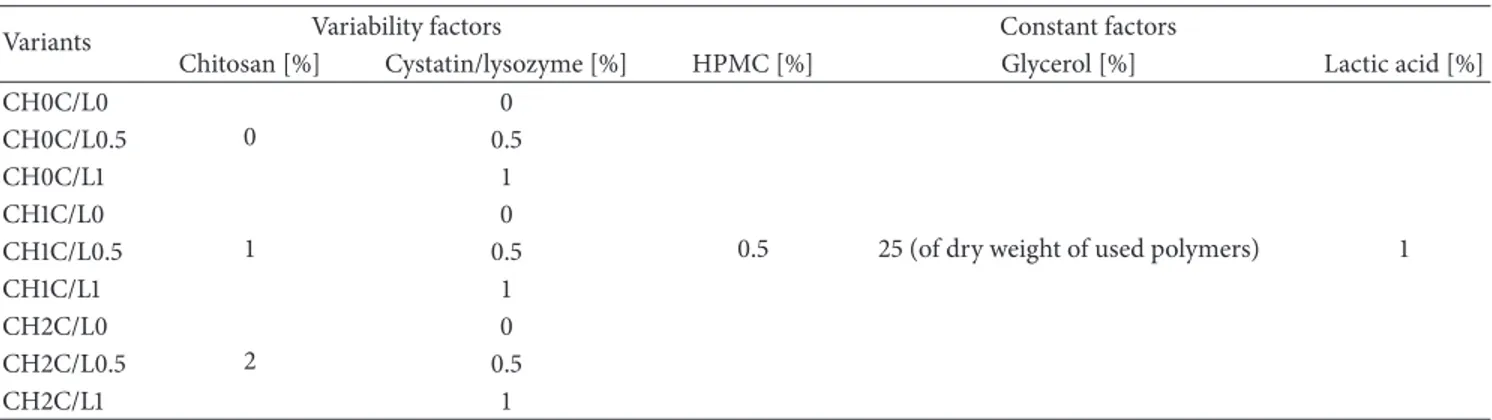
same procedure was applied to prepare protein solution. Glycerol was used as plasticizer in amount of 25% of polymers dry mass. Thus prepared solutions of hydrocolloids, prepara-tion of C/L, equivalent of proteins, and glycerol were mixed in suitable proportions to obtain final concentrations of the components shown in Table 1. The HPMC and CH solutions at three different levels, 0%, 1%, and 2%, were blended with 25% wt/wt of glycerol (of dry weight of the used polymers) and C/L mixture (at three different levels: 0%, 0.5%, and 1%, which correspond to 0/0, 0.35/96, and 0.7/192 U/g activity of C/L, resp.) in different ratio (wt/wt). All experimental
samples were centrifuged at 8400×g for 20 minutes to remove
air bubbles. Twelve mL of film forming solutions was then
cast on leveled, coated by Teflon glass plate on an area of 66×
77 mm, and dried at 25∘C and 60% RH for 48 hours. The dried
biocomposites were peeled from plates and cut into pieces for the measurements of thermal and mechanical properties.
2.5. Dynamic Mechanical Analysis. Strips of HPMC films
(5 mm length and 7 mm width) were subjected to Dynamic Mechanical Analysis using TRITEC 2000 DMA from Triton
Technology. Samples were heated from−80∘C to 70∘C at a
heating rate of 2∘C/min and frequency of 1 Hz. 𝐸, storage
modulus, and tan𝛿, loss factor, were recorded.
2.6. Tensile Test. Tensile test was performed in a universal
testing machine HSKT (Tinius Olsen). The samples were cut
into 12 mm× 65 mm dumbbell-shaped test specimen with
contraction of the following dimensions: 7 mm × 15 mm.
The film strips were uniaxially stretched immediately after
removing from the chamber (25∘C, 60% RH) to minimize
moisture content variability. Tensile strength (TS, Pa) values were obtained from equation of measured maximum force
(N) divided by film cross-section (thickness × width) and
Table 1: Experimental design of film forming hydrosols.
Variants Variability factors Constant factors
Chitosan [%] Cystatin/lysozyme [%] HPMC [%] Glycerol [%] Lactic acid [%] CH0C/L0
0
0
0.5 25 (of dry weight of used polymers) 1
CH0C/L0.5 0.5 CH0C/L1 1 CH1C/L0 1 0 CH1C/L0.5 0.5 CH1C/L1 1 CH2C/L0 2 0 CH2C/L0.5 0.5 CH2C/L1 1
reported as deformation at break divided by the initial length and multiplying by 100.
2.7. Fourier Transform Infrared Spectrometry. Infrared
spec-tra were registered in FTIR-460 Plus, Specac spectrometer.
The transmission spectra were collected at 4 cm−1resolution
and by 32 scans, directly on films with a golden bridge reflection apparatus. The reference background was air.
2.8. Water Vapour Permeability. Water vapour permeability
of the film was determined following ASTM E-96 method
[31]. The cups were filled with 100 cm3of distilled water each.
A sample was placed in between the cup and the ring cover
of each cup. Then, they were stored at 4∘C and 60% RH. Cups
were weighed every hour for 6 h. Water vapour transmission rate (WVTP) was estimated using the following equation:
WVTR= 𝐺
(𝑡 × 𝐴), (1)
where𝐺 is the change in weight (g), 𝑡 is the time (h), and 𝐴 is
the test area (m2).
Water vapour permeability (WVP) was calculated as
WVP= (WVTR× 𝑇)
Δ𝑃 , (2)
where𝑇 is the thickness of the test specimen (mm) and Δ𝑃 is
the partial pressure difference of the water vapour across the film.
2.9. Antimicrobial Activity. Disc diffusion test was used to
determine the antibacterial activity of biocomposites. B.
cereus B3p, M. flavus ATCC10240, P. fluorescens PCM 1994, E. coli PCM 2560, and C. famata MI1a were selected for
determination. Gram-positive and Gram-negative bacteri-ainoculums were prepared by growing cells in enriched broth
for 24 h at 37∘C. Yeast was inoculated in YM broth for 24 h at
30∘C. Optical density of the bacterial culture was measured in
a Ray Leigh UV 1800 spectrophotometer at𝜆 = 550 nm. The
inoculum containing 106CFU/mL was evenly spread on agar
plates. The uniform discs with 20 mm diameter of appropriate films were sterilized by UV for 2 minutes and placed on
the surface of the agar plates. The plates were incubated at
37∘C for bacteria and at 30∘C for yeast for 24 h. The diameter
of the inhibition zone (mm) was measured using the GIMP 2 program.
2.10. Statistical Analysis. Data were analyzed by analysis of
variance (ANOVA). The differences between means were established with Duncan Test. Statistical analyses were per-formed using the Software STATISTICA (Version 7.1, Statsoft,
Inc.). Significance of differences was defined at𝑝 ≤ 0.05. All
experiments were performed in triplicate.
3. Results and Discussion
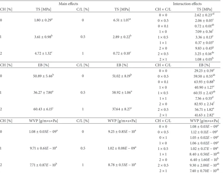
3.1. Tensile Test. The main and interaction effects of CH and
C/L blend on tensile properties of HPMC films are showed in Table 2. There was significant impact of chitosan, C/L preparation, and interaction of blended factors on tensile strength (TS) and elongation at break (EB). TS of the films significantly increased (𝑝 < 0.05) from 1.80, 3.61 to 4.72 MPa with the addition of 0, 1, and 2% of chitosan, respectively. On the contrary, it decreased with the 0, 0.5, and 1% addition of cystatin/lysozyme preparation, from 6.51, 2.89 to 0.72 MPa. Chitosan films with protein addition provide much more tensile resistance than without it [32]. Lysozyme and cystatin are both proteins, but the brittleness was caused by the presence of lysozyme. Lysozyme is an enzyme with degraded
properties towards 𝛽-(1 → 4) linkages of
polysaccha-rides and could hydrolyze both chitosan and hydroxypropyl methylcellulose [33]. Simultaneously, implicated in our study, chitosan with high deacetylation degree is less susceptible to enzymatic degradation [34] but cannot be stopped com-pletely and the products, oligomers, can still have inter-mediate viscosities [18]. On the other hand, the reason of weaker structure and integrity of films could be caused by disruption of crystalline structure formation during drying and weakening intermolecular hydrogen bonds of chitosan and HPMC [34]. Existence of seven and six homogenous groups in interactional effects was showed and proved high complicity of CH and C/L impact on TS and EB, respectively. The elongation at break was enlarged with chitosan 2% (60.42%), compared to its 0 and 1% addition (50.89 and
Table 2: Mechanical properties of films.
Main effects Interaction effects
CH [%] TS [MPa] C/L [%] TS [MPa] CH× C/L TS [MPa]
0 1.80± 0.29a 0 6.51± 1.07a 0× 0 2.62± 0.23cd 0× 0.5 2.06± 0.05c 0× 0.1 0.72± 0.01ab 1 3.61± 0.98b 0.5 2.89± 0.22b 1× 0 7.09± 0.36f 1× 0.5 3.36± 0.13e 1× 1 0.37± 0.03a 2 4.72± 1.32c 1 0.72± 0.10c 2× 0 9.83± 0.45g 2× 0.5 3.25± 0.16de 2× 1 1.08± 0.05b CH [%] EB [%] C/L [%] EB [%] CH× C/L EB [%] 0 50.89± 5.46b 0 51.02± 8.19b 0× 0 29.23± 0.59e 0× 0.5 59.50± 0.55ab 0× 0.1 63.93± 0.88b 1 36.27± 7.80a 0.5 58.92± 1.06a 1× 0 40.90± 1.27c 1× 0.5 60.55± 2.43ab 1× 1 7.36± 0.55d 2 60.43± 6.15c 1 37.64± 8.27c 2× 0 82.93± 2.34f 2× 0.5 56.71± 1.82a 2× 1 41.63± 2.82c
CH [%] WVP [g/m∗s∗Pa] C/L [%] WVP [g/m∗s∗Pa] CH× C/L WVP [g/m∗s∗Pa] 0 1.08± 0.03E − 09a 0 9.25± 0.85E − 10a 0× 0 1.08± 0.03E − 09a 0× 0.5 1.12± 0.11E − 09a 0× 1 1.05± 0.02E − 09a 1 9.71± 0.61E − 10a 0.5 1.02± 0.08E − 09a 1× 0 1.06± 0.02E − 09a 1× 0.5 1.02± 0.17E − 09a 1× 1 8.40± 0.50E − 10ab 2 7.71± 0.87E − 10b 1 8.78± 0.53E − 10a 2× 0 6.40± 1.60E − 10b 2× 0.5 9.30± 2.00E − 10ab 2× 1 7.40± 0.70E − 10ab Values with different letters (a–g) within the same column differ significantly (𝑝 < 0.05).
36.27%, resp.). EB decreased with 1% addition of C/L prepa-ration. The highest TS and EB were registered for samples with the highest amount of CH. Hosseini et al. have reported 12.2 MPa tensile strength of film with 2% chitosan and 0.5% of glycerol [35]. However, 6% of HPMC film with no plasticizer was registered at 63 MPa [36], which is very high value because of the HPMC concentration and lack of glycerol. The same authors noted that with the increased concentration of plasticizer the TS decrease significantly. Worse resistance of our films is caused by addition of plasticizer and hydrophilic compounds, which create lower density of films and help in polymers chains movements. On the other hand, chitosan films made with lactic acid exhibit lower tensile strength and higher elongation than those made with acetic acid, which was registered by Kumar et al. [37].
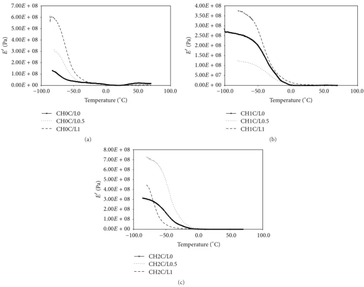
3.2. Dynamic Mechanical Analysis. Glass transition𝑇𝑔
iden-tifiers of DMA test are large drop in storage modulus𝐸and
a peak in tan𝛿 (Figures 1(a), 1(b), 1(c), 2(a), 2(b), and 2(c)).
Storage modulus is an index of rigidity of polysaccharides [38]. The location of the glass transition was shifted to the
higher temperature with addition of CH and C/L. The𝐸
curves increased from 120 MPa for CH0C/L0 to 600 MPa for CH0C/L1 and to 450 MPa for CH2C/L1 films. One drop in
𝐸 was observed in DMA curves of polysaccharides films
at −40–5∘C. Wu et al. have presented study on
cellulose-chitosan blend and cellulose-chitosan; the curves of storage modulus
of chitosan showed a drop at 0–30∘C [15]. The authors
suggested that drop is related with hydration of side groups
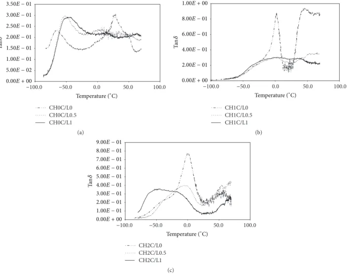
(–CH2OH) on chitosan. On the other hand, tan𝛿 curves
showed two peaks; the first of them is characterized as the 𝛽 relaxation attributed to local mode in amorphous phase,
and another one is designed as the𝛼 relaxation assigned to
glass transition of amorphous phase [39]. Tan𝛿 is a measure
of polysaccharide viscoelasticity. Wetton has also suggested
that tan𝛿 peak size is correlated with the polymer in the
composition of sample [40]. The 𝛼 relaxation peaks were
reduced after C/L addition, which confirms degradative effect of lysozyme on main chain of polymers. The viscoelastic
CH0C/L0 CH0C/L0.5 CH0C/L1 −50.0 0.0 50.0 100.0 −100.0 Temperature (∘C) 0.00E + 00 1.00E + 08 2.00E + 08 3.00E + 08 4.00E + 08 5.00E + 08 6.00E + 08 7.00E + 08 E (P a) (a) CH1C/L0 CH1C/L0.5 CH1C/L1 −50.0 0.0 50.0 100.0 −100.0 Temperature (∘C) 0.00E + 00 5.00E + 07 1.00E + 08 1.50E + 08 2.00E + 08 2.50E + 08 3.00E + 08 3.50E + 08 4.00E + 08 E (P a) (b) CH2C/L0 CH2C/L0.5 CH2C/L1 E (P a) −50.0 0.0 50.0 100.0 −100.0 Temperature (∘C) 0.00E + 00 1.00E + 08 2.00E + 08 3.00E + 08 4.00E + 08 5.00E + 08 6.00E + 08 7.00E + 08 8.00E + 08 (c)
Figure 1: Storage modulus (𝐸) of HMPC, CH, and C/L preparation films of (a) CH0C/L0, CH0C/L0.5, and CH0C/L1; (b) CH1C/L0,
CH1C/L0.5, and CH1C/L1; and (c) CH2C/L0, CH2C/L0.5, and CH2C/L1 variants.
behavior of films was confirmed by loss factor values, which
were between 0.1 and 0.9. The CH0C/L0 film represented𝛽
relaxation around −80–(−50)∘C and 𝑇𝑔 at 5–50∘C samples
with CH and C/L were characterized by tan𝛿 peaks at 0∘C
and between 40 and 60∘C. The shift of𝛽 and 𝛼 transition
temperature is the result of CH addition. Addition of chitosan
has changed𝑇𝑔of HPMC to higher temperature but has not
changed the width and height of the peak, which suggest that both polymers were mixing well. If the peak got broadness and dumping decrease, it will mean that polysaccharides are incompatible and semicompatible [41].
3.3. Fourier Transform Infrared Spectrometry. FTIR spectra
of HPMC films had absorption bands at 3373.85 cm−1,
2885.95 cm−1, 1649.80 cm−1, and 1041.37 cm−1assigned to
O-H stretch, combination of methyl groups and C-O-H stretch, C=O, and ether C-O-C stretch, respectively (Figure 3(a)). The position of the peaks of HPMC film spectrum is similar to those described by Gustafsson et al. [42]. Hydrogen bonding or other interactions between chemical groups on dissimilar polymers should theoretically cause a shift in peak position
of the participating groups [43]. This kind of behavior was observed for the OH stretching since this peak shifted
from 3373.85 cm−1 for HPMC film toward 3293.82 cm−1 for
HPMC-CH mixtures (Figures 3(b) and 3(c)). Also increased C/L preparation concentration changed position of the OH
group from 3373.85 cm−1 for pure HPMC to 3350.71 cm−1
for HPMC-0.5% C/L and to 3331.43 cm−1for HPMC-1% C/L
blends. According to Wu et al. [16], FTIR spectra of CH films
had absorption bands at 3400–3480 cm−1 that responded
to OH-3 and CH2OH intra- and intermolecular hydrogen
bonds, 1650 cm−1 for amide I, and 1557 cm−1 for amide II
vibrational mode (Figures 3(b) and 3(c)).
3.4. Water Vapour Permeability. The results of statistical
anal-ysis of water vapour permeability are presented in Table 2. No significant effect of C/L preparation on water permeability was noted. Lopes, Martins, Fonseca, and Vicente (2011) also noticed that WVP of chitosan films were not affected by the enzyme, glucose oxidase incorporation [44]. Cellulase did not change water resistance of chitosan-hydroxypropyl
CH0C/L0 CH0C/L0.5 CH0C/L1 −50.0 0.0 50.0 100.0 −100.0 Temperature (∘C) Ta n𝛿 0.00E + 00 5.00E − 02 1.00E − 01 1.50E − 01 2.00E − 01 2.50E − 01 3.00E − 01 3.50E − 01 (a) CH1C/L0 CH1C/L0.5 CH1C/L1 −50.0 0.0 50.0 100.0 −100.0 Temperature (∘C) Ta n𝛿 0.00E + 00 2.00E − 01 4.00E − 01 6.00E − 01 8.00E − 01 1.00E + 00 (b) CH2C/L0 CH2C/L0.5 CH2C/L1 −50.0 0.0 50.0 100.0 −100.0 Temperature (∘C) Ta n𝛿 0.00E + 00 1.00E − 01 2.00E − 01 3.00E − 01 4.00E − 01 5.00E − 01 6.00E − 01 7.00E − 01 8.00E − 01 9.00E − 01 (c)
Figure 2: Tan𝛿 of HPMC, CH, and C/L preparation films of (a) CH0C/L0, CH0C/L0.5, and CH0C/L1; (b) CH1C/L0, CH1C/L0.5, and CH1C/L1; and (c) CH2C/L0, CH2C/L0.5, and CH2C/L1 variants.
methylcellulose films [10]. However, interactional effect of C/L and CH blend as well as chitosan influence was sig-nificant. Water resistance of tested films was higher with
increasing concentration of chitosan from 1.08𝐸 − 09 to
7.71𝐸 − 10 g/m ∗ s ∗ Pa for 0 and 2% dose, respectively. Those results were confirmed by significant impact of simultaneous
action of CH and C/L. The lowest WVP of6.40𝐸−10 g/m∗s∗
Pa was noticed for film with the highest addition of chitosan and lack of lysozyme/cystatin preparation.
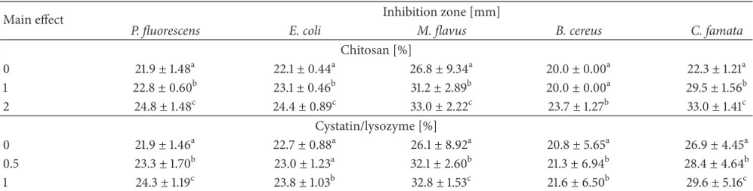
3.5. Antibacterial Activity. Antimicrobial effect of chitosan
and cystatin/lysozyme preparation incorporated into the filmogenic hydrosol composition against M. flavus, B. cereus,
E. coli, P. fluorescens, and C. famata was presented in Tables
3 and 4. Incorporation of C/L and CH exhibited a clear inhibitory zone by the absence of bacterial and yeast growth. CH showed maximum inhibition of 33.0 mm against Gram-positive bacteria, M. flavus, in the highest concentration of 2%. These bacteria were selected for our test because of their high susceptibility to the various antimicrobial agents. Similar inhibiting effect of 32.8 against the same
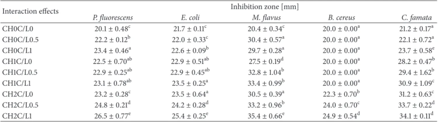
bacteria was observed with 1% addition of C/L. The highest concentration of C/L statistically influenced inhibition of yeast growth (29.6 mm). Kolaczkowska et al. have proved antifungal activity of cystatin, affinity-purified from chicken egg white, against the most frequent human fungal pathogens of the genus Candida [25]. The mechanism of fungal growth inhibition by cystatin is not related to its protease inhibitory properties as Wesierska et al. suggested in their study regard-ing antibacterial activity of cystatin [24]. The purified recom-binant proteins of tarocystatin from Colocasia esculenta such as N-terminus peptide have a greater antifungal activity than full-length peptide. In addition, C-terminus peptide has not showed antifungal effect, which is proof of lack of connection to the inhibitory activity [45]. Significant reduction of two Gram-negative bacteria by every tested concentration of CH and C/L was also noted. Characteristic intensification of antibacterial activity is also possible by the addition of hydrolytic enzymes, such as lysozyme to the chitosans films, which increases the inhibiting effect on Escherichia coli growth [33], which was also confirmed in our study. Micro-biological test showed that chitooligomers obtained after
30 40 50 60 70 80 90 100 CH0C/L0 CH0C/L0.5 CH0C/L1 T (%) 3150 2650 2150 1650 1150 650 3650 Wavenumber (cm−1) (a) 60 70 80 90 100 T (%) CH1C/L0 CH1C/L0.5 CH1C/L1 3150 2650 2150 1650 1150 650 3650 Wavenumber (cm−1) (b) 60 70 80 90 100 T (%) CH2C/L0 CH2C/L0.5 CH2C/L1 3150 2650 2150 1650 1150 650 3650 Wavenumber (cm−1) (c)
Figure 3: FTIR spectra of HMPC and C/L preparation films of (a) CH0C/L0, CH0C/L0.5, and CH0C/L1; (b) CH1C/L0, CH1C/L0.5, and CH1C/L1; and (c) CH2C/L0, CH2C/L0.5, and CH2C/L1 variants.
Table 3: Antibacterial activity of edible films modified by CH and C/L preparation (main effects).
Main effect Inhibition zone [mm]
P. fluorescens E. coli M. flavus B. cereus C. famata
Chitosan [%] 0 21.9± 1.48a 22.1± 0.44a 26.8± 9.34a 20.0± 0.00a 22.3± 1.21a 1 22.8± 0.60b 23.1± 0.46b 31.2± 2.89b 20.0± 0.00a 29.5± 1.56b 2 24.8± 1.48c 24.4± 0.89c 33.0± 2.22c 23.7± 1.27b 33.0± 1.41c Cystatin/lysozyme [%] 0 21.9± 1.46a 22.7± 0.88a 26.1± 8.92a 20.8± 5.65a 26.9± 4.45a 0.5 23.3± 1.70b 23.0± 1.23a 32.1± 2.60b 21.3± 6.94b 28.4± 4.64b 1 24.3± 1.19c 23.8± 1.03b 32.8± 1.53c 21.6± 6.50b 29.6± 5.16c Values with different letters (a–c) within the same column differ significantly (𝑝 < 0.05).
lysozyme hydrolysis exhibit higher antilisterial activity than chitosan [46]. Interaction effects of statistical analysis showed that inhibition growth of E. coli and P. fluorescens is the result of simultaneous addition of tested bioactive substance. Both species are the food poisoning bacteria in chilled foods. The lack of reduction zone of B. cereus growth (20.0 mm, the same
as film diameter) was observed for variants with 0 and 1% chitosan with C/L addition in various concentrations. It confirms what Mellegard et al. have noted that antimicrobial activity of chitosan is concentration-dependent towards B.
cereus [47]. They have also found out that chitosan with low
Table 4: Antibacterial activity of edible films modified by CH and C/L preparation (interaction effects).
Interaction effects Inhibition zone [mm]
P. fluorescens E. coli M. flavus B. cereus C. famata
CH0C/L0 20.1± 0.48c 21.7± 0.11c 20.4± 0.34c 20.0± 0.00a 21.2± 0.17a CH0C/L0.5 22.2± 0.12b 22.0± 0.33c 30.4± 0.57a 20.0± 0.00a 22.1± 0.72a CH0C/L1 23.4± 0.46a 22.6± 0.09b 29.7± 0.28a 20.0± 0.00a 23.7± 0.58e CH1C/L0 22.5± 0.70ab 22.9± 0.51ab 27.5± 0.19d 20.0± 0.00a 28.2± 0.47b CH1C/L0.5 22.9± 0.25ab 22.9± 0.45ab 32.8± 1.04b 20.0± 0.00a 29.4± 1.62b CH1C/L1 23.1± 0.78ab 23.5± 0.25a 33.4± 0.99b 20.0± 0.00a 30.9± 1.09c CH2C/L0 23.2± 0.28c 23.5± 0.64a 30.5± 0.39a 22.3± 0.70b 31.2± 0.63c CH2C/L0.5 24.8± 0.21d 24.2± 0.28d 33.2± 0.96b 24.0± 0.70c 33.7± 0.22d CH2C/L1 26.5± 0.77e 25.4± 0.25e 35.4± 0.66e 24.9± 0.54d 34.1± 0.11d
Values with different letters (a–e) within the same column differ significantly (𝑝 < 0.05).
B. cereus growth more effectively than with high acetylation
degree and low molecular weight, which also explain very low reduction of these bacteria in our study.
4. Conclusions
HPMC films were successfully prepared with chitosan and cystatin/lysozyme preparation. Thermal properties of the HPMC-CH-C/L biocomposites are almost dominated by hydroxypropyl methylcellulose and protein preparation. Hydrolyzed impact of cystatin/lysozyme preparation was
observed only in𝛼 main chain of HPMC. C/L preparation
induces significant changes of tensile strength and elongation at break but has no influence on water vapour permeability. Elongation of films was improved by CH addition, which is important in coating process of elastic materials, such as raw meat. C/L preparation addition results in higher elasticity of
obtained biocomposites, because very high𝑇𝑔 temperature
provides a brittle material. Simultaneous effect of CH and C/L in microbial reduction of every tested microorganism was obtained. Implication of HPMC biocomposites with CH and bioactive preparation in packaging production system is possible. We also assumed that designed composition is suitable for meat and meat products, due to simplicity of the process to obtain film components and to achieve desired thermomechanical, barrier, and antimicrobial properties.
Conflict of Interests
The authors declare that there is no conflict of interests regarding the publication of this paper.
Acknowledgment
This paper is supported by Wroclaw Centre of Biotechnology, programme The Leading National Research Centre (KNOW) for years 2014–2018.
References
[1] M. Z. Elsabee and E. S. Abdou, “Chitosan based edible films and coatings: a review,” Materials Science and Engineering C, vol. 33, no. 4, pp. 1819–1841, 2013.
[2] F. A. Osorio, P. Molina, S. Matiacevich, J. Enrione, and O. Skurtys, “Characteristics of hydroxy propyl methyl cellulose (HPMC) based edible film developed for blueberry coatings,”
Procedia Food Science, vol. 1, pp. 287–293, 2011.
[3] S. Mathew, M. Brahmakumar, and T. E. Abraham, “Microstruc-tural imaging and characterization of the mechanical, chemical, thermal, and swelling properties of starch-chitosan blend films,”
Biopolymers, vol. 82, no. 2, pp. 176–187, 2006.
[4] A. Hambleton, M.-J. Fabra, F. Debeaufort, C. Dury-Brun, and A. Voilley, “Interface and aroma barrier properties of iota-carrageenan emulsion-based films used for encapsulation of active food compounds,” Journal of Food Engineering, vol. 93, no. 1, pp. 80–88, 2009.
[5] J. G´omez-Estaca, L. Bravo, M. C. G´omez-Guill´en, A. Alem´an, and P. Montero, “Antioxidant properties of tuna-skin and bovine-hide gelatin films induced by the addition of oregano and rosemary extracts,” Food Chemistry, vol. 112, no. 1, pp. 18– 25, 2009.
[6] S. D. Figueir´o, J. C. G´oes, R. A. Moreira, and A. S. B. Sombra, “On the physico-chemical and dielectric properties of glutaraldehyde crosslinked galactomannan—collagen films,”
Carbohydrate Polymers, vol. 56, no. 3, pp. 313–320, 2004.
[7] G. Y. Park, S. Mun, Y. Park et al., “Influence of encapsulation of emulsified lipids with chitosan on their in vivo digestibility,”
Food Chemistry, vol. 104, no. 2, pp. 761–767, 2007.
[8] R. Porta, L. Mariniello, P. di Pierro, A. Sorrentino, and C. V. L. Giosafatto, “Transglutaminase crosslinked pectin- and chitosan-based edible films: a review,” Critical Reviews in Food
Science and Nutrition, vol. 51, no. 3, pp. 223–238, 2011.
[9] S. Rawdkuen, P. Suthiluk, D. Kamhangwong, and S. Benjakul, “Mechanical, physico-chemical, and antimicrobial properties of gelatin-based film incorporated with catechin-lysozyme,”
Chemistry Central Journal, vol. 6, article 131, 2012.
[10] A. Zimoch-Korzycka, J. Ambrozik-Haba, D. Kulig, and A. Jar-moluk, “Modification effect of cellulase on the physicochemical characteristic of polysaccharides edible films,” International
Journal of Polymer Science, vol. 2015, Article ID 184616, 7 pages,
2015.
[11] S. Kamel, N. Ali, K. Jahangir, S. M. Shah, and A. A. El-Gendy, “Pharmaceutical significance of cellulose: a review,” eXPRESS
Polymer Letters, vol. 2, no. 11, pp. 758–778, 2008.
[12] M. M. Al-Tabakha, “HPMC capsules: current status and future prospects,” Journal of Pharmacy and Pharmaceutical Sciences, vol. 13, no. 3, pp. 428–442, 2010.
[13] S. Jain, P. S. Sandhu, R. Malvi, and B. Gupta, “Cellulose derivatives as thermoresponsive polymer: an overview,” Journal
of Applied Pharmaceutical Science, vol. 3, no. 12, pp. 139–144,
2013.
[14] M. J. Akhtar, M. Jacquot, E. Arab-Tehrany, C. Ga¨ıani, M. Linder, and S. Desobry, “Control of salmon oil photo-oxidation during storage in HPMC packaging film: influence of film colour,” Food
Chemistry, vol. 120, no. 2, pp. 395–401, 2010.
[15] Y.-B. Wu, S.-H. Yu, F.-L. Mi et al., “Preparation and char-acterization on mechanical and antibacterial properties of chitsoan/cellulose blends,” Carbohydrate Polymers, vol. 57, no. 4, pp. 435–440, 2004.
[16] T. Wu, S. Zivanovic, F. A. Draughon, W. S. Conway, and C. E. Sams, “Physicochemical properties and bioactivity of fungal chitin and chitosan,” Journal of Agricultural and Food Chemistry, vol. 53, no. 10, pp. 3888–3894, 2005.
[17] C. Valenzuela, L. Abugoch, and C. Tapia, “Quinoa protein-chitosan-sunflower oil edible film: mechanical, barrier and structural properties,” LWT: Food Science and Technology, vol. 50, no. 2, pp. 531–537, 2013.
[18] J. Rhoades and S. Roller, “Antimicrobial actions of degraded and native chitosan against spoilage organisms in laboratory media and foods,” Applied and Environmental Microbiology, vol. 66, no. 1, pp. 80–86, 2000.
[19] G. G. Buonocore, A. Conte, M. R. Corbo, M. Sinigaglia, and M. A. Del Nobile, “Mono- and multilayer active films containing lysozyme as antimicrobial agent,” Innovative Food Science &
Emerging Technologies, vol. 6, no. 4, pp. 459–464, 2005.
[20] A. Conte, G. G. Buonocore, M. Sinigaglia, and M. A. Nobile, “Development of immobilized lysozyme based active film,”
Journal of Food Engineering, vol. 78, no. 3, pp. 741–745, 2007.
[21] E. D. N. S. Abeyrathne, H. Y. Lee, and D. U. Ahn, “Egg white proteins and their potential use in food processing or as nutraceutical and pharmaceutical agents-a review,” Poultry
Science, vol. 92, no. 12, pp. 3292–3299, 2013.
[22] W. Kopec and T. Trziszka, “Lysozyme and its characteristics. Part II. Isolation and practical applications,” Food Industry, vol. 3, pp. 36–37, 1997 (Polish).
[23] Y. M. C. Henskens, E. C. L. Veerman, and A. V. N. Amerongen, “Cystatins in health and disease,” Biological Chemistry
Hoppe-Seyler, vol. 377, no. 2, pp. 71–86, 1996.
[24] E. Wesierska, Y. Saleh, T. Trziszka, W. Kopec, M. Siewinski, and K. Korzekwa, “Antimicrobial activity of chicken egg white cystatin,” World Journal of Microbiology and Biotechnology, vol. 21, no. 1, pp. 59–64, 2005.
[25] A. Kolaczkowska, M. Kolaczkowski, A. Sokolowska et al., “The antifungal properties of chicken egg cystatin against Candida yeast isolates showing different levels of azole resistance,”
Mycoses, vol. 53, no. 4, pp. 314–320, 2010.
[26] A. R. Collins and A. Grubb, “Cystatin D, a natural salivary cys-teine protease inhibitor, inhibits coronavirus replication at its physiologic concentration,” Oral Microbiology & Immunology, vol. 13, no. 1, pp. 59–61, 1998.
[27] T. Skiba, W. Kope´c, and Ł. Bobak, “Chemo-thermal method of initial isolation of bioactive proteins from egg white,” Przemysl
Chemiczny, vol. 91, no. 4, pp. 527–530, 2012 (Polish).
[28] G. Lesnierowski and J. Kijowski, “Lysozyme,” in Bioactive Egg
Compounds, R. Huopalahti, R. Lopez-Fandino, M. Anton, and
R. Schade, Eds., pp. 35–37, Springer, Berlin, Germany, 2007. [29] M. Siewi´nski, “Method of purification of thiol proteinase
inhibitors from human urine,” Cancer Biochemistry Biophysics, vol. 12, no. 1, pp. 33–43, 1991.
[30] M. A. Daeschel, T. Musafija-Jeknic, Y. Wu, D. Bizzarri, and A. Villa, “High-performance liquid chromatography analysis of lysozyme in wine,” American Journal of Enology and Viticulture, vol. 53, no. 2, pp. 154–157, 2002.
[31] ASTM, “Standard test methods for water vapor transmission of materials,” ASTM E96/E96M-13, ASTM International, West Conshohocken, Pa, USA, 2013.
[32] M. Y. Arancibia, A. Alem´an, M. E. L´opez-Caballero, M. C. G´omez-Guill´en, and P. Montero, “Development of active films of chitosan isolated by mild extraction with added protein concentrate from shrimp waste,” Food Hydrocolloids, vol. 43, pp. 91–99, 2015.
[33] R. J. Nordtveit, K. M. V˚arum, and O. Smidsrød, “Degradation of partially N-acetylated chitosans with hen egg white and human lysozyme,” Carbohydrate Polymers, vol. 29, no. 2, pp. 163–167, 1996.
[34] S.-I. Park, M. A. Daeschel, and Y. Zhao, “Functional properties of antimicrobial lysozyme-chitosan composite films,” Journal of
Food Science, vol. 69, no. 8, pp. M215–M221, 2004.
[35] M. H. Hosseini, S. H. Razavi, and M. A. Mousavi, “Antimicro-bial, physical and mechanical properties of chitosan-based films incorporated with thyme, clove and cinnamon essential oils,”
Journal of Food Processing and Preservation, vol. 33, no. 6, pp.
727–743, 2009.
[36] M. Imran, S. El-Fahmy, A.-M. Revol-Junelles, and S. Desobry, “Cellulose derivative based active coatings: effects of nisin and plasticizer on physico-chemical and antimicrobial properties of hydroxypropyl methylcellulose films,” Carbohydrate Polymers, vol. 81, no. 2, pp. 219–225, 2010.
[37] M. N. V. R. Kumar, R. A. A. Muzzarelli, C. Muzzarelli, H. Sashiwa, and A. J. Domb, “Chitosan chemistry and pharmaceu-tical perspectives,” Chemical Reviews, vol. 104, no. 12, pp. 6017– 6084, 2004.
[38] A. Hashemi Doulabi, H. Mirzadeh, and M. Imani, “Interaction and miscibility study of fumarate-based macromers with chi-tosan,” Materials Chemistry and Physics, vol. 139, no. 2-3, pp. 515–524, 2013.
[39] M. Mucha and A. Pawlak, “Thermal analysis of chitosan and its blends,” Thermochimica Acta, vol. 427, no. 1-2, pp. 69–76, 2005. [40] R. E. Wetton, “Dynamic mechanical thermal analysis of poly-mers and related systems,” in Developments in Polymer
Charac-terization, J. V. Dawkins, Ed., pp. 179–222, Springer, New York,
NY, USA, 1986.
[41] Y. Wan, H. Wu, A. Yu, and D. Wen, “Biodegradable polylac-tide/chitosan blend. Membranes,” Biomacromolecules, vol. 7, no. 4, pp. 1362–1372, 2006.
[42] C. Gustafsson, C. Nystr¨om, H. Lennholm, M. C. Bonfer-oni, and C. M. Caramella, “Characteristics of hydroxypropyl methylcellulose influencing compactibility and prediction of particle and tablet properties by infrared spectroscopy,” Journal
[43] R. K. Wanchoo and P. K. Sharma, “Viscometric study on the compatibility of some water-soluble polymer-polymer mix-tures,” European Polymer Journal, vol. 39, no. 7, pp. 1481–1490, 2003.
[44] M. I. Lopes, J. T. Martins, L. P. Fonseca, and A. A. Vicente, “Effect of glucose oxidase incorporation in chitosan edible films properties,” in Proceedings of the 6th International CIGR
Symposium on Towards a Sustainable Food Chain: Food Process, Bioprocessing and Food Quality Management, Nantes, France,
April 2011.
[45] K.-M. Wang, S. Kumar, Y.-S. Cheng, S. Venkatagiri, A.-H. Yang, and K.-W. Yeh, “Characterization of inhibitory mechanism and antifungal activity between group-1 and group-2 phytocystatins from taro (Colocasia esculenta),” The FEBS Journal, vol. 275, no. 20, pp. 4980–4989, 2008.
[46] A. Zimoch-Korzycka, C. Gardrat, A. Castellan, V. Coma, and A. Jarmoluk, “The use of lysozyme to prepare biologically active chitooligomers,” Pol´ımeros, vol. 25, no. 1, pp. 35–41, 2015. [47] H. Melleg˚ard, C. From, B. E. Christensen, and P. E. Granum,
“Inhibition of Bacillus cereus spore outgrowth and multiplica-tion by chitosan,” Internamultiplica-tional Journal of Food Microbiology, vol. 149, no. 3, pp. 218–225, 2011.
Submit your manuscripts at
http://www.hindawi.com
Scientifica
Hindawi Publishing Corporation
http://www.hindawi.com Volume 2014 Hindawi Publishing Corporation
http://www.hindawi.com Volume 2014 Hindawi Publishing Corporation
http://www.hindawi.com Volume 2014
Hindawi Publishing Corporation
http://www.hindawi.com Volume 2014
Ceramics
Journal ofHindawi Publishing Corporation
http://www.hindawi.com Volume 2014
Nanoparticles
Journal ofHindawi Publishing Corporation
http://www.hindawi.com Volume 2014
Hindawi Publishing Corporation
http://www.hindawi.com Volume 2014
International Journal of
Biomaterials
Hindawi Publishing Corporation
http://www.hindawi.com Volume 2014
Nanoscience
Journal ofTextiles
Hindawi Publishing Corporation
http://www.hindawi.com Volume 2014
Journal of
Hindawi Publishing Corporation
http://www.hindawi.com Volume 2014
Crystallography
Journal ofHindawi Publishing Corporation
http://www.hindawi.com Volume 2014
The Scientific
World Journal
Hindawi Publishing Corporationhttp://www.hindawi.com Volume 2014
Hindawi Publishing Corporation
http://www.hindawi.com Volume 2014
Coatings
Journal ofAdvances in
Materials Science and Engineering
Hindawi Publishing Corporation
http://www.hindawi.com Volume 2014
Hindawi Publishing Corporation
http://www.hindawi.com Volume 2014
Hindawi Publishing Corporation
http://www.hindawi.com Volume 2014
Metallurgy
Journal ofHindawi Publishing Corporation
http://www.hindawi.com Volume 2014
BioMed
Research International
Materials
Journal ofHindawi Publishing Corporation
http://www.hindawi.com Volume 2014
N
a
no
ma
te
ria
ls
Hindawi Publishing Corporation
http://www.hindawi.com Volume 2014
Journal of