Any correspondence concerning this service should be sent to the repository administrator:
[email protected]
To link to this article : DOI:
10.1016/j.otsr.2016.01.019
URL :
https://doi.org/10.1016/j.otsr.2016.01.019
This is an author-deposited version published in:
http://oatao.univ-toulouse.fr/
Eprints ID: 18541
O
pen
A
rchive
T
oulouse
A
rchive
O
uverte (
OATAO
)
OATAO is an open access repository that collects the work of Toulouse
researchers and makes it freely available over the web where possible.
To cite this version: Obert, Laurent and Saadnia, Rachid and Tournier,
Clément and Bonnevialle, Nicolas and Saragaglia, Dominique and
Sirveaux, François and Société Française de Chirurgie Orthopédique et
Traumatologie, Four-part fractures treated with a reversed total shoulder
prosthesis: Prospective and retrospective multicenter study. Results and
complications. (2016) Orthopaedics & Traumatology: Surgery &
Four-part fractures treated with a reversed total shoulder prosthesis:
Prospective and retrospective multicenter study. Results and
complications
L. Obert
a,∗, R. Saadnia
a, C. Tournier
b, N. Bonnevialle
c, D. Saragaglia
d,
F. Sirveaux
e, La SOFCOT
faChirurgie orthopédique, traumatologique et plastique, centre hospitalier de Besanc¸on, 2, boulevard Fleming, 25030 Besanc¸on, France bService d’orthopédie-traumatologie, place Amélie-Raba-Léon, 33076 Bordeaux cedex, France
cCentre hospitalier universitaire de Toulouse, Institut de l’appareil locomoteur, place du Dr-Baylac, 31059, Toulouse, France dService d’orthopédie-traumatologie, CHRU de Grenoble, 38000 Grenoble, France
eService d’orthopédie-traumatologie, centre chirurgical E.-Galle, 49, rue Hermite, 54000 Nancy, France fSociété franc¸aise de chirurgie orthopédique et traumatologie, 56, rue Boissonnade, 75014 Paris, France
Keywords: Fractures Proximal humerus Four-part fracture Multicenter study Reversed prosthesis Osteosuture Elderly patients
a b s t r a c t
Introduction: The reversed shoulder prosthesis is becoming the gold standard for cases of complex frac-ture of the proximal humerus after 70 years of age.
Material and methods: The French Society of Orthopaedic and Traumatology Surgery (SOFCOT) conducted a prospective and retrospective multicenter study to evaluate the results of the reversed shoulder pros-thesis implanted in patients with a four-part fracture in nine centers. In the retrospective study (n = 41 patients, 78 years of age, 14% ASA grade 3, 21% associated fractures) and in the prospective study (n = 32 patients, 79 years of age, 37% ASA grade 3, 21% associated fractures) evaluation by independent surgeons was conducted to measure the QuickDASH score, the Constant score, the SSV (subjective shoulder value), and complications to correlate these measurements with radiological results.
Results: In both studies, use of an autograft (75%) to perform an osteosuture of tuberosities (90%) and no postoperative immobilization (75%) were similar. In the retrospective study at 39 months (range: 24–62 months) of follow-up, the QuickDASH reached 28 (range: 0–59), the Constant scores (raw Constant = 57, weighted Constant = 83.4%), and SSV 75 (range: 35–100). Complications after the 1st month (7%) were nonunion or ossifications. In the prospective study at 11 months (range: 5–16.5 months) of follow-up, the QuickDASH reached 40 (range: 1–75), the Constant scores 50 (raw Constant) and 74.6% (weighted Constant), and SSV 69 (range: 10–100). Complications after the 1st month (21%) were stiffness and dislo-cation, with two patients who underwent revision surgery. In both studies, early complications reached 6% (palsy, dislocation).
Conclusion: This double (retrospective and prospective) study confirms the good results with a low level of complications of the reversed implant in cases of fracture but with osteosuture of tuberosities.
1. Introduction
Management of four-part fractures in the elderly subject is a veritable therapeutic challenge. The incidence of these fractures is on the rise because of the aging of the population. The func-tional demand of these patients has also increased. After 70 years of age and in case of a displaced four-part fracture of the proximal
∗ Corresponding author, Chirurgie orthopédique, traumatologique et plastique, centre hospitalier de Besanc¸on, 2, boulevard Fleming, 25030 Besanc¸on, France.
E-mail address:[email protected](L. Obert).
humerus, hemiarthroplasty is logically preferred to osteosynthesis, but the clinical results, related to anatomic union of the tuberosi-ties, are difficult to predict[1,2]. In these situations, age greater than or equal to 75 years, female gender, and the presence of osteo-porosis are predictive factors of poor functional results[3,4]. This is why for several years the reversed prosthesis, first developed by Grammont for irreparable rotator cuff rupture, has a place in the elderly subject who presents negative prognostic factors for hemi-arthroplasty. The objective of this prospective and retrospective multicenter study was to report the short- and long-term func-tional results of reversed shoulder prostheses implanted in patients presenting a displaced four-part fracture of the proximal humerus.
2. Material and methods
The clinical research protocol included a prospective study and a retrospective study with the same inclusion criteria. SOFCOT (the French Society of Orthopaedic and Traumatology Surgery) promoted the study in collaboration with the Lille Regional Hos-pital Biostatics unit. This was a study protocol on routine care and underwent CPP (Comité de Protection des Personnes) review with approval by the CCTIRS (Consulting committee on data processing in healthcare research) and the CNIL (national personal data protec-tion agency) (CNIL agreement: 26/06 no. 913192). All evaluaprotec-tions were accessible and completed on a secure site with access to the images (KLETEL). The inclusion criteria for the retrospective study were being an adult 18 years or older presenting a four-part proxi-mal humerus fracture (CT4), displaced or not, with a maximum time from injury of 3 weeks, occurring between 1st January 2009 and 31st December 2011. The main endpoint was the weighted Con-stant score, which, when it was less than 70%, was considered as a poor result. The secondary criteria were the QuickDASH and SSV scores, range of motion, complications, and revisions. The prospec-tive study was conducted from 1st January 2013 to 31st December 2013 with the same inclusion criteria, but required a 2D and 3D CT scan. Follow-up information at 3 months, 6 months, and at the last follow-up was required. The advantage of the retrospective study was to assess the late complications. In the initial series that grouped 113 patients, 70 in the retrospective study (CT4retro) and 43 in the prospective study (CT4pro), only 73 were followed up (41 in the CT4retro study and 32 in the CT4pro study). Of the 11 inves-tigating centers, nine implanted reversed shoulder prostheses. Five centers out of nine had implanted more than five prostheses. 2.1. The overall series
Ninety percent of the patients were female, with a mean age of 79 years± 5 years (range: 60–95 years) and a mean BMI of 27± 9 kg/m2(range: 19–51 kg/m2); 52% of the patients presented comorbidities (diabetes, Parkinson disease, or heart disease); 70% had osteoporosis and 22% an associated fracture in the upper limb, lower limb, or spine (Tables 1 and 2).
The approach was superolateral in 75% of the cases with implantation of a reversed prosthesis manufactured by Tornier® (Saint-Ismier, France) in 90% of the cases, by FX®(Bourg-en-Bresse, France) or Depuy Synthes®(Saint-Priest, France) in 10% of cases. In 96% of the patients, a 36-mm-diameter glenoid sphere was used. Osteosuture fixation of the tuberosities was performed in 93% of the cases, tenodesis of the long head biceps brachii in 75% of the
Table 1
Epidemiological variables of the two series.
Retrospective series (n = 41) 1% = 2.4 9 centers 5 centers > 6 poses Prospective series (n = 32) 1% = 3.1 9 centers 4 centers > 4 poses Follow-up (months) 39 (24–62) 11.3 (5–16.5) Age 78.2 (60–88) 79.4 (72–91) Gender F: 90% F: 90.6% Weight (kg) 70.8 (45–147) 66.3 (50–83) Height (cm) 159.4 (146–175) 157.9 (145–167) BMI 27 (19–51) 26.4 (20–34) ASA > 3 14.6% 37%, P = 0.02 Diabetes 17% 6.2% Parkinson 0%?! 9.3% Alcoholism 0%?! 5.8%
Medical history in addition to diabetes, etc.
53.6% 50%
Osteoporosis 64% 75%
Table 2
Fracture, associated lesions, treatment, and immobilization of 2 series.
Retrospective series (n = 41) 1% = 2.4 9 centers 5 centers > 6 poses Prospective series (n = 32) 1% = 3.1 9 centers 4 centers > 4 poses Fracture dominant side
right
89% 96.6%
Associated fractures Upper limb, lower limb, spine 21.6% 21.7% Percentage of head dislocated 13% 32% Percentage of head broken 23.7% 9.6% Superior approach 78% 71.8% Diameter 36 100% 91% 3 > 40
Type of prosthesis Tornier Tornier except 4 FX solutions implants, 4 Depuy implants Bone graft 65% 84.3% Tuberosity fixation 90% 97% Postoperative tuberosity reduction 82% (both) 85% (both) Tenodesis 74.3% 84.3% Acromioplasty 5.4% 3% Intraoperative complications 2.5% 1 fracture per op 3% 1 fissure per op Immobilization 4S 17% 25%
Type of immobilization Internal rotation: 63.8% N rot: 16.6%
Internal rotation: 61.3% N rot: 32.2%
Return to home 39% 37.5%
Rehabilitation center 47.2% 40.6% N rot: neutral rotation; per op: peroperative.
cases, and acromioplasty in 4%. An autologous bone graft was used in 75% of the cases. The tuberosities were deemed reduced after surgery in 83% of the cases. Two intraoperative complications were observed, one in each series: 2.5% in the retrospective series and 3% in the prospective series, a fissure or an intraoperative diaphyseal fracture.
Four-week immobilization was deemed useful in 21% of the cases, which was in internal rotation in 60% of the cases; all the other patients were mobilized immediately. Thirty-eight percent of the patients were able to return home with a self-rehabilitation program and 43% were transferred to a rehabilitation center. The remaining 20% returned home with a rehabilitation protocol to follow on an out-patient basis or with an independent physical therapist.
The CT4pro and CT4retro series were comparable except for the American Society of Anesthesiologists (ASA) score: 14% of the patients were ASA 3 or greater in the retrospective series and 37% in the prospective series (P = 0.02) (Table 2).
3. Results
3.1. Functional and radiographic results 3.1.1. Short-term results (prospective series)
At 3 months of follow-up, the percentage of satisfied and very satisfied patients was 79%, increasing to 87% at 11 months (Table 3). The rate of excellent and good results (weighted Con-stant score > 80%) was 35% at 3 months and 53% at 11 months, showing an increase from the 3rd month and the 11th month with the Constant score gaining 10 points and a weighted Constant score that gained 10%. Anterior elevation of the arm progressed by 10◦ and external rotation by 6◦. The QuickDASH score improved slightly as did the SSV between 3 and 11 months. The failures (weighted
Table 3
Functional and radiographic results of 2 series not considering complications.
Retrospective series (n = 41) 1% = 2.4 9 centers 5 centers > 6 poses Prospective series (n = 32) 1% = 3.1 9 centers 4 centers > 4 poses Follow-up (months) 39 (24–62) 11.3 (5–16.5) Weighted Constant score 83.4 (36–120) 3 M: 66 (32.8–100) 11 M: 74.6 (28–110) Raw Constant score 57 (23–90) 3 M: 44.5 (19–71),
P = 0.04 11 M: 50 (15–76) Weighted Constant
score excellent and good > 80% 42% 3 M: 35% 11 M: 53% Weighted Constant score < 70% 21%: 9 cases 30%: 10 cases QuickDASH 28 (0–59) 3 M: 44.6 (4.5–81.8), P = 0.009 11 M: 40 (1–75) SSV 75 (35–100) 3 M: 62.8 (30/90) 11 M: 69 (10–100) Active flexion 130◦(50–180) 3 M: 97◦(30–160), P = 0.011 11 M: 111◦(30–180) Active flexion < 80◦ 5% 11 M: 9%, P = 0.011 Active ER1 23◦(−20/70) 3 M: 10◦(−10/50) 11 M: 16◦(−10/50) Active IR 4.09 sacrum 3 M: 3.66 sacrum
11 M: 4 Percentage satisfied
and very satisfied
95% 3 M: 78.7% 11 M: 87% Union of great tuberosities Anat: 73% Lysis: 17% Anat: 51.7% Lyse: 27.5% Union lesser tuberosities Anat: 65.8% Lyse: 22% Anat: 69% Lysis: 12% Loss of reduction major
tubercle
3% 1%
Humeral radiolucent line
14.6% but 2 complete 18.5% but only 1 complete
Grooves 23% 6%
5 grade 1 2 grade 2 Peg radiolucent line 12.7% 0 ER: external rotation; IR: internal rotation; Anat: anatomical;
Constant score < 70%) accounted for 30% of the population at 11 months.
3.1.2. Long-term results (retrospective series)
At the mean follow-up of 39 months (± 10.1; range: 24–62 months), the weighted Constant score was 83% (range: 36–120%) with active anterior elevation at 130◦± 30◦(range: 50–180◦), exter-nal rotation with the elbow against the body of 23◦± 20◦(range: −20 to 70◦) and internal rotation allowing the patient to put her hand on her sacrum. The QuickDASH score was 28± 14 (range: 0–59) and the SSV 75%± 15% (range: 35–100%). At the last follow-up visit, 95% of the patients were satisfied or very satisfied and 42% had an excellent or good objective result (weighted Con-stant score > 80%). Twenty-one percent were considered failures (weighted Constant score < 70%).
3.1.3. Radiographic results
The radiographic results were assessed for the prospective and retrospective series. Union of the greater tubercle was judged anatomic in 62% of the cases and union of the lesser tubercle in 67% of the cases. In 2% of the cases, a loss of reduction of the lesser tubercle was observed. Humeral radiolucent lines were found in 16% of the cases and a humeral groove on the inferior side of the neck of scapula was found in 23% for the retrospective study and 6%
Table 4
Complications in two series.
Retrospective series (n = 41) 1% = 2.4 9 centers 5 centers > 6 poses Prospective series (n = 32) 1% = 3.1 9 centers 4 centers > 4 poses Follow-up (months) 39 (24–62) 11.3 (5–16.5) Weighted Constant score 83.4 (36–120) 3 M: 66 (32.8–100) 11 M: 74.6 (28–110) Early complications < 1 month 6% 1 hematoma 1 plexus paralysis 6% 1 plexus 1 dislocation Late complications 7%, n = 3 2 ossifications 1 malunion No infection 21%, n = 7 1 humerus fracture 2 cases of true stiffness < 40◦ 1 additional dislocation 1 plexus, the same 2 ossifications No infection
Death 1 case, 3%
Surgical revision None 2 for dislocation
for the prospective study (P = 0.003). At the metaglene, there was no radiolucent line around the peg in the prospective study; however, it was visible in 12% of the cases in the retrospective study. 3.2. Complications
Concerning the early complications (< 1 month), two were found in each series: one case of plexus brachial paralysis in each series, one hematoma in the retrospective series, and one dislocation in the prospective series, corresponding to 6% early complications (Table 4). Late complications were three times more frequent in the prospective series: stiffness, ossifications, dislo-cation, and one humerus fracture. Two surgical revisions were necessary for dislocation.
3.3. The factors associated with a better result
The only factor that was associated with a better result is the superolateral approach compared to the deltopectoral approach since the patients operated via this approach had a weighted Constant score 10 points higher; on the other hand, no other rela-tions were observed between the superior approach and the other parameters of the score. In addition, no relation was observed between the functional results and the ASA score, the diameter of the glenoid sphere, the type of immobilization, return home, or rehabilitation in a center. Finally, there was no relation between the humeral groove and the neck of scapula and the BMI.
However, late complications indisputably influenced the results. Out of the 25% failures in the overall series (weighted Constant score < 70%), we found 20% failures in the “no late com-plications” subgroup and 67% in the “with late comcom-plications” subgroup. As soon as there was a late complication, a 26% loss in the weighted Constant score was observed as well as a loss of 20 points on the QuickDASH: 40◦in anterior elevation of the arm and 17◦in internal rotation.
4. Discussion
The two series presented are some of the largest series reported in the literature since they vary from seven cases for Levy and Badman[11]to 40 cases for Bufquin et al.[5](Tables 5 and 6). In addition, 249 cases were published through 12 series (Table 5). The epidemiological variables are comparable to the published series. The mean age was 77 years (range: 72–86 years). The proportion
Table 5
Analytic joint range of motion (when reported) of published series on reversed protheses in traumatology.
Number of patients
Age (years) Follow-up (months)
Anterior elevation Abduction ER1 IR1
Reversed arthroplasties Cazeneuve and Cristofari[4] 16 75.5 86 > 120◦ > 120◦ – 2.4
Bufquin et al.[5] 40 78 22 97◦ 86◦ 8◦ –
Gallinet et al.[6] 16 74 12.4 97.5◦ 91◦ 9◦ –
Sirveaux et al.[7] 11 78 46 107◦ – 10◦ 4.6
Klein et al.[8] 20 75 33 122.7◦ 112.5◦ 25◦ L 4
Boileau et al.[9] 13 80 10.2 126◦ – 22◦ 5
Cazeneuve and Cristofari[10] 28 75 71 > 120◦ > 120◦ – –
Levy and Badman[11] 7 86 14 117◦ 80◦ 19◦
Lenarz et al.[12] 30 77 23 139◦ – 27◦
Mattiassich et al.[13] 16 72 20 115.6◦ 106.9◦ 20.6◦ –
Valenti et al.[14] 27 78 22.5 112◦ 97◦ 12.7◦ –
Baudi et al.[15] 25 77.3 27 131◦ 128◦ 15◦ –
ER: external rotation; IR: internal rotation.
of female patients varied from 70% for Klein et al.[5]to 100% for Hubert et al.[16]; the mean follow-up reported in the literature is 33 months (range: 10–86 months).
The superolateral and deltopectoral approaches can be used in reversed arthroplasty with fracture. In previous studies, the super-olateral approach remains the most widely used. Bufquin et al.[5]
found no difference in the results for these two approaches. For Sirveaux et al.[7], the superolateral approach is preferable because it results in fewer dislocations. However, this approach presents the disadvantage of weakening the deltoid muscle, the main motor of the prosthesis, whereas in a fracture situation the subscapular tendon does not need to be resected. Osteosuture of the tuberosi-ties should be attempted whenever possible. Bufquin et al.[5]and Gallinet et al.[6]showed that this reinsertion was possible and effective in restoring rotation. The literature does not report criteria for defining anatomic union of the tuberosities in reversed pros-theses. In another study, Gallinet et al.[18] showed significant improvement of the Constant score and throughout range of move-ment when anatomic union of the tuberosities was observed. Cuff et al.[19]nuanced these observations, considering that reinsertion of the tuberosities and their union significantly influences external rotation but not the functional scores.
Of the 12 series reported in the literature, the functional range of motion reached 118.6◦(range: 97–139◦) for anterior elevation, 107.4◦(range: 80–128◦) for abduction, and 16◦(range: 8–27◦) for external rotation with the elbow against the body. The mean raw Constant score varied from 44 points for Bufquin et al.[5]to 67.9 points for Klein et al.[8]. The precise analysis of all the published series is reported inTable 6.
In the two series presented at the symposium, the only factors significantly associated with a poor result were intra- and postop-erative complications. These are rare but serious, even if over time they tend to decrease in the published studies.
Intraoperative fractures are exceptional but they aggravate the prognosis. Bufquin et al.[5]reported fractures of the glenoid (2.3% of the complications). In these symposium series, we found no acromion or spine of scapula fractures. Dislocation is one of the most stressful complications for the patient. The risk of recurrence is even higher when the first episode occurs early. Its rate has been reported to be 16.6% in recent injuries[20]. The factors of instability described in the literature are the approach and most particularly the deltopectoral approach, which may be related to an increased risk of dislocation compared to the superolateral approach[21], because of the anteroinferior capsular release (more frequently used in scheduled surgery); a humeral length defect and therefore deltoid tension[22]; fat degeneration of the subscapularis muscle
[23]; and finally, use of a small-diameter glenoid sphere (36 mm)
[24].
Neurological lesions are not rare. The distinction between post-traumatic and iatrogenic neurological impairment remains subject to a preoperative examination that is often difficult. In the lit-erature, the axillary nerve is the most frequently injured. The neurological lesion rate was 12.5% for Bufquin et al.[5].
The infection rate in series of reversed prostheses with frac-tures was 10% for Klein et al.[8]. The series of the 2006 SOFCOT symposium found 5.1% infections in scheduled surgery[24]. Pele-gri et al.[25]found 3% infections, all indications combined, with an 80% healing rate.
The rate of a groove on the lower side of the neck of scapula varies from 0% for Cuff et al. and Valenti et al. [14,19] to 96% for Werner et al.[26]. This is a specific radiological anomaly of the reversed prosthesis, but the analysis of this phenomenon can be delicate because it depends on the quality of the X-ray. Most frequently, the groove is partially hidden by the glenoid sphere when the patient is positioned too frontal. Use of a horizontal or slightly ascending ray coupled with radioscopy makes it possible to assess the presence of a groove. This scapular groove generally appears in the 1st year after surgery. In the 2006 SOFCOT series
[24], the groove rate was 48% at 1 year of follow-up. These changes remain difficult to assess because it varies from case to case. Lévi-gne et al.[27]found a tendency toward progression of the groove over time. This notion of aggravation over time was also observed by Cazeneuve and Cristofari[10]and may stem from two related phenomena: mechanical impingement between the medial edge of the humeral cup and the lower edge of the neck of scapula and an osteolytic reaction to the polyethylene particles released in the joint capsule. The groove’s clinical and radiological consequences continue to be debated in the literature. According to Lévigne et al.
[27], the presence of a groove has no significant incidence on the Constant-Murley score or joint range of movement. On the other hand, it is associated with a significant decrease in strength. The 2006 SOFCOT symposium series[24]showed that the presence of a groove was significantly correlated with lower anterior elevation and less strength. For Sirveaux et al.[28], presence of a stage 3 or 4 groove is significantly associated with a lower Constant-Murley score. Even in cases of fracture, prevention of the groove seems logical to improve the longevity of reversed prostheses. The means to reduce its incidence are well known: low implantation of the glenoid insert flush with the lower edge of the glenoid[26], posi-tioning the glenoid insert with at least 10% downward inclination, polyethylene cup depth as low as possible to preserve satisfac-tory stability, lateralization of the glenoid implant distancing the humerus from the neck of scapula[29], and reduction (verticaliza-tion) of the neck-shaft angle of the humeral implant[30]. Given the age of the patients concerned by the indication of a reversed pros-thesis for fracture, their life expectancy, and the absence of clinical
Table 6
Review of the literature of reversed prostheses implanted for fracture.
Series
n, age (years), females (%), follow-up (months)
Type of fracture, approach, tuberosity osteosuture
Raw Constant score (RC), weighted Constant score (WC) QuickDASH
Tuberosity union Clinical complications Radiological complication Comments
Hubert et al.[16] n = 14 Age: 80 14 females (100%) Follow-up: 14 4-part fract: 13 3-part fract: 1
Superior external approach
WC: 80% 10/14 (71%) Capsulitis: 2 (14%) Loosening: 0
Radiolucent line on sup metaglene screw: 1 (7%) Groove: 1 (7%) Inferior spur: 10 (71%)
1st series reported
tuberosities stabilized after excision of supraspinal
Technique not specified Cazeneuve and Cristofari[4]
n = 23 Age: 75.5 21 females (91%) Follow-up: 86 7 deaths, 16 revised 4-part fract: 18 5 fract dislocation Superior external approach Tuberosity osteosuture: 5
RC: 60 Not evaluated Algodystrophy: 2 (8.7%)
Infection: 1 (4.3%) Dislocation: 1 (4.3%) Glenoid Loosening: 1 (4.3%) Groove: 11 (50%) Spur: 9 (39%) Humerus
Proximal medial lysis w/out loosening: 3 (13%)
1st series with long follow-up Better external and internal rotation in 5 cases with reinserted tuberosities
Bufquin et al.[5] n = 43 Age: 78 41 females (95%) follow-up: 22 3-part fract: 5 4-part fract: 38, with 12 dislocations
(20 patients)
Deltopectoral approach (23 others) Tuberosity reinsertions with cerclage wires: 36 No vertical osteosuture RC: 44 WC: 66% 17 anatomic union (42%) 5 malunion (13.9%) 14 pseudarthrosis (38.8%) Intraoperative glenoid fract: 1 (2.3%) Regressive neurological complications: 5 Acromion fracture at 12 months: 1 (2.3%) Algodystrophy: 3 (7%) Dislocation: 1 (2.3%) Secondary displacement of tuberosities: 19/36 (53%) Malunion: 5 (11.6%) Pseudarthrosis: 14 (32.5%) Groove: 10 (25%) (Sirveaux) Calcification: 36 (90%)
Results not influenced by approach or tuberosity union
Better active external rotation if tuberosity union anatomic, but not significant (P = 0.07) Gallinet et al.[6] n = 19 (3 lost to follow-up) Age: 74 13 females (81%) Follow-up: 12.4 3-part fract: 4 4-part fract: 15
Superior external approach 1 reinsertion, others: resected tuberosities
RC: 53 QuickDASH: 37.4
Not evaluated Deep infection: 1 (5.2%)
Superficial infection: 1 (5.2%) Algodystrophy: 1 (5.2%) Glenoid Groove: 15 Humerus
Proximal medial lysis w/out loosening: 5
1st series comparing 19 reversed implants to 21 hemiarthroplasty: Constant and DASH significantly better for
Abduction and external anterior rotation significantly better for reversed implant
ER1 better but not significantly for hemi (but tuberosity reinserted for hemi and not reversed)
Sirveaux et al.[7] n = 15 revised Age: 78 Follow-up: 46
RC: 55 Comparison with hemiarthroplasty
group – for reversed group, Constant score 10 points better and active flexion 40◦
Klein et al.[8] Age: 74.8 14 females (70%) Follow-up: 33.3
Superior external approach Tuberosity resection RC: 67.9 QuickDASH: 46.9 Dislocations: 2 in same patient (D9 and D10) Deep infections: 2 (10%)
Nerot I groove: 1 (5%) Surprising rotation results because of
absence of tuberosity reinsertion.
Boileau et al.[9] Age: 80.2 28 females (90%) Follow-up: 10.6 13 revised 4-part fract: 22 3-part fract: 9
Superior external approach Tuberosity osteosuture with 4 horizontal and 2 vertical cerclage wires and metaphyseal cancellous grafting
RC: 61 WC: 91%
12/13 92%
None Very good tuberosity union rate
despite mean age of series Importance of cancellous bone graft Surprising given graft position
Cazeneuve and Cristofari [10] n = 36 Age: 75 34 females (94%) Follow-up: 6.6 3- or 4- part fract: 26 10 fract-dislocation Superior external approach Tuberosity resection RC: 53 WC: 69.3% Dislocation (11%) Deep infection: 1 (3%) Algodystrophy: 2 (6%)
Aseptic glenoid loosening: 1 (3%)
Glenoid groove: 19 (53%) Spur: 14 (39%)
Tuberosities resected to prevent impingement, source of instability Series w/nil rotation, propose adding latissimus dorsi transfer to correct this
Table 6 (Continued)
Series
n, age (years), females (%), follow-up (months)
Type of fracture, approach, tuberosity osteosuture
Raw Constant score (RC), weighted Constant score (WC) QuickDASH
Tuberosity union Clinical complications Radiological complication Comments
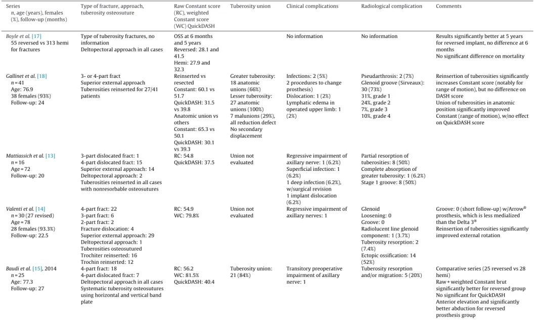
Boyle et al.[17]
55 reversed vs 313 hemi for fractures
Type of tuberosity fractures, no information
Deltopectoral approach in all cases
OSS at 6 months and 5 years Reversed: 28.1 and 41.5 Hemi: 27.9 and 32.3
No information No information Results significantly better at 5 years
for reversed implant, no difference at 6 months
No significant difference on mortality
Gallinet et al.[18] n = 41 Age: 76.9 38 females (93%) Follow-up: 24 3- or 4-part fract Superior external approach Tuberosities reinserted for 27/41 patients Reinserted vs resected Constant: 60.1 vs 51.7 QuickDASH: 31.5 vs 39.8 Anatomic union vs others Constant: 65.3 vs 50.1 QuickDASH: 30.1 vs 39.3 Greater tuberosity: 18 anatomic unions (66%) Lesser tuberosity: 27 anatomic unions (100%) 7 malunions (29%), all reduction defect No secondary displacement Infections: 2 (5%) 2 procedures to change prosthesis) Dislocation: 1 (2%) Lymphatic edema in operated upper limb: 1 (2%)
Pseudarthrosis: 2 (7%) Glenoid groove (Sirveaux): 30 (73%)
31%, grade 1 24%, grade 2 7%, grade 3 10%, grade 4
Reinsertion of tuberosities significantly increases Constant score (notably for range of motion), but no difference on DASH score
Union of tuberosities in anatomic position significantly improved Constant (range of motion), w/no effect on QuickDASH score
Mattiassich et al.[13] n = 16
Age = 72 Follow-up: 20
3-part dislocated fract: 1 4-part dislocated fract: 15 Superior external approach: 14 Deltopectoral approach: 2 Tuberosities reinserted in all cases with nonresorbable osteosutures
RC: 54.8 QuickDASH: 37.5 Union not evaluated Regressive impairment of axillary nerve: 1 (6.2%) Superficial infection: 1 (6.2%) 1 deep infection (6.2%), w/surgical revision 1 implant dislocation (6.2%) Partial resorption of tuberosities: 8 (50%) Complete absorption of greater tuberosity: 1 (6.2%) Stage 1 groove: 8 (50%) Valenti et al.[14] n = 30 (27 revised) Age = 78 28 females (93.3%) Follow-up: 22.5 4-part fract: 22 3-part fract: 6 2-part fract: 2 Fracture dislocation: 4 Superior external approach: 29 Deltopectoral approach: 1 Tuberosities osteosutured Trochiter reinserted: 16 Trochin reinserted: 12 RC: 54.9 WC: 79.8% Union not evaluated Regressive impairment of axillary nerves: 1 Glenoid Loosening: 0 Groove: 0
Radiolucent line glenoid component: 1 (3.7%) Tuberosity resorption: 2 (7.4%)
Ectopic ossification: 14 (52%)
Groove: 0 (short follow-up) w/Arrow®
prosthesis, which is less medialized than the Delta 3®
Reinsertion of tuberosities significantly improved external rotation
Baudi et al.[15], 2014 n = 25
Age: 77.3 Follow-up: 27
4-part fract: 18 4-part dislocated fract: 7 Deltopectoral approach in all cases Systematic tuberosity osteosutures using horizontal and vertical band plate RC: 56.2 WC: 81.5% QuickDASH: 40.4 Tuberosity union: 21 (84%) Transitory preoperative impairment of axillary nerve: 1 Tuberosity resorption and/or migration: 5 (20%)
Comparative series (25 reversed vs 28 hemi)
Raw + weighted Constant brut significantly better for reversed group No significant for QuickDASH Anterior elevation and significantly better abduction for reversed prosthesis group
repercussions, it is difficult to consider the groove as a complication in this study.
5. Conclusion
In experienced hands, the reversed prosthesis implanted for a four-part fracture provides regular and good results, with a weighted Constant score at 80%, 110–130◦anterior elevation, and active external rotation with the elbow against the body reaching 15–20◦. Even if it passes through the deltoid muscle, the superolat-eral approach provides good visibility on the glenoid in the fracture setting. Union of the tuberosities is possible (in 64% of cases) and effective. However, one patient out of four will not have a good result. The complications in these two series are rare but they are serious and will reduce recuperation of function.
Disclosure of interest
Laurent Obert is a consultant for the following companies: FX SOLUTION, ZIMMER, MEDARTIS, ARGO, EVOLUTIS, BIOTECH-WRIGHT.
Nicolas Bonnevialle is a consultant for TORNIER. Francois Sirveaux is a consultant for TORNIER.
Rachid Saadnia, Clément Tournier, Dominique Saragaglia declare that they have no competing interest.
Acknowledgements:
We extend our thanks to the investigators: P. Boileau, P. Clavert, C. Cuny, L. Doursounian, L. Favard, L. Obert, P. Mansat, H. Thomazeau, T. Fabre, X. Ohl, D. Saragaglia, F. Sirveaux; to the sym-posium participants: D. Block, R. Bouchet, F. Gadea, T. d’Ollonne, C. Tournier, N. Bonnevialle; acknowledge the scientific contributions of A. Berrichi, M.O. Gauci, J. Mayer, P. Mangin, C. Nérot, R. Saadnia, X. Clement, G. Dillmann, B.J. Chedal Bornu, G. Boudard, J. Lombard, Y. Knaffo, C. Goetz, L. Decroocq, Y. Bouju, J. Berhouet, G. Bacle, A. Erbland, B. Dunet, H. Demezon, J. Rigal, A. Adam, E. Jardin, T. Zap-aterra, N. Gasse, S. Rochet, D. Ancelin, T. Trang, J. Lebon, B. Aisene, H. Coudane, D. Mainard; and are grateful to Ms. A. Frégéac of AER-COT and Ms. N. Ramdane of the statistics department of the Lille University Hospital.
References
[1]Castricini R, Benedetto M, Pirani P, Panfoli N, Pace N. Shoulder hemiarthro-plasty for fractures of the proximal humerus. Musculoskelet Surg 2011;95(1): 49–54.
[2]Boileau P, Krishnan SG, Tinsi L, Walch G, Coste JS, Molé D. Tuberosity mal-position and migration: reasons for poor outcomes after hemiarthroplasty for displaced fractures of the proximal humerus. J Shoulder Elbow Surg 2002;11(5):401–12.
[3]Reuther F, Mühlhäusler B, Wahl D, Nijs S. Functional outcome of shoulder hemi-arthroplasty for fractures: a multicentre analysis. Injury 2010;41(6):606–12.
[4]Cazeneuve JF, Cristofari DJ. Grammont reversed prosthesis for acute complex fracture of the proximal humerus in an elderly population with 5 to 12 years follow-up. Rev Chir Orthop Reparatrice Appar Mot 2006;92(6):543–8.
[5]Bufquin T, Hersan A, Hubert L, Massin P. Reverse shoulder arthroplasty for the treatment of three-and four-part fractures of the proximal humerus in the elderly. A prospective review of 43 cases with a short-term follow-up. J Bone Joint Surg Br 2007;89(4):516–20.
[6]Gallinet D, Clappaz P, Garbuio P, Tropet Y, Obert L. Three or four parts com-plex proximal humerus fractures: hemiarthroplasty versus reverse prosthesis: a comparative study of 40 cases. Orthop Traumatol Surg Res 2009;95(1): 48–55.
[7]Sirveaux F, Navez G, Favard L, Boileau P, Walch G, Mole D. Reverse prosthesis for acute proximal humerus fracture, the multicentric study. Reverse shoulder arthroplasty. Sauramps médical; 2006. p. 73–80.
[8]Klein M, Juschka M, Hinkenjann B, Scherger B, Ostermann PA. Treatment of comminuted fractures of the proximal humerus in elderly patients with the Delta III reverse shoulder prosthesis. J Orthop Trauma 2008;22(10):698–704.
[9]Boileau P, Moineau G, Brassart N, Clavert P, Favard L, Sirveaux F. Reverse shoulder fracture prothesis for the treatment of proximal humeral fractures in elderly patients: early clinical and radiological results. Shoulder concept 2010: arthroscopy and arthroplasty. Sauramps médical; 2010. p. 231–43.
[10]Cazeneuve J-F, Cristofari DJ. Delta III reverse shoulder arthroplasty: radiolog-ical outcome for acute complex fractures of the proximal humerus in elderly patients. Orthop Traumatol Surg Res 2009;95(5):325–9.
[11]Levy JC, Badman B. Reverse shoulder prosthesis for acute four-part fracture: tuberosity fixation using a horseshoe graft. J Orthop Trauma 2011;25(5):318–24.
[12]Lenarz C, Shishani Y, McCrum C, Nowinski RJ, Edwards TB, Gobezie R. Is reverse shoulder arthroplasty appropriate for the treatment of fractures in the older patient?: early observations. Clin Orthop Relat Res 2011;469(12):3324–31.
[13]Mattiassich G, Marcovici LL, Krifter RM, Ortmaier R, Wegerer P, Kroepfl A. Delta III reverse shoulder arthroplasty in the treatment of complex 3- and 4-part fractures of the proximal humerus: 6 to 42 months of follow-up. BMC Muscu-loskelet Disord 2013;14(1):1–10.
[14]Valenti P, Katz D, Kilinc A, Elkholti K, Gasiunas V. Mid-term outcome of reverse shoulder prostheses in complex proximal humeral fractures. Acta Orthop Belg 2012;78:442–9.
[15]Baudi P, Campochiaro G, Serafini F, Gazzotti G, Matino G, Rovesta C, et al. Hemiarthroplasty versus reverse shoulder arthroplasty: comparative study of functional and radiological outcomes in the treatment of acute proximal humerus fracture. Musculoskelet Surg 2014;98(1):19–25.
[16]Hubert L, Lahogue J-F, Hersan A, Gournay A, Massin P. 116 prothèse inversée Delta®en traumatologie de l’épaule : résultats préliminaires. Rev Chir Orthop
Reparatrice Appar Mot 2004;90(6):83 [Supplement 1].
[17]Boyle MJ, Youn S-M, Frampton CMA, Ball CM. Functional outcomes of reverse shoulder arthroplasty compared with hemiarthroplasty for acute proximal humeral fractures. J Shoulder Elbow Surg 2013;22(1):32–7.
[18]Gallinet D, Adam A, Gasse N, Rochet S, Obert L. Improvement in shoulder rota-tion in complex shoulder fractures treated by reverse shoulder arthroplasty. J Shoulder Elbow Surg 2013;22(1):38–44.
[19]Cuff DJ, Pupello DR. Comparison of hemiarthroplasty and reverse shoul-der arthroplasty for the treatment of proximal humeral fractures in elshoul-derly patients. J Bone Jt Surg Am 2013;95(22):2050.
[20]Van Seymortier P, Stoffelen D, Fortems Y, Reynders P. The reverse shoulder prosthesis (Delta III) in acute shoulder fractures: technical considerations with respect to stability. Acta Orthop Belg 2006;72(4):474–7.
[21]Molé D, Wein F, Dézaly C, Valenti P, Sirveaux F. Surgical technique: the antero-superior approach for reverse shoulder arthroplasty. Clin Orthop Relat Res 2011;469(9):2461–8.
[22]Walch G, Nové-Josserand L. Instabilité de la prothèse totale inversée de l’épaule [Instability of the reverse shoulder prosthesis]. In: Boileau P, Walch G, editors. Prothèses d’épaule état actuel. Paris: Elsevier Masson; 2008. p. 407–14 [cité 6 juin 2014].
[23]Edwards TB, Williams MD, Labriola JE, Elkousy HA, Gartsman GM, O’Connor DP. Subscapularis insufficiency and the risk of shoulder dislocation after reverse shoulder arthroplasty. J Shoulder Elbow Surg 2009;18(6):892–6.
[24]Baulot E, Valenti P, Garaud P, Boileau P, Neyton L, Sirveaux F, et al. Résultats des prothèses inversées. Rev Chir Orthop Reparatrice Appar Mot 2007;93(6):63–92 [Supplement 1].
[25]Pelegri C, Jacquot N, Coste J-S, Boileau P. Infection des prothèses d’épaule : spécificité et prise en charge [Infection in shoulder prostheses : specificities and management]. In: Boileau P, Walch G, editors. Prothèses d’épaule état actuel. Paris: Elsevier Masson; 2008. p. 382–90 [Internet, cité 3 sept 2014].
[26]Werner CML, Steinmann PA, Gilbart M, Gerber C. Treatment of painful pseudo-paresis due to irreparable rotator cuff dysfunction with the Delta III reverse-ball-and-socket total shoulder prosthesis. J Bone Jt Surg 2005;87(7):1476–86.
[27]Lévigne C, Boileau P, Favard L, Garaud P, Molé D, Sirveaux F, et al. Scapular notch-ing in reverse shoulder arthroplasty. J Shoulder Elbow Surg 2008;17(6):925–35.
[28]Sirveaux F, Favard L, Oudet D, Huquet D, Walch G, Mole D. Grammont inverted total shoulder arthroplasty in the treatment of glenohumeral osteoarthritis with massive rupture of the cuff results of a multicentre study of 80 shoulders. J Bone Joint Surg Br 2004;86(3):388–95.
[29]Boileau P, Balg F. Prothèse totale d’épaule inversée : principes biomécaniques, concept et évolution [The reverse shoulder prosthesis: biomechanical princi-ples, concept and evolution]. In: Boileau P, Walch G, editors. Prothèses d’épaule état actuel. Paris: Elsevier Masson; 2008. p. 153–68 [Internet].
[30]Kempton LB, Balasubramaniam M, Ankerson E, Wiater JM. A radiographic anal-ysis of the effects of prosthesis design on scapular notching following reverse total shoulder arthroplasty. J Shoulder Elbow Surg 2011;20(4):571–6.