UNIVERSITÉ CLERMONT AUVERGNE
UFR DE MÉDECINE ET DES PROFESSIONS PARAMÉDICALES
THÈSE D’EXERCICE pour le
DIPLÔME D’ÉTAT DE DOCTEUR EN MÉDECINE par
Elsa BESSE-PINOT
Présentée et soutenue publiquement le 28 Mai 2020
Rôle prédictif de la présence de troubles du comportement en sommeil paradoxal en préopératoire sur l’évolution motrice, cognitive, psycho-comportementale et sur la qualité de vie un an après stimulation cérébrale
profonde dans la Maladie de Parkinson
Directeur de thèse :
Madame Ana Raquel MARQUES, Docteur, CHU Clermont-Ferrand Président du jury :
Monsieur Franck DURIF Antoine, Professeur, UFR de Médecine et des Professions paramédicales de Clermont-Ferrand
Membres du jury :
Monsieur Bruno PEREIRA, Biostatisticien, Direction de la Recherche Clinique et de l’Innovation
Madame Maria Livia FANTINI, Professeur, UFR de Médecine et des professions paramédicales de Clermont-Ferrand.
Monsieur Pierre CLAVELOU, Professeur, UFR de Médecine et des professions paramédicales de Clermont-Ferrand
3
UNIVERSITÉ CLERMONT AUVERGNE
UFR DE MÉDECINE ET DES PROFESSIONS PARAMÉDICALES
THÈSE D’EXERCICE pour le
DIPLÔME D’ÉTAT DE DOCTEUR EN MÉDECINE par
Elsa BESSE-PINOT
Présentée et soutenue publiquement le 28 Mai 2020
Rôle prédictif de la présence de troubles du comportement en sommeil paradoxal en préopératoire sur l’évolution motrice, cognitive, psycho-comportementale et sur la qualité de vie un an après stimulation cérébrale
profonde dans la Maladie de Parkinson
Directeur de thèse :
Madame Ana Raquel MARQUES, Docteur, CHU Clermont-Ferrand Président du jury :
Monsieur Franck DURIF Antoine, Professeur, UFR de Médecine et des Professions paramédicales de Clermont-Ferrand
Membres du jury :
Monsieur Bruno PEREIRA, Biostatisticien, Direction de la Recherche Clinique et de l’Innovation
Madame Maria Livia FANTINI, Professeur, UFR de Médecine et des professions paramédicales de Clermont-Ferrand.
Monsieur Pierre CLAVELOU, Professeur, UFR de Médecine et des professions paramédicales de Clermont-Ferrand
4
UNIVERSITE CLERMONT AUVERGNE
___________________
PRESIDENTS HONORAIRES : JOYON Louis
UNIVERSITE D’AUVERGNE : DOLY Michel
: TURPIN Dominique : VEYRE Annie : DULBECCO Philippe : ESCHALIER Alain
PRESIDENTS HONORAIRES : CABANES Pierre
UNIVERSITE BLAISE PASCAL : FONTAINE Jacques
: BOUTIN Christian : MONTEIL Jean-Marc : ODOUARD Albert PRESIDENT DE L'UNIVERSITE et : LAVIGNOTTE Nadine
PRESIDENT DU CONSEIL ACADEMIQUE PLENIER : BERNARD Mathias PRESIDENT DU CONSEIL ACADEMIQUE RESTREINT : DEQUIEDT Vianney VICE-PRESIDENT DU CONSEIL D'ADMINISTRATION : WILLIAMS Benjamin VICE-PRESIDENT DE LA COMMISSION DE LA RECHERCHE
VICE PRESIDENTE DE LA COMMISSION DE LA
: HENRARD Pierre FORMATION ET DE LA VIE UNIVERSITAIRE : PEYRARD Françoise
DIRECTEUR GENERAL DES SERVICES : PAQUIS François
²²²²²
UFR DE MEDECINE
ET DES PROFESSIONS PARAMEDICALES
DOYENS HONORAIRES : DETEIX Patrice
: CHAZAL Jean
DOYEN : CLAVELOU Pierre
5
LISTE DU PERSONNEL ENSEIGNANT
PROFESSEURS HONORAIRES :
MM. BACIN Franck - BEGUE René-Jean - BOUCHER Daniel - BOURGES Michel - BUSSIERE Jean-Louis - CANO Noël - CASSAGNES Jean - CATILINA Pierre - CHABANNES Jacques – CHAZAL Jean - CHIPPONI Jacques - CHOLLET Philippe - COUDERT Jean - DASTUGUE
Bernard - DEMEOCQ François - DE RIBEROLLES Charles - ESCANDE Georges - Mme FONCK Yvette - MM. GENTOU Claude - GLANDDIER Gérard - Mmes GLANDDIER Phyllis - LAVARENNE Jeanine - MM. LAVERAN Henri - LEVAI Jean-Paul - MAGE Gérard -
MALPUECH Georges - MARCHEIX Jean-Claude - MICHEL Jean-Luc - MOLINA Claude -
MONDIE Jean-Michel - PERI Georges - PETIT Georges - PHILIPPE Pierre - PLAGNE Robert - PLANCHE Roger - PONSONNAILLE Jean - RAYNAUD Elie - REY Michel - Mme RIGAL Danièle - MM. ROZAN Raymond - SCHOEFFLER Pierre - SIROT Jacques - SOUTEYRAND Pierre - TANGUY Alain - TERVER Sylvain - THIEBLOT Philippe - TOURNILHAC Michel - VANNEUVILLE Guy - VIALLET Jean-François - Mle VEYRE Annie
PROFESSEURS EMERITES :
MM. - BEYTOUT Jean - BOITEUX Jean-Paul - BOMMELAER Gilles - CHAMOUX Alain - DAUPLAT Jacques - DETEIX Patrice - ESCHALIER Alain - IRTHUM Bernard - JACQUETIN Bernard - KEMENY Jean-Louis – Mme LAFEUILLE Hélène – MM. LEMERY Didier - LESOURD Bruno - LUSSON Jean-René - RIBAL Jean-Pierre
PROFESSEURS DES UNIVERSITES-PRATICIENS HOSPITALIERS
PROFESSEURS DE CLASSE EXCEPTIONNELLE
M. VAGO Philippe Histologie-Embryologie Cytogénétique M. AUMAITRE Olivier Médecine Interne
M. LABBE André Pédiatrie
M. AVAN Paul Biophysique et Traitement de l'Image
M. DURIF Franck Neurologie
M. BOIRE Jean-Yves Biostatistiques, Informatique Médicale
et Technologies de Communication
M. BOYER Louis Radiologie et Imagerie Médicale
option Clinique
M. POULY Jean-Luc Gynécologie et Obstétrique M. CANIS Michel Gynécologie-Obstétrique
6
M. BAZIN Jean-Etienne Anesthésiologie et Réanimation
Chirurgicale
M. BIGNON Yves Jean Cancérologie option Biologique
M. BOIRIE Yves Nutrition Humaine
M. CLAVELOU Pierre Neurologie
M. DUBRAY Claude Pharmacologie Clinique
M. GILAIN Laurent O.R.L.
M. LEMAIRE Jean-Jacques Neurochirurgie
M. CAMILLERI Lionel Chirurgie Thoracique et Cardio-Vasculaire M. DAPOIGNY Michel Gastro-Entérologie
M. LLORCA Pierre-Michel Psychiatrie d’Adultes
M. PEZET Denis Chirurgie Digestive
M. SOUWEINE Bertrand Réanimation Médicale
M. BOISGARD Stéphane Chirurgie Orthopédique et Traumatologie M. CONSTANTIN Jean-Michel Anesthésiologie et Réanimation Chirurgicale
Mme DUCLOS Martine Physiologie
M. SCHMIDT Jeannot Thérapeutique
PROFESSEURS DE 1ère CLASSE
M. DECHELOTTE Pierre Anatomie et Cytologie Pathologique M. CAILLAUD Denis Pneumo-phtisiologie
M. VERRELLE Pierre Radiothérapie option Clinique M. CITRON Bernard Cardiologie et Maladies Vasculaires M. D’INCAN Michel Dermatologie -Vénéréologie Mme JALENQUES Isabelle Psychiatrie d'Adultes
Mle BARTHELEMY Isabelle Chirurgie Maxillo-Faciale
M. GARCIER Jean-Marc Anatomie-Radiologie et Imagerie Médicale M. GERBAUD Laurent Epidémiologie, Economie de la Santé
et Prévention
M. SOUBRIER Martin Rhumatologie
M. TAUVERON Igor Endocrinologie et Maladies Métaboliques
M. MOM Thierry Oto-Rhino-Laryngologie
M. RICHARD Ruddy Physiologie
M. RUIVARD Marc Médecine Interne
M. SAPIN Vincent Biochimie et Biologie Moléculaire M. BAY Jacques-Olivier Cancérologie
M. BERGER Marc Hématologie
M. COUDEYRE Emmanuel Médecine Physique et de Réadaptation Mme GODFRAIND Catherine Anatomie et Cytologie Pathologiques M. ROSSET Eugénio Chirurgie Vasculaire
M. ABERGEL Armando Hépatologie
M. LAURICHESSE Henri Maladies Infectieuses et Tropicales M. TOURNILHAC Olivier Hématologie
7
M. FILAIRE Marc Anatomie – Chirurgie Thoracique et
Cardio-Vasculaire
M. GALLOT Denis Gynécologie-Obstétrique
M. GUY Laurent Urologie
M. TRAORE Ousmane Hygiène Hospitalière
M. ANDRE Marc Médecine Interne
M. BONNET Richard Bactériologie, Virologie
M. CACHIN Florent Biophysique et Médecine Nucléaire
M. COSTES Frédéric Physiologie
M. FUTIER Emmanuel Anesthésiologie-Réanimation Mme HENG Anne-Elisabeth Néphrologie
M. MOTREFF Pascal Cardiologie
Mme PICKERING Gisèle Pharmacologie Clinique
PROFESSEURS DE 2ème CLASSE
Mme CREVEAUX Isabelle Biochimie et Biologie Moléculaire M. FAICT Thierry Médecine Légale et Droit de la Santé Mme KANOLD LASTAWIECKA Justyna Pédiatrie
M. TCHIRKOV Andréï Cytologie et Histologie M. CORNELIS François Génétique
M. DESCAMPS Stéphane Chirurgie Orthopédique et Traumatologique M. POMEL Christophe Cancérologie – Chirurgie Générale
M. CANAVESE Fédérico Chirurgie Infantile
M. LESENS Olivier Maladies Infectieuses et Tropicales M. RABISCHONG Benoît Gynécologie Obstétrique
M. AUTHIER Nicolas Pharmacologie Médicale
M. BROUSSE Georges Psychiatrie Adultes/Addictologie
M. BUC Emmanuel Chirurgie Digestive
M. CHABROT Pascal Radiologie et Imagerie Médicale M. LAUTRETTE Alexandre Néphrologie Réanimation Médicale M. AZARNOUSH Kasra Chirurgie Thoracique et Cardiovasculaire Mme BRUGNON Florence Biologie et Médecine du Développement et
de la Reproduction
Mme HENQUELL Cécile Bactériologie Virologie M. ESCHALIER Romain Cardiologie
M. MERLIN Etienne Pédiatrie
Mme TOURNADRE Anne Rhumatologie
M. DURANDO Xavier Cancérologie
M. DUTHEIL Frédéric Médecine et Santé au Travail Mme FANTINI Maria Livia Neurologie
M. SAKKA Laurent Anatomie – Neurochirurgie M. BOURDEL Nicolas Gynécologie-Obstétrique
M. GUIEZE Romain Hématologie
8
M. SOUTEYRAND Géraud Cardiologie
PROFESSEURS DES UNIVERSITES
M. CLEMENT Gilles Médecine Générale
Mme MALPUECH-BRUGERE Corinne Nutrition Humaine M. VORILHON Philippe Médecine Générale
PROFESSEURS ASSOCIES DES UNIVERSITES
Mme BOTTET-MAULOUBIER Anne Médecine Générale
M. CAMBON Benoît Médecine Générale
MAITRES DE CONFERENCES DES UNIVERSITES -
PRATICIENS HOSPITALIERS
MAITRES DE CONFERENCES HORS CLASSE
Mme CHAMBON Martine Bactériologie Virologie Mme BOUTELOUP Corinne
Nutrition
MAITRES DE CONFERENCES DE 1ère CLASSE
M. MORVAN Daniel Biophysique et Traitement de l’Image Mle GOUMY Carole Cytologie et Histologie, Cytogénétique Mme FOGLI Anne Biochimie Biologie Moléculaire Mle GOUAS Laetitia Cytologie et Histologie, Cytogénétique
9
M. MARCEAU Geoffroy Biochimie Biologie Moléculaire Mme MINET-QUINARD Régine Biochimie Biologie Moléculaire M. ROBIN Frédéric Bactériologie
Mle VERONESE Lauren Cytologie et Histologie, Cytogénétique
M. DELMAS Julien Bactériologie
Mle MIRAND Andrey Bactériologie Virologie
M. OUCHCHANE Lemlih Biostatistiques, Informatique Médicale
et Technologies de Communication
M. LIBERT Frédéric Pharmacologie Médicale
Mle COSTE Karen Pédiatrie
M. EVRARD Bertrand Immunologie
Mle AUMERAN Claire Hygiène Hospitalière M. POIRIER Philippe Parasitologie et Mycologie Mme CASSAGNES Lucie Radiologie et Imagerie Médicale M. LEBRETON Aurélien Hématologie
MAITRES DE CONFERENCES DE 2ème CLASSE
Mme PONS Hanaë Biologie et Médecine du Développement
et de la Reproduction
M. JABAUDON-GANDET Matthieu Anesthésiologie – Réanimation Chirurgicale M. BOUVIER Damien Biochimie et Biologie Moléculaire
M. BUISSON Anthony Gastroentérologie M. COLL Guillaume Neurochirurgie
Mme SARRET Catherine Pédiatrie
M. MAQDASY Salwan Endocrinologie, Diabète et Maladies
Métaboliques
Mme NOURRISSON Céline Parasitologie - Mycologie
MAITRES DE CONFERENCES DES UNIVERSITES
Mme Mme
BONHOMME Brigitte
VAURS-BARRIERE Catherine
Biophysique et Traitement de l’Image Biochimie Biologie Moléculaire M. BAILLY Jean-Luc Bactériologie Virologie
Mle AUBEL Corinne Oncologie Moléculaire
M. BLANCHON Loïc Biochimie Biologie Moléculaire Mle GUILLET Christelle Nutrition Humaine
10
M. MARCHAND Fabien Pharmacologie Médicale M. DALMASSO Guillaume Bactériologie
M. SOLER Cédric Biochimie Biologie Moléculaire M. GIRAUDET Fabrice Biophysique et Traitement de l’Image Mme VAILLANT-ROUSSEL Hélène Médecine Générale
Mme LAPORTE Catherine Médecine Générale
M. LOLIGNIER Stéphane Neurosciences – Neuropharmacologie Mme MARTEIL Gaëlle Biologie de la Reproduction
M. PINEL Alexandre Nutrition Humaine
MAITRES DE CONFERENCES ASSOCIES DES UNIVERSITES
M. TANGUY Gilles Médecine Générale
M. BERNARD Pierre Médecine Générale
Mme ESCHALIER Bénédicte Médecine Générale
11
REMERCIEMENTS
Je tiens à remercier les membres de mon jury :
Monsieur le Professeur Franck Durif,
Vous me faites l’honneur de présider ce jury. Merci pour l’écoute et la disponibilité dont vous avez fait preuve à mon égard. Je vous suis reconnaissante de m’avoir accordé votre confiance et de m’avoir encouragé à partir à Paris pour concrétiser mes projets professionnels.
Madame le Docteur Ana Raquel Marques,
Je te remercie de l’aide précieuse que tu m’as apportée pour réaliser ce travail et ce même lorsque tu étais à des milliers de kilomètres. Je tiens également à te remercier pour ta bonne humeur et ta gentillesse qui ont accompagné tout mon internat.
Monsieur Bruno PEREIRA,
Merci de m’avoir soutenue plusieurs mois dans ce travail. Tes conseils, ton expertise, et ta bonne humeur constante ont rendus nos échanges chaque jour plus constructifs.
Madame le Professeur Maria Livia Fantini,
Je tiens à t’exprimer toute ma gratitude pour la disponibilité dont tu as fait preuve et pour tes conseils toujours très avisés. C’est toujours un plaisir d’apprendre à tes côtés.
Monsieur le Professeur Pierre Clavelou,
Je vous suis très reconnaissante d’avoir accepté de juger mon travail. De l’externat jusqu’à la fin de mon internat vous étiez présent, votre enseignement et vos conseils m’ont toujours été bénéfiques. Pour tout cela je vous remercie.
12
Table des matières
INTRODUCTION ... 13
Préambule, contexte scientifique et clinique ... 13
Hypothèses de travail et objectifs ... 16
Références bibliographiques ... 17
ARTICLE ... 20
CONCLUSION ... 51
13
INTRODUCTION
Préambule, contexte scientifique et clinique
La maladie de Parkinson représente la deuxième pathologie neurodégénérative après la maladie d’Alzheimer (1). Sa prévalence est estimée à 0.3/1000 dans la population générale des pays industrialisés (2) et concernerait 1% des sujets de plus de 60 ans (3). Son pronostic est grevé par la progression du handicap moteur et l’apparition dans 25 à 40 % des cas d’une démence, responsables d’une réduction de l’espérance de vie (4).
Historiquement, les études de neuropathologie ont mis en évidence une dégénérescence précoce des neurones dopaminergiques de la pars compacta de la substance noire, responsable des symptômes moteurs de la maladie associés à des dépôt anormaux de protéines alpha synucléines agrégées, formant des inclusions intra neuronales appelées corps de Lewy (5), (6). Plus tard, un schéma évolutif de la maladie a été proposé, comparable à celui des maladies à Prions, avec une progression rostro-caudale depuis le système nerveux périphérique jusqu’au système nerveux central (7). Ce modèle de progression rostro-caudale est néanmoins actuellement débattu. L’hypothèse actuelle est celle d’une atteinte neurodégénérative multi systémique hétérogène expliquant la coexistence de nombreux signes moteurs et non moteurs. Ce modèle a également permis d’expliquer l’existence d’une phase prodromale comportant une série de symptômes non moteurs tels que la constipation (8), l’anosmie (9), les troubles du comportement en sommeil paradoxal (TCSP) (10), la dysautonomie (11) ou la dépression (9), pouvant parfois précéder de plusieurs années l’apparition de la maladie. Ainsi, la triade parkinsonienne associant rigidité extra pyramidale, bradykinésie et tremblement de repos, s’explique par la déplétion dopaminergique des neurones de la substance noire se projetant sur le putamen dorsal du striatum. La dénervation dopaminergique mésolimbique et mésocorticale serait quant à elle à l’origine des troubles psycho-comportementaux et cognitifs tandis que la dénervation tubero-infandibulaire serait responsable de la dysautomie (11) La dénervation intéresse également des systèmes non dopaminergiques comprenant l’atteinte des systèmes sérotoninergiques [14],
14
cholinergiques [15], noradrénergiques et adrénergiques qui participerait respectivement à la genèse des troubles de l’humeur, des troubles du sommeil et troubles moteurs axiaux [16], symptômes qui s’avèrent alors non dopa-sensible.
Les troubles du comportement en sommeil paradoxal (TCSP) sont définis, selon la classification internationale des troubles du sommeil, comme des parasomnies du sommeil paradoxal et sont caractérisés par la perte de l’atonie musculaire, normalement présente, au cours du sommeil paradoxal, à l’origine d’une activité motrice involontaire semblant mimer le rêve (12). Les TCSP peuvent être idiopathiques ou associés à des pathologies neurologiques, essentiellement aux alpha-synucléopathies. Cependant, il a été montré que 81% des TCSP dit « idiopathiques » évoluent vers une alpha-synucléopathie après 16 ans de suivi, remettant en cause l’existence de forme idiopathiques (13). Alors que la prévalence des TCSP idiopathiques est faible, de l’ordre de 0.5 à 2 % (14,15) celle-ci est beaucoup plus élevée chez les patients atteints d’alpha-synucléinopathies, comprenant la maladie de Parkinson idiopathique, la démence à corps de Lewy (DCL) et l'atrophie multisystématisée (AMS) (16). Dans le cas de la maladie de Parkinson, la prévalence des TCSP irait de 19 à 70% (17).
Plusieurs études ont suggéré que la présence de TCSP dans la maladie de Parkinson pourrait constituer un sous-groupe distinct de patients avec un phénotype plus sévère, en particulier avec un déclin cognitif, des hallucinations et la présence plus marquée de troubles du contrôle des impulsions (18– 20). Celle-ci pourrait également être associée à des symptômes axiaux plus importants et à une progression plus rapide des symptômes moteurs de la maladie (21), (22). La présence de TCSP chez les patients parkinsoniens pourrait donc conditionner le pronostic ainsi que la prise en charge, en suggérant par exemple une utilisation plus prudente des agonistes dopaminergiques chez ces patients-là, en raison du risque plus élevé d’hallucinations et de troubles du contrôle des impulsions.
La stimulation cérébrale profonde des noyaux subthalamiques (SCP-NST) a montré son efficacité pour le traitement de la MPI chez des patients présentant des fluctuations motrices sévères malgré une adaptation optimale du traitement médicamenteux (23). Elle consiste en l’implantation d’électrodes
15
au niveau du noyau sous-thalamique, qui sont reliées à un stimulateur réglable en intensité, en fréquence et en amplitude. Elle a prouvé son efficacité dans l’amélioration de la fonction motrice d’environ 50%, dans la diminution des complications motrices jusqu’à 70% et dans la réduction d’un peu plus de la moitié des doses de traitements dopaminergiques (24,25). Son effet sur la qualité de vie a également été bien démontré (26). Toutefois, il existe aussi des effets indésirables rencontrés au décours de la chirurgie. En dehors des rares complications directement liées à la procédure, les effets indésirables stimulo-induits concernent surtout l’apparition ou l’aggravation de troubles de la marche (freezing), de troubles posturaux (27) et d’une dysarthrie (24). Son effet sur les signes non moteurs reste à ce jour débattu. En effet, plusieurs publications rapportent une amélioration des symptômes hyper-dopaminergiques (28), qui serait essentiellement liée à la diminution du traitement dopaminergique, mais une aggravation de l’apathie et des troubles cognitifs (29). Néanmoins, il semble qu’il existe une grande variabilité dans ces résultats et ceci pourrait être directement lié au positionnement des électrodes de stimulation au sein du noyau subthalamique (30).
A l’heure actuelle, il reste difficile d’identifier en préopératoire les patients à risque d’une évolution péjorative après SCP-NST. Puisque les TCSP dans la maladie de Parkinson semblent associés à un plus fort risque de déclin cognitif (31), d’hallucinations (32), de trouble du contrôle des impulsions (33) et à des symptômes axiaux plus importants (21),(22), leur présence en préopératoire pourrait également être associée a de moins bons résultats de la stimulation cérébrale profonde subthalamique sur les signes moteurs et non moteurs. Or, les données de la littérature sur cette question sont rares et discordantes.
En effet, deux études (34) ,(35) ont évalué l’évolution après SCP-NST de patients présentant un TCSP en préopératoire (preopTCSP+) par rapport à des patients sans TCSP (preopTCSP-). D’un côté, Zibetti
et al ont montré, sur une cohorte de 41 patients, que les patients présentant des preopTCSP+ avaient
une évolution motrice significativement moins bonne 3 ans après SCP-NST, en particulier pour les symptômes axiaux (34). D’un autre côté, Bargiotas et al n’ont pas retrouvé de différence d’évolution
16
motrice 1 an après SCP-NST sur une cohorte de 50 patients, entre les patients avec et sans preopTCSP. Dans cette cohorte, il existait en revanche une amélioration des symptômes dépressifs, de l'apathie et des activités de la vie quotidienne chez les patients preopCSP+ comparativement aux patients preopTCSP-.
Hypothèses de travail et objectifs
Notre hypothèse est que la présence de TCSP en préopératoire chez les patients avec une maladie de Parkinson idiopathique, serait associée à une moins bonne évolution un an après SCP-NST sur le plan moteur, cognitif et psycho-comportemental, comparativement à des patients sans TCSP préopératoire.
L’objectif de notre étude est de comparer l’évolution des symptômes parkinsoniens moteurs, des complications motrices, des fonctions cognitives (efficience globale, attention et mémoire de travail, mémoire épisodique, fonctions exécutives, fonctions visuospatiales, langage), des fonctions psycho-comportementales et de la qualité de vie 1 ans après la SCP-NST chez les patients parkinsoniens avec pTCSP+ par rapport aux patients pTCSP-.
17
Références bibliographiques
1. Dorsey ER, Constantinescu R, Thompson JP, Biglan KM, Holloway RG, Kieburtz K, et al. Projected number of people with Parkinson disease in the most populous nations, 2005 through 2030. Neurology. 30 janv 2007;68(5):384-6.
2. de Lau LML, Breteler MMB. Epidemiology of Parkinson’s disease. Lancet Neurol. juin 2006;5(6):525-35.
3. Nussbaum RL, Ellis CE. Alzheimer’s Disease and Parkinson’s Disease. Guttmacher AE, Collins FS, éditeurs. N Engl J Med. 3 avr 2003;348(14):1356-64.
4. Emre M. Dementia associated with Parkinson’s disease. Lancet Neurol. avr 2003;2(4):229-37.
5. Dickson DW, Braak H, Duda JE, Duyckaerts C, Gasser T, Halliday GM, et al. Neuropathological assessment of Parkinson’s disease: refining the diagnostic criteria. Lancet Neurol. déc 2009;8(12):1150-7.
6. Goedert M, Spillantini MG, Del Tredici K, Braak H. 100 years of Lewy pathology. Nat Rev Neurol. 2013;9(1):13-24.
7. Braak H, Del Tredici K, Rüb U, de Vos RAI, Jansen Steur ENH, Braak E. Staging of brain pathology related to sporadic Parkinson’s disease. Neurobiol Aging. avr 2003;24(2):197-211.
8. Savica R, Carlin JM, Grossardt BR, Bower JH, Ahlskog JE, Maraganore DM, et al. Medical records documentation of constipation preceding Parkinson disease: A case-control study. Neurology. 24 nov 2009;73(21):1752-8.
9. Przuntek H, Müller T, Riederer P. Diagnostic staging of Parkinson’s disease: conceptual aspects. J Neural Transm Vienna Austria 1996. févr 2004;111(2):201-16.
10. Schenck CH, Bundlie SR, Mahowald MW. Delayed emergence of a parkinsonian disorder in 38% of 29 older men initially diagnosed with idiopathic rapid eye movement sleep behaviour disorder. Neurology. févr 1996;46(2):388-93.
11. Ziemssen T, Reichmann H. Cardiovascular autonomic dysfunction in Parkinson’s disease. J Neurol Sci. 15 févr 2010;289(1-2):74-80.
12. Thorpy MJ. Classification of sleep disorders. Neurother J Am Soc Exp Neurother. oct 2012;9(4):687-701.
13. Schenck CH, Boeve BF, Mahowald MW. Delayed emergence of a parkinsonian disorder or dementia in 81% of older men initially diagnosed with idiopathic rapid eye movement sleep behavior disorder: a 16-year update on a previously reported series. Sleep Med. août 2013;14(8):744-8.
14. Ohayon MM, Caulet M, Priest RG. Violent behavior during sleep. J Clin Psychiatry. août 1997;58(8):369-76; quiz 377.
18
15. Kang S-H, Yoon I-Y, Lee SD, Han JW, Kim TH, Kim KW. REM sleep behavior disorder in the Korean elderly population: prevalence and clinical characteristics. Sleep. 1 août 2013;36(8):1147-52.
16. Barone DA, Henchcliffe C. Rapid eye movement sleep behavior disorder and the link to alpha-synucleinopathies. Clin Neurophysiol Off J Int Fed Clin Neurophysiol. 2018;129(8):1551-64.
17. Zhang X, Sun X, Wang J, Tang L, Xie A. Prevalence of rapid eye movement sleep behavior disorder (RBD) in Parkinson’s disease: a meta and meta-regression analysis. Neurol Sci. janv 2017;38(1):163-70.
18. Postuma RB, Bertrand J-A, Montplaisir J, Desjardins C, Vendette M, Rios Romenets S, et al. Rapid eye movement sleep behavior disorder and risk of dementia in Parkinson’s disease: a prospective study. Mov Disord Off J Mov Disord Soc. mai 2012;27(6):720-6. 19. Fereshtehnejad S-M, Romenets SR, Anang JBM, Latreille V, Gagnon J-F, Postuma RB.
New Clinical Subtypes of Parkinson Disease and Their Longitudinal Progression: A Prospective Cohort Comparison With Other Phenotypes. JAMA Neurol. 1 août 2015;72(8):863.
20. Fantini ML, Macedo L, Zibetti M, Sarchioto M, Vidal T, Pereira B, et al. Increased risk of impulse control symptoms in Parkinson’s disease with REM sleep behaviour disorder. J Neurol Neurosurg Psychiatry. févr 2015;86(2):174-9.
21. Postuma RB, Gagnon JF, Vendette M, Charland K, Montplaisir J. REM sleep behaviour disorder in Parkinson’s disease is associated with specific motor features. J Neurol Neurosurg Psychiatry. oct 2008;79(10):1117-21.
22. Duarte Folle A, Paul KC, Bronstein JM, Keener AM, Ritz B. Clinical progression in Parkinson’s disease with features of REM sleep behavior disorder: A population-based longitudinal study. Parkinsonism Relat Disord. mai 2019;62:105-11.
23. Movement Disorder Group, Chan AYY, Yeung JHM, Mok VCT, Ip VHL, Wong A, et al. Subthalamic nucleus deep brain stimulation for Parkinson’s disease: evidence for effectiveness and limitations from 12 years’ experience. Hong Kong Med J Xianggang Yi Xue Za Zhi. déc 2014;20(6):474-80.
24. Krack P, Batir A, Van Blercom N, Chabardes S, Fraix V, Ardouin C, et al. Five-Year Follow-up of Bilateral Stimulation of the Subthalamic Nucleus in Advanced Parkinson’s Disease. N Engl J Med. 13 nov 2003;349(20):1925-34.
25. Limousin P, Krack P, Pollak P, Benazzouz A, Ardouin C, Hoffmann D, et al. Electrical stimulation of the subthalamic nucleus in advanced Parkinson’s disease. N Engl J Med. 15 oct 1998;339(16):1105-11.
26. Schuepbach WMM, Rau J, Knudsen K, Volkmann J, Krack P, Timmermann L, et al. Neurostimulation for Parkinson’s Disease with Early Motor Complications. N Engl J Med. 14 févr 2013;368(7):610-22.
19
27. van Nuenen BFL, Esselink RAJ, Munneke M, Speelman JD, van Laar T, Bloem BR. Postoperative gait deterioration after bilateral subthalamic nucleus stimulation in Parkinson’s disease. Mov Disord. 15 déc 2008;23(16):2404-6.
28. Lhommée E, Wojtecki L, Czernecki V, Witt K, Maier F, Tonder L, et al. Behavioural outcomes of subthalamic stimulation and medical therapy versus medical therapy alone for Parkinson’s disease with early motor complications (EARLYSTIM trial): secondary analysis of an open-label randomised trial. Lancet Neurol. 2018;17(3):223-31.
29. Wang Y, Li Y, Zhang X, Xie A. Apathy following Bilateral Deep Brain Stimulation of Subthalamic Nucleus in Parkinson’s Disease: A Meta-Analysis. Park Dis. 2018;2018:1-7. 30. Petry-Schmelzer JN, Krause M, Dembek TA, Horn A, Evans J, Ashkan K, et al.
Non-motor outcomes depend on location of neurostimulation in Parkinson’s disease. Brain J Neurol. 1 nov 2019;142(11):3592-604.
31. Postuma RB, Bertrand J-A, Montplaisir J, Desjardins C, Vendette M, Rios Romenets S, et al. Rapid eye movement sleep behavior disorder and risk of dementia in Parkinson’s disease: A prospective study. Mov Disord. mai 2012;27(6):720-6.
32. Fereshtehnejad S-M, Romenets SR, Anang JBM, Latreille V, Gagnon J-F, Postuma RB. New Clinical Subtypes of Parkinson Disease and Their Longitudinal Progression: A Prospective Cohort Comparison With Other Phenotypes. JAMA Neurol. 1 août 2015;72(8):863.
33. Fantini ML, Macedo L, Zibetti M, Sarchioto M, Vidal T, Pereira B, et al. Increased risk of impulse control symptoms in Parkinson’s disease with REM sleep behaviour disorder. J Neurol Neurosurg Psychiatry. févr 2015;86(2):174-9.
34. Bargiotas P, Debove I, Bargiotas I, Lachenmayer ML, Ntafouli M, Vayatis N, et al. Effects of bilateral stimulation of the subthalamic nucleus in Parkinson’s disease with and without REM sleep behaviour disorder. J Neurol Neurosurg Psychiatry. déc 2019;90(12):1310-6. 35. Zibetti M, Rizzi L, Colloca L, Cinquepalmi A, Angrisano S, Castelli L, et al. Probable REM sleep behaviour disorder and STN-DBS outcome in Parkinson’s Disease. Parkinsonism Relat Disord. mai 2010;16(4):265-9.
20
ARTICLE
Predictive role of REM sleep behavior disorder for subthalamic deep brain stimulation outcomes in Parkinson's disease
Elsa Besse-Pinota, Bruno Pereirab, Franck Durifa, Livia Fantinia, Elodie Duranda, Béréngère
Debillya, Philippe Derosta, Caroline Moreauc, Jean Christophe Corvold, David Maltêtee,
Tiphaine Rouaudf, Mylène Meyerg, Jean Francois Houvenaghelh, Claire Marséi, Christine
Tranchantj, Elodie Hainquek, Béchir Jarrayal, Solène Ansquerm, Marie Bonnetn, Lhaouas
Belamrio, Mélissa Tirp, Christine Brefel-Courbonq, Teodor Danailar, Alexandre Eusebios,
David Devosc and Ana Marquesa for the PREDI-STIM study group
a Université Clermont Auvergne, EA7280, Clermont-Ferrand University Hospital, Neurology
Department, 63000 Clermont-Ferrand, France
b Clermont-Ferrand university Hospital, Biostatistics department, 63000 Clermont-Ferrand,
France
c CHU of Lille, Univ. Lille, Inserm UMRS_1171, Licend, Department of Medical
Pharmacology, Neurology and Movement Disorders Department, Referent center of Parkinson’s disease, F-59000 Lille, France
21
e Department of Neurology, Rouen University Hospital and University of Rouen, France;
INSERM U1239, Laboratory of Neuronal and Neuroendocrine Differentiation and Communication, Mont-Saint-Aignan, France
f Clinique Neurologique, Hôpital Guillaume et René Laennec, Boulevard Jacques Monod,
44093 Nantes Cedex
g Neurology Department, Nancy University Hospital, 54000 Nancy, France
h Behavior and Basal Ganglia" Research Unit (EA 4712), University of Rennes 1, Rennes,
France; Department of Neurology, Rennes University Hospital, 35033 Rennes Cedex, France.
i CHU Nice, Centre Expert Parkinson, Service de Neurologie, 06002 Nice, France
j Service de Neurologie, Hôpitaux Universitaires de Strasbourg, Strasbourg, France ; Institut de
Génétique et de Biologie Moléculaire et Cellulaire (IGBMC), INSERM-U964/CNRS-UMR7104/Université de Strasbourg, Illkirch, France ; Fédération de Médecine Translationnelle de Strasbourg (FMTS), Université de Strasbourg, Strasbourg, France
k Département de Neurologie, Hôpital Pitié-Salpêtrière, AP-HP, Faculté de Médecine de
Sorbonne Université, UMR S 1127, Inserm U 1127, and CNRS UMR 7225, and Institut du Cerveau et de la Moëlle épinière, F-75013, Paris, France
l Pôle Neurosciences, Foch Hospital, Suresnes, France ; Université de Versailles Paris-Saclay,
INSERM U992, CEA Neurospin, Paris, France
m Service de Neurologie, Centre Expert Parkinson, CIC-INSERM 1402, CHU Poitiers, 86000
Poitiers, France
n CHU de Bordeaux, Centre expert Parkinson, Institut des maladies neuro-dégénératives,
F-33000 Bordeaux, France
22
p Department of neurology, Department of neurosurgery, Expert centre for Parkinson's disease,
Amiens University Hospital, EA 4559 Laboratoire de Neurosciences Fonctionnelles et Pathologie (LNFP) Université de Picardie Jules Verne, University of Picardy Jules Verne (UPJV), Amiens, France
q Department of Clinical Pharmacology and Neurosciences, Centre d'Investigation Clinique
CIC1436, University Hospital of Toulouse, Toulouse, France
r Centre Expert Parkinson, Hôpital Neurologique "Pierre Wertheimer", Hospices Civils de
Lyon, 69677 Bron, France
s Aix Marseille Université, AP-HM, Hôpital de La Timone, Service de Neurologie et Pathologie
du Mouvement, and UMR CNRS 7289, Institut de Neuroscience de La Timone, Marseille, France
* Correspondence to:
Elsa Besse-Pinot, Neurology department, Clermont-Ferrand University hospital, Place henri Dunant, 63000 Clermont-Ferrand, France; Tel: 0473751666; E-mail: ebesse@chu-clermontferrand.fr
Glossary:
PD = Parkinson Disease; RBD = Rapid eye movement sleep Behavior Disorder; STN-DBS = Subthalamic nucleus Deep Brain Stimulation; LEDD = Levodopa Equivalent Daily Dose; UPDRS = Unified Parkinson’s disease Rating Scale; MoCA = Montreal Cognitive Assessment; LEDD = Levodopa Equivalent Daily Dose; ASBPD = Ardouin Scale of Behavior in Parkinson's disease; ICD = impulse control disorders.
23
ABSTRACT
Backgrounds: Identifying PD patients at risk of negative outcomes after STN-DBS remains a challenge, and there are currently few clinical indicators allowing to target indications in patients that will most likely benefit of this treatment. Since the presence of REM sleep behavior disorder (RBD) in PD is associated with a more severe phenotype of the disease, we aimed to determine whether PD patients with RBD preoperatively (preop-RBD) could be more at risk of poorer motor and non-motor outcomes 12 months after STN-DBS
Methods: Data were obtained from the French multicentric cohort study Predi-Stim. Four hundred and forty-eight patients awaiting STN-DBS and screened for the presence of probable preop-RBD (RBD) were included. All these patients were assessed before STN-DBS, and 242 patients were also assessed 12 months after surgery. We compared parkinsonian symptoms, cognitive functions, psycho-behavioral profile, and quality of life between PD patients with (RBD+) and without probable preop-RBD (RBD-) at baseline and 12 months after DBS-STN.
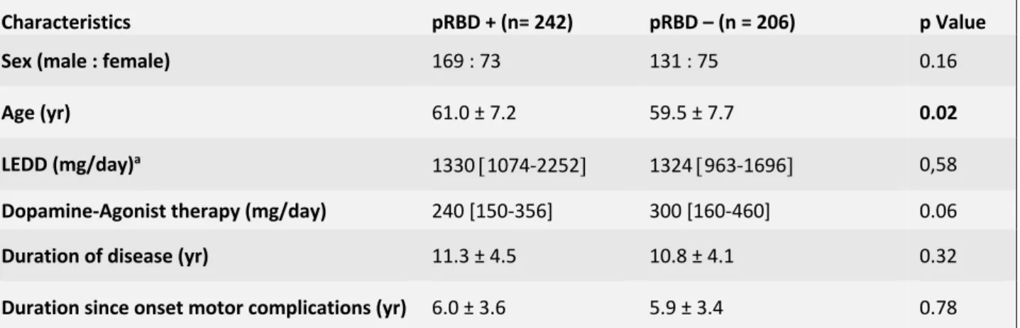
Results: Among 448 PD patients at baseline, 242 (54%) were RBD+. Before surgery, RBD+ were older (p= 0.02), had better disabilities motor score (p= 0.03) but exhibited greater non-motor aspects of experiences of daily living (p<0,001) with more signs such as anxiety (p=0.01), depression (p= 0.01) and apathy (p=0.01), and poorer quality of life (p= 0.03), as compared to RBD-. Chronic bilateral STN-DBS improved parkinsonian symptoms and quality of life in both groups at 12 months, whereas it worsened cognitive function in both groups, with no between group differences of variation. At postoperative evaluation, LEDD dose was lower (p= 0.05) in RBD+ compared to RBD-. Hypodopaminergic symptoms did not differ anymore between group while and ICDs improved in RBD+ (p=0.05). Despite positive outcomes of psycho-behavioral, QoL remained poorer in RBD+ (p= 0.04).
24
Conclusions: The presence of RBD preoperatively is not associated with different motor and cognitive outcomes 1 year after STN-DBS, but seems to be associated with better psycho-behavioral outcomes, either for hypo-dopaminergic symptoms and ICDs, as compared to patients without RBD before surgery.
INTRODUCTION
Rapid eye movement (REM) sleep behavior disorder (RBD) is a parasomnia characterized by the loss of normal muscular atonia during REM sleep, with complex motor behavior and dreaming (1). This disordered is frequently associated with Parkinson Disease (PD) and others α-Synucleinopathy (2–4), and can even be present several years before the onset of motor symptoms (5). Recent evidences suggest that PD patients with RBD, might exhibit a more severe phenotype with greater motor disabilities, especially for axial symptoms, and more non-motors signs such as impulse control disorders, hallucination and increased cognitive impairment (6,7).
Subthalamic nucleus Deep Brain Stimulation (STN-DBS) is a well-documented treatment for severe PD with motor fluctuations and levodopa-induced dyskinesias (LIDs), allowing a dramatic improvement of quality of life and motor symptoms and a reduction in Levodopa equivalent daily doses (LEDD) (7,8,11). STN-DBS has also be shown to be effective in improving non-motor symptoms such sensory, dysautonomic and cognitive fluctuations, but also neuropsychiatric fluctuations and impulse control disorders (10,11). However, some patients also demonstrate worsening of other symptoms following STN-DBS with increased apathy (10), increased axial symptoms including freezing of gait, postural instability and dysarthria (12), and could also impact non-motor features outcomes (12,13). Identifying PD patients at risk of negative outcomes after STN-DBS remains a challenge, and there are
25
currently few clinical indicators allowing to target indications in patients that will most likely benefit of this treatment. Since the presence of RBD in PD is associated with a more severe phenotype of the disease, including higher risk of postural instability and freezing of gait,(14,15) dysautonomia,(16) impaired cognitive profile,(17,18) visual hallucinations,(19) and impulse control disorders,(20,21) we hypothesized that PD patients with RBD preoperatively (preop-RBD) could be more at risk of poorer motor and non-motor outcomes after STN-DBS. Up to now, only few studies assessed outcomes of PD patients with preop-RBD undergoing STN-DBS but reported conflicting results : the first one, focusing on motots symptoms, reported less improvement for parkinsonian symptoms in PD patients with preop-RBD, one year and three years after STN-DBS (21); conversely, another study did not show any difference for motor outcomes after STN-DBS between PD patients with and without preop-RBD, but highlighted greater improvement of hypodopaminergic symptoms in PD patients with preop-RBD compared to patients without RBD (22).
Thus, the aim of this study was to investigate whether the presence of RBD before STN-DBS could impact the motor, cognitive, psycho-behavioral outcomes and quality of life in PD patients 12 months after STN-DBN.
MATERIALS AND METHODS
This is an ancillary study of a multicentric prospective study PREDI-STIM whose main objective is to identify the predictive factors of quality of life response after Deep Brain Stimulation of the Sub-Thalamic Nucleus in PD patients (clinicalTrial.gov n°NCT02360683).
26
All patients included met the condition of the Movement Disorder Society Clinical Diagnostic Criteria for the diagnosis of Parkinson's Disease (24) and the standard surgical criteria defined by moderate to severe levodopa related motor fluctuations despite optimal treatment adjustment. Patients were not included if they had a contraindication to surgery : age greater than 75 years, atypical Parkinson's disease, course of the disease less than 5 years, severe cognitive impairment or dementia (MoCA score <24 and DSMIV criteria), parkinsonian psychosis, dopa-sensibiliy<30%, severe cerebral atrophy or MRI abnormality, presence of another serious terminal pathology. The study was approved by the ethics committee (CPP Nord-Ouest IV) and all patients gave their informed consent.
Study design and clinical scores:
The diagnosis of probable RBD (RBD) was assessed preoperatively using the RBD-1Q and RBDSQ scores, both validated as sensitive and specific tools for detecting RBD (25,26).
Patients were divided in two groups depending on the presence of preop-RBD screened with a positive response for RBD-1Q and / or a score ≥6 for RBDSQ at preoperative evaluation (preop-RBD) (25,27). The two groups were compared at baseline and 12 months after STN-DBS (postoperative evaluation).
Parkinsonism evaluation was performed using the Unified Parkinson's Disease Rating Scale (UPDRS) (5) which assess non-motor and motor experiences of daily living (UPDRS I, II), motor examination (UPRDS III) and motor complications (UPDRS IV).
At baseline motor assessment was performed using UPDRS Part III in the ‘Off’ state after 12-h wit12-hdrawal of antiparkinsonian medication (Med Off), and in t12-he ‘On’ state after taking 1.5 times the usual morning Levodopa dose using dispersible Levodopa (Med On). Twelve months after surgery, the acute efficacy of STN-DBS and of Levodopa was assessed in the morning after at least 12 h of withdrawal of antiparkinsonian medication using UPDRS Part III in three
27
conditions: ‘medication off – stimulation off’ (Med Off / Stim Off), ‘medication off – stimulation on’ (Med Off / Stim On), and ‘medication on – stimulation on’ (Med On / Stim On).
Comparison between baseline and 12 months after surgery were performed for UPDRS III Off defined by the conditions “Med Off” preoperatively vs “Med Off / Stim Off” postoperatively and for UPDRS III On defined by the conditions “Med On” preoperatively vs “Med On / Stim On” postoperatively. The effect of chronic STN-DBS was also evaluated comparing UPDRS III in “Med Off” condition preoperatively vs “Med Off / Stim On” postoperatively.
Cognitive functions were assessed using 11 validated tests in Parkinson's disease according to the French consensus for the evaluation of cognitive functions in Parkinson's disease (28) : Montreal Cognitive Assessment (MoCA) (29) for global efficiency; Symbol Digit Modalities Test (SDMT) for attention and working memory; Digit Span Forward and Backward, Subtests 16-item Free/Cued Recall Test and 10/36 Spatial Recall Test for episodic memory; Oral Letter– Number Sequencing task and D-KEFS Color–Word Interference Test for executive function; Benton Judgement of Line Orientation and CLOX clock-drawing test for visuospatial function; Boston Naming Test and Animal fluency test for language. Cognitive results were not normalized thus, we could not realize between groups comparisons of absolute value. However, intra group comparisons were performed between baseline and follow up, as well as between-groups comparisons of the percentage of variations.
Regarding psycho-behavioral symptoms, we used the french version of Ardouin Scale of Behavior in Parkinson's Disease (ASBPD) to assess hypo and hyperdopaminergic disorders, non-motor fluctuations and control disorder (ICD) (30). The presence of impulse-control disorders (ICDs) was defined by a scores ≥ 2 for at least one of the following items: eating behavior, hobbyism, punding, compulsive shopping, pathological gambling,
28
hypersexuality, dopaminergic addiction (subscores 5, 7, 8, 10, 11, 12, or 13) (16). For these two scales, higher scores indicate greater apathy and severe mood and behavioral disturbances.
The Parkinson's Disease Questionnaire (PDQ 39) scale was used to assess quality of life. PDQ39 summary index (PDQ39-SI) ranges from 0 to 100; being the higher the score, the worse the quality of life (31).
The Levodopa Equivalent Daily Dose (LEDD) was automatically provided for each patient taking account of levodopa treatments, dopamine agonists, and other antiparkinsonian drugs such as MAO-B inhibitors, COMT inhibitors and amantadine (32).
Statistical analysis
Statistical analyses were performed using Stata software, version 15 (StataCorp, College Station, TX, US). The tests were two-sided, with a type I error set at 5%. Continuous data were expressed as mean and standard-deviation or median and [interquartile range] according to statistical distribution. The assumption of normality was assessed by using the Shapiro-Wilk test. Concerning non-repeated measures, continuous variables were compared between groups by Student t-test or Mann-Whitney test when assumptions of t-test were not met. The homoscedasticity was analyzed using the Fisher-Snedecor’s test. Categorical parameters were compared between groups using chi-squared or Fisher’s exact tests. The relationships between continuous variables were studied estimating correlation coefficients (Pearson or Spearman, according to statistical distribution with the Sidak’s type I error correction due to multiple comparisons). Random-effects models for correlated data were performed to measure time and group effects and their interaction time x group, taking into account between and within patient variability (subject as random-effect). The normality of residuals from these models was studied using the Shapiro-Wilk test. When appropriate, a logarithmic transformation has been proposed to achieve the normality of dependent outcome. Then, multivariable analyses have been
29
performed using adjustment on covariates fixed according to the univariate results and to the clinical relevance.
RESULTS
Characteristics of the patients
Among 448 patients awaiting STN-DBS who completed RBD-SQ and/or RBD-1Q, 330 patients underwent surgery when carrying out study. Two hundred and six patients were excluded from analysis either because of missing data (n=25), still waiting for surgery (n=93) or did not reach 12 months follow up after surgery (n= 88). Thus, baseline analysis was conducted on 448 patients, while 12 months analysis and comparison between baseline and 12 months were performed on 242 patients. (Supplemental data. Flow chart)
Our total population had a mean age of 63.3±7.4 years, with a sex ratio of 300 men for 148 women and disease duration was 11.0±4.3 years at baseline. In the whole group, STN-DBS allowed a mean decrease of UPDRS III Off from 52%, of UPDRS IV from 36% and a decrease of dopaminergic treatments from 52% at postoperative evaluation (12 months after STN-DBS) compared to baseline.
Among 448 PD patients at baseline, 242 (54%) had probable preop-RBD (RBD+), while 206 did not have probable preop-RBD (RBD-). At baseline, RBD+ were older (p= 0.02), but did not differ from RBD- for sex ratio, and disease duration (Table 1). Motors symptoms, non-motors symptoms and quality of life of RBD+ and RBD- preoperatively and 12-months after STN-DBS are resumed in Table 2 and Table 3.
30
Parkinsonian, cognitive, psycho-behavioral symptoms and quality of life at baseline:
UPDRS III score assessed in “Med Off” condition was significantly lower in RBD+ compared to RBD- (p =0,003), but there was no significant difference between groups for the UPDRS III scores in “Med On” condition. UPDRS I score was higher in RBD+ (p < 0.001) compared to RBD-. There was no significant between-groups difference for UPDRS II scores neither for UPDRS IV scores, neither for total LEDD dose.
The two groups were comparable for MOCA scores at baseline.
ASBPD total score was significantly higher in RBD- groups (p =0.03). When considering specifically each part of scale, we noticed significant differences between groups with RBD+ who exhibited more apathy, (p = 0.019), anxiety (p= 0.01) and depression signs (p = 0.001). In contrast, hyperdopaminergic behaviors, neuropsychiatric fluctuation and ICD subscores did not differ between groups.
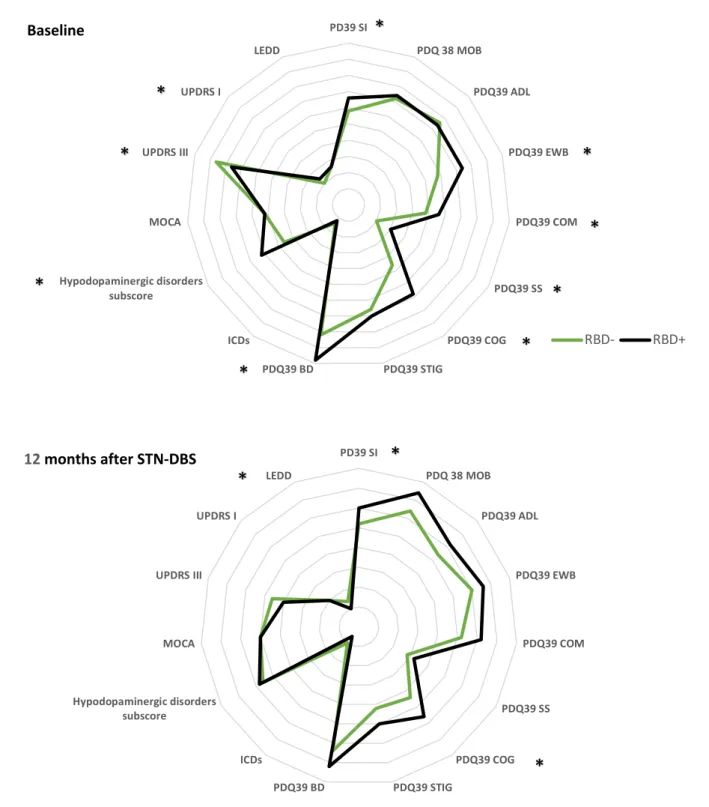
PDQ-39 SI score was significantly higher in RBD+ comparing to RBD- (p= 0.03), especially for “emotional well-being” (p<0.01), “communication” (p=0.02), “social support” (<0.01), “cognition” (p<0.01), and “bodily discomfort” (p<0.01) (Figure 1A).
Parkinsonian, cognitive, psycho-behavioral symptoms and quality of life 12 month after STN-DBS
Twelve months after STN-DBS, UPDRS III score assessed in “Med Off / Stim Off” condition was lower in RBD+ (p= 0.04) compared to RBD-, with lower LEDD (p = 0.05). UPDRS I scores, UPDRS II scores in “Med Off / Stim On” condition, UPDRS III scores in “Med Off / Stim On“ and “Med On / Stim On” condition, UPDRS IV scores, MOCA and ASBPD total scores and sub-scores were not different in RBD+ compared to RBD-after STN-DBS.
31
PDQ 39 SI, and cognition subscore were significantly higher in RBD+ (respectively p=0.04 and p=0.01) compared to RBD-, but no significant difference was found for other PDQ 39 dimensions (Figure 1B).
Parkinsonian, cognitive, psycho-behavioral symptoms and quality of life variations between baseline and postoperative evaluation at 12 months
STN-DBS allowed a mean decrease of UPDRS III Off scores by 53% in RBD- (p<0.001) and by 52.2 % in RBD+ (p<0.01), while no postoperative variation of UPDRS III On compared to baseline was observed in both groups. UPDRS I score significantly decreased by 16% (p<0.01) in RBD+ group, whereas no significant variation was observed in RBD- group.
UPDRS II Off and UPDRS IV scores significantly decreased in both groups (respectively p = 0,01 for RBD- and p < 0,01 for RBD +; p < 0,01 for RBD- and RBD +).
MOCA score compare to baseline decreased in RBD- (p< 0.05) and in RBD+ (p<0.05). There was no significant change whatever the group for attentional, instrumental and memory functions assessed by the use of 11 cognitive tests (Supplemental Table 4).
ASBPD total score, hyperdopaminergic behaviors and neuropsychiatric fluctuation subscores significantly decreased in both groups. While, hypodopaminergic subscore worsened in RBD- (p = 0.03), it tended to be improved in RBD+ (p = 0.65).
PDQ39 SI scores decreased by 16% in RBD- (p< 0.01) and by 8% in RBD+ (p<0.01). Considering sub scores, “Activities of daily living”, “bodily discomfort” and “Stigma” significantly decreased in both groups (respectively p < 0,01 for RBD- and RBD+; p < 0,01 for RBD- and RBD+; p < 0,01 for RBD- and RBD+) whereas “Cognition” sub score only decreased in RBD+ (p<0,01) (Figure 2).
32
Finally, when comparing the percentage of variation from baseline to 12 months between groups, no significant difference was found and multivariate analysis adjusted on age, sex, LEDD and UPDRS III Off scores confirmed the provided results (Supplemental Table 5).
DISCUSSION
Among PD candidates to STN-DBS, RBD+ patients are older, present milder motor symptoms, but poorer non motor aspects of experiences of daily living with more hypodopaminergic psycho-behavioral symptoms and decreased quality of life compared to RBD- at preoperative evaluation. Twelve months after STN-DBS, RBD+ show milder motor symptoms when “Med Off / Stim Off” and no difference when “Med Off / Stim On”, with a smaller postoperative LEDD dose, compared to RBD-. In spite of a greater postoperative decrease of total LEDD in RBD+, there is no longer between group difference for hypodopaminergic symptoms 12 months after STN-DBS. Indeed, hypodopaminergic symptoms worsen after STN-DBS in RBD- while they remain unchanged in RBD+. ICDs improve postoperatively in RBD+, while they remain unchanged in RBD-. Quality of life appears poorer in RBD+ before surgery compared to RBD- and, even if it improves in both groups similarly, remains poorer in RBD+12 month after STN-DBS.
To our knowledge, this is the first study to assess motor and non-motor outcomes of STN-DBS depending on the presence of probable preop-RBD in such a large multicentric cohort of PD patients. So far, two prospective studies compared the outcome of PD patients with and without preop-RBD and reported conflicting results. The first one, was conducted on 41 PD patients who underwent STN-DBS and were screened for probable preop-RDB (12% of patients) using a semistructured clinical interview.(22) This study focused on motor outcomes one years and
33
three years after STN-DBS. The second one assessed motor and non-motor outcomes, and was conducted on 50 PD patients who underwent surgery. In that latter study, preop-RBD diagnosis was confirmed by a night-polysomnography (48% of patients).(23)
Those studies did not report any significant difference at baseline in motors symptoms assessed by UPDRS III Off between PD patients with and without preop-RBD, whereas in our study RBD+ patients have better motor performance than RBD-. These findings are surprising since studies generally report greater motor disabilities and more axial involvement in PD patients with RDB (33,34). Yet, it is likely that we selected as candidates for surgery a specific subgroup of PD patients with RBD because those with axial signs such as freezing of gait or postural instability were not meeting STN-DBS criteria, thereby excluding a large range of PD with associated RBD. The relatively low percentage of PD with preop-RBD observed in our study (54%) and in previous ones (12% and 48%), compared to the generally observed prevalence of RBD in PD population (60%) reinforces this hypothesis.(35) Moreover, the use of UPDRS III scores might not allow to assess axial symptoms, while the use of composite axial score, such as Zibetti et al did, highlighted difference in severity of axial symptoms between groups.(36)
The first study mentioned above investigated whether groups exhibited differences in the percentage of improvement of motor symptoms between baseline and postoperative evaluation (at 1 and 3 years) with STN stimulation alone or in combination with levodopa treatment. (22) They reported less improvement of UPDRS III scores in PD patients with preop-RBD, but only for “Med On/ Stim On” condition, whilst no difference compared to patients without preop-RBD for “Med Off /Stim On” condition. This can be partly explained by the presence of increased axial features in PD patients with preop-RBD in this study, which are less likely to
34
be improve with dopaminergic medication. The second one compared UPDRS II and UPDRS III scores absolute values at baseline (« Med On » condition), and postoperatively (“Med On / Stim On” condition one year after surgery). (23) After STN-DBS, UPRDS III On score improved significantly in both groups, but the change was comparable between the two groups, unlike reported in the aforementioned study. Moreover, UPDRS II On score did significantly improved in PD patient with preop-RBD contrary to PD patients with preop-RBD. Those apparently conflicting results are probably explained by the heterogeneity of methodology used to assess motors outcomes in these different studies.
Otherwise, while previous studies did not show any difference in the post-operative treatment variation between groups, we highlight greater decrease in dopaminergic treatment in RBD+ compare to RBD-. The postoperative decreased dopaminergic treatment in RBD+ cannot be explained by a better efficiency of stimulation alone on motor symptoms since these patients are not different from RBD- in “Med Off/ Stim On” condition (Table 2. Supplemental Figure 3). Neither do they differ for treatment induced complications such as dyskinesia/ fluctuations (UPDRS IV) or ICDs symptoms. Yet RBD- present increased hypodopaminergic symptoms after DBS-STN, whereas RBD+ stay stable on this aspect, which could explain the relative lack of decrease of treatment in the RBD- group, in an attempt to prevent worsening of this psycho-behavioral manifestation.
Regarding cognitive functions, there is no difference between groups at baseline and 12 months after surgery. However, RBD+ exhibit poorer subjective assessment in cognitive dimension of PDQ39 compared to RBD-, either before or after surgery. This suggest a more severe impact of a similar cognitive impairment in RBD+ on daily life functioning. Previous studies assessing
35
STN-DBS outcome of PD with RBD did not investigate cognitive functions, but the cognitive dimension of PDQ39 assessed by Bargiotas et al, was worst in RBD+ preoperatively, in line with our results, but did not differ compared to RBD- after STN-DBS. These discrepancies could be explained by a clear improvement in depression scores after STN-DBS in Bargiotas
et al study, whereas no significant postoperative variation of depression score was observed in
our study. Thus, alleviation of depressive symptoms may have diminished the negative impact of cognitive impairment in daily life functioning in that previous study.
Even if RBD+ have greater apathy, anxiety and depression preoperatively, they have no postoperative variation of severity for these symptoms, whereas RBD- patients worsen for total dopaminergic subscore. This finding suggests no deleterious effect of surgery on hypo-dopaminergic symptoms in RBD+, in spite of a greater decrease of hypo-dopaminergic treatment in that subgroup. These data are in line with Bargiotas et al study showing that RBD+ have greater apathy and depression symptoms than RBD- at baseline, whereas no difference remained one year after STN-DBS. (23) Alltogether, hypodopaminergic symptoms appear to be relatively preserved from worsening in RBD+ patients after DBS-STN compared to RBD-. In the general population of PD, the effect of STN-DBS on hypodopaminergic symptoms is debated, with studies reporting improvement (13,37) and others describing worsening (38–41) of hypo dopaminergic signs, and the postoperative decrease of dopaminergic treatment is not likely to solely explain the increase of apathy that can occur after STN-DBS (42). Thus, predictive factor for the occurrence of hypo-dopaminergic outcomes such as apathy after surgery are of crucial importance, and the presence of RBD before STN-DBS could indicate a reduced risk for such an evolution.
36
In regard to hyperdopaminergic symptoms, we found no between-group difference preoperatively, but our results reveal a postoperative decrease of ICDs in RBD+ whereas no significant variation for the presence of ICDs is reported after STN-DBS for RDB-. Previous studies did not assess hyperdopaminergic outcomes after STN-DBS in PD patients with and without preop-RBD. The lack of association with ICD preoperatively in our study for the RBD+ group goes against the generally described association between RBD and ICDs in PD (35), and could be due to the selection of a non-representative “less severe” subgroup of PD patients with RBD as candidates for DBS-STN. The decrease of ICDs after STN-DBS in the PD-RBD+ group compared to the RBD- could be related to greater reduction of dopaminergic treatment in the RBD+ group.
In both groups, global QoL improve after surgery, yet it remains worst in RBD+ either pre or post operatively compare to RBD-. One of our assumptions to explain that lack of improvement, is that even if surgery seems to provide benefit effect on non-motors signs especially hypodopaminergic signs, in RBD+, some others symptoms not clearly emphasized by UPDRS I such as dysautonomia, other sleep-wake disorders, could remain debilitating on daily life functioning. The use of another scale such as NMSS could have highlighted difference (43).
A number of limitations of this study must be identified. First, the lack of polysomnography confirmed diagnosis of RBD could lead to some PD patients having been mis assigned. Yet, the screening of probable RBD was conducted using a validated scale with 72.6% of sensitivity, 65.7% of specificity for the diagnosis of RBD.(44) Furthermore, the limited access to video-polysomnography in clinical practice did not allow to perform this exam on such an important multicentric cohort. Another limitation is that RBD status of patients was not controlled after
37
STN-DBS, though STN-DBS has been reported to have an effect on RBD symptoms and could modify their prevalence postoperatively (45,46). Indeed, RBD symptoms may fluctuate along the evolution of PD (47) but in spite of these clinical variations, a specific long-term trait is associated with RBD, probably in relation with a specific pattern of neurodeneration in those patients, and is what we aimed to assess as a predictor for STN-DBS outcome in that study. The lack of data regarding the location of electrodes and stimulation parameters should also be acknowledged and could interfere with postoperatively results. However, we noted in our whole population an improvement of 52% for motor UPDRS scores in the “Off” medication state, a reduction of 36% for dyskinesia and a reduction of antiparkinsonian drugs by approximately 36% after STN-DBS. Those results close to generally observed motor improvement, pleads for the correct location of the effective contacts (8,11).
CONCLUSION:
Altogether, our results suggest that the presence of preop-RBD is not associated with different motor and cognitive outcomes 1 year after STN-DBS, but could indicate relatively better psycho-behavioral outcomes, as RBD+ patients appear to be relatively spared from hypo-dopaminergic worsening and present a decrease of ICDs, as compared to RBD-. STN-DBS improves quality of life in PD patients regardless to the presence of preop-RBD but does not allow to erase the difference observed between those groups before surgery, suggesting other factors leading to worse quality of life are associated with RBD. Further studies, assessing long-term outcomes associated with the presence of RBD preoperatively 3 years and 5 years after surgery, will improve our comprehension of the specific prognosis associated with the presence of RBD in PD candidates to STN-DBS.
38
REFERENCE
1. Thorpy MJ. Classification of Sleep Disorders. Neurotherapeutics. oct 2012;9(4):687‑701. 2. Schenck CH, Bundlie SR, Mahowald MW. Delayed emergence of a parkinsonian disorder in 38% of 29 older men initially diagnosed with idiopathic rapid eye movement sleep behaviour disorder. Neurology. févr 1996;46(2):388‑93.
3. Barone DA, Henchcliffe C. Rapid eye movement sleep behavior disorder and the link to alpha-synucleinopathies. Clin Neurophysiol. 2018;129(8):1551‑64.
4. Zhang X, Sun X, Wang J, Tang L, Xie A. Prevalence of rapid eye movement sleep behavior disorder (RBD) in Parkinson’s disease: a meta and meta-regression analysis. Neurol Sci. janv 2017;38(1):163‑70.
5. Schenck CH, Mahowald MW. REM sleep behavior disorder: clinical, developmental, and neuroscience perspectives 16 years after its formal identification in SLEEP. Sleep. 15 mars 2002;25(2):120‑38.
6. Duarte Folle A, Paul KC, Bronstein JM, Keener AM, Ritz B. Clinical progression in Parkinson’s disease with features of REM sleep behavior disorder: A population-based longitudinal study. Parkinsonism & Related Disorders. mai 2019;62:105‑11.
7. Fantini ML, Macedo L, Zibetti M, Sarchioto M, Vidal T, Pereira B, et al. Increased risk of impulse control symptoms in Parkinson’s disease with REM sleep behaviour disorder. Journal of Neurology, Neurosurgery & Psychiatry. 1 févr 2015;86(2):174‑9.
8. Limousin P, Krack P, Pollak P, Benazzouz A, Ardouin C, Hoffmann D, et al. Electrical stimulation of the subthalamic nucleus in advanced Parkinson’s disease. N Engl J Med. 15 oct 1998;339(16):1105‑11.
9. Schuepbach WMM, Rau J, Knudsen K, Volkmann J, Krack P, Timmermann L, et al. Neurostimulation for Parkinson’s Disease with Early Motor Complications. N Engl J Med. 14 févr 2013;368(7):610‑22.
10. Abbes M, Lhommée E, Thobois S, Klinger H, Schmitt E, Bichon A, et al. Subthalamic
stimulation and neuropsychiatric symptoms in Parkinson’s disease: results from a long-term follow-up cohort study. J Neurol Neurosurg Psychiatry. 2018;89(8):836‑43.
11. Azulay J-P, Witjas T, Eusebio A. Effect of subthalamic deep brain stimulation on non-motor fluctuations in Parkinson’s disease. J Neural Transm (Vienna). avr 2013;120(4):655‑7.
12. Krack P, Batir A, Van Blercom N, Chabardes S, Fraix V, Ardouin C, et al. Five-Year Follow-up of Bilateral Stimulation of the Subthalamic Nucleus in Advanced Parkinson’s Disease. N Engl J Med. 13 nov 2003;349(20):1925‑34.
13. Lhommée E, Wojtecki L, Czernecki V, Witt K, Maier F, Tonder L, et al. Behavioural outcomes of subthalamic stimulation and medical therapy versus medical therapy alone for Parkinson’s disease with early motor complications (EARLYSTIM trial): secondary analysis of an open-label randomised trial. Lancet Neurol. 2018;17(3):223‑31.
39
14. Videnovic A, Marlin C, Alibiglou L, Planetta PJ, Vaillancourt DE, Mackinnon CD. Increased REM sleep without atonia in Parkinson disease with freezing of gait. Neurology. 17 sept 2013;81(12):1030 ‑5.
15. Duarte Folle A, Paul KC, Bronstein JM, Keener AM, Ritz B. Clinical progression in Parkinson’s disease with features of REM sleep behavior disorder: A population-based longitudinal study. Parkinsonism Relat Disord. 2019;62:105‑11.
16. Kim J-S, Park H-E, Oh Y-S, Lee S-H, Park J-W, Son B-C, et al. Orthostatic hypotension and cardiac sympathetic denervation in Parkinson disease patients with REM sleep behavioral disorder. J Neurol Sci. 15 mars 2016;362:59‑63.
17. Marques A, Dujardin K, Boucart M, Pins D, Delliaux M, Defebvre L, et al. REM sleep behaviour disorder and visuoperceptive dysfunction: a disorder of the ventral visual stream? J Neurol.
2010;257:383‑91.
18. Jozwiak N, Postuma RB, Montplaisir J, Latreille V, Panisset M, Chouinard S, et al. REM Sleep Behavior Disorder and Cognitive Impairment in Parkinson’s Disease. Sleep. 01 2017;40(8).
19. Lenka A, Hegde S, Jhunjhunwala KR, Pal PK. Interactions of visual hallucinations, rapid eye movement sleep behavior disorder and cognitive impairment in Parkinson’s disease: A review. Parkinsonism Relat Disord. janv 2016;22:1‑8.
20. Fantini M, Macedo L, Zibetti M, Sarchioto M, Vidal T, Pereira B, et al. Increased risk of impulse control symptoms in Parkinson’s disease with REM sleep behaviour disorder. J Neurol Neurosurg Psychiatry. 2015;86:174‑9.
21. Lu H-T, Shen Q-Y, Zhao Q-Z, Huang H-Y, Ning P-P, Wang H, et al. Association between REM sleep behavior disorder and impulsive-compulsive behaviors in Parkinson’s disease: a systematic review and meta-analysis of observational studies. J Neurol. 21 oct 2019;
22. Zibetti M, Rizzi L, Colloca L, Cinquepalmi A, Angrisano S, Castelli L, et al. Probable REM sleep behaviour disorder and STN-DBS outcome in Parkinson’s Disease. Parkinsonism & Related Disorders. mai 2010;16(4):265‑9.
23. Bargiotas P, Debove I, Bargiotas I, Lachenmayer ML, Ntafouli M, Vayatis N, et al. Effects of bilateral stimulation of the subthalamic nucleus in Parkinson’s disease with and without REM sleep behaviour disorder. J Neurol Neurosurg Psychiatry. 17 août 2019;jnnp-2019-320858.
24. Goetz CG, Tilley BC, Shaftman SR, Stebbins GT, Fahn S, Martinez-Martin P, et al. Movement Disorder Society-sponsored revision of the Unified Parkinson’s Disease Rating Scale (MDS-UPDRS): Scale presentation and clinimetric testing results. Mov Disord. 15 nov 2008;23(15):2129‑70. 25. Postuma RB, Arnulf I, Hogl B, Iranzo A, Miyamoto T, Dauvilliers Y, et al. A single-question screen for rapid eye movement sleep behavior disorder: A multicenter validation study. Mov Disord. juin 2012;27(7):913‑6.
26. Stiasny-Kolster K, Mayer G, Schäfer S, Möller JC, Heinzel-Gutenbrunner M, Oertel WH. The REM sleep behavior disorder screening questionnaire-A new diagnostic instrument. Mov Disord. 25 sept 2007;22(16):2386‑93.