Comparison of procalcitonin and CrP in the postoperative
course after lung decortication
Giovanni L. Carboni
a, Rene Fahrner
a, Amiq Gazdhar
a, Gert Printzen
b,
Ralph Alexander Schmid
a,*
, Beatrix Hoksch
aa
Division of General Thoracic Surgery, University Hospital Berne, 3010 Berne, Switzerland
bInstitute for Clinical Chemistry, University Hospital Berne, 3010 Berne, Switzerland
Received 30 August 2007; received in revised form 4 February 2008; accepted 6 February 2008
Abstract
Objective: The objective of this prospective study was to compare the clinical value of procalcitonin (PCT) and C-reactive protein (CrP) plasma concentrations in their postoperative course after decortication. Methods: Twenty-two patients requiring surgery for pleural empyema were chosen for this prospective study. Routine blood samples including CrP and PCT plasma concentrations were taken before the operation and on the 1st, 2nd, 3rd, and 7th postoperative day. Results: Due to infection PCT and CrP were elevated preoperatively. In the postoperative course both PCT and CrP reached peak-levels on day 2 with values up to 43.55 ng/ml and 384.00 mg/l, respectively. In PCT the rise was followed by a clear decrease in 20 (90.9 %) patients until day 7. In contrast the CrP levels decreased slowly and only seven (54.5%) patients had values of 100 mg/l or below on day 7. PCT showed a better correlation with the clinic in case of septic course than CrP does. Conclusions: PCT reflects postoperative clinical course more accurately than CrP. Therefore, PCT is a more appropriate laboratory parameter to monitor patients after surgery for pleural empyema.
#2008 European Association for Cardio-Thoracic Surgery. Published by Elsevier B.V. All rights reserved.
Keywords: Pleural empyema; PCT; CRP
1. Introduction
The main goal of surgery in the treatment of pleural empyema is to control the infection[1]. Therefore, a rapidly obtainable and accurate laboratory marker capable of distinguishing persistent infection from SIRS or sepsis in the postoperative course would be clinically useful. After surgery C-reactive protein (CrP) is widely used to monitor the postoperative course, especially with regard to infection. However, several trials showed a lower sensitivity in the (early) postoperative period for CrP in comparison to procalcitonin (PCT) that has become increasingly popular as a marker of infection after surgical procedures[2,3].
Until now no study has evaluated the time course and clinical relevance of changes in PCT in patients undergoing surgery for pleural empyema. The aim of this prospective pilot study was to investigate the accuracy of PCT in comparison to CrP as a marker of postoperative course and complications.
2. Methods
Twenty-two patients undergoing surgery for pleural empyema at the Division of Thoracic Surgery, Inselspital Bern, were chosen for this prospective pilot study. Inclusion criteria consisted of clinically proven pleural empyema following pneumonia and secondary infected posttraumatic pleural effusion. Definition of pleural empyema was based on the following criteria: presence of micro-organisms in pleural fluid Gram stain or culture, pleural pH less than 7.2, radiographic features of empyema as pleural enhancement in CT scan, or intraoperative findings of purulent liquid in the pleural space. Patients with empyema after lung resections or with tuberculous empyema and patients on immunosuppressive therapy were excluded from the trial.
Complete lung decortication was performed through a muscle-sparing lateral thoracotomy without rib resection and with preservation of the latissimus and serratus muscles. The whole fibrous peel encasing the lung and diaphragm was removed. Resection of the parietal peel was done by an extrapleural dissection. To ensure adequate drainage of the pleural space three chest tubes were placed.
www.elsevier.com/locate/ejcts European Journal of Cardio-thoracic Surgery 33 (2008) 777—780
* Corresponding author. Tel.: +41 31 6323096; fax: +41 31 6322327. E-mail address:[email protected](R.A. Schmid).
1010-7940/$ — see front matter # 2008 European Association for Cardio-Thoracic Surgery. Published by Elsevier B.V. All rights reserved. doi:10.1016/j.ejcts.2008.02.013
Routine blood examinations including CRP and PCT were sampled before operation and on 1st, 2nd, 3rd, and 7th postoperative day.
2.1. Assays
PCTwas measured using the quantitative Kryptor-Test PCT (Brahms Diagnostica AG, Germany) with a lower detection limit of 0.06 ng/ml. All blood samples were collected, prepared, and frozen according to the manufacturers instructions. The PCT measurement was carried out at a later date. Therefore the PCT values were not available at time of hospitalisation.
3. Statistical analysis
For statistical analysis the Wilcoxon rank sum test was used. To compensate for the multiple testing situation the p values were adjusted using the procedure according to Holm.
4. Results
The median age of the 22 patients (10 woman and 12 men) was 58.0 years and ranged from 24 to 84 years. The leading cause of the pleural empyema was pneumonia (n = 17) followed by a thoracic trauma in five cases.
4.1. PCT
In the postoperative course PCTrose to a peak level on day 2 (median 0.85 range: 0.14—43.55 ng/ml) followed by a decrease until day 7 (median 0.17 ng/ml, range: 0.04— 2.66 ng/ml) (Fig. 1). On day 3, 54.5% of the patients had reached PCT levels of 0.5 ng/ml or below. On day 7 the number of patients with PCT values <0.5 ng/ml was 19 (86.4%) (Fig. 1).
In some patients PCT was elevated excessively: 11 patients (50.00%) had PCT levels of higher than 0.5 ng/ml on day 1, 14 patients (63.6%) on day 2, and 10 (45.4%) on day 3. Values of more than 2 ng/ml were observed in four (18.2%), seven (31.8 %), and three (13.6%) cases on day 1, 2, and 3 respectively. Four patients (18.2%) developed levels of over 10 ng/ml in the postoperative course. In three of these four patients the levels decreased to less than 10 ng/ml by day 7. One patient had a PCT of 2.66 ng/ ml on day 7, another patient a PCT of 36.39 ng/ml. This patient developed recurrent pleural effusion (see below). In patients with a PCT level of 10.0 ng/ml or higher the decrease was slower than in the patients with PCT values below 10.0 ng/ml. Six of seven patients (85.7%) with values of more than 2 or even 10 ng/ml in the postoperative course also had the clinical signs of a sepsis (according to the guidelines of the Consensus Conference of American College of Chest Physicians and of the Society of Critical Care Medicine) (Table 1)[4,5]. PCT values in case of sepsis were significant higher than in the other patients on day 1, 2, and 3 ( p = 0.001).
4.2. CrP
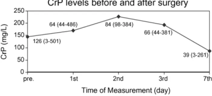
CrP showed a median value of 144.50 mg/l before surgery (range: 3.00—501.00 mg/l). CrP levels also peaked on the second postoperative day (median 226.50, range: 84.00— 390.00 mg/l). Thereafter the values decreased slowly. On day 3 after surgery, 10 patients (45.4%) still had levels of 200 mg/l and more, eight patients (36.4%) levels of 100 mg/l or more. On day 7 only 12 patients (54.5%) had CrP levels of 100 mg/l or below whereas in 10 patients the levels were 100 mg/l or higher (up to 261 mg/l) (Fig. 2,Table 1). In the patients with septic course and very high PCT values the CrP did not show significantly different values to the other ones ( p = 0.30).
G.L. Carboni et al. / European Journal of Cardio-thoracic Surgery 33 (2008) 777—780 778
Fig. 1. PCT levels before and after surgery.
Fig. 2. CrP levels before and after surgery.
Table 1
PCT and CrP course in patients with and without septic signs
Preoperative 1st 2nd 3rd 7th
PCT 1 0.29 (0.03—2.89) 2.25 (1.94—43.55) 16.4 (1.94—43.55) 1.71 (0.54—36.81) 0.27 (0.17—36.39)
PCT 2 0.12 (0.04—1.29) 0.26 (0.15—1.66) 0.30 (0.15—1.40) 0.22 (0.07—0.92) 0.11 (0.03—0.43)
CrP 1 144 (5—477) 198 (64—262) 224 (84—312) 180 (66—285) 112 (39—261)
CrP 2 136 (3—501) 128 (44—486) 236 (98—390) 198 (44—381) 56 (3—207)
PCT 1: patients with septic signs; PCT 2: patients without septic signs; CrP 1: patients with septic signs; CrP 2: patients without septic signs median (range) preoperative and postoperative days 1, 2, 3, and 7.
4.3. Complications
No patient died. Complications occurred in two patients (9.1%): one required wound-revision due to a wound infection 14 days postoperatively and one patient was readmitted with fever and recurrent pleural effusion, which was treated conservatively. In the first case both the PCT and the CrP showed decreasing values until the day of discharge. The wound infection occurred 2 weeks later. In the other patient a continuously high level of PCT (36.39 ng/ml) was observed whereas CrP values fell clearly (Fig. 3). The patient was discharged in good clinical condition. The readmission occurred one week later. A conservative therapy was successfully carried out with antibiotics.
4.4. Microbiology/antibiotics
The microbiological detection of bacteria succeeded in seven cases (31.8%) only.
Antibiotic therapy was continued in all patients after operation. Fourteen patients (63.6%) were also treated with antibiotics after discharge due to persistently high CrP-values. In nine of these 14 cases (64.3%) the CrP was above 100 mg/l. At the same time, the median PCT of the 14 patients was 0.17 ng/ml. Only two of them (14.3%) had higher PCT-levels, which might be interpreted as a sign of persistent infection. One of these patients had to be admitted later in the follow-up. The course of the other patient was uneventful.
5. Discussion
Today non-tuberculotic bacterial pneumonia is the leading cause of pleural empyema. It is estimated that nearly 5% of the 1.2 million annual cases of pneumonia worldwide are complicated with a pleural empyema, which could lead to sepsis with serious consequences if not adequately treated[6]. About 40—65% of the patients are not cured of their empyema by conservative therapy, i.e. chest tube drainage, antibiotics, urokinase, and therefore require surgical intervention (dec-ortication) [7,8]. In the postoperative course clinical and laboratory parameters are needed to reflect the response to therapy in the follow-up. The aim of this trial was to investigate the course of PCT in comparison to CrP after
decortication. Several studies have underscored the value of PCT in a variety of clinical conditions. Simon et al. and Castelli et al. used PCT for identifying infectious processes[9,10]. PCT and its characteristic course for critically ill patients including sepsis was examined in studies[11]. PCT guided therapy was the object of the trials by Christ-Crain et al. and Sandek et al. in 2004[12,13]. All these trials confirm that PCT is a suitable parameter to detect and to evaluate the course of bacterial, fungal or parasitic infections. It appears to be more helpful in the early diagnosis of postoperative infections and in the differentiation of SIRS and infection than CrP[14]. However to date, little is known about the value of PCT measurements in thoracic surgery. Meisner et al. in 1998 and Molter et al. in 2003 examined the postoperative course of PCT values including patients undergoing different types of thoracic procedures
[3,14]. A study dealing exclusively with thoracic surgery was published in 2005 by Falcoz et al.[15]. In this trial PCT was significantly higher in patients with a postoperative infection than in patients with no postoperative infection. The authors found that PCT can provide more accurate information about the postoperative course than CRP does, and that pathological values are detected before the occurrence of clinical infection
[15]. In pleural empyema nothing is known about the postoperative course of PCT in comparison to CrP. The situation in pleural empyema is different to all the mentioned studies dealing in particular with the occurrence of an infection after an operation: in pleural empyema an elevated CrP and PCT value is expected preoperatively because of the infection. But after the removal of damaged and infectious tissue both the CrP and the PCT values should decrease rapidly. The amount and the pattern of decrease could act as indicators for the success of the operation more than radiological changes. In general PCT was elevated preoperatively with a range up to 2.89 ng/ml as sign of a systemic infection. Only two (9.1%) of the patients showed PCT levels lower than 0.05 ng/ ml. In the postoperative course PCTrose to a peak-level on day 2 and of 43.55 ng/ml followed by a clear decrease until day 7. High PCT levels over 10 ng/ml are an indicator for severe systemic inflammation or sepsis due to bacteraemia. In these patients with high levels of over 10 ng/ml the decrease of PCT was slower than in the group of patients with values between 2 ng/ml and 10 ng/ml. Several studies have suggested the correlation between PCT levels and the severity of infection
[16—20]. In the presented trial PCTalso had a good correlation with the postoperative course in case of SIRS or sepsis: Patients with very high PCT values showed clinical signs of a SIRS or sepsis which may be due to transient or persistent bacteraemia after decortication. In case of septic course the PCT levels were significant higher than in patients without septic events. One of two patients with elevated levels of PCT on day 7 had a complication. Until now it has been difficult to recommend universal cut-off points for PCT, which clearly differentiate a normal from a complicated postoperative course. In the literature the cut-off points range from 1 ng/ml to 5 ng/ml
[16,21,22]. Sponholz et al.[16]advise interpreting PCT levels according to the clinical context. In general, dynamics of PCT levels, rather than the absolute values, may be more important for identifying patients with persistent infection or infectious complications after surgery. Because all PCT values were not available during hospitalisation the clinical decisions were made with the help of the CrP values only. CrP
G.L. Carboni et al. / European Journal of Cardio-thoracic Surgery 33 (2008) 777—780 779
values were elevated before surgery (81.8%) but in contrast to PCT they decreased very slowly. High CrP values are the cause of why more than half of the patients (63.6%) received antibiotics at time of discharge for the next 1 or 2 weeks. In 12 of these 14 patients the PCT level was lower than 0.5 ng/ml. If the PCT values had been available most of these patients (up to 85.7%!) would not have received antibiotics. So the use of PCT instead of CrP in the postoperative course after decortication could save at least 7 days of antibiotic treatment. A meta-analysis by Simon et al. showed that the PCT level was more sensitive (88% vs 75%) and more specific (81% vs 67%) than the CRP level in differentiating bacterial from non-infective causes of inflammation[9].
Furthermore in patients with a sepsis after the operation CrP was not more elevated than in all the other patients. Several trials have proved the poor diagnostic value of CRP in the postoperative period especially if compared with PCT. Macrina et al.[2]used CrP to follow septic patients and found that CrP was unable to predict the outcome of disease or severity.
6. Conclusions/summary
Although there are only a small number of patients with pleural empyema due to different causes in this trial PCT seems to describe more accurately the postoperative course after successful decortication than CrP. After surgery for pleural empyema the main focus should be on the dynamics of PCT levels, rather than on the absolute values. By using the Kryptor method a lower detection concentration of 0.06 ng/ml allows a higher accuracy. PCT is a more appropriate laboratory diagnostic parameter than CRP to monitor the course of patients after surgery for pleural empyema.
Acknowledgment
The authors like to thank Dirk Klingbiel, Institute of Mathematical Statistics and Actuarial Science, University of Berne, for reviewing the statistical work.
References
[1] Solaini L, Prusciano F, Bagioni P. Video-assisted thoracic surgery in the treatment of pleural empyema. Surg Endosc 2007;21(2):280—4. [2] Macrina F, Tritapepe L, Pompei F, Sciangula A, Evangelista E, Toscano F,
Criniti A, Branacaccio G, Puddu PE. Procalcitonin is useful whereas C-reactive protein is not, to predict complications following coronary artery bypass surgery. Perfusion 2005;20(3):169—75.
[3] Meisner M, Adina H, Schmidt J. Correlation of procalcitonin and C-reactive protein to inflammation, complications, and outcome during the intensive care unit course of multiple-trauma patients. Crit Care 2006;10(1):R1.
[4] Brunkhorst FM. Diffusely composed traditional definitions — definition and diagnosis of sepsis with current criteria. Klinikarzt 2004;33:167—72. [5] Members of the American College of Chest Physicians/Society of Critical Care Medicine Consensus Conference Committee. Definitions for sepsis and organ failure and guidelines for the use of innovative therapies in sepsis. Crit Care Med 1992;20:864—74.
[6] Cheng Y-J, Wu H-H, Chou S-H, Kao E-L. Video-assisted thoracoscopic surgery in the treatment of chronic empyema thoracis. Surg Today 2002;32:19—25.
[7] Okada M, Tsubota N, Yoshimura M, Miyamoto Y, Yamagishi H, Satake S. Surgical treatment for chronic pleural empyema. Surg Today 2000;30:506—10.
[8] Cameron RJ. Management of complicated parapneumonic effusions and thoracic empyema. Intern Med J 2002;32:408—14.
[9] Simon L, Gauvin F, Amre DK, Saint-Louis P, Lacroix J. Serum procalcitonin and C-reactive protein levels as markers of bacterial infection: a sys-tematic review and meta-analysis. Clin Infect Dis 2004;39:206—17. [10] Castelli GP, Pognani C, Meisner M, Stuani A, Bellomi D, Sgarbi L.
Procal-citonin and C-reactive protein during systemic inflammatory response syndrome, sepsis and organ dysfunction. Crit Care 2004;8:R234—42. [11] Uzzan B, Cohen R, Nicolas P, Cucherat M, Perret GY. Procalcitonin as a
diagnostic test for sepsis in critically ill adults and after surgery or trauma: a systematic review and meta-analysis. Crit Care Med 2006;34:1996—2003.
[12] Christ-Crain M, Jaccard-Stolz D, Bingisser R, Gencay MM, Huber PR, Tamm M, Muller B. Effect of procalcitonin-guided treatment on antibiotic use and outcome in lower respiratory tract infections: cluster-randomised, single-blinded intervention trial. Lancet 2004;363:600—7.
[13] Sandek A, Springer J, Habedank D, Brunkhorst FM, Anker SD. Procalci-tonin-guided antibiotic treatment in heart failure. Lancet 2004;363:1555—6.
[14] Mokart D, Merlin M, Sannini A, Brun JP, Delpero JR, Houvenaeghel G, Moutardier V, Blache JL. Procalcitonin, interleukin 6 and systemic inflam-matory response syndrome (SIRS): early markers of postoperative sepsis after major surgery. Br J Anaesth 2005;94(6):767—73.
[15] Falcoz PE, Laluc F, Toubin MM, Puyraveau M, Clement F, Mercier M, Chocron S, Etievent JP. Usefulness of procalcitonin in the early detection of infection after thoracic surgery. Eur J Cardiothorac Surg 2005;27(6):1074—8.
[16] Sponholz C, Sakr Y, Reinhart K, Brunkhorst F. Diagnostic value and prognostic implications of serum procalcitonin after cardiac surgery: a systematic review of the literature. Crit Care 2006;10(5):R145. [17] Boeken U, Feindt P, Micek M, Petzold T, Schulte HD, Gams E. Procalcitonin
(PCT) in cardiac surgery: diagnostic value in systemic inflammatory response syndrome (SIRS), sepsis and after heart transplantation (HTX). Cardiovasc Surg 2000;8:550—4.
[18] Sablotzki A, Borgermann J, Baulig W, Friedrich I, Spillner J, Silber RE, Czeslick E. Lipopolysaccharide-binding protein (LBP) and markers of acute-phase response in patients with multiple organ dysfunction syn-drome (MODS) following open heart surgery. Thorac Cardiovasc Surg 2001;49:273—8.
[19] Rau BM, Frigerio I, Bu¨chler MW, Wegscheider K, Bassi C, Puolakkainen PA, Beger HG, Schilling MK. Evaluation of procalcitonin for predicting septic multiorgan failure and overall prognosis in secondary peritonitis. Arch Surg 2007;142:134—42.
[20] Dahaba AA, Hagara B, Fall A, Rehak PH, List WF, Metzler H. Procalcitonin for early prediction of survival outcome in postoperative critically ill patients with severe sepsis. Br J Anaesth 2006;97(4):503—8.
[21] Aouifi A, Piriou V, Bastien O, Blanc P, Bouvier H, Evans R, Ce´leard M, Vandenesch F, Rousson R, Lehot JJ. Usefulness of procalcitonin for diagnosis of infection in cardiac surgical patients. Crit Care Med 2000;28:3171—6.
[22] Rothenburger M, Markewitz A, Lenz T, Kaulbach HG, Marohl K, Kuhlmann WD, Weinhold C. Detection of acute phase response and infection. The role of procalcitonin and C-reactive protein. Clin Chem Lab Med 1999;37:275—9.
G.L. Carboni et al. / European Journal of Cardio-thoracic Surgery 33 (2008) 777—780 780