Université de Montréal
Brain tumour and brain endothelial cells’ response to ionizing radiation and phytochemical treatments
Par
Nancy McLaughlin
Département de physiologie Faculté des études supérieures
Thèse présentée à la Faculté des études supérieures en vue de l’obtention du grade de
Ph.D. en physiologie
Janvier 2009
Faculté des études supérieures
Cette thèse intitulée :
Brain tumour and brain endothelial cells’ response to ionizing radiation and phytochemical treatments
présentée par : Nancy McLaughlin
a été évaluée par un jury composé des personnes suivantes :
Dr Rémi Sauvé président rapporteur Dr Richard Béliveau directeur de recherche Dr Louis Gaboury membre du jury Dr David Fortin examinateur externe Dr Richard Robitaille représentant du doyen de la FES
i
RÉSUMÉ EN FRANÇAIS
Le glioblastome multiforme (GBM) représente la tumeur cérébrale primaire la plus agressive et la plus vascularisée chez l’adulte. La survie médiane après le diagnostic est de moins d’un an en l’absence de traitement. Malheureusement, 90% des patients traités avec de la radiothérapie après la résection chirurgicale d’un GBM développent une récidive tumorale. Récemment, le traitement des GBM avec radiothérapie et témozolomide, un agent reconnu pour ses propriétés antiangiogéniques, a permis de prolonger la survie médiane à 14,6 mois. Des efforts sont déployés pour identifier des substances naturelles capables d’inhiber, de retarder ou de renverser le processus de carcinogenèse. Epigallocatechin-3-gallate (EGCG), un polyphénol retrouvé dans le thé vert, est reconnu pour ses propriétés anticancéreuses et antiangiogéniques. L’EGCG pourrait sensibiliser les cellules tumorales cérébrales et les cellules endothéliales dérivées des tumeurs aux traitements conventionnels.
Le chapitre II décrit la première partie de ce projet de doctorat. Nous avons tenté de déterminer si l’EGCG pourrait sensibiliser la réponse des GBM à l’irradiation (IR) et si des marqueurs moléculaires spécifiques sont impliqués. Nous avons documenté que les cellules U-87 étaient relativement radiorésistantes et que Survivin, une protéine inhibitrice de l’apoptose, pourrait être impliquée dans la radiorésistance des GBM. Aussi, nous avons démontré que le pré-traitement des cellules U-87 avec de l’EGCG pourrait annuler l’effet cytoprotecteur d’une surexpression de Survivin et potentialiser l’effet cytoréducteur de l’IR.
Au chapitre III, nous avons caractérisé l’impact de l’IR sur la survie de cellules endothéliales microvasculaires cérébrales humaines (HBMEC) et nous avons déterminé si l’EGCG pouvait optimiser cet effet. Bien que les traitements individuels avec l’EGCG et l’IR diminuaient la survie des HBMEC, le traitement combiné diminuait de façon synergique la survie cellulaire. Nous avons documenté que le traitement combiné augmentait la mort cellulaire, plus spécifiquement la nécrose.
Au chapitre IV, nous avons investigué l’impact de l’IR sur les fonctions angiogéniques des HBMEC résistantes à l’IR, notamment la prolifération cellulaire, la migration cellulaire en présence de facteurs de croissance dérivés des tumeurs cérébrales, et la capacité de tubulogenèse. La voie de signalisation des Rho a aussi été étudiée en relation avec les propriétés angiogéniques des HBMEC radiorésistantes. Nos données suggèrent que l’IR altère significativement les propriétés angiogéniques des HBMEC. La réponse aux facteurs importants pour la croissance tumorale et l’angiogenèse ainsi que la tubulogenèse sont atténuées dans ces cellules.
En conclusion, ce projet de doctorat confirme les propriétés cytoréductrices de l’IR sur les gliomes malins et propose un nouveau mécanisme pour expliquer la radiorésistance des GBM. Ce projet documente pour la première fois l’effet cytotoxique de l’IR sur les HBMEC. Aussi, ce projet reconnaît l’existence de HBMEC radiorésistantes et caractérise leurs fonctions angiogéniques altérées. La combinaison de molécules naturelles anticancéreuses et antiangiogéniques telles que l’EGCG avec de la radiothérapie pourrait améliorer l’effet de l’IR sur les cellules tumorales et sur les cellules endothéliales associées, possiblement en augmentant la mort cellulaire. Cette thèse supporte l’intégration de nutriments avec propriétés
iii anticancéreuses et antiangiogéniques dans le traitement des gliomes malins pour sensibiliser les cellules tumorales et endothéliales aux traitements conventionnels.
MOTS CLÉS
Astrocytome Angiogenèse Epigallocatechin-3-gallate Glioblastome multiforme Irradiation RadiothérapieRho, voie de signalisation Survivin
v
SUMMARY
Glioblastoma multiform (GBM) represents the most aggressive and vascularised primary cerebral neoplasm in adults. Median length of survival without further therapy is usually less than one year from the time of diagnosis. Unfortunately, 90% of patients receiving radiotherapy following GBM resection develop a tumor recurrence. More recently, treatment of GBM with combined radiotherapy and temozolomide, an agent recognized for its antiangiogenic activity, increased the median survival to 14,6 months. Efforts have been oriented towards identifying naturally occurring substances capable of inhibiting, delaying or reversing the multi-stage carcinogenesis process. Epigallocatechin-3-gallate (EGCG), a green tea polyphenol, has been recognized for its anticancerous and antiangiogenic property. EGCG may represent a potential agent capable of sensitizing brain tumor cells and their derived endothelial cells (ECs) to conventional treatments.
In chapter II, the first part of this doctorate project aimed at determining if EGCG, in synergy with radiotherapy, can sensitize GBM’s response to radiation and whether specific molecular markers are involved. We documented that U-87 cells were relatively radioresistant and that Survivin, an inhibitor of apoptosis protein, may be involved in GBM’s radioresistance. We also found that pre-treatment of U-87 cells with EGCG could overcome the cytoprotective effect of Survivin overexpression and potentiate the cytoreductive effect of irradiation (IR).
In chapter III, we characterized the impact of IR on human brain microvascular endothelial cell (HBMEC) survival and determined whether EGCG, could optimize this effect. We found that although EGCG treatment and IR individually decreased
HBMEC survival, the combined treatment synergistically reduced survival. We documented that the combined treatment increased cell death, more specifically necrosis.
In chapter IV, we investigated the impact of IR exposure on the angiogenic functions i.e. cell proliferation, cell migration in response to brain tumor-derived growth factors, and capacity for tubulogenesis of surviving human brain tumor-derived ECs. The Rho signalling pathway was also investigated in relation to the functional properties of radioresistant HBMEC. Our data suggests that IR significantly alters radioresistant HBMEC migration response to tumor-secreted growth factors and tubulogenesis. Response to growth factors important for tumor expansion and angiogenesis is significantly attenuated in these cells.
In conclusion, this doctorate project confirmed IR’s cytoreductive properties on malignant gliomas. We proposed a novel mechanism to explain GBMs’ radioresistance. This project documented for the first time IR’s cytotoxic effect in HBMEC. It also described the existence of radioresistant HBMEC and characterized their altered angiogenic functions. The combination of natural anticancerous and antiangiogenic molecules such as EGCG with radiotherapy could improve IR’s effect on human malignant glioma cells and microvascular ECs, especially through increased necrosis of HBMEC. The thesis supports integrating nutrients bearing anticancerous and antiangiogenic properties, such as EGCG, in the management of gliomas to sensitize tumor and tumor-associated ECs to conventional therapies.
vii
KEY WORDS
Astrocytoma Angiogenesis Epigallocatechin-3-gallate Glioblastoma multiform Irradiation RadiotherapyRho, signalisation pathway Survivin
TABLE DES MATIÈRES
Résumé i
Mots clés en français iii
Summary iv
Key words vi
Table des matières vii
Liste des tableaux xiii
Liste des figures xiv
Liste des sigles et abbréviations xvii
Dédicace xxii
Remerciements xxiii
CHAPTER I INTRODUCTION 1
1.1 The brain 1
1.1.1 General anatomy 1
1.1.2 Brain cellular components and their function 2
1.1.2.1 Nerve cells 2
1.1.2.2 Glial cells 4
ix
1.2 Primary brain tumors 6
1.2.1 Generalities 6
1.2.1.1 Epidemiology of primary brain tumors 6 1.2.1.2 Classification of primary brain tumors 9 1.2.2 Classification and grading of astrocytomas 10
1.2.3. Cellular origins of gliomas 12
1.3 Glioblastoma multiforme 16
1.3.1 Epidemiology of glioblastoma 16
1.3.2 Risk factors 17
1.3.2.1 Hereditary syndromes and familial aggregation 17 1.3.2.2 Polymorphisms in genes relevant to cancer 18
causation or prevention
1.3.2.3 Ionizing radiation 19
1.3.3 Clinical presentation of glioblastomas 19
1.3.4 Radiological characteristics 21
1.3.5 Histological characteristics 23
1.3.6 Primary and secondary glioblastomas 25
1.3.7 Overview of altered molecular pathways in glioblastoma 27
1.3.7.1 Signal transduction 27
1.3.7.2 Cell cycle 27
1.4 Angiogenesis 30 1.4.1 Distinction between vasculogenesis and angiogenesis 30 1.4.2 Steps in angiogenesis and implicated molecular players 31
1.4.2.1 The angiogenic switch 31
1.4.2.2 Mechanism of sprouting angiogenesis 34
1.4.3 Tumor vasculature 35
1.4.3.1 Types of tumor angiogenesis 35
1.4.3.2 Macroscopic and microscopic characteristics of 37 tumor blood vessels
1.4.3.3 Cellular characteristics of brain tumor endothelial cells 38
1.4.4 Angiogenesis as a therapeutic target 40
1.5 Therapeutic management of glioblastomas 41
1.5.1 Surgery 41
1.5.2 Radiotherapy 42
1.5.2.1 Overview of radiobiology 42
1.5.2.1.1 Types of rays 42
1.5.2.1.2 Effects of radiation : physical, cellular, tissular 43
1.5.2.1.3 Types of radiotherapy 46
xi
1.5.3 Chemotherapy 49
1.5.3.1 Principles of CNS pharmacology and 49 obstacles to chemotherapy
1.5.3.2 Description of frequently used agents 50
1.6 Nutratherapy 53
1.6.1 Diet and cancer prevention 53
1.6.2 Nutritional sciences 54
1.6.3 Dietary polyphenolic phytochemicals 57
1.6.3.1 Polyphenol catechins 57
1.6.3.2 Epigallocatechin-3-gallate anticancerous properties 59
1.6.3.3 Mechanisms of action of EGCG 62
1.7 Hypothesis, goals, and cellular models 63
1.7.1 Hypothesis 63
1.7.2 Goals 67
1.7.3 Cellular models used 67
1.7.3.1 Brain tumor-derived cell lines 67
CHAPTER II The Survivin-mediated radioresistant phenotype of
glioblastomas is regulated by RhoA and inhibited by the green tea polyphenol (-)-epigallocatechin-3-gallate. (Brain Res. 2006;1071(1):1-9)
69
Abstract 70
Introduction 72
Material and Methods 75
Results 79
Discussion 84
Acknowledgements 88
References 89
Figures 98
CHAPTER III Combined low dose ionizing radiation and green tea-derived epigallocatechin-3-gallate treatment induces human brain endothelial cells death. (J Neurooncol. 2006 Nov;80(2):111-21)
106
Abstract 107
Introduction 109
Material and Methods 112
Results 117
Discussion 121
Acknowledgements 127
References 128
xiii CHAPTER IV The response to brain tumor-derived growth factors is altered in
radioresistant human brain endothelial cells. (Cancer Biol Ther. 2006 Nov;5(11):1539-45)
147
Abstract 148
Introduction 150
Material and Methods 153
Results 159 Discussion 163 Acknowledgements 168 References 169 Figures 177 CHAPTER V DISCUSSION 184 5.1 Summary 184 5.2 Discussion 185 5.2.1 Radioresistance of cells 185
5.2.2 Tumor-associated endothelial cells as a therapeutic target 191 5.2.3 Necrosis, an important goal of adjuvant therapies 198 5.2.4 Nutritherapy as a complementary therapeutic modality 203
5.3 Conclusion 208
LISTE DES TABLEAUX
CHAPTER ITable 1. Distribution and incidence rate of primary brain and 8 central nervous system tumors by histology
Table 2. Classification of malignant gliomas and prognostic 11
significance Table 3. Principal tumor suppressor genes altered in brain tumors 18 and involved in hereditary tumoral syndromes Table 4. Karnofsky performance scale for brain tumor patients 20
Table 5. Cell death pathway characteristics 29
Table 6. Total dose of irradiation tolerated by normal organs 45
Table 7. Partial list of bioactive food components that may 55 influence cancer risk and tumor behavior
Table 8. Mechanistic findings of EGCG against tumor development 61 and progression
xv
LISTES DES FIGURES
CHAPTER I
Figure 1. The central nervous system 1
Figure 2. Neurons, the functional cellular unit of the nervous system 3 Figure 3. Relationships between astrocytes and other 5
central nervous system cells.
Figure 4. Distribution of all primary brain and central nervous 7 system tumors by location
Figure 5. Current concept of the stem cell niche 13 Figure 6. Comparison of the niches under normal and 14
cancerous conditions
Figure 7. Incidence rates of primary brain tumors by major neuro- 17 epithelial tissue and meningeal histologic types and age group Figure 8. Characteristic radiological features of glioblastomas 22 Figure 9. Characteristic histopathological features of glioblastomas 24 Figure 10. Formation of primary and secondary glioblastoma multiform 26
Figure 11. The angiogenic balance 33
Figure 12. Mechanisms of tumor angiogenesis 35 Figure 13. Glomeruloid vascular proliferation in glioblastoma multiform 37 Figure 14. Mechanisms of action of ionizing radiations 44 Figure 15. Diet may influence genetic & epigenetic events 56
associated with several cancer processes
Figure 16. Structures of the major polyphenolic catechins 58 present in green tea
CHAPTER II
Figure 1. Effects of ionizing radiation on the proliferation rates of 98 malignant glioma and medulloblastoma cell lines
Figure 2. Ionizing radiation effects on caspase-3 activity in 99 malignant glioma and medulloblastoma cell lines.
Figure 3. Expression of prosurvival proteins in malignant glioma 100 cells exposed to ionizing radiation.
Figure 4. Effect of ionizing radiation on the proliferation rates of 101 malignant glioma cells transfected with proteins involved in
radioresistance.
Figure 5. Survivin overexpression does not antagonize EGCg’s 103 anti-proliferative effect.
Figure 6. Effect of combined EGCg and low dose IR on U-87 105 glioma cell proliferation.
CHAPTER III
Figure 1. Effects of EGCg treatment or ionizing radiation exposure 137 on HBMECs’ survival.
Figure 2. Ionizing radioation’s influence on HBMEC cell cycle 139 phase distribution in not modulated by EGCg pre-treatment
Figure 3. Induction of cyclin kinase inhibitors p21 and p27 by 140 EGCg treatment and IR
xvii
Figure 4. Combined EGCg and ionizing radiation treatments 142 increased sub-G1 population
Figure 5. IR-induced caspase-dependant mechanisms are not 143 increased by EGCg pre-treatment.
Figure 6. Cell necrosis significantly increases following EGCg/IR 145 combined treatments
CHAPTER IV
Figure 1. Ionizing radiation decreases HBMEC survival 177 Figure 2. Ionizing radiation inhibits HBMECs’ migratory response 178
to brain tumor-derived growth factors
Figure 3. Ionizing radiation induces RhoA, ROK and Caveolin-1 179 expression in radioresistant HBMEC
Figure 4. Inhibition of RhoA/ROK signalling pathway produces 180 similar diminution in migration responses of RhoA-transfected and of irradiated HBMEC.
Figure 5. Ionizing radiation and RhoA overexpression decrease 182 HBMEC capacity to form tubes in vitro
CHAPTER V
Figure 1. Differences between apoptosis and necrosis 199
LISTE DES SIGLES ET DES ABRÉVIATIONS
67LR 67-kDa laminin receptor
Akt/PKB Thymoma viral proto-oncogene / protein kinase B
Ang1 Angiopoietin-1
Ang-2 Angiopoietin-2
α-SMA Alpha-smooth muscle actin
ATP Adenosine triphosphate
BAEC Bovin aortic endothelial cells
BBB Blood brain barrier
Bcl-2 B-cell leukemia/lymphoma 2 Bcl-x(L) B-cell lymphoma-extra large BFGF Basic fibroblast growth factor
bFGFR Basic Fibroblast growth factor receptor
BTSC Brain tumor stem cells
CD31 Cluster designation 31, PECAM-1 (Platelet Endothelial Cell Adhesion Molecule-1)
CD34 Cluster designation 34
CD105 Cluster designation 105/ endoglin CD133 Cluster designation 133/prominin-1
CD144 Cluster designation 144/ vascular endothelial (VE)-cadherin
cdk4 Cyclin-dependent kinase 4
cdk6 Cyclin-dependent kinase 6
xix
Chk2 Checkpoint kinase - 2
c-kit Receptor of stem cell factor (SCF)
CNS Central nervous system
COX-2 Cyclooxygenase-2
Crm1 Chromosome region maintenance
CT Computed tomography
DNA Deoxyribonucleic acid
DXM Dexamethasone
EC (-)Epicatechin
ECs Endothelial cells
EGC (-)Epicatechin-3-gallate
EGCG (-)Epigallocatechin-3-gallate
EGF Epidermal growth factor
EGFR Epidermal growth factor receptor/ErbB-1/HER1 EPC Endothelial progenitor cell
ErbBR Tyrosine kinase receptors ERK Extracellular-related kinase
ET-1 Endothelin-1
GBM Glioblastoma multiform
GC Gallocatechin
Gy Gray
HBMEC Human brain microvascular endothelial cells
HGF Hepatocyte growth factor HIF-1 Hypoxia inducible factor-1
HUVEC Human umbilical vein endothelial cells IAP Inhibitor of apoptosis protein
IGFR-1 Insulin-like growth factor-1 receptor iNOS Inducible nitric oxide synthase Interleukin-8 IL-8
IR Irradiation
JNK Jun N-terminal kinase
KPS Karnofsky performance scale
LOH Loss of heterozygosity
MAPK Mitogen-activated protein kinase
MDM2 Murine double minute 2
MDR1 Multidrug resistance gene
MGMT O6-methylguanine-DNA methyltransferase MMP-2 Collagenase type IV/gelatinase A
MMP-9 Collagenase type IV/gelatinase B MMPs Matrix metalloproteinases
MRI Magnetic resonance imaging
mRNA Messenger Ribonucleic acid
mTOR Mammalian target of rapamycin NAD b-nicotinamide adenine dinucleotide
xxi
NSC Neural Stem cells
p14/ARF Cyclin-dependent kinase inhibitor, produced by an alternative reading frame (ARF) of the human INK4 locus
p16(INK4a) Cyclin-dependent kinase inhibitor p21/WAF1/Cip1 Cyclin-dependent kinase inhibitor p27/Kip1 Cyclin-dependent kinase inhibitor PARP-1 Poly(ADP-ribose) polymerase member 1 PDGF Platelet derived growth factor
PDGFR Platelet derived growth factor receptor
Pgp P-glycoprotein
PI3K Phosphatidylinositol-3'-OH kinase PTEN Phosphatase and tensin homolog
PVC Multiagent regimen including Procarbazine – Lomustine – Vincristine
Rb Retinoblastoma gene
Rho Small GTP-binding proteins (GTPases)
RhoA Small GTP-binding proteins (GTPases), type A RhoB Small GTP-binding proteins (GTPases), type B RhoBN19 Inducible dominant negative form of RhoB
RNA Ribonucleic acide
ROK Rho Kinase
ROS Reactive oxygen species
S1P(3) receptor Receptor of sphingosine-1-phosphate, subtype 3
SAPK/JNK Stress activated protein kinase or c-Jun N-terminal kinase
SCID Severe combined immunodeficiency
SCF Stem cell factor
siRNA Small interfering RNA
SRS Stereotactic radiosurgery
SV40-LT Simian virus 40, large T antigens
TMZ Temozolomide
U-87 Human glioblastoma cell line U-87 GF Brain tumor-derived growth factors uPA Urokinase-plasminogen activator VE-cadherin Vascular endothelial cadherin VEGF Vascular endothelial growth factor
VEGFR Vascular endothelial growth factor receptor,
vWF Von Willebrand factor
xxiii
To my mother and father, for their unconditional love To my husband, for every sunrise and sunset shared, for his eternal love
REMERCIEMENTS
L’idée d’un doctorat pendant une résidence en neurochirurgie a germé dans le bureau d’un grand homme.
La réalisation de ce projet n’aurait jamais été possible sans la présence du Dr Richard Béliveau, directeur de recherche, et du Dr Borhane Annabi, chercheur associé. Je désire les remercier chaleureusement pour leur patience, leur support, leur disponibilité, leur encouragement, leur confiance et leur amitié. Ils sont des exemples de persévérance, d’ingéniosité et de réussite.
J’aimerais remercier les collègues du laboratoire pour m’avoir accueillie à bras ouverts. Merci à Mounia Bouzeghrane, Jean-Christophe Curie, Anissa Belkaid, Marguerite Buchanan, Marie-Paule Lachambre, Julie Poirier, Isabelle Lavallée. Leur enseignement des techniques de laboratoire, leur esprit de collaboration et de camaraderie ont favorisé la progression du travail.
Je remercie les collaborateurs Dr Jean-Paul Bahary, Dr Kwang Sik Kim, Dr Robert Moumdjian et Dr Achim Temme pour leur participation au projet.
Un merci tout spécial aux techniciennes de la résonance magnétique, Diane et Marie-Josée, pour leur grande disponibilité, leur patience et leurs doux mots d’encouragement.
Je tiens à exprimer ma gratitude à mes collègues résidents de neurochirurgie qui m’ont encouragée tout au long de ce projet. Merci au Dr Mathieu Laroche pour son amitié sincère et son support inestimable. Mille fois merci à mon cher mentor, Dr Michel Bojanowski, ce grand homme aux grandes idées mais par-dessus tout au grand
xxv cœur, pour l’écoute attentive, le support et l’encouragement manifestés tout au long de mon projet de doctorat et de ma résidence en neurochirurgie.
Toute ma reconnaissance aux membres de mon comité de thèse, les Drs Rémi Sauvé, Louis Gaboury, David Fortin et Richard Robitaille, pour leur dévouement à la science et leurs précieux conseils.
Je tiens à remercier l’Université de Montréal et le Centre hospitalier de l’université de Montréal pour leur soutien financier. Merci particulièrement à la bourse Claude Bertrand devenue la chaire Claude Bertrand en neurochirurgie, soutenant les résidents en neurochirurgie dans les projets de recherche fondamentale.
Enfin, je remercie chaleureusement toute ma famille et mes amis qui sont une source d’inspiration et de motivation. Ceci s’adresse particulièrement à mes parents qui ont toujours cru en moi et qui m’encouragent toujours à me dépasser. Merci à ma belle-maman et à ma cousine Antoinette pour la lecture minutieuse de ma thèse et à Gislaine pour son aide dans les tableaux et graphiques. Un merci tout spécial à mon époux, Laurent, pour son regard scintillant, sa confiance infinie, son support inconditionnel lors des moments plus difficiles, son écoute attentive et son grand amour.
1.1 The brain
1.1.1 General anatomy
The human central nervous system (CNS) is divided in the spinal cord, the cerebellum, and the brain. The brain is further subdivided in two cerebral hemispheres, a diencephalon, a brain stem, and a cerebellum (Figure 1). Each specific region has general and more specific functions, working in unison to realize the most complex movements and translate perceptions, emotions, and thoughts.
Figure 1. The central nervous system. A) Location of the central nervous system in the body. B) The major divisions of the central nervous system are: (1) cerebral hemispheres, (2) diencephalon, the brain stem composed of the(3) midbrain, (4) pons, and (6) medulla, (5) the cerebellum and (7) spinal cord [1].
2 1.1.2 Brain cellular components and their function
1.1.2.1 Nerve cells
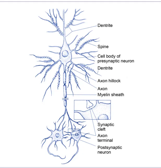
Three types of cells compose the nervous tissue: nerve cells, glial cells, and endothelial cells. These cells interact together within the extracellular matrix. The nerve cell, or neuron, is the functional cellular unit of the nervous system. There is a great diversity of structural and functional characteristics possessed by the various cells that are named neuron. However, they have properties related to their function in common. Neurons are polarized cells derived from epithelial origins. They are composed of dendrites, cell body and axon terminals (Figure 2). Dendrites and the cell body receive information from other neurons at specialized contact sites called synapses. The nucleus and cellular organelles reside in the cell body. The axon conducts electrical information encoded in action potentials to axon terminals.
Dendrites and neuronal cell bodies in the CNS are located in cortical areas and in nuclei located beneath the cortical surface, giving the appearance of grey matter. Regions rich in axons surrounded by cells rich in myelin form the white matter.
Figure 2. Neurons, the functional cellular unit of the nervous system. A schematic nerve cell is shown, illustrating the dendrites, cell body, and axon. The presynaptic terminals of the neuron are shown synapsing on the cell body of the postsynaptic neurons. The inset showns the spatial relation of three components of the synapse, the axon terminal, the synaptic cleft, and the cell body of the postsynaptic neuron. Adapted from [1]
4 1.1.2.2 Glial cells
Glial cells outnumber neurons in a 10:1 ratio. They are divided in two major classes: microglia and macroglia. Most microglia originate from bone marrow-derived monocytes which enter the brain during early development. They function as resident immune cells and phagocytes in the CNS, exerting a scavenger role in response to nervous system infection, trauma, and inflammation. Macroglia, including oligodendrocytes, Schwann cells, ependymal cells, and astrocytes, subserve support and nutritive functions. Oligodendrocytes form the myelin shealth around axons in the CNS. In addition to myelinization and axonal support in the peripheral nervous system, Schwann cells can promote axonal regeneration. Ependymal cells line the ventricles and the central canal in the spinal cord. Astrocytes are the most abundant cell within the CNS. Although there are many subtypes of astrocytes, the presence of a unique population of intermediate filaments enriched in glial fibrillary acidic protein is common to all astrocytes. These cells are important for structural support, homeostatic regulation of the CNS microenvironment and regulation of energy metabolism. Astrocytes processes surround many other CNS (Figure 3) such as blood vessels, implicating them in the blood-brain barrier (BBB) [2]. In the presence of CNS pathology, reactive astrocytes’ may secrete a variety of substances that can inhibit or promote axonal regeneration, brain repair, and neuronal function [2].
Figure 3. Relationships between astrocytes and other central nervous system cells. Astrocyte processes surround blood vessels (BV), synapses, nodes of Ranvier, neuronal cell bodies (N), and groups of myelinated axons (A). Myelin (m) Adapted from [2]
1.1.2.3 Endothelial cells
In the adult brain, the total surface area of microvasculature is 12m2. A BBB is present in more than 99% of the brain capillaries [2]. Brain capillary endothelial cells (ECs) are surrounded by astrocytes, pericytes, neurons, microglia and extracellular matrix. In addition to composing the BBB, they have highly specialized properties and functions. Brain capillary ECs constitute a continuous lining given the lack of fenestrations and the presence of tight junctions between each ECs [3], hindering the passage of small and large molecules between blood and brain. Also, brain capillary ECs are characterized by low pinocytic activity. Only highly lipophylic molecules can
6 relatively easily pass the BBB, other molecules depend on selective specialized transport mechanisms.
1.2 Primary brain tumors
1.2.1 Generalities
1.2.1.1 Epidemiology of primary brain tumors
Primary brain tumors arise from intrinsic cellular elements found in the CNS. In contrast, secondary tumors originate outside the nervous system and reach neural tissues either by contiguity or hematogenous spread. In the most recent compilation of primary brain and CNS tumors in the United States, the overall annual incidence rate of primary brain and CNS tumors was 11.5/100 000 persons/year. The calculated annual incidence rates for benign tumors varied from 2.1-8.9/100 000 persons/year throughout the various regions analyzed. The calculated annual incidence rates for malignant tumors varied from 5.9-7.8/100 000 persons/year [4].
Tumors most frequently occur within the brain parenchyma with 39% located in the supratentorial region [4] (Figure 4).
Figure 4. Distribution of all primary brain and central nervous system tumors by location, CBTRUS 1990-1994. CBTRUS, Central Brain Tumor Registry of the United States; not otherwise specified (NOS) [4].
The most frequently reported histopathologies are meningiomas (24.0%), glioblastomas (22.6%), and astrocytomas (13,7%, including diffuse, anaplastic, not otherwise specified) [4] (Table 1). The highest mean age at diagnosis is found in meningiomas (62 years) and glioblastomas (62 years). The lowest mean age at diagnosis is found in pilocytic astrocytomas (17 years) and medulloblastomas (14 years) [4].
8 Table 1. Distribution and incidence rate of primary brain and central nervous system tumors by histology
1.2.1.2 Classification of primary brain tumors
Many classification systems have been proposed to organize tumors of the CNS. In the nineteenth century, Cohnheim proposed that neoplasms develop from nests of embryonic cells [5]. The suffix –oma was added to the name of the cell from which the tumor was believed to originate. This cytogenetic concept was used in the first tumor classification scheme written by Bailey and Cushing [5]. They attributed a histopronostic value to each tumor depending on its cellular differentiation.
Later on, Kernohan proposed that tumor cells originate from differentiated cells that undergo anaplasia. He introduced in his classification grades of malignancy depending on their differentiation and degree of anaplasia.
The most recent classification, the 2007 World Health Organization (WHO) classification of tumours of the CNS, builds on the previous classification schemes [6]. It is based on the consensus of an international working group of pathologists and geneticists and represents the standard for the definition of brain tumors [6]. Two basic concepts are used in the WHO system to classify tumors. First, recognition of the cellular component of the tumor either by histology or immunocytochemical methods [7]. Primary CNS tumors may origin from neuroepithelial tissue, cranial and paraspinal nerves, meninges, germ cells, lymphomas and haematopoetic neoplasm, sellar region [7].
10 Second, each tumor is graded according to a scheme that is a malignancy scale, predicting the biological behavior of the neoplasm [5, 6]. Grade I is reserved for lesions with low proliferative potential and the possibility of cure following surgery. Grade II designates infiltrative lesions with low proliferative activity. Grade III is attributed to lesions with anaplastic characteristics including nuclear atypia and brisk mitotic activity. Grade IV is reserved for cytologically malignant lesions with high mitotic activity, endothelial proliferation and necrosis [6]. Adequate sampling of the tumor is important in order to determine its type and judge its malignant potential
1.2.2 Classification and grading of astrocytomas
Tumors that originate from glial cells are grouped together as gliomas. Gliomas include tumors that originate from astrocytes (astrocytomas), oligodendrocytes (oligodendrogliomas), ependymal cells (ependymomas) and choroids plexus. Furthermore, some tumors of glial origin contain more than one type of neoplastic cells and are referred to as mixed tumors (oligoastrocytomas) [6].
Numerous grading systems have been systematically evaluated and successfully applied to astrocytomas. Currently, the two most commonly used classifications are the St. Anne/Mayo, also named the Daumas-Duport system [8] and the WHO [7] grading systems. The St. Anne/Mayo classification system is restricted to fibrillary, protoplasmic, gemistocytic, anaplastic astrocytomas and glioblastomas. It assesses the presence or absence of four morphologic criteria: nuclear atypia, mitoses, endothelial proliferation, and necrosis. The summary score is translated into a grade
as follows: 0 criterion = grade 1, 1 criterion = grade 2, 2 criteria = grade 3, 3 or 4 criteria = grade 4 [8]. Grade I is attributed to diffuse astrocytoma without atypia.
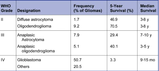
The WHO classification includes in grade I pilocytic astrocytoma and subependymal giant cell astrocytoma. The diffusely infiltrative astrocytic tumors are graded II through IV. Grade II astrocytic tumors (diffuse astrocytoma) present little cellularity and minimal pleomorphic changes. Grade III (anaplastic astrocytoma) is reserved for tumors showing moderate cellularity and pleomorphism but no necrosis. Grade IV (glioblastoma) is attributed to highly cellular tumors with nuclear and cellular pleomorphism, numerous mitotic figures, endothelial proliferation or glomeruloid microvascular proliferations and necrosis [6, 9]. Tumor grade has an important prognostic significance, influencing the choice of adjuvant therapies such as radiation and chemotherapy (Table 2) [10].
Table 2. Classification of malignant gliomas and prognostic significance
12 1.2.3. Cellular origins of gliomas
Although the WHO classification scheme implies a cell of origin for most brain tumors, that cell of origin has not been unequivocally identified for any of them. As such, astrocytomas and oligodengrogliomas are believed to arise from astrocytic or oligodendroglial precursors. Stem cells, progenitor cells and differentiated cells may acquire genetic alterations leading to the development of neoplastic cells.
Analysis of the genetic profile of both cellular components of mixed tumors oligoastrocytomas has revealed that both cell types present loss of heterozygosity of 1p and 19q, suggesting that oligodendroglial and astrocytic cells derived from a single precursor cell [11]. Similarly, genetic analysis of the glial and sarcomatous elements revealed that both components carried identical p53 mutations, suggesting a common origin of the two cellular components from a multipotent neural stem cell or an early glial progenitor cell [12].
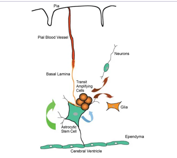
Neural stem cells have been identified in the adult CNS within its specialized microenvironment named the niche which can regulate stem cell quiescence, asymmetrical self-renewal, and differentiation into mature astrocytes and neurons (Figure 5) [13].
Figure 5. Current concept of the stem cell niche. The multipotent stem-cell like astrocytes are closely opposed to the ventricular lining and basal lamina associated with the pial microvasculature. Asymmetric division gives rise to self-renewal (green arrow) and a transient amplifying population (blue arrow). These cells can migrate out of the germinal niche and differentiate into neurons and glia (brown arrow) [13].
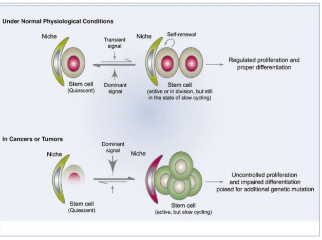
14 The occurrence of a genetic mutation in neural stem cells (NSC) or early glial progenitors could lead the cells to become independent of growth signals or to resist antigrowth signal. These cells could undergo uncontrolled proliferation and possible tumorigenesis (Figure 6) [14].
Figure 6. Comparison of the niches under normal and cancerous conditions. The stem cell niche under normal physiological conditions provides an environment that predominantly inhibits proliferation and differentiation. A transient signal is required to support ongoing tissue regeneration. In cancer, internal mutations or change in the niche’s signals results in abnormal cell proliferation and growth [14].
Brain tumor stem cells (BTSC) have been identified in human brain tumors of different phenotypes from both children and adults [15]. Studies have shown that BTSC isolated from astrocytomas, when explanted into naive mouse brains, result in the development of tumors identical to the parent tumor [16]. Isolated tumor stem cells form neurospheres, possess the capacity for self-renewal, express genes associated with NSC such as the neural precursor cell surface marker CD133, generate daughter cells of different phenotypes from one mother cell, and differentiate into the phenotypically diverse populations of cells similar to those present in the initial GBM [16]. Also it has been found that CD133(+) and CD133(-) primary glioblastoma-derived cancer stem cells show molecular differences and different biological growth pattern in vitro an in vivo [17]. This might suggest that CD133(+) and CD133(-) cancer stem cell lines might reflect two biologically different glioblastoma subtypes in primary glioblastomas [17].
Another possible source of transformed glial cell with properties resembling those of stem cells is the mature astrocyte or oligodendrocyte that may be brought to dedifferentiate in response to extrinsic signals or genetic mutations [18]. Recent studies have shown that explanted mature astrocytes isolated from embryonic cortical tissue are able to dedifferentiate in the recipient brain into an earlier glia-like phenotype and acquire proliferative and migratory capabilities that these progenitor cells possess early in CNS development [18].
16 1.3 Glioblastoma multiforme
1.3.1 Epidemiology of glioblastoma
Every year, approximately 18,000 patients are diagnosed with malignant primary brain tumors in the United States [19]. More than half of these patients have glioblastoma multiforme (GBM), making this the most common malignant brain tumor in adults [4, 19]. The incidence of GBM is highest in elderly patients, peaking at age 65 to 74 years (Figure 7) [20]. GBM seem to occur more frequently in men and Caucasians [4, 20].
Figure 7. Incidence rates of primary brain tumors by major neuroepithelial tissue and meningeal histologic types and age group. CBTRUS, 1992-1997. The astrocytoma category includes diffuse astrocytomas, anaplastic astrocytomas, unique astrocytomas and astrocytomas not otherwise specified. Adapted from [20].
1.3.2 Risk factors
1.3.2.1 Hereditary syndromes and familial aggregation
In most cases, malignant gliomas occur sporadically. However, the inheritance of certain genes may influence the risk of developing primary brain tumors. Patients with some hereditary syndromes – such as tuberous sclerosis, neurofibromatosis type 1 and 2, nevoid basal cell carcinoma syndrome, Li-Fraumeni syndrome, and syndromes involving adenomatous polyps – seem to be predisposed to malignant gliomas [20] (Table 3). Interestingly, primary brain tumors can also occur in families
18 without a known predisposing hereditary disease [21]. Environmental exposures may be important in the etiology of this familial aggregation [21].
Table 3. Principal tumor suppressor genes altered in brain tumors and involved in hereditary tumoral syndromes.
Adapted from [22]
1.3.2.2 Polymorphisms in genes relevant to cancer causation or prevention
It has been proposed that polymorphisms in genes might influence the susceptibility to brain tumors in concert with other external factors [20]. Alteration in genes involved in oxidative metabolism, detoxification of carcinogens, DNA stability and repair might confer genetic susceptibility to brain tumors [20].
1.3.2.3 Ionizing radiation
Therapeutic ionizing radiation (IR) has been recognized as a risk factor for brain tumors [20, 23, 24]. Authors observed a high prevalence (17%) of previous radiation therapy with an average dose of 48.5 Gray (Gy) and an average latency period of 15 years between initial therapy and GBM diagnosis [23]. Studies have found that radiation-induced gliomas are nearly all astrocytic in their differentiation, present as high grade lesions, and occur in a younger patient population than would be expected [24, 25]. Unfortunately, little is known regarding the susceptibility of individuals within the general population to radiation-induced tumorigenesis [26] Diagnostic radiation techniques have not been associated with an increased risk of glioma [27].
1.3.3 Clinical presentation of glioblastomas
Glioblastomas become clinically eloquent due to infiltration, compression and destruction of normal brain structures by tumor, edema, and sometimes hemorrhage. Cerebral spinal fluid flow may be compromised by the tumor, leading to further increased intracranial pressure. Most often, symptoms install progressively, over weeks to months. However, acute apoplectic clinical presentations do occur, for example following intratumoral hemorrhage or de novo seizures due to cortical stimulation. The signs and symptoms depend on the tumor’s mass effect and its location. General manifestations may occur such as mental changes, headaches, nausea, vomiting and generalized seizures. Focal manifestations include focal seizures, weakness, sensory abnormalities, speech disturbances, and visual deficits [2].
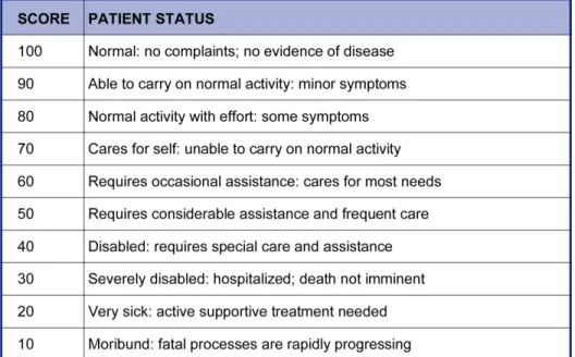
20 The clinical features at presentation may be used as a measure of the outcome of treatment. The karnofsky performance scale (KPS) is a standardized tool describing the patient’s capabilities (Table 4) [2]. Although a KPS of 70 or more at diagnosis has favourable impact on survival, clinical parameters do not fully account for the variation of survival rates [28].
Table 4. Karnofsky performance scale for brain tumor patients
1.3.4 Radiological characteristics
Magnetic resonance imaging (MRI) is more sensitive and accurate than computed tomography (CT) in studying gliomas [29, 30]. Glioblastomas typically present as irregular supratentorial white matter masses with infiltrated margins, poor demarcation and extensive edema. Necrosis, cysts and thick irregular margin are common findings [31]. Spontaneous hemorrhage can be seen and is believed to correlate with histologic grade [32]. Following contrast injection, a thick ring of enhancement surrounding central necrosis is commonly found, although a solid, nodular or patchy appearance may be visualized. The enhancement seen on MRI and CT following contrast injection correlates with the area of greatest vascularity [2, 31] (Figure 8). The aggressiveness of GBM may be suggested by its infiltration through the adjacent brain parenchyma, best approximated by the hyperintensity on T2-weighted images [33] (Figure 8). However, tumor cells are found infiltrating the brain beyond the hyperintensity signal changes in T2-weighted images when results of stereotactic biopsies are compared with MRI [33, 34]. Newer techniques are being developed to improve diagnosis accuracy, to determine the most appropriate biopsy site, and to evaluate tumor recurrence [31] such as MRI perfusion, magnetic resonance spectroscopy, positron emission tomography, diffusion tensor imaging.
22
Figure 8. Characteristic radiological features of glioblastoma.
A) Axial contrast enhanced CT shows strong, heterogeneous, irregular rim enhancement surrounding a hypodense necrotic core. B) Axial T2 weighted image MR shows a heterogeneous mass with surrounding hyperintense signal compatible with tumor infiltration and vasogenic edema. C) Axial T1 weighted image MR with gadolineum demonstrating a hazy, heterogeneous peripheral enhancement.
The most common differential diagnosis include brain metastasis, enhancing anaplastic astrocytoma, primary CNS lymphoma, and non-neoplastic disorders such as an abscess, multiple sclerosis, cerebral infarction, vascular malformation [31]. The final diagnosis can be confirmed only by histopathological examination.
1.3.5 Histological characteristics
Intraneoplasm and interneoplasm heterogeneity is characteristic of GBM. Although some areas have the characteristics of a low grade astrocytoma or anaplastic astrocytoma, the significant amount of microvascular proliferation and the presence of necrosis are key features distinguishing GBM from grade 2 and 3 astrocytomas [9].
The cellular morphology of GBM is highly variable. A spectrum ranging from small tightly packed, round or elongated cells to giant elements with significant nuclear atypia can be encountered within the same tumor. GBM are highly cellular tumors with cellular pleomorphism and numerous mitotic figures with corresponding elevated proliferation marker indexes. Endothelial proliferation or glomeruloid microvascular proliferations are highly suggestive of GBM. Necrosis on the other hand is characteristic of grade IV astrocytomas as it results of a pathological tumor microvasculature [9] (Figure 9).
GBM cells tend to infiltrate adjacent brain by spreading along white matter tracts, through the adjacent extracellular matrix or along the basement membranes [2]. Therefore islets of GBM cells can be found at distance from the primary tumor site.
24
Figure 9. Characteristic histopathological features of glioblastomas.
A) Cellular pleomorphism seen as variable nuclear:cytoplasmic ratio. B) Neovascularisation seen as buds of capillary endothelial proliferation among neoplastic cells. C) Pseudopalisading of cells around necrotic areas. Adapted from [9].
1.3.6 Primary and secondary glioblastomas
GBM seem to develop from one of two ways. The majority of cases (>90%) are primary glioblastomas that develop rapidly de novo from glial cells without clinical or histological evidence of a less malignant precursor lesion. Typically, they become clinically manifest within less than 6 months of diagnosis. Approximately 50% of patients with primary GBM have a clinical history of less than 3 months [35]. Primary GBM is most common in older patients [36]. Secondary GBM develops over months to years from pre-existing low-grade astrocytomas [37]. Neuroimaging or histologic evidence of evolution from a less malignant precursor lesion is mandatory to diagnose secondary GBM. This subtype of GBM predominantly affects younger patients, average age at diagnosis is 39 years [36].
Primary and secondary GBM represent distinct entities that arise through different genetic pathways. The development of primary GBM is associated with over-expression or amplification of the epidermal growth factor (EGF) gene. Additional genetic alterations leading to this subtype of GBM include amplification or over-expression of murine double minute 2 (MDM2), loss of heterozygosity (LOH) 10q, deletion or mutation of the phosphatase and tensin homolog deleted from chromosome 10 (PTEN), p16(INK4a) deletion [37]. In the pathway to secondary glioblastoma, p53 mutations are the most frequent and earliest detectable genetic alteration, already present more than 60% of precursor low-grade astrocytomas. It is also associated with overexpression of platelet-derived growth factor (PDGF) ligands and receptors and LOH 10q [35, 37, 38] (Figure 10). Although histological characteristics of primary and secondary glioblastomas are reported identical [39],
26 differences in their pattern of promoter methylation and in expression profiles at RNA and protein levels seem to have implications for therapeutic options [37].
Figure 10. Formation of primary and secondary glioblastoma multiform (GBM). Multiple genetic changes are involved in the development of primary and secondary glioblastomas.Abbreviation: DCC, deleted in colorectal cancer; EGFR, epidermal growth factor receptor; LOH, loss of heterozygosity; MDM2, murine double minute 2; PDGF, platelet-derived growth factor; PTEN, phosphate and tensin homolog deleted on chromosome 10; RB1, retinoblastoma 1 gene; WHO, World Health Organization. Adapted from [38]
1.3.7 Overview of altered molecular pathways in glioblastoma
1.3.7.1 Signal transduction
Altered signal transduction may contribute to gliomatogenesis. Critical signals are initiated in GBM by members of the tyrosine kinase receptor family involving, for instance, EGF receptor (EGFR), PDGF receptor (PDGFR), basic fibroblast growth factor receptor (bFGFR). Increased production of the growth factor or increased production or activity of the growth factor receptor may occur through various mutations. As a result, downstream pathways exhibit increased activity such as increased PI3K, Akt, or Ras which may contribute to tumor formation and tumor progression [40]. Also, mutations of tumor suppressor genes may result in an oncogenic signal. For example loss of PTEN function in glioma cells may participate in astrocytic tumor pathogenesis and progression through activation of the prosurvival PI3K/Akt pathway [39, 40].
1.3.7.2 Cell cycle
In glioblastoma, regulatory controls of the cell cycle are often disrupted, leading to uncontrolled proliferation of transformed cells. The Rb and p53 pathways normally maintain cells in G1 arrest. Other cell cycle regulators such as p21/WAF1/Cip1 and p27/Kip1 also regulate the progression from the G1 to the S phase. Loss of function of such regulators promotes accelerated growth and malignant transformation in astrocytes [2, 41].
28 1.3.7.3 Programmed cell death
Apoptosis, a specialized mechanism of cell death, is an important mechanism for maintaining genetic stability. If damage is beyond repair, p53 activates a series of pro-apoptotic members. In glioblastoma cells, altered signal transduction pathways modify the balance between pro-apoptotic and anti-apoptotic activities. An example of such a protein is Survivin, an inhibitor of apoptosis protein (IAP), that has been reported to increase survival in GBM by suppressing caspase-mediated apoptosis [42-44]. Survivin can bind specifically to the terminal effector cell death proteases, caspase-3 and caspase-7 [45]. Studies with a transgenic mouse model of transgenic expression of Survivin in the skin have shown that this IAP can also be antiapoptotic by inhibiting the intrinsic caspase-9-dependent pathway [46]. Recently Survivin has been recognized to also inhibit cell death in various cell lines through a caspase-independent pathway although the mechanisms have not yet been elucidated [47, 48]. In addition to being involved in the modulation of apoptosis[42-44, 49]. Survivin is implicated in the regulation of cell growth [42], in the regulation of mitotic events such as chromosomal segregation and cytokinesis [50, 51], and in the process of angiogenesis [52, 53].
Other types of cell death less well studied in glioblastoma possibly contribute to the survival versus death balance such as autophagy, necrosis, and mitotic catastrophe [54]. In the context of cancer therapy, senescence consisting in permanent growth arrest, is also considered a type of cell death. The biological and morphological characteristics of each cell death pathway are presented in table 5.
Table 5. Cell death pathway characteristics
Adapted from [54]. Abbreviations: ER, endoplasmic reticulum; HDGF, hepatoma-derived growth factor; HMGB1, high-mobility group box 1 protein; LC3, microtubule-associated protein 1, light chain 3.
30 1.4 Angiogenesis
In addition to their agressive character, malignant gliomas are also recognized as the most intensively vascularized solid tumors [2]. The process of angiogenesis is essential for tumor progression. Indeed, malignant gliomas become more angiogenic with increasing tumor grade, suggesting that the vascular component plays an important role in their malignant progression [55].
1.4.1 Distinction between vasculogenesis and angiogenesis
New vessel formation occurs via two distinct processes: vasculogenesis and angiogenesis [56]. Vasculogenesis is defined as the formation of blood vessels from endothelial-cell precursors, the angioblasts. During embryologic and fetal development, angioblasts arise in the mesoderm and differentiate into endothelial cells that proliferate to form a primitive vascular network in an avascular tissue [56, 57]. In the postnatal period, circulating bone marrow-derived endothelial progenitor cells (EPCs) may home to sites of physiological and pathological neovascularisation [58, 59]. Postnatal vasculogenesis is subject to regulation by many factors, including cytokines such as vascular endothelial growth factor (VEGF) and growth factors such as PDGF [59].
The primitive vascular network is modified by the process of angiogenesis leading to the formation of new vascular segments. Normal angiogenesis occurs in physiological conditions such as wound healing and female reproduction system [56]. Aberrant angiogenesis is present in pathological non-malignant conditions such as ischemia and inflammatory reactions as well as in malignancies [56].
1.4.2 Steps in angiogenesis and implicated molecular players 1.4.2.1 The angiogenic switch
Initially, it was proposed that tumors exist in two phases: avascular and vascular. In the avascular phase, solid tumors of 2-3 mm3 or less obtain the necessary oxygen and nutrient supplies required for growth and survival and eliminate metabolic waste products by simple passive diffusion. However, tumors larger than 2mm3 require blood supply for further growth. This concept has been challenged by the discovery of a subset of tumors that initiate growth by co-opting existing host vessels. Co-opted vessels overexpress angiopoietin-2 (Ang-2) which leads to vessel regression [60]. Vessel collapse and increased tumor growth occurs when anaplastic astrocytomas progress to glioblastoma. Hypoxic conditions induce the activation of a inducible factor (HIF-1). This transcription factor binds to hypoxia-responsive elements and induces the transcription of many angiogenic factors such as VEGF, PDGF, Angiopoietin (Ang)-1 and -2 [61], stem cell factor (SCF) [62]. In addition, hypoxia increases VEGF messenger RNA (mRNA) stability through binding of several RNA-binding proteins [56]. Hypoxia and subsequent necrosis is pathognomonic of glioblastoma and is the principal stimulus for new vessels formation. Interestingly, high SCF expression has been documented in glioma cells but also in normal host neurons, possibly in response to the glioma-induced damage in normal brain parenchyma [62]. The areas of neuronal and glioma SCF overexpression correspond to areas of sprouting angiogenesis. Therefore, normal brain cells may also participate to induce pathological angiogenesis and support tumor growth and infiltration [62]. Activation of SCF/c-Kit signalling pathway in ECs has been found to
32 enhance ECs’ proliferation, survival and migration, even in the absence of growth factors believed to be obligate such as VEGF [62]. Most probably, various pro-angiogenic factors have complementary roles in tumors with significant angiogenesis such as gliomas.
In all circumstances, tumor progression requires the induction of a tumor vasculature, termed the angiogenic switch [63]. The angiogenic switch can occur at different stages of the tumor-progression pathway, depending on the tumor type and its environment [57]. Although angiogenesis is necessary, it is not in itself sufficient for tumor growth [63]. Induction of the angiogenic switch depends on the balance between angiogenic stimulators and inhibitors, termed the angiogenic balance (Figure 11) [63].
Figure 11. The angiogenic balance. Angiogenesis is orchestrated by a variety of activators and inhibitors – only a few of which are listed above. Activators are mainly receptor tyrosine kynase ligands such as vascular endothelial growth factor (VEGF), fibroblast growth factors (FGFs), platelet-derived growth factor (PDGF) and epidermal growth factor (EGF), but can also be of various origin such as lysophosphatic acid (LPA). Inhibitors include thrombospondin-1 and the statins. Adapted from [57].
Expression of such regulators may be in response to physiological stimuli, such as hypoxia, resulting from increased tissue mass, and also to oncogene activation or tumor suppressor mutation in tumor cells [57]. Tumor cells may upregulate expression of angiogenic activators or downregulate expression of angiogenic inhibitors, mobilize angiogenic proteins from the extracellular matter, recruit host cells such as macrophages which produce their own angiogenic proteins [63].
34 1.4.2.2 Mechanism of sprouting angiogenesis
Angiogenesis may occur via sprouting and non-sprouting mechanisms. Sprouting angiogenesis is a multi-step process involving interplay between cells, soluble factors, and extracellular matrix components. Angiogenic activators such as VEGF, placental growth factor and Ang-1 may initiate angiogenesis [57]. Angiogenesis begins with vasodilation and increased vascular permeability in response to VEGF and loosening of pericytes covering host vessels. The vascular basement membrane and extracellular matrix are locally degraded by proteolytic enzymes such as cathepsin B and matrix metalloproteinases (MMPs) [57]. Endothelial cells may migrate into the interstitial space towards chemotactic angiogenic stimuli. Endothelial cells proliferate at the migrating tip, forming a solid vessel sprout. The sphingolipid sphingosine-1-phosphate (S1P) and S1P(3) receptors enhance ECs proliferation and migration, playing a key role in angiogenesis [64]. Endothelial cells change shape and adhere to each other to form a lumen. A new basement membrane is produced around the newly formed blood vessel with recruitment of pericytes. Finally vascular sprouts fuse with other sprouts to form loops and blood may flow in the newly vascularized area [57].
1.4.3 Tumor vasculature
1.4.3.1 Types of tumor angiogenesis
Tumor vasculature is not necessarily derived from sprouting angiogenesis: it can also occur through non-sprouting processes [65]. Cancer tissue can acquire its vasculature by intussusceptive angiogenesis, recruitement of circulating endothelial precursor cells, co-option of pre-existing vessels, mosaic vessel formation, and vasculogenic mimicry [65] (Figure 12).
Figure 12. Mechanisms of tumor angiogenesis. The mechanisms of tumor angiogenesis are: A) Sprouting; B) Intussuceptive angiogenesis; C) Recruitment of precursor cells; D) Vessel co-option; E) Mosaic vessel formation; F) Vasculogenic mimicry. Adapted from [65].
36 Intussusceptive angiogenesis refers to vessel network formation by insertion of connective tissue columns, called tissue pillars into the vessel lumen and to subsequent growth of these pillars, resulting in partitioning of the vessel lumen [66]. Vessel co-option may occur as cancer cells proliferate along pre-existing microvessels, without a tumor capsule, eliciting an invasive character [57, 60, 66]. Mosaic vessels refers to the presence of tumor cells within the walls of tumor vasculature. Vasculogenic mimicry may contribute to tumor angiogenesis in various malignancies such as melanomas, breast, prostate and lung cancers. These cancers have the ability to express an endothelial cell phenotype and to form three dimentional vessel-like networks, mimicking the pattern of embryonic vascular networks [67, 68].
Astrocytomas also feature another mechanism of tumor angiogenesis named glomeruloid angiogenesis. In fact, glomeruloid bodies are best known in high grade astrocytomas where they are one of the diagnostic histopathological features of GBM [69]. These bodies are tufted collections of newly formed microvessels surrounded by variably thickened basement membrane with an incomplete layer of pericytes [66, 69]. It has been proposed that hypoxic astrocytoma cells in areas surrounding central necrosis up-regulate the expression and secretion of VEGF, which induces vascular hyperplasia of nearby vessels that develops in glomeruloid bodies (Figure 13) [70].
Figure 13. Glomeruloid vascular proliferation in glioblastoma multiform. Hypoxic astrocytoma cells in zones surrounding central necrosis up-regulate the expression and secretion of VEGF, which acts on nearby vessels to cause vascular hyperplasia, inducing glomeruloid vascular proliferation. Adapted from [70].
1.4.3.2 Macroscopic and microscopic characteristics of tumor blood vessels
The appropriate balance between activators and inhibitors is lost in tumor angiogenesis, resulting in extensive blood vessel growth and failure to mature. Histological examination of glioblastoma tissue reveals an architecturally specific vasculature, different from normal brain counterparts. Macroscopically, two main types of vascular patterns have been described in glioblastoma [71, 72]. The glomeruloid/garland like type, which refers to glomeruloid angiogenesis, is characterized by unevenly distributed vascular formations. The ‘classic’ capillary like
38 vascular pattern shows irregularly shaped, dilated and tortuous vessels, with occasional dead ends [57]. Tumor blood vessels share characteristics of venules, arterioles and capillaries. Blood flow is irregular in tumor vessels, moving slowly, sometimes oscillating and even flowing in reverse sense [73]. The vascular network is often leaky and hemorrhagic [74]. The perivascular cells that are usually in contact with endothelial cells are more loosely apposed and less abundant than in normal vasculature [57]. Endothelial cells derived from GBM have a flat appearance, with large nuclei, abundant cytoplasm and multiple nucleoli [75]. Normal brain endothelial cells are smaller, with limited cytoplasm [76].
It has been proposed that vascular patterns influence clinical outcome of patients with astroglial brain tumors [77]. However, other authors observed that poor observer agreement on vascular patterns in patients with glioblastoma limits the clinical utility of these factors [78]. Improved methodologies for morphologic assessment of glioblastoma vascularization need to be identified. In addition, regional tumor heterogeneity may limit the clinical relevance of these histopathological assessments [79].
1.4.3.3 Cellular characteristics of brain tumor endothelial cells
GBM-associated ECs express typical endothelial markers such as vWF, CD105, CD31, similarly to normal ECs. For some markers, the level of expression and the distribution of expression varies between studies given the heterogeneity of ECs sampled [75]. For instance, the expression of CD34, a marker for EPCs, has been reported elevated, similar, or decreased in comparison to control ECs [75]. Expression
of CD144 (VE-cadherin), a tight junction protein, is reduced in GBM-associated ECs [76]. This may contribute to the leakiness of GBM’s vessels [80]. The reduced expression of other tight junction proteins in tumor microvessels such as claudin-1, claudin-5, and occludin may also contribute to the tumor’s leakiness [81]. Interestingly, 50% of GBM-associated ECs express α-SMA, a cytosqueletal protein implicated in initiation of cell contraction that is mostly expressed by mural cells and absent from control ECs [75]. Its expression may be related to tumor-associated ECs’ enhanced migratory potential.
Although some authors have found that GBM endothelial cells proliferate faster than normal ECs [80], others have documented a slower rate of replication [75, 76]. This discrepancy might be explained by tumor vasculature heterogeneity: endothelial cells isolated from the periphery of a tumor may proliferate more than those obtained close to hypoxic or necrotic regions [75]. Furthermore, glioblastoma-derived ECs were found to migrate more than those in normal brain, based on results of modified Boyden chamber migration assays. [76]. Glioblastoma-derived ECs also have enhanced survival properties. Authors have documented that these cells undergo less apoptosis after serum starvation [76]. Migration activity and proliferative and apoptotic activities might be mutually exclusive behaviours in tumor ECs, as was proposed for astrocytoma cells [82].
Tumor endothelial cells also produce angiogenic growth factors [83]. Studies have documented an increased expression of VEGF, endothelin-1 (ET-1) and interleukin-8 (IL-8) in comparison to normal counterparts [76, 83]. Also, the expression of VEGF receptors VEGFR-1 (Flt-1) and VEGFR-2 (Flk-1/KDR), as well
40 as that of IL-8 receptors CXCR1 and CXCR2, was documented on GBM-derived endothelial cells [83]. Endothelial cells derived from GBM maintain angiogenic properties, they have the capacity to form tubules [84].
1.4.4 Angiogenesis as a therapeutic target
Given the requirement of angiogenesis for growth and progression of tumors, the tumor vasculature is an attractive target for tumor therapy. Therapeutic vascular targeting is of two types: anti-antiangiogenic approaches which aims to prevent neovascularization in tumors and vascular disrupting approaches which aims to disrupt established tumor vasculature [85], both approaches culminate in tumor cell death.
Many targets have been explored and the VEGF/VEGFR-2 signalling has been strongly suggested as the primary target [86]. However, studies have shown that targeting only one signalling pathway may result in the activation of alternative pro-angiogenic pathways [86]. Therefore, targeting multiple pro-angiogenic signalling pathways by polyvalent inhibitors or combinations of agents with distinct antiangiogenic properties has been attempted to optimize antiangiogenic agents [86].
1.5 Therapeutic management of glioblastomas
1.5.1 Surgery
Depending on the clinical state of the patient and the radiological characteristics, surgical goals should be adjusted to each patient. Surgery should enable to obtain a tissue diagnosis. Although the diagnosis of a malignant glioma is highly suspected with the presence of contrast enhancement on CT and MRI, up to approximately 40% of malignant gliomas do not enhance following contrast injection [87]. Given that a radiological diagnosis is not reliable, a tissue sample is required in essentially all cases [88]. For patients in whom resection is not possible because of advanced age, multiple or severe comorbidities, tumor location and/or extent, a biopsy should be performed to determine the histology of the tumor [88]. Biopsies may be performed either openly, through a mini-craniotomy or through a burr hole with stereotaxy or neuronavigation guidance.
When surgical resection is possible, total tumor resection should be the goal. Decreasing local tumor mass can improve neurological function, reduce steroid dependence, and prevent early death [2]. Decrease in tumor burden has been reported to be a significant prognostic indicator for survival. In deed, gross-total tumor resection is associated with longer survival in patients with GBM. Functional mapping has been used to maximize the extent of tumor removal and avoid injury to cortex essential for language, motor and sensory functions. Intraoperative magnetic resonance guidance, fluorescence-guided surgery, and neuronavigation systems have been used to achieve a more complete removal of deep-seated tumor than with