Cyclodextrins as a potential carrier in drug nebulization
B. Evrarda1, P. Bertholeta1, M. Guedersc, M.-P. Flamentb,G. Piela,L. Delattrea, A. Gayotb, P. Letermeb, J.-M.
Foidartc, D. Cataldoc
aLaboratory of Pharmaceutical Technology, Department of Pharmacy, University of Liege, CHU-Tour 4, Bat B36, Avenue de V Hδpital 1, 4000, Liege 1, Belgium
bLabor atoire de Pharmacotechnie Industrielle, Faculte des Sciences Pharmaceutiques et Biologiques, Rue du Professeur Laguesse, BP 83, F-59006 Lille, France
cDepartment of Tumors and Developmental Biology, University of Liège, Avenue de I'Hôpital 1,4000, Liege 1, Belgium
Abstract
The inhalation route is widely studied for many drug applications focusing on either local or systemic distributions. One matter of concern is the solubilization of hydrophobic drugs.
We have studied the feasibility of using different cyclodextrins (CDs) to elaborate pharmaceutical formulations for the inhalation route and tested the short-term toxicity of such formulations administered by inhalation to C57BL/6 mice.
We have shown that HP-β-CD, γ-CD, as well as RAMEB aqueous solutions can undergo aerosolization and that the resulting droplet-size ranges are compatible with pulmonary deposition. In vivo, we have demonstrated that short-term exposure to inhaled HP-β-CD, γ-CD and RAMEB solutions are non-toxic after assessing
bronchoalveolar lavage (BAL), lung and kidney histology, bronchial responsiveness to methacholine and blood urea. The only change noted is a slight increase in lymphocyte count in the BAL after HP-β-CD and γ-CD inhalation.
We conclude that CDs are useful in significantly enhancing the solubility of apolar drugs with a view to inhalation therapy although an increase in lymphocyte counts in the BAL after CDs inhalations needs further investigations.
Keywords: Cyclodextrins; Drug delivery; Inhalation therapy; Lung
1. INTRODUCTION
The respiratory tract has become an increasingly attractive route of application for a wide range of active substances with either topic or systemic pharmacological activity and the trend towards a systemic application of drug compounds via the lung is rapidly increasing [1]. New drug delivery systems, such as microparticles, nanoparticles or liposomes, may enhance the systemic bioavailability of substances applied to the lung by offering relative protection against degrading factors, as well as by exhibiting controlled release properties [2]. Recently, pulmonary delivery has emerged as a possibility for non-invasive delivery of protein and
macromolecular drugs. A major obstacle to the widespread use of pulmonary drug delivery is the relative impermeability of the lung to many of these peptide and macromolecular drugs when they are administered without an absorption enhancer. Cyclodextrins (CDs) have been reported as potential absorption enhancer in pulmonary protein delivery [3],
Cyclodextrins are cyclic oligosaccharides consisting of covalently linked glucopyranose rings. α-, β-, and γ-CDs are naturally occurring CDs combining six, seven or eight glucopyranose units. The CDs and their hydrophilic derivatives are used in pharmaceutical formulations to enhance solubility, dissolution rate, stability and bioavailability. The fact that they enhance solubility may be explained by the formation of water-soluble inclusion complexes in which the hollow, truncated, cone-like CD structure encapsulates hydrophobic drug molecules in the apolar interior. In contrast, the outer, hydrophilic region of the CD induces solubilization through interaction with water molecules [4]. Little has yet been published concerning the pulmonary
applications of cyclodextrins [5-8]. It was found that hydrophilic cyclodextrin derivatives could be used in liquid formulation to be administered by inhalation thus increasing drugs solubility, but very little data was given as to
the potential toxicity of CD formulations on the lung parenchyma and bronchial tree. It is generally accepted that aerosol particles of 0.5-5 µm are required for deposition in the alveolar region of the lung. The primary factors influencing aerosol particle size and, ultimately, the site of aerosol deposition include the design of the inhalation device, as well as the physico-chemical properties of the formulation [9]. Both the viscosity of the solution and surface tension characteristics play an important part in the formation of the droplets during nebuliza-tion [10,11]. To date, there are no data available regarding the characterization of aerosols obtained with such cyclodextrins solutions.
To determine the potential advantage of using CD-drugs complexes for pulmonary administration, several CDs have been considered in this work for their ability to form respirable aerosol droplets from liquid formulation containing different concentrations of CD. Among the hydrophilic CDs, 2-hydroxypropyl-β-cyclodextrin (HP-β-CD), methylated-β-cyclodextrin (RAMEB) and gamma-cyclodextrin (γ-CD) appear to be especially useful — considering their safety profiles in humans [12]. In this work, we also address the potential lung toxicity of cyclodextrins inhalation in vivo, by administering inhalations of different CDs at various dosages to mice. In this way, bronchial responsiveness, airway inflammation and histological changes, blood urea and renal histology can be assessed.
2. MATERIALS AND METHODS 2.1. Materials
HP-β-CD (degree of substitution = 0.64) was a gift from Roquette (France). Randomly methylated-β-CD (RAMEB) and γ-CD were generously given by Wacker Chemie (Germany). Apyrogenic phosphate-buffered saline (PBS) was purchased from Bio-Wit-taker (Verviers, Belgium). Methacholine was from Sigma-Aldrich (Germany).
All other materials were of analytical grade. Sterile water for injection was used throughout this study. Sterile, apyrogenic and isotonic CD solutions were prepared at 20, 50 and 75 mM for HP-β-CD and RAMEB and at 20, 50 and 100 mM for γ-CD. Cyclodextrins were tested following the Bacterial Endotoxin Test described in USP XXVI using Limulus Amebocyte Lysate (LAL). Osmolalities of all the solutions were measured by a Knauer Automatic semi-micro Osmometer and adjusted to the value of 300 mOsm/kg by the addition of an adequate amount of NaCl. A terminal sterilization of the solutions was performed by steam sterilization process.
2.2. Surface tension, density, and viscosity measurements
The surface tension measurement method consists in measuring the force that has to be exerted on a
platine/iridium stirrup piece, which is in contact with the solution surface, to stretch the interfacial liquid film. Surface tension measurements were made with a Lauda TD1 tensiometer (Prolabo, Paris, France), the measuring range of which is 0-100 mN/m, precision 0.1 mN/m and sensitivity 0.001 mN/m. All experiments were carried out at 25 °C
The density of each solution was measured by using a densitimeter Anton Paar type DMA45A Viscosity of the solutions, the behavior of which is Newtonian, was measured with a capillary viscosimeter following the method that is described in the European Pharmacopeia (Micro Ostwald viscosimeter for the determination of kinematics viscosity, Schott-Gerate, Germany). All experiments were carried out at 25 °C.
2.3. Production of the aerosol
The aerosol was produced by using an ultrasonic nebulizer SYSTAM (Système Assistance Medical, Le Ledat, France), the vibration frequency of which is 2.4 MHz with variable vibration intensity and ventilation levels. Vibration intensity was fixed in position 6 and the ventilation level was 25(v1/2) l/min. Those conditions were
used for the characterization of the aerosols as well as for the animal exposure.
2.4. Characterization of nebulized aerosol
Aerosol size distribution emitted from CDs solutions was determined with a laser size analyzer Mastersizer (Malvern, Orsay, France). Ten milliliters of each solution was directly nebulized in the laser beam. The mouthpiece was held at 1 cm from the center of the laser beam. The resulting aerosol was aspirated on the opposite side of the beam. Environmental temperature and relative humidity were maintained constant, that is to say at 20 °C and 40-45%. Triplicates of each measurement were performed and compared to controls of PBS.
The results are expressed as the percentage of droplets comprised in the range 0.5-5.79 µm and the median diameter. The concentration of droplets in the air evaluated by the obscuration percentage of the laser beam was in the same range for each experiment (15-25%). The mass loss after nebulization was also determined after 5 min of nebulization and was used as a measure of the nebulization rate.
2.5. Animals and in vivo exposure
Four groups of eight C57BL/6 mice have been tested for each investigated CDs. Three of the groups were exposed daily for 7 days to the inhalation of one CD concentration dispensed by the ultrasonic nebulizer, while the fourth received a placebo solution (PBS). The duration of inhalation was set at 30 min/day and the mice were exposed to aerosols in a Plexiglas exposure chamber (30 x 20 x 15 cm). The estimated amount of inhaled cyclodextrin was ranging from 1.3 to 4.1 µg/min/kg of body weight.
2.6. Airway responsiveness measurement
Twenty-four hours after the final exposure, bronchial hyperresponsiveness was determined by measuring the Penh using a barometric plethysmograph as proposed by Hamelmann et al. [13]. The Penh was measured at baseline and 5 min after the inhalation of increasing doses (25, 50, 75 and 100 mM) of methacholine (Mch).
2.7. Bronchoalveolar lavage (BAL) and histology
Immediately after the assessment of airway responsiveness, mice were sacrificed and 1 ml of PBS free of ionised calcium and magnesium but supplemented with 0.05 mM sodium EDTA was instilled four times via a tracheal cannula and recovered by gentle manual aspiration. The recovered bronchoalveolar lavage fluid (BALF) was centrifuged (1800 rpm for 10 min at 4 °C). The supernatant was processed for protein assessments whereas the cell pellet was washed twice and finally resuspended in 1 ml of PBS. A total cell count was performed in a Thoma chamber and the differential cell counts on at least 400 cells were performed on cytocentrifuged preparations (Cytospin 2; Cytospin, Shandon td., Runcorn, Cheshire, UK) using standard morphologic criteria after staining with Diff-Quick (Dade, Germany). After BAL, the thorax was opened and the left main bronchus was clamped. The left lung was excised and frozen immediately in liquid N2 for protein chemistry and mRNA
extraction while the right lung was processed for histology. As previously described [14], the right lung was infused with 4% paraformaldehyde and embedded in paraffin. Sections of 4-µm thickness from all lobes were stained with haematoxylin and eosin. The extent of peribronchial infiltrates was estimated by an inflammation score. Slides were coded and the peribronchial inflammation was graded in a blinded fashion using a
reproducible scoring system described elsewhere [14]. A value from 0 to 3 per criteria was adjudged to each tissue section scored. A value of 0 was adjudged when no inflammation was detectable, a value of 1 for
occasional cuffing with inflammatory cells, a value of 2 when most bronchi were surrounded by a thin layer (1 to 5 cells) of inflammatory cells and a value of 3 when most bronchi were surrounded by a thick layer (>5 cells) of inflammatory cells. As 5-7 randomly selected tissue sections per mouse were scored, inflammation scores could be expressed as a mean value per animal and could be compared between groups. The left lung was snap frozen in liquid nitrogen and crushed using a Mikro-Dismembrator S (Braun Biotech International, Melsungen, Germany). Kidneys were excised and paraffin embedded, sections of 4 µm were stained by haematoxylin and eosin. Blood was sampled by cardiac puncture and serum was stored at -80°C until analyses were performed. Serum urea was measured using a spectrophotometer measuring the absorbance of the samples at 520 nM after adding an aqueous solution of diacetylmonoxime, thiosemicarbazide, sulfuric acid, phosphoric acid as previously described [15].
All in vivo manipulations were approved by the local Veterinarian Ethics Committee.
3. RESULTS
3.1. Determination of the physico-chemical characteristics of the solution
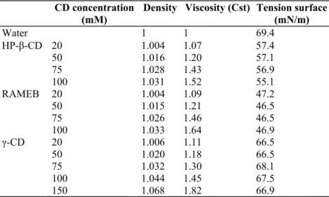
The measured density values in the CDs concentration range tested were close to that of water while an increase in the viscosity of the solutions was related to the increase of in CDs concentration. Table 1 shows the density, the viscosity and the surface tension data obtained for water and solutions of HP-β-CD, RAMEB and γ-CD at five different concentration levels. At the same concentration, γ-CD solutions displayed lower viscosity than the β-CD derivatives. It confirms the results reported by Müller et al. [16] and Thompson [17] which showed that the substitution of native CDs reduces the surface activity of the molecule and that the methylated compounds show the greatest lowering.
The influence of the CDs concentration on the surface tension was negligible in the range of the CDs concentrations studied in the present work.
3.2. Influence of CDs concentration on droplets size
The CDs solutions density close to 1 g/cm3 and the spherical shape of the droplets measured meant that the
percentage of droplets in the range of 0.5-5.79 µm could be assimilate to respirable fraction (RF) and median diameter to mass median aerodynamic diameter (MMAD) [18,19]. Table 2 shows the results for the percentage of droplets comprised in the range of 0.5-5.79 µm, the median diameter and the geometric standard deviation (GSD). A comparison of the MMAD, the GSD and the percentage of droplets comprised in the range of 0.5-5.79 µm of all the CDs solutions with the corresponding values for PBS demonstrated that the presence of CDs in the solution did not influence the droplet size distribution in the aerosols. A fraction of droplets comprised in the range of 0.5-5.79 µm close to 65% was obtained in each experiment.
Table 1: Characteristics of aqueous solutions of different CDs CD concentration
(mM) Density Viscosity (Cst) Tension surface(mN/m)
Water 1 1 69.4 HP-β-CD 20 1.004 1.07 57.4 50 1.016 1.20 57.1 75 1.028 1.43 56.9 100 1.031 1.52 55.1 RAMEB 20 1.004 1.09 47.2 50 1.015 1.21 46.5 75 1.026 1.46 46.5 100 1.033 1.64 46.9 γ-CD 20 1.006 1.11 66.5 50 1.020 1.18 66.5 75 1.032 1.30 68.1 100 1.044 1.45 67.5 150 1.068 1.82 66.9
Table 2: Percentage of droplets in the range of 0.5-5.79 µm, mass median aerodynamic diameter (MMAD) and geometric standard deviation (GSD)
Sol. Concentration of CD
(mM) % of droplets (0.5-5.79 µm) MMAD GSD
Mean S.D. Mean S.D. Mean S.D.
Water 0 58.58 1.03 5.75 0.16 1.62 0.03 HPBCD 0 58.58 1.03 5.75 0.16 1.62 0.03 20 68.11 0.95 5.55 0.56 1.40 0.03 50 67.31 0.74 5.46 0.30 1.39 0.16 75 62.35 0.36 5.83 0.05 1.67 0.01 100 63.56 0.39 5.70 0.08 1.64 0.01 RAMEB 0 58.58 1.03 5.75 0.16 1.62 0.03 20 66.38 1.00 5.31 0.07 1.30 0.03 50 68.72 0.44 5.22 0.06 1.34 0.02 75 70.68 2.55 5.50 0.78 1.37 0.10 100 69.11 2.38 5.76 1.04 1.55 0.14 Gamma 0 58.58 1.03 5.75 0.16 1.62 0.03 20 58.91 0.35 6.00 0.11 1.69 0.02 50 64.34 3.41 5.70 0.23 1.66 0.07 75 70.70 3.06 5.03 0.20 1.35 0.09 100 69.39 0.58 5.14 0.08 1.48 0.02
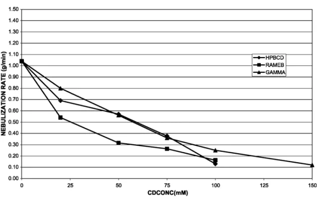
Fig. 1: Influence of CDs concentration on the nebulization rate.
3.3. Influence of CDs concentration on the rate of aerosol production
Nebulization rate decreased with the CDs concentration. This decrease was greater for RAMEB solutions displaying higher viscosity values. Fig. 1 shows nebulization rate as a function of the concentration of CDs for HP-β-CD, RAMEB and γ-CD. Solutions with a viscosity higher than 1.45 led to nebulization rates lower than 0.2 g/min resulting in aerosol's duration which are not compatible with human administration. The maximal CDs concentrations for in vivo experiments were then fixed at 75 mM for both HP-β-CD and RAMEB and to 100 mM for γ-CD.
3.4. In vivo evaluation 3.4.1. BAL cellularity
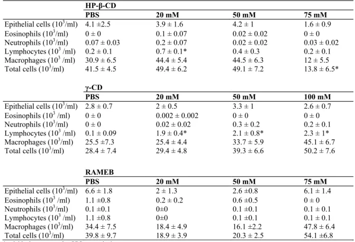
When compared to PBS exposed control mice there, was a slight decrease in total number of cells recovered by BAL in the HP-β-CD-treated group only at the dose of 75 mM (p<0.05) (Table 3). The number of BAL epithelial cells was similar to controls in all CDs and concentrations tested, indicating that the inhalation of the
formulations did not induce any significant epithelial shedding. As regards inflammatory cells, the only
significant difference is a slight increase in the number of lymphocytes compared to controls in mice exposed to 20 mM HP-β-CD and at all doses tested for γ-CD (20, 50, 100 mM) (Table 3).
3.4.2. Histology
There were no significant changes in lung histology after a 1-week daily inhalation of HP-β-CD, γ-CD and RAMEB solutions. In particular, the integrity of the epithelium was verified by observation and the presence of inflammation was assessed by calculating an inflammation score which showed no difference between the CD-exposed mice and controls.
3.4.3. Bronchial responsiveness
Bronchial responsiveness was assessed in each mice by measurement of the Penh for 5 min after inhaling different concentrations of MCh (20, 50 and 100 mM). HP-β-CD, HP-γ-CD, and RAMEB exposure for 7 days did not significantly influence bronchial responsiveness when considering the values of the Penh or some
previously published indices proposed as indicators of bronchial responsiveness (PC200 which is the MCh concentration at which the lung resistance display a 200% increase).
3.4.4. Kidney histology
Based on morphological criteria, no difference was noted in kidney histology studied in standard light microscopy between all different groups exposed to CDs inhalation and controls.
3.4.5. Blood urea levels
In every experimental group, there were no significant differences in blood urea levels between the CD-exposed and control mice.
Table 3: BAL cell counts after placebo (PBS) or CDs exposure (n = 8 per experimental condition) HP-β-CD PBS 20 mM 50 mM 75 mM Epithelial cells (103/ml) 4.1 ±2.5 3.9 ± 1.6 4.2 ± 1 1.6 ± 0.9 Eosinophils (103/ml) 0 ± 0 0.1 ± 0.07 0.02 ± 0.02 0 ± 0 Neutrophils (103/ml) 0.07 ± 0.03 0.2 ± 0.07 0.02 ± 0.02 0.03 ± 0.02 Lymphocytes (103 /ml) 0.2 ± 0.1 0.7 ± 0.1* 0.4 ± 0.3 0.2 ± 0.1 Macrophages (103 /ml) 30.9 ± 6.5 44.4 ± 5.4 44.5 ± 6.3 12 ± 5.5 Total cells (103/ml) 41.5 ± 4.5 49.4 ± 6.2 49.1 ± 7.2 13.8 ± 6.5* γ-CD PBS 20 mM 50 mM 100 mM Epithelial cells (103/ml) 2.8 ± 0.7 2 ± 0.5 3.3 ± 1 2.6 ± 0.7 Eosinophils (103 /ml) 0 ± 0 0.002 ± 0.002 0 ± 0 0 ± 0 Neutrophils (103/ml) 0 ± 0 0.02 ± 0.02 0.3 ± 0.2 0.2 ± 0.1 Lymphocytes (103 /ml) 0.1 ± 0.09 1.9 ± 0.4* 2.1 ± 0.8* 2.3 ± 1* Macrophages (103/ml) 25.5 ±7.3 25.4 ± 4.4 33.7 ± 5.9 45.1 ± 6.7 Total cells (103/ml) 28.4 ± 7.4 29.4 ± 4.8 39.3 ± 6.6 50.2 ± 7.6 RAMEB PBS 20 mM 50 mM 75 mM Epithelial cells (103/ml) 6.6 ± 1.8 2 ± 1.3 2.6 ±0.8 6.1 ± 1.4 Eosinophils (103 /ml) 1.1 ±0.8 0.2 ± 0.2 0.6 ±0.5 0 ± 0 Neutrophils (103/ml) 0.1 ±0.1 0±0 0.1 ±0.1 0.1 ± 0.1 Lymphocytes (103 /ml) 1.1 ±0.8 0±0 0.1 ±0.1 0.1 ± 0.1 Macrophages (103/ml) 34.4 ± 7.5 18.4 ± 4.9 16.1 ±2.2 47.8 ± 6.4 Total cells (103/ml) 39.8 ± 9.7 18.9 ± 3.9 20.3 ± 2.5 54.1 ±6.8
*p<0.05 when compared to PBS exposed mice.
4. DISCUSSION
We have studied the feasibility of using CDs for the elaboration of pharmaceutical formulations intended for inhalation. We have described that HP-β-CD, γ-CD, as well as RAMEB aqueous solutions can undergo aerosolization and that the resulting droplet-size is compatible with pulmonary deposition. In vivo, we have demonstrated that inhaled HP-β-CD, γ-CD and RAMEB solutions are non toxic as assessed by the study of BAL, lung and kidney histology, bronchial responsiveness to methacholine and blood urea. The only change noted is a slight increase in lymphocyte count after HP-β-CD and γ-CD inhalation.
New vehicles are required for the pulmonary administration of drugs displaying a low aqueous solubility. Indeed, considerable efforts are being made to find ways to make use of the pulmonary route for systemic therapy of drugs such as peptides which are metabolized by the digestive tract, as well as to administer drugs such as anti-inflammatory agents and bronchodilators.
Previous authors studied the pulmonary absorption of CD (β-CD, dimethyl-β-CD (DM-β-CD) and 2-Hydroxypropyl-β-cyclodextrin (HP-β-CD)) in rabbits through a crossover pharmacokinetics study following intratracheal (IT) instillation and IV injection [6] and found that serum values after IT instillations were considerably higher than CD absorption by other non-parenteral routes indicating that toxicity at both local and systemic levels was a matter of concern. 2-Hydroxypropyl-β-cyclodextrin (HP-β-CD) was investigated for pulmonary applications of salbutamol in a dry powder formulation, in an attempt to modify its pharmacokinetics properties and a rapid dissociation of the drug from the complex was observed in vivo leading the hypothesis that CDs can promote lung delivery of drugs [5]. CDs have been evaluated as potential drug carrier in DPI formulations which are the most convenient for a daily use and indicate that formulations containing CDs enhanced drug delivery to the lower stage of the impactor. In these studies, no data are available on the potential lung toxicity of CD [8]. A very recent study showed that cyclosporin A-maltosyl-α-CD complexes were useful as therapy for allergen-induced inflammation with an apparently low toxicity [7]. Using high doses of intravenously administered HP-γ-CDs (> 600 mg/kg), the occurrence of a pulmonary accumulation of macrophages
(histiocytosis) was demonstrated [20]. In our study, the topic administration of CDs at dosages between 20 and 100 mM in the lungs did not induce any accumulation of macrophages at all as demonstrated by BAL
differential cell counts and histology. Interestingly, our results show an increase in lymphocyte counts in BAL after inhalation of HP-β-CD and HP-γ-CD. This finding could be related to interactions of CDs and some cholesterol/sphingolipid-rich plasma membrane microdomains present at the surface of lymphocytes and referred to as "lipid rafts" as demonstrated for methyl-β-CDs [21]. These lipid rafts have been demonstrated to be
regulatory elements in a wide spectrum of cellular events such as signaling and cell adhesion [21,22]. A potential interaction of CDs with regulatory elements of lymphocytes trafficking could explain the increased lymphocyte counts in the BAL of mice exposed to HP-β-CD and γ-CD. Further in vitro studies are needed to explore more in depth the potential interaction with lymphocyte functions.
In conclusion, we have demonstrated the feasibility of cyclodextrin inhalation from a technical point of view. The short-term safety of such inhaled formulations is accurate in terms of lung and renal integrity and function. An increase in lymphocyte counts in the BAL after CDs inhalations needs further investigation.
Acknowledgements
This work was financially supported by the Fonds national de la recherche scientifique (FNRS, communaute francaise de Belgique). D. Cataldo is a postdoctoral researcher from the Fonds national de la recherche scientifique (FNRS). This work was also supported by grants from the Fonds de la Recherche Scientifique Médicale, the Fonds Spéciaux de la Recherche (University of Liège), the D.G.T.R.E. from the "Région Wallonne". We acknowledge the precious technical help of Fabienne PERIN-RASQUIN.
References
[1] R.U. Agu, M.I. Ugwoke, M. Armand, R. Kinget, N. Verbeke, The lung as a route for systemic delivery of therapeutic proteins and peptides, Respir. Res. 2 (2001) 198-209.
[2] X.M. Zeng, G.P. Martin, C. Mariott, The controlled delivery of drug to the lung, Int. J. Pharm. 124 (1995) 149-164.
[3] K.H. Frömming, J. Szejtli, Cyclodextrins in Pharmacy, Kluwers Academic Publishing, Dodrecht, 1994.
[4] A. Hussain, J. Arnold, M. Khan, F. Ahsan, Absorption enhancers in pulmonary protein delivery, J. Control Release 94 (2004) 15-24.
[5] H.M. Cabral Marques, J. Hadgraft, I.W. Kellaway, G. Taylor, Studies of cyclodextrin inclusion complexes: IV. The pulmonary absorption of salbutamol from a complex with 2-hyd-roxypropyl-cyclodextrin in rabbits, Int. J. Pharm. 77 (1991) 303-307.
[6] H.M. Cabral Marques, J. Hadgraft, I.W. Kellaway, G. Taylor, Studies of cyclodextrin inclusion complexes: III. The pulmonary absorption of β-, DM-β-and HP-β-cyclodextrins in rabbits, Int. J. Pharm. 77 (1991) 297-302.
[7] H. Fukaya, A. Iimura, K. Hoshiko, T. Fuyumuro, S. Noji, T. Nabeshima, A cyclosporin A/maltosyl-alpha-cyclodextrin complex for inhalation therapy of asthma, Eur. Respir. J. 22 (2003) 213-219.
[8] T. Srichana, R. Suedee, W. Reanmongkol, Cyclodextrin as a potential drug carrier in salbutamol dry powder aerosols: the in-vitro deposition and toxicity studies of the complexes, Respir. Med. 95 (1991) 513-519.
[9] O.N. Mccallion, K.M. Taylor, M. Thomas, A.J. Taylor, Neb-ulization of fluids of different physico-chemical properties with air-jet and ultrasonic nebulizers, Pharm. Res. 12 (1995) 1682-1688.
[10] O.N. Mccallion, M.J. Patel, Viscosity effects on nebulisation of aqueous solutions, Int. J. Pharm. 130 (1996) 245-246.
[11] O.N. Mccallion, K.M. Taylor, M. Thomas, A.J. Taylor, The influence of surface tension on aerosols produced by medical nebulisers, Int. J. Pharm. 129 (1996) 123-136.
[12] T. Irie, K. Uekama, Pharmaceutical applications of cyclodextrins: III. Toxicological issues and safety evaluation, J. Pharm. Sci. 86 (1997) 147-162.
[13] E. Hamelmann, J. Schwarze, K. Takeda, A. Oshiba, G.L. Larsen, C.G. Irvin, E.W. Gelfand, Noninvasive measurement of airway responsiveness in allergic mice using barometric plethysmography, Am. J. Respir. Crit. Care Med. 156 (1997) 766-775.
[14] D.D. Cataldo, K.G. Tournoy, K. Vermaelen, C. Munaut, J.M. Foidart, R. Louis, A. Noel, RA. Pauwels, Matrix metallopro-teinase-9 deficiency impairs cellular infiltration and bronchial hyperresponsiveness during allergen-induced airway inflammation, Am. J. Pathol. 161 (2002) 491-498.
[15] M. Rahmatullah, T.R. Boyde, Improvements in the determination of urea using diacetyl monoxime; methods with and without deproteinisation, Clin. Chim. Acta 107 (1980) 3-9.
[16] B.W. Müller, U. Brauns, H. Backensfeld, Cyclodextrins derivatives for solubilization, stabilization, and absorption of drugs, in: O. Huber, J. Szejtli (Eds.), Proceedings of the fourth International Symposium on Cyclodextrins, Kluwer Academic Publishing, Dodrecht, 1988, pp. 369-382.
[17] D.O. Thompson, Cyclodextrins-enabling excipients: their present and future use in pharmaceuticals, Crit. Rev. Ther. Drug Carr. Syst. (1997) 1-104.
[18] A.R. Clark, The use of laser diffraction for the evaluation of the aerosol clouds generated by medical nebulizers, Int. J. Pharm. 115 (1995)69-78.
[19] M.P. Flament, P. Leterme, A. Gayot, Influence of the technological parameters of ultrasonic nebulisation on the nebulisation quality of alphal protease inhibitor (alphalPI), Int. J. Pharm. 189 (1999) 197-204.
[20] H.H. Donaubauer, H. Fuchs, K.H. Langer, A. Bar, Subchronic intravenous toxicity studies with gamma-cyclodextrin in rats, Regul. Toxicol. Pharmacol. 27 (1998) 189-198.
[21] D.R. Drake III, T.J. Braciale, Cutting edge: lipid raft integrity affects the efficiency of MHC class I tetramer binding and cell surface TCR arrangement on CD8+ T cells, J. Immunol. 166 (2001) 7009-7013.
[22] M.R. Marwali, J. Rey-Ladino, L. Dreolini, D. Shaw, F. Takei, Membrane cholesterol regulates LFA-1 function and lipid raft heterogeneity, Blood 102 (2003) 215-222.