Changes in Matrix Gene and Protein Expressions After Single or Repeated
Exposure to One Minimal Erythemal Dose of Solar-simulated Radiation in
Human Skin In Vivo
Sophie Seite1, Alain Colige2, Christophe Deroanne2, Charles Lambert2, Pascale Piquemal-Vivenot3, Christiane Montastier3, Anny Fourtanier1, Charles Lapière2 and Betty Nusgens2
1L'Oréal Recherche, Clichy, France;
2University of Liège, Laboratory of Connective Tissues Biology, CHU Sart Tilman, Liège, Belgium and 3L'Oréal, Recherche Appliquée et Développement, Chevilly-Larue, France
ABSTRACT
Damage to the skin extracellular matrix (ECM) is the hallmark of long-term exposure to solar UV radiation. The aim of our study was to investigate the changes induced in unexposed human skin in vivo after single or repeated (five times a week for 6 weeks) exposure to 1 minimal erythemal dose (MED) of UV solar-simulated radiation. Morphological and biochemical analyses were used to evaluate the structural ECM components and the balance between the degrading enzymes and their physiologic inhibitors. A three-fold increase in matrix
metalloproteina.se 2 messenger RNA (mRNA) (P < 0.02, unexposed versus exposed) was observed after both single and repeated exposures. Fibrillin 1 mRNA level was increased by chronic exposure (P < 0.02) and unaltered by a single MED. On the contrary, a single MED significantly enhanced mRNA levels of interleukin-1α (IL-interleukin-1α), IL-1β (P < 0.02) and plasminogen activator inhibitor-1 (P < 0.05). Immunohisto-chemistry
demonstrated a significant decrease in Type-I procollagen localized just below the dermal-epidermal junction in both types of exposed sites. At the same location, the immunodetected tenascin was significantly enhanced, whereas a slight increase in Type-IΠ procollagen deposits was also observed in chronically exposed areas. Although we were unable to observe any change in elastic fibers in chronically exposed buttock skin, a significant increase in lysozyme and alpha-1 antitrypsin deposits on these fibers was observed. These results demonstrate the existence of a differential regulation, after chronic exposure compared with an acute one, of some ECM components and inflammatory mediators.
INTRODUCTION
Aging of the skin encompasses two clinically and biologically independent processes that occur simultaneously. Chronological or intrinsic aging affects skin by slow and irreversible tissue changes, whereas extrinsic aging or photoaging that results from exposure to environmental factors including primarily ultraviolet radiations (UVR) leads to major visible alterations. In areas exposed to sun, skin damages called photoaging are superimposed on tissue defects resulting from chronological aging. Photoaging induces marked cutaneous alterations clinically characterized by wrinkles, roughness, sallowness, mottled dyspigmentation, telangectasia and a variety of benign or malignant neoplasms (1). The two types of aging processes evolve slowly over several decades probably as a result of cumulative minute changes. The ultimate stage of the processes is mainly found in the dermal
connective tissue, as observed by histological and ultrastructural investigations (2,3). This is characterized by rarefaction and disorganization of collagen fibers framework and deposits of elastotic material. It is now recognized that both UV-A and UV-B range of wavelengths and daily cumulative exposure to solar UVR contribute to chronic photodamage of human skin (4).
The extracellular matrix (ECM) in the dermis is composed primarily of Type-I collagen, associated with a lower amount of Type-Ill collagen, elastin and associated microfibrils, proteoglycans and fibronectin. Sun-exposed sites usually display a loss of mature Type-I collagen fibers and an increased collagen III— collagen I ratio. A significant correlation was found between the reduced level of Type-I collagen and the severity of photodamage in human skin (5). The histopathological hallmark of photoaging is a massive accumulation of elastotic material in the upper and mid dermis (6). This process, known as solar elastosis, has been ascribed to changes in elastin network, the principal component of elastic fibers. Alterations in the microfibrillar components of the elastic fibers, in particular fibrillin 1, have also been reported (7). The accumulation of elastotic material is
accompanied by degeneration of the surrounding collagen meshwork (5). Matrix-degrading metalloproteinases (MMP), a family of proteolytic enzymes that collegially degrade all matrix proteins forming the dermal connective tissue, are critical for matrix remodeling during development and wound healing (8). The activity of MMP is tightly controlled by several transcriptional and posttranscriptional mechanisms regulating their expression and activation. Their proteolytic activity is also modulated by the presence of specific inhibitors, the
tissue inhibitor of matrix metalloproteinase (TIMP) (9). MMP are also involved in many pathological conditions, such as tumoral progression and metastasis, rheumatoid arthritis, aortic aneurysms and plaque instability in vascular occlusive diseases. They are obviously implicated in UV-induced photodamage in vivo (10,11). Their modulation by UV light has been extensively investigated in skin cells in vitro (10).
Table 1. Identification and characterization of the volunteers
No. Symbol Sex Age Skin type
1 ○ F 21 ΠI-A 2 ● M 23 II 3 □ M 21 ΠI-A 4 ■ F 23 IΠ-A 5 ∆ F 35 II 6 ▲ M 23 II
Our study was designed to evaluate on a comparative basis the regulation of the main ECM macromolecules and their degrading enzymes, activators and inhibitors, as well as inflammatory and angiogenic mediators in human skin in vivo, at one time point after exposure to environmental doses of UV solar-simulated radiation (SSR). The main goal was to investigate differences in expression of these proteins by measuring their messenger RNA (mRNA) levels using a quantitative reverse transcription-polymerase chain reaction (RT-PCR) procedure after single (acute SSR) or repeated (chronic SSR) exposure to 1 minimal erythemal dose (MED) of the previously unexposed skin of young volunteers. These measurements were completed by immunomophological analyses.
MATERIALS AND METHODS
Study population. Six healthy Caucasian volunteers (three males and three females) were enrolled in the study
after signing an informed consent that was approved by a medical ethics committee. The characteristics of the volunteers are detailed in Table 1. They were 21-35 years old with skin Types II or III. The untanned upper buttock region was used as the site for exposure.
UV source and dosimetry.
UV SSR source was an Oriel® 1000 W xenon arc solar simulator equipped with a Schott® WG 320/2 mm filter and two-step plate dichroic mirrors to deliver a UV solar-simulating spectrum. A 2 mm thick filter was chosen because it cuts off more UV-B radiations than usual WG320 filters used to simulate zenithal UV sun spectrum. The spectral power distribution at the skin level (40 cm far from the lamp) was measured with a calibrated spectroradiometer (Macam 3010, Livingston, Scotland) (Fig. 1). The daily output was monitored with a Centra Osram (Miinchen, Germany) UV meter equipped with UV-A and UV-B sensors.
Exposure regimen.
Three areas (3×3 cm) were randomized and delineated with a template on the buttock of each volunteer. One area received no exposure (recorded as control). The second one (recorded as acute SSR) was exposed once to 1 MED of SSR and served as positive control. The third zone (recorded as chronic SSR) was exposed for 5 days.week-1 for 6 weeks to 1 MED of SSR per exposure. Individual MED determined before exposure or suntanning on each volunteer averaged 0.86 J·cm-2 UV-B and 11 J·cm-2 UV-A (UV-A/UV-B = 13) (spectroradio-metrically measured).
Clinical aspect and skin pigmentation assessments.
Erythema and pigmentation were evaluated visually and graded each week between Day 1 and Day 43 using a 12-point grading scale. Skin pigmentation was also assessed using a CR200 chromameter (Minolta, Osaka, Japan) with an illuminating C system as described previously (12). The output was given in terms of L*, a* and b* chromaticity coordinates (means of quadruplicate measurements), and individual typological angles (ITA° = arc tangent (L* -50)/b*) were calculated. ITA° characterize the current degree of skin melanization (basic or
acquired). Ranges of ITA° values allows defining skin color categories ("very light" with ITA° values >55° to "black" with ITA° < -10°).
Figure 1. Spectral power distribution at skin level of radiations emitted from the filtered xenon arc solar simulator. Spectrum was measured with a calibrated spectroradiometer Macam 3010. Relative irradiance is expressed in watts per square meter per nanometer.
Quantitative RT-PCR.
Total RNA was extracted and purified by ultracentrifugation on a cesium chloride cushion (13) from snap
frozen biopsy samples collected from six volunteers as described previously (14). Specific mRNA and 28S ribosomal RNA (rRNA) were measured by quantitative noncompetitive RT-PCR. Pairs of RT-PCR primers were selected as described previously (14-16). For each investigated mRNA, a synthetic RNA (sRNA) was generated, according to previously published procedures (15,17), to monitor in each reaction tube the efficiency of both reverse transcription and amplification reactions (PCR). RT-PCR was performed using an automated system (GeneAmp PCR System 9600, Perkin Elmer) with the GeneAmp Thermostable rTth Reverse Transcriptase RNA PCR kit (Perkin Elmer, Boston, MA), specific pairs of primers (5 pmol each), 5 ng of total cellular RNA and a known copy number of sRNA per 25 µL reaction mixture. The RT and PCR conditions were as described previously (15,17). The RT-PCR products were resolved on 10% polyacrylamide gels and quantified (Fluor-S-Multilmager, BioRad, Hercules, CA) after staining (GelStar dye, FMC BioProducts, Philadelphia, PA). For each mRNA of interest, the 18 samples were analyzed in triplicate in the same ran. The signals for the endogenous RNA were normalized by the value of the sRNA and expressed per unit of 28S rRNA. This quantitative assay allows a comparative analysis of samples, displaying an extended range in expression level. The accuracy, linearity of response and reliability of this procedure have been reported previously (14,15).
Histology and immunohistochemistry.
Biopsies were performed under local anesthesia 24 h after the single exposure (positive control) and 72 h after the last repeated exposure for timing convenience. For histological studies, skin samples were fixed in 10% buffered formalin, embedded in paraffin and processed for light microscopy. Hematoxylin-phloxin-saffron was used for overall morphological evaluation by a pathologist. Sections were also stained with Luna's aldehyde fuchsin as well as with orcein for observation and quantification of elastic fibers.
For indirect immunofluorescence analysis, eryostat sections were air dried, rinsed in phosphate-buffered saline (PBS), pH 7.2 (Biomerieux Laboratories, Marcy l'étoile France), and immunolabeled at room temperature with antisera diluted in PBS as follows: 1:50 for guinea pig antiserum against human elastin (IPL, Lyons, France), 1:200 for rat anti-human procollagen I amino-terminal monoclonal antibody (Chemicon, Temecula, CA), 1:2000 for rabbit anti-bovine procollagen III amino-terminal polyclonal antibody (18) and 1:10 for rabbit antiserum
against human tenascin (Life Technologies, Cergy Pontoise, France). Rabbit antisera against human alpha-1 antitrypsin (Immunon, Pittsburgh, PA) or human lysozyme (Zymed, San Francisco, CA) were used undiluted. Cryostat sections (5 µm) were incubated with each specific antibody for 60 min at room temperature or
overnight at 4°C, washed with PBS, incubated with fluorescein isothiocyanate-conjugated goat anti-guinea pig or swine anti-rabbit IgG or rhodamine (tetramethyl rhodamine isothiocyanate)-conjugated swine anti-rabbit IgG from Dako (Glostrup, Denmark) (dilution 1:50 or 1:100) for 60 min, washed again with PBS, incubated with propidium iodide 1:10 for nucleus staining, washed with PBS and mounted. In negative controls the primary antibody was omitted or nonimmune IgG was used in replacement. For quantitative evaluation, visual
assessment was performed or computer-based software (Quantimet 570 system, version 2.02, Leica, Cambridge Ltd, 1993) was used. The same operator analyzed all samples. For visual evaluation (lysozyme and alpha-1 antitrypsin deposits), we used a grading scale from 0 (normal deposition) to 3 with a 0.25 increment. The results obtained with the image analysis system were expressed in arbitrary units taking into account information about the number of pixels within the measurement frame and the level of illumination they represent (256 discrete gray levels).
Statistical analysis.
All results are expressed as means ± standard deviation. For each gene, the ratios of the mRNA level between control and acute (acute-control) or control and chronic exposed area (chronic-control) were calculated for each subject. Then, a Student's f-test was performed on the ratio to calculate whether the mean ratio of these gene products was statistically different from 1.0. For immunohistochemical evaluations, statistical analysis was based on a mixed model of variance analysis, including subject as random factor and treatment as fixed factor (SAS, PROC MIXED), which was followed by a Dunnett's test to compare each exposed area with the control site. All these comparisons were made at a two-tailed significant level of 5%.
Table 2. The biopsy samples from the control and exposed areas contained a similar proportion of dermis and epidermis*
T-test Control (C) Acute (A) Chronic (Ch) A-C Ch-C Vimentin 970 ± 253 1149 ± 400 1036 ± 241 NS NS Keratin 10 2464 ± 447 2473 ± 349 2443 ± 584 NS NS VIM/K10 0.40 ± 0.11 0.46 ± 0.12 0.43 ± 0.05 NS NS
*Results are expressed in arbitrary units per unit of 28S rRNA. NS, nonsignificant.
RESULTS
Clinical aspect and skin pigmentation
Chronic SSR exposure for 6 weeks induced a significant increase in the clinical scores for pigmentation. The measurements showed that ITA° decreased steadily throughout the study. From very light color (ITA° > 55°), the chronic SSR-exposed areas turned to "matt" color (28° > ITA° > 10°).
Gene expression profiling
The relative contribution of dermis and epidermis in the RNA collected from the biopsy samples was estimated by measuring the steady-state level of two representative mRNA, e.g. vimentin (VIM), the protein of the intermediate filaments of mesenchymal cells, and keratin 10 (K10) to represent the epidermis (Table 2). The mean ratio VIM/K10 was similar in the biopsy samples of unexposed control and SSR-exposed skin, indicating a similar contribution of epidermal and dermal RNA in the samples. This allows a reliable estimation of the mRNA of interest based on a unit amount of 28S rRNA.
Four groups of genes known or suspected to be modulated by UVR in vivo or in vitro were investigated in this study, i.e. ECM proteins (collagens Type I and Type III, elastin, fibrillin 1 and decorin), the MMP participating
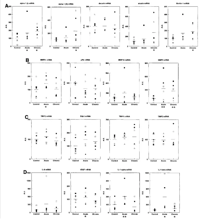
in their remodeling (MMP1, MMP2, MMP3, MMP8, MMP9, MMP12 and MMP14) and their specific inhibitors (TIMP1, TIMP2 and TIMP3), some components of the plasmin-plasminogen system (urinary-type plasminogen activator [uPA] and plasminogen activator inhibitor-1 [PAI-1]) (19) and inflammatory (interleukin-lα [lα], IL-lβ, IL-6) and angiogenic (vascular endothelial growth factor [VEGF]) mediators. The number of PCR cycles used to obtain a clear signal after electrophoresisis indicated to provide a rough estimate of the relative abundance of each specific mRNA (Table 3). Among them, MMP1, MMP3, MMP8 and MMP12 were not detected even using the highest acceptable number of cycles (n = 35). Individual values of the steady-state level of the expressed mRNA in the control, acute SSR and chronic SSR skin biopsy samples are illustrated in Fig. 2. In this figure, the mean value for each mRNA has been normalized to 100, and the individual values have been identified by the symbols shown in Table 1. This method allows showing the variability of the studied
population. For the control values, the interindividual variability is low for ECM proteins (α1 I and α1 III collagen chains, elastin, fibrillin 1 and decorin), constitutively expressed MMP2, TIMP, IL-lβ and VEGF, lying around ± 34%. It is about two-fold higher for the other investigated mRNA. The average standard deviation for the whole series of mRNA is 42 ± 18%.
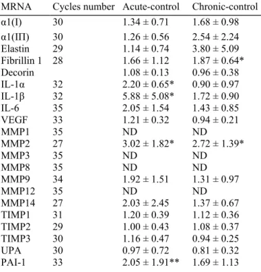
Table 3. Ratio of the mRNA steady-state level between control and exposed areas
MRNA Cycles number Acute-control Chronic-control
α1(I) 30 1.34 ± 0.71 1.68 ± 0.98 α1(IΠ) 30 1.26 ± 0.56 2.54 ± 2.24 Elastin 29 1.14 ± 0.74 3.80 ± 5.09 Fibrillin 1 28 1.66 ± 1.12 1.87 ± 0.64* Decorin 1.08 ± 0.13 0.96 ± 0.38 IL-1α 32 2.20 ± 0.65* 0.90 ± 0.97 IL-1β 32 5.88 ± 5.08* 1.72 ± 0.90 IL-6 35 2.05 ± 1.54 1.43 ± 0.85 VEGF 33 1.21 ± 0.32 0.94 ± 0.21 MMP1 35 ND ND MMP2 27 3.02 ± 1.82* 2.72 ± 1.39* MMP3 35 ND ND MMP8 35 ND ND MMP9 34 1.92 ± 1.51 1.31 ± 0.97 MMP12 35 ND ND MMP14 27 2.03 ± 2.45 1.37 ± 0.67 TIMP1 31 1.20 ± 0.39 1.12 ± 0.36 TIMP2 29 1.00 ± 0.43 1.08 ± 0.37 TIMP3 30 1.16 ± 0.47 0.94 ± 0.25 UPA 30 0.97 ± 0.72 0.81 ± 0.32 PAI-1 33 2.05 ± 1.91** 1.69 ± 1.13 *P < 0.02. **P < 0.05. ND, not detected.
A single MED SSR exposure increased the level of all expressed mRNA, with the exception of TIMP2 and uPA, as illustrated in Table 2 by the mean of the individual ratios between acute SSR and control. A significantly increased mean ratio was observed for IL-1α (P < 0.02), ΓL-1 β (P < 0.02), MMP2 (P < 0.02) and PAI-1 (P < 0.05). The illustration of the individual values (Fig. 2) clearly demonstrates that the variation of most mRNA differs in amplitude in the different volunteers and does not affect similarly all the tested mRNA. The average standard deviation is significantly higher than that recorded in the control sites (68 ± 24%, P = 0.02).
Chronic exposure to SSR for 6 weeks barely modified the level of most of the expressed mRNA when compared with a single exposure. Both IL-1α and IL-1β as well as PAI-1 returned to the control level. By contrast, MMP2 remained significantly increased upon chronic exposure. Interestingly, the fibrillin 1 gene that was weakly induced in acute SSR exposure was significantly increased in the chronically exposed skin. As observed in the 1 MED exposure, the average standard deviation was also higher than that in the control sites (56 ± 24%, P = 0.07).
Figure 2. Gene expression profiting. Individual values (% of control at 100) of the steady-state level of the specific mRNA. See Table 1 for identification of the volunteers (○,●, ■, ∆, ▲ and □). Mean (—) is significantly different from the control with **P < 0.05 or P < 0.10.
Figure 3. ECM proteins expression. Immunofluorescence microscopy using antisera against: tenasin C (250×), Type-I and Type-IΠ procollagens (125×), elastin (125×), lysozyme (250×) and alpha-1 antitrypsin (125×). Fluorescence was quantified (arbitrary units) with a computer-assisted color image analysis system, as
described in Materials and Methods, or performed by visual assessment using a grading scale from 0 to 3 with a 0.25 increment (lysozyme and alpha-1 antitrypsin). Data are means ± standard deviations; *P < 0.05 compared with control site.
Histology and immunohistochemistry
No dermal infiltrate was observed after the single MED (acute exposure) or after a 6 week SSR exposure either at histological level or by visual assessment of the immunostaining pattern obtained using an antiserum directed against human vimentin or Factor XIII-A (data not shown). Among the various matrix components investigated by immunohistochemistry, tenascin, a large glycoprotein minimally deposited just below the dermal-epidermal junction (DEJ) in normal skin (20), was slightly but significantly decreased in the acute exposure but markedly increased after chronic exposure to SSR. It remained clearly localized just below the DEJ (Fig. 3). Type-I and Type-III procollagens were predominantly located in the papillary dermis underneath the epidermal basement membrane as usually reported. After single or repeated exposure, the staining associated with Type-I procollagen decreased significantly (Fig. 3), whereas that of Type III was increased (Fig. 3), leading to a higher Type-III to Type-I procollagen ratio. Using a double-staining procedure, we examined simultaneously the elastin component of the elastic fibers and lyso-zyme or alpha-1 antitrypsin, which have been described to be associated with damaged elastic fibers (5,21). Chronic SSR exposure did not induce any significant change in the elastin network (Fig. 3). Lysozyme and alpha-1 antitrypsin (Fig. 3) deposits on the elastin fibers were significantly increased after 6 weeks of SSR exposure, whereas acute exposure did not induce any significant change.
DISCUSSION
Repeated solar exposure is the major environmental insult that contributes to clinical and histological changes in sun-exposed skin areas (face, hands, etc.) of an individual. The dermal damages induced by repeated UV irradiation are mainly reflected histologically by a disorganization of the bundles of collagen fibers (22) and an accumulation of abnormal elastin-containing material (5). These alterations are involved in the wrinkled appearance of sun-exposed skin. Biochemical evidence of connective tissue alterations in photoaged skin includes reduced levels of Type-I and Type-III collagen precursors (23), an increased number of crosslinks (24) and an increased Type-III to Type-I collagen ratio (25) and elastin content. Our immunohistological findings evidenced a significant decrease in the fluorescence associated with Type-I procollagen but a significant increase in Type-III procollagen, both located at the DEJ. This site has been reported to be the location for new collagen synthesis (26). Using a quantitative RT-PCR methodology, we have observed in the same biopsy sample a trend toward an increase in Type-I and Type-III collagen mRNA levels. These results are in agreement with those described by Chung et al. (27), showing a higher expression of Type-I procollagen mRNA in photoaged human skin associated with a lower extracellular expression of Type-I procollagen protein.
Acute and chronic SSR exposures induced a significant increase in MMP2 (72 kDa gelatinase—Type-IV collagenase) mRNA level. Fisher et al. (28) have shown that in human buttock skin MMP2 mRNA was detectable but increased only 1.6-fold (P < 0.05) at 24 h after UV-B (2 MED) exposure and that neither protein expression nor protein activation of latent activity was induced. These observations are in agreement with our own results because the increased MMP2 mRNA observed in both single and multiple SSR exposures was not accompanied by a similar stimulation of MMP14, the membrane-bound MMP involved in the activation of MMP2. Furthermore, TIMP2, the specific inhibitor of MMP2, was not modified. This TIMP also participates in the activation of the metalloproteinase. However, IL-1α and IL-1β were induced mostly after a single MED, perhaps in the epithelial cells. An increased vascular permeability after a single MED is, however, possible because the VEGF mRNA level tends to be increased (P = 0.09). As reported by Marionnet et al. (29), in human epidermis after UV-B exposure, a single MED of SSR increased the PAI-1 mRNA level in human skin in vivo, and this might be the result of collagen degradation (19).
As opposed to what has been published by others (28,30), we did not observe the expression of MMP1 and MMP3 or that of the inflammation-related MMP (9 and 12). Histological observation showed an absence of inflammatory cells in both the control and exposed skin samples. Our procedure for measuring the MMP was very sensitive because amplification at 35 cycles clearly detected MMP1 mRNA in one-tenth of the RN A content of one fibroblast (2 pg) in a retracting collagen gel (17). It has been reported previously that MMP1 expression may be influenced by aging (lower level of MMP1 protein in young skin) (27), skin type (higher UV-induced MMP1 mRNA induction in light skin) (11) or smoking status (higher level of MMP1 mRNA in the skin of smokers-10-20 cigarettes a day for 3-25 years) (31). In our study, a criterion for selection of the light-skinned (L* > 65) young volunteers (21-35 years old) was smoking less than 10 cigarettes per day. Furthermore, in most of the reports describing a MMP1 mRNA induction by UV, the UV dose was higher, often 2 MED (28,30), than the 1 MED SSR used in our study. Finally, in our SSR radiation source, to better simulate zenithal sun spectrum (irradiance ratio UV-A/ UV-B = 17), we used a 2 mm thick filter that cuts off more UV-B (UV-A/UV-B = 13)
than the 1.0 mm most commonly used (UV-A/ UV-B = 11). All these facts may be responsible for the lack of response of our volunteers after acute exposure.
The extent of progressive accumulation of elastic fibers containing in particular elastin and fibrillin 1 is dependent on the degree of sun exposure (32). In our volunteers, only the mRNA of fibrillin 1 was significantly increased after repeated exposures, whereas the level of elastin mRNA did not change upon one or repeated SSR exposure, which further supports fibrillin 1 as a marker of photoreaction. A change in the network of elastic fibers in the young volunteers enrolled in this study might be insufficient to be observed by immunomorphology. However, using immuno-histochemical techniques, we detected increased lysozyme and alpha-1 antitrypsin deposits on photodamaged elastic fibers, as already observed by other investigators (21). The intensity of staining is known to positively correlate with the degree of sun damage (21,33). The components of elastic fibers to which lysozyme or alpha-1 antitrypsin are bound are not yet known but seem different (staining just below the DEJ for alpha-1 antitrypsin). Because lysozyme or alpha-1 antitrypsin at high concentrations inhibits the activity of collagenase and elastase (34), these proteins could protect elastic fiber components from proteolysis (35). Deposits of lysozyme or alpha-1 antitrypsin were enhanced after repeated SSR exposure. An intimate
relationship between ultrastructural alterations and deposits of these proteins on the degenerative elastic fibers in sun-exposed skin has been demonstrated previously (21) and might be an early event leading to solar elastosis. To conclude, our study demonstrates that at one time point after irradiation single, as well as repeated, 1 MED exposure induces clear alterations in matrix gene and protein expressions. Some of the biochemical changes associated with chronic exposure and related to photoaging (increased procollagen III to 1 ratio or lysozyme deposition on elastin fibers) can be induced by low UV exposure even if a strongly induced pigmentation arose than inducing progressive skin autoprotection. Although the volunteers were of similar complexion and age, some variations were observed in their basal status and their reactivity to SSR exposure. These individual differences, at least in part, account for the different degrees of photodamage inflicted to humans when exposed to sunlight.
Acknowledgements
We thank Dr. Parneix-Spake and N. Goupil for biopsy sampling of the volunteers, Mrs. Lombard and Miss Verdier for histological and immunohistological procedures, Mrs. Tison for anatomohistopathology and Mrs. Compan and Ressayre for statistical evaluations. The skillful technical assistance of M.J. Nix and T. Heyeres for performing the molecular biology assays is gratefully acknowledged.
REFERENCES
1. Griffiths, C. E. M. (1992) The clinical identification and quantification of photodamage. Br. J. Dermatol. 127, 37-42.
2. Smith, J. G„ E. A. Davidson, W. M. Sams and R. D. Clark (1962) Alterations in human dermal connective tissue with age and chronic sun damage. J. Investig. Dermatol. 39, 347-350.
3. Warren, R., V. Garstein, A. M. Kligman, W. Montagna, R. A. Allendorf and G. M. Ridder (1991) Age, sunlight and facial skin: a histologic and quantitative study. J. Am. Acad. Dermatol. 25, 751-760.
4. Gray, J. and J. Hawk, editors (1998) The benefit of lifetime photoprotection. In International Congress and Symposium, Series 231. Royal Society of Medicine Press Limited, London, UK.
5. Lavker, R. M. (1995) Cutaneous aging: chronologic versus photoaging. In Photodammage (Edited by B. A. Gilchrest), pp. 123-135. Blackwell Science, Cambridge, New York.
6. Kligman, A. M. and R. M. Lavker (1988) Heliodermatitis. In Cutaneous aging (Edited by A. M. Kligman and Takasy), pp. 353-360. University of Tokyo Press, Tokyo.
7. Watson, R. E., C. E. Griffiths, N. M. Craven, C. A. Shuttleworth and C. M. Kielty (1999) Fibrillin-rich microfibrils are reduced in photoaged skin. Distribution at the dermal-epidermal junction. J. Investig. Dermatol. 112, 782-787.
8. Kähäri, V. M. and U. Saarialho-Kere (1997) Matrix metalloproteinases in skin. Exp. Dermatol. 6, 199-213.
9. Tschesche, H. (1998) Bimolecular interaction of matrix metalloproteinases and their inhibitors TIMPS. J. Protein Chem. 17, 549-551.
11. Fisher, G. J., S. Kang, J. Varani, Z. Bata-Csorgo, Y. Wan, S. Datta and J. J. Voorhees (2002) Mechanisms of photoaging and chronological skin aging. Arch. Dermatol. 138, 1462-1470.
12. Seité, S., D. Moyal, S. Richard, J. de Rigal, J. L. Lévêsque, C. Hourseau and A. Fourtanier (1998) Mexoryl SX: a broad absorption UVA filter protects human skin from the effects of repeated suberythemal doses of UVA. J. Photochem. Photobiol. B 44, 69-76.
13. Chirgwin, J. M., A. E. Przybyla, R. J. MacDonald and W. J. Rutter (1979) Isolation of biologically active ribonucleic acid from sources enriched in ribonuclease. Biochemistry 18, 5294-5299.
14. Nusgens, B. V., P. Humbert, A. Rougier, A. C. Colige, M. Haftek, C. A. Lambert, A. Richard, P. Creidi and C. M. Lapiere (2001) Topically applied vitamin C enhances the mRNA level of collagens I and III, their processing enzymes and tissue inhibitor of matrix metalloproteinase 1 in the human epidermis. J. Investig. Dermatol. 116, 853-859.
15. Lambert, C. A., A. C. Colige, C. Munaut, C. M. Lapiere and B. V. Nusgens (2001) Distinct pathways in the over-expression of matrix metalloproteinases in human fibroblasts by relaxation of mechanical tension. Matrix Biol. 20, 397-408.
16. Deroanne, C. F., K. Bonjean, S. Servotte, L. Devy, A. Colige, N. Clausse, S. Blacher, E. Verdin, J. M. Foidart, B. V. Nusgens and V. Castronovo (2002) Histone deacetylases inhibitors as anti-angiogenic agents altering vascular endothelial growth factor signaling. Oncogene 21, 427-436.
17. Lambert, C. A., A. C. Colige, C. M. Lapiere and B. V. Nusgens (2001) Coordinated regulation of procollagens I and IΠ and their post-translational enzymes by dissipation of mechanical tension in human dermal fibroblasts. Eur. J. Cell. Biol. 80, 479-485.
18. Pierard, D., B. V. Nusgens and C. M. Lapiere (1984) Radioimmunoassay for the amino terminal sequences of type III procollagen in human body fluids measuring fragmentated precursor sequences. Anal. Biochem. 141, 127-136.
19. Jones, J. M., R. L. Cohen and D. A. Chambers (2002) Collagen modulates gene activation of plasminogen activator system molecules.
Exp. Cell. Res. 280, 244-254.
20. Lightner, V. A., F. Gumkowski, D. D. Bigner and H. P. Erickson (1989) Tenascin/hexabrachion in human skin: biochemical identification and localization by light and electron microscopy. J. Cell. Biol. 108, 2483-2493.
21. Suwabe, H., A. Serizawa, H. Kajiwara, M. Ohkido and Y. Tsutsumi
(1999) Degenerative processes of elastic fibers in sun-protected and sun-exposed skin: immunoelectron microscopic observation of elastin, fibrillin-1, amyloid P component, lysozyme and alpha 1-antitrypsin. Pathol. Int. 49, 391-402.
22. Bernstein, E. F., Y. Q. Chen and J. B. Koop (1996) Long-term sun exposure alters the collagen of the papillary dermis: comparison of sun-protected and photoaged skin by northern analysis, immunohistochem-ical staining and confocal laser microscopy.J. Am. Acad.
Dermatol. 34, 209-218.
23. El-Domayati, M., S. Attia, F. Saleh, D. Brown, D. E. Birk, F. Gasparro, H. Ahmad and J. Uitto (2002) Intrinsic aging vs. photoaging: a comparative histopathological, immunohistochemical, and ultrastruc-tural study of skin. Exp. Dermatol. 11, 398-405.
24. Yamauchi, M„ P. Prisavanh, Z. Haque and D. T. Woodley (1991) Collagen cross-linking in sun-exposed and unexposed sites of aged human skin. J. Investig. Dermatol. 97, 938-941.
25. Schwartz, E., F. A. Cruickshank, C. C. Christensen, J. S. Perlish and M. Lebwohl (1993) Collagen alterations in chronically sun-damaged human skin. Photochem. Photobiol. 58, 841-844.
26. Fleischmajer, R., J. S. Perlish and R. Timpl (1985) Collagen fibrillogenesis in human skin. Ann. N. Y. Acad. Sci. 460, 246-257.
27. Chung, J. H., J. Y. Seo and H. R. Choi, M. K. Lee, C. S. Youn, G. Rhie, K. H. Cho, K. H. Kim, K. C. Park and H. C. Eun (2001) Modulation of skin collagen metabolism in aged and photoaged human skin in vivo. J. Investig. Dermatol. 117, 1218-1224.
28. Fisher, G. J., S. C. Datta, H. S. Talwar, Z. Q. Wang, J. Varani, S. Kang and J. J. Voorhees (1996) Molecular basis of sun-induced premature skin ageing and retinoid antagonism. Nature 379, 335-339.
29. Marionnet, C, K. Mollier, F. Moch, F. Galisson and N. Basset-Seguin
(2000) PAI-1 expression is induced in human skin keratinocytes after repeated UVB irradiation. J. Investig. Dermatol. 115, 516.
30. Lahmann, C, A. R. Young, K. P. Wittern and J. Bεrgemann (2001) Induction of mRNA for matrix metalloproteinase 1 and tissue inhibitor of metalloproteinase 1 in human skin in vivo by solar simulated radiation. Photochem. Photobiol. 73, 657-663.
31. Lahmann, C, J. Bergemann, G. Harrison and A. R. Young (2001) Matrix metalloproteinase-1 and skin ageing in smokers. Lancet 357, 935-936.
32. O'Brian, J. P. and W. Regan (1991) A study of elastic tissue and actinic radiation in "aging", temporal arteries, polymyalgia, rheumatica and arteriosclerosis. J. Am. Acad. Dermatol. 24, 765-776.
33. Mera, S. L„ C. R. Lovell, R. Russell Jones and J. D. Davies (1987) Elastic fibers in normal and sun-damaged skin: an immunohistochemical study. Br. J. Dermatol. 117, 21-27.
34. Davies, J. D., E. W. Young, S. L. Mera and K. Barnard (1983) Lysozyme is a component of human vascular elastic fibers. Exper-imentia 39, 382-383.
35. Park, P. W., K. Biedermann, L. Mecham, D. L. Bissett and R. P. Mecham (1996) Lysozyme binds to elastin and protects elastin from elastase-mediated degradation. J. Investig. Dermatol. 106, 1075-1080.