Staging of Regional Nodes in AJCC Stage I and II
Melanoma:
18
FDG PET Imaging versus Sentinel
Node Detection
TARIK
BELHOCINE,
aGÉRALD
PIERARD,
bMICHEL DE
LABRASSINNE,
cTHIERRY
LAHAYE,
dPIERRE
RIGO
aaDivision of Nuclear Medicine and the Departments of bPathology, cDermatology, and dSurgery,
University Hospital of Liège, Liège, Belgium
Key Words. Cutaneous melanoma · Sentinel node · Lymphatic mapping · 18FDG PET
ABSTRACT
Primary Purpose. The staging of regional nodes by means of sentinel node detection has been shown to accu-rately detect subclinical nodal metastases from cutaneous melanoma. On the other hand, the oncological applications of 18F-fluoro-2-deoxy-D-glucose positron emission tomog-raphy (18FDG PET) are, nowadays, firmly established. However, the sensitivity of such metabolic imaging for stag-ing the regional nodes in primary melanoma remains debat-able. We prospectively assessed the actual value of PET for detecting sentinel node metastases in 21 consecutive patients presenting with early-stage melanoma.
Materials and Methods. Twenty-one melanoma patients scheduled for lymphatic mapping and sentinel lym-phadenectomy underwent fully corrected whole-body PET using 18FDG. In all cases, the disease was initially classified as either stage I or II, from the latest version of the American Joint Committee on Cancer staging system. The sentinel node detection was systematically performed within the week following the PET scan. Serial sections of the sentinel nodes were analyzed by both conventional pathology and immunohistochemical staining. Metastatic sentinel nodes were also assessed for the size of tumor
deposits and the degree of nodal involvement (focal, partial, or massive). The median follow-up time was 12 months.
Results. Six of the 21 patients (28.5%) had an involved sentinel node. PET was positive in only one case with a sentinel node >1 cm. In the five other cases, the sentinel nodes missed by PET were <1 cm with focal and/or par-tial involvements. One patient, free of regional nodal metastases in both sentinel node detection and PET imag-ing, had, however, a same-basin recurrence 3 months later. In another case, PET had one false positive result. Overall, the sentinel detection of subclinical nodal metas-tases had a sensitivity of 86%. PET detected only 14% of sentinel node metastases.
Conclusions. Sentinel node detection remains the pro-cedure of choice for detecting subclinical lymph node involvement from primary cutaneous melanoma. Owing to its limited spatial resolution, PET appears insufficiently sensitive to identify microscopic nodal metastases. As a practical consequence, metabolic imaging is not recom-mended as a first-line imaging strategy for staging regional lymph nodes in patients with stage I or II melanoma. The Oncologist 2002;7:271-278
The Oncologist 2002;7:271-278
www.TheOncologist.com
Correspondence: Tarik Belhocine, M.D., University Hospital of Liège, Division of Nuclear Medicine, Sart Tilman -Bâtiment 35, 4000 Liège, Belgium. Telephone: 324-366-7931; Fax: 324-366-7933; e-mail: tarik.bel@swing.be Received January 28, 2001; accepted for publication March 4, 2002. ©AlphaMed Press 1083-7159/2002/$5.00/0
INTRODUCTION
Malignant melanoma of the skin remains a critical public health problem worldwide despite earlier diagnosis due to screening and public education [1]. As melanoma cells pri-marily spread through the lymphatic way, the N classification
of such lymphophilic cancer is a key step for treatment planning and patient prognosis [2, 3].
According to the sentinel node theory, the disease pro-gresses in an orderly fashion, and thus, affects the regional nodes draining the primary tumor before disseminating to
T
he
O
ncologist
®at faculte on March 25, 2009
www.TheOncologist.com
distant sites [4, 5]. Subsequently, the lymphatic mapping followed by sentinel lymphadenectomy (LM/SL) has become a valuable tool for detecting subclinical lymph node metastases from melanoma [6-8]. Though even the thera-peutic benefit of complete selective lymph node dissection (CSLND) has not yet been proven, the clinical usefulness of LM/SL in terms of staging accuracy and prognostic significance is widely recognized nowadays [7, 9, 10].
On the other hand, the utility of a sophisticated metabolic imaging technology, such as positron emission tomography
(PET) using 18F-fluoro-2-deoxy-D-glucose (18FDG) for
stag-ing various malignancies, is firmly established. Many
pub-lished data report the capability of 18FDG PET in detecting
lymph node involvement missed by morphological imaging
procedures. Indeed, 18FDG PET has been shown to localize
metastatic deposits in normal-sized nodes [11-14]. Nonethe-less, in pretreatment nodal staging of malignant melanoma, the actual value of PET imaging is debatable. While initial reports emphasized the high sensitivity of PET for detecting impalpable metastatic nodes [15-19], more recent studies underlined its low efficiency for diagnosing microscopic nodal involvement [20-24]. For this purpose, we prospec-tively evaluated the contribution of metabolic imaging for detecting sentinel node metastases in 21 consecutive patients presenting with early-stage primary melanoma,
which underwent both 18FDG PET imaging and LM/SL.
MATERIALS ANDMETHODS Patient Population
Twenty-one consecutive melanoma patients (10 men, 11 women, mean age = 58 ± 11 years, Breslow’s thickness
= 1.89 mm) scheduled for a LM/SL underwent 18FDG PET
imaging in pretreatment staging. All patients included into this institutional protocol were classified at stage I or II cutaneous melanoma (T1-4 N0 M0) according to the latest version of the American Joint Committee on Cancer (AJCC) staging system [25]. The patients with histologi-cally unproven primary melanoma or those with confirmed but more advanced disease (AJCC stages III or IV), as well as subjects previously treated for malignant melanoma or other malignancies were systematically excluded from the study group. Additionally, in our staging protocol, only the patients who underwent LM/SL within the week following the metabolic imaging were taken into account. The LM/SL technique included, in a 1-day protocol, a preoperative lym-phoscintigraphy followed by an intraoperative lymphatic mapping using gamma probe guidance. Each patient was individually followed with a median follow-up time of 12 months. Post-therapy surveillance included complete phys-ical examination at the control visits and oriented imaging
procedures, such as computed tomography or PET, when clinically indicated. All cases were managed by a multidisci-plinary melanoma group study including a surgeon, a derma-tologist, a nuclear physician, a pathologist, and an oncologist. Patient characteristics are detailed in Table 1.
PET Imaging Procedure
All patients underwent fully corrected whole-body PET imaging using either a PENN PET 240H scanner (UGM; Philadelphia, PA), in 13 cases, or a C-PET scanner (Adac, Philips Medical Systems; Milpitas, CA) in eight patients. The spatial resolution of these scanners in clinical practice approaches 1 cm. Patients fasted at least 4 hours before injection time to reduce serum glucose and insulin levels to near basal concentrations. Intravenous injection of diuretics (furosemide, 10 mg) was performed to avoid bladder and ureter artifacts. In addition, the patients received 10 mg of diazepam orally about 1 hour prior to starting the study in order to provide muscle relaxation. Patients were kept at rest and were asked to void just before starting the acquisi-tion. PET scans were performed 60 to 90 minutes after i.v.
injection of 259 to 333 MBq of 18FDG. Scanning started at
the inguinal region and proceeded to the neck. Optional views were focused over the neck, the groin, or the popliteal fossea in order to verify all physiological nodal stations potentially draining the primary tumor. In all cases, a whole-body segmented attenuation correction was per-formed post-injection using singles transmission scans with
a 137Cesium external point source. The total scanning time
was, on average, 60 minutes. The data were reconstructed by using an iterative method based on an ordered subset-expec-tation maximization algorithm. UGM software was used for data acquisition and processing. Finally, the images were displayed on transversal, coronal, and sagittal slices and were visually interpreted in routine clinical fashion. LM/SL Procedure
Preoperative Lymphoscintigraphy
A mean activity of 37 MBq (1 mCi) of 99mTc-ultrafiltered
sulfur-colloid (Lymphoscint®; Nycomed Amersham Sorin®;
Milan, Italy; mean size = 50 nm) divided into four insulin syringes (23-gauge) was injected intradermally (0.1 to 0.15 cc per syringe) in two to four points around the primary site. Massage and compression of the sites of injection for 1 to 2 minutes were applied to stimulate the lymphatic flow. Dynamic sequences followed by static spot views over the lymphatic basins were performed using a DSX rectangular gamma camera (Sopha Medical; GE Medical Systems
Benelux, Diegem, Belgium). A 57Cobalt flood was also placed
under the gamma camera performing a virtual transmission in
at faculte on March 25, 2009
www.TheOncologist.com
order to define the body contours, and thus, to localize more accurately the site of the sentinel lymph nodes (SLNs). For the axillary areas, optional views were acquired mimicking the surgical position of the patients. According to the standard definition, the first lymph node identified on the initial scans and confirmed on at least two static images was considered as the SLN [8]. The skin covering the SLN was tattooed to guide the surgical excision.
Intraoperative Gamma Probe Guidance
Three to four hours after the preoperative lymphoscintig-raphy, the patients were referred to the operating room. Intra-operative lymphatic mapping was performed by using a
hand-held gamma probe (Navigator®using a CdTe detector;
Autosuture®, Tyco Healthcare; Mechelen, Belgium).
Radio-activity (in counts/sec) of the SLNs and the adjacent non-SLNs was measured in vivo and verified ex vivo after removal. A signal to background ratio higher than 2 to 3 in vivo and higher than 10 ex vivo was considered significant. After excision of the SLNs, the lymphatic basin was rechecked for radioactivity. The use of blue dye was left at the discretion of the surgeon. In all cases, the LM/SL procedure was performed by the same surgical team.
Histologic Analysis of the SLNs
Serial sections of the SLNs were examined by the same pathologist using, in all cases, both conventional pathology (hematoxylin and eosin) and immunohistochemical stains with antibodies to S-100 protein, HMB-45, and NKIC3 anti-gens. In cases of involved SLNs, the tumor deposits were microscopically measured and the lymph node involvement was described as massive, partial, or focal following the degree of lymph node invasion.
Statistical Analysis
The results of 18FDG PET and LM/SL were expressed in
terms of sensitivity, specificity, diagnostic accuracy, and posi-tive and negaposi-tive predicposi-tive values, respecposi-tively. Estimates are presented with a 95% confidence interval (95% CI). Any hypermetabolic focus detected by PET in the lymphatic basin draining the primary tumor was considered as true positive when the result matched with an SLN histologically involved. Otherwise, all hypermetabolic foci not confirmed by the his-tology or corresponding to inflammatory nodes were inter-preted as false positive results. A negative PET scan matching with an SLN histologically free of tumor was considered as true negative. Conversely, the result of PET was considered as Table 1. Patient characteristics
Patient Age Gender Primary Breslow’s Clark’s level AJCC
n (year) tumor sites depth (mm) Stage
1 52 M Head and neck 2.4 IV II
2 62 M Head and neck 3.6 IV II
3 46 F Lower extremity 0.6 II Ia 4 49 F Trunk 2.8 IV II 5 49 M Lower extremity 1.7 IV Ib 6 39 F Trunk 0.56 III Ia 7 65 F Lower extremity 4.25 V II 8 58 F Trunk 1.33 IV Ib 9 66 M Trunk 1.4 IV Ib 10 76 M Upper extremity 3.85 IV II 11 44 F Upper extremity 0.5 I Ia 12 50 F Trunk 1.3 IV II
13 78 M Upper extremity 0.7 III Ia
14 48 F Upper extremity 2.1 IV II
15 50 M Upper extremity 1.18 IV Ib
16 74 M Trunk 1.1 IV Ib
17 77 F Upper extremity 1.6 IV Ib
18 73 F Head and neck 4.56 V II
19 48 M Lower extremity 2.35 IV II
20 63 F Lower extremity 0.73 IV Ia
21 59 M Trunk 1.4 IV Ib
Abbreviation: AJCC = American Joint Committee on Cancer (Last version 2001).
at faculte on March 25, 2009
www.TheOncologist.com
falsely negative when the SLN detected by LM/SL was histo-logically involved. The same-basin recurrences were also con-sidered as false negative results of PET and/or LM/SL when the initial interpretation did not conclude to a nodal involvement. RESULTS
In the present series, 6 of the 21 melanoma patients (28.5%) had an involved SLN, which was easily localized in both preoperative and intraoperative detection. PET was posi-tive in only one patient, who had a metastatic SLN measuring 1.8 cm with massive involvement and capsular infiltration (Fig. 1). This patient was clinically classified at AJCC stage II dis-ease at the time of the PET scan and had progressed to AJCC stage IV melanoma after the diagnosis of microscopic lung metastases on a thoracic computed tomography. In the five other cases, the positive SLNs missed by the metabolic imag-ing were <1 cm, and most often harbored microscopic and focal tumor deposits (Fig. 2). Two of these (2/5) were only detected by using immunohistochemical stains. Another patient with AJCC stage I disease had a confirmed same-basin recurrence 3 months after intervention, while both LM/SL and PET were initially negative. In addition, PET had one false positive result and 13 true negatives (SLNs free of tumor and no subsequent recurrence) with a median follow-up of 12
months. The patients having a positive SLN underwent CSLND followed by high-dose systemic adjuvant interferon α-2B according to the scheme of Kirkwood et al. [26]. Otherwise, the patients with negative SLNs had no subsequent treatments and were clinically followed up at the control visits. Overall, in pretreatment staging of subclinical melanoma nodal metastases, LM/SL had an 86% sensitivity, a 100% specificity, a 95% diagnostic accuracy, a 100% positive pre-dictive value, and a 93% negative prepre-dictive value. For detect-ing SLN metastases, PET had a 14% sensitivity, a 93% specificity, a 67% diagnostic accuracy, a 50% positive predic-tive value, and a 68% negapredic-tive predicpredic-tive value. The perfor-mances of each procedure with their 95% confidence intervals are summarized in Table 2.
DISCUSSION
In melanoma patients presenting with AJCC stage I or II disease, the histological status of the regional lymph nodes is the most important prognostic factor [27]. This explains why it is crucial to achieve an accurate nodal stag-ing in patients at risk of metastases. Such a need is even more warranted by the survival benefit obtained in patients with nodal involvement treated by high-dose systemic adjuvant
interferon α-2B [26].
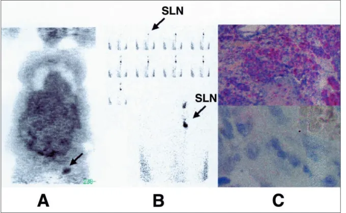
Figure 1. A case of malignant melanoma of the left foot (Breslow’s depth = 4.5 mm). 18FDG PET imaging (A) accurately detected an involved sentinel node harvested at the left groin (B). Histology revealed that the sentinel node measured 1.8 cm and had massive tumor involvement with capsular infiltration (C). 18FDG PET = 18F-fluoro-2-deoxy-D-glucose positron emission tomography; SLN = sentinel lymph nodes.
at faculte on March 25, 2009
www.TheOncologist.com
Introduction of LM/SL in malignant melanoma was a substantial advance for the detection of subclinical nodal metastases. The technique is, nowadays, standardized and allows for the selection of a subset of patients presenting with clinically unsuspected involvement of their sentinel nodes for complete lymphadenectomy and systemic adjuvant treat-ments. Moreover, the procedure is useful to avoid the cost and morbidity of unnecessary elective lymph node dissections in the wide majority of the patients with an SLN free of tumor [6-10]. Owing to the determinant data provided by LM/SL in the management of patients with stage I and II melanoma, the results of this procedure recently have been incorporated into the last version of the revised AJCC staging system [25].
On the other hand, the value of 18FDG PET for staging
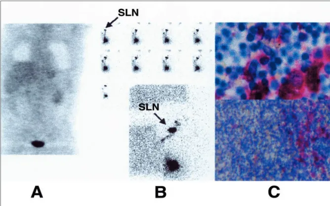
various malignancies is firmly established [11-14]. In pre-treatment nodal staging of malignant melanoma, however, the role of metabolic imaging is less evident. Despite the opti-mistic conclusions of initial reports, more recent prospective and retrospective studies are less enthusiastic about the actual performance of PET for detecting occult lymph node metas-tases. For instance, in 13 patients, Gritters et al. reported the detection of melanoma metastases in 100% of the lymph node basins [15]. Similarly, in 11 melanoma patients submitted to complete lymphadenectomy of 14 nonpalpable lymph node basins, Wagner et al. reported a sensitivity and a specificity of 100% for the detection of nodal metastases by means of Figure 2. A case of malignant melanoma of the back (Breslow’s depth = 1.33 mm). 18FDG PET imaging was negative (A). The sentinel node located at the right axilla was involved (B). Histology showed focal tumor deposits measuring 0.6 cm (C). 18FDG PET = 18 F-fluoro-2-deoxy-D-glucose positron emission tomography; SLN = sentinel lymph nodes.
Table 2. Overall results
PET LM/SL
Sensitivity (95% CI) 14% (0-28) 86% (72-100)
Specificity (95% CI) 93% (83-103) 100% (100-100)
Diagnostic accuracy (95% CI) 67% (47-87) 95% (86-104) Positive predictive value (95% CI) 50% (29-71) 100% (100-100) Negative predictive value (95% CI) 68% (49-88) 93% (83-103) Abbreviations: PET = positron emission tomography; LM/SL = lymphatic mapping/sentinel lymphadenectomy; CI = confidence interval.
at faculte on March 25, 2009
www.TheOncologist.com
18FDG PET [18]. Several other studies also confirmed the
per-formance of 18FDG PET for the detection of occult or residual
lymph node metastases [12, 14, 16, 17, 19]. Nonetheless, only a few studies recently addressed the value of metabolic imag-ing for stagimag-ing subclinical nodal metastases detected by the LM/SL procedure. In a prospective study of 70 patients pre-senting with AJCC stage I, II, or III cutaneous melanoma,
Wagner et al. first compared PET imaging of regional lymph
node basins with sentinel node biopsy results (SNB). They concluded that PET had a sensitivity of 16.7% and a speci-ficity of 95.8% versus 94.4% and 100%, respectively, for SNB [20]. Similar observations were reported by Acland et al. in a prospective study of 50 patients with primary melanoma >1 cm thick or lymphatic invasion [21]. Of the 14 patients with positive SLNs, PET detected none of them. More recently, Kokoska et al. prospectively evaluated the value of PET and lymphoscintigraphy in the management of 18 head and neck melanoma patients [24]. Sentinel nodes were found in 94.4% of the patients, in which 5/18 (27.8%) had metas-tases. PET detected nodal metastases in three patients (16.7%), and only one of them had a positive SLN. In another study, Acland et al. retrospectively showed that the sensitivity of PET was correlated with the AJCC stage of melanoma dis-ease [22]. They found a high sensitivity of 93% for stage III, but the value dramatically fell to 50% and 33% for stages I and II, respectively. They concluded that PET is a valuable procedure for staging the patients with known regional spread but is suboptimal in stage I or stage II disease. Similarly, in a prospective study of 95 patients, Tyler et al. confirmed the
clinical utility of 18FDG PET imaging in melanoma patients
with AJCC stage III disease [28].
To determine the factors that may influence the sensitivity of the metabolic imaging for detecting melanoma nodal metas-tases, Crippa et al. analyzed the value of PET in terms of lymph node size [29]. In a retrospective study of 36 patients presenting with confirmed lymph node metastases from melanoma, the authors showed that PET had only a 23% sen-sitivity for lymph node metastases ≤5 mm, while the sensitiv-ity increased to 83% and 100%, respectively, for metastases that were 6-10 mm and ≥10 mm. More recently, Wagner et al. retrospectively studied the tumor volume threshold for suc-cessful PET imaging of melanoma nodal metastases in 45 patients with 49 pathologically positive regional nodal basins [23]. The sensitivity of PET for detection of all tumor volumes was 49%. The observed 90% sensitivity threshold of nodal
metastases was ≥78 mm3. PET sensitivity fell to 14% for
detection of tumor volumes <78 mm3. Indeed, the PET
sensi-tivity differed by prescan AJCC stage: 0% (stage I), 24% (stage II), 81% (stage III), and 100% (stage IV).
Although these prospective and retrospective trials assessed the sensitivity of metabolic imaging for detecting
sentinel node metastases on large patient populations, they had technical biases regarding their PET protocol. For instance, a correction of attenuation and an iterative recon-struction, two key parameters affecting the quality of the images, and thus, the sensitivity of the technique, were inconsistently or never performed [30-32]. Despite the rel-atively limited sample size of our study group, the results were enhanced by fully corrected whole-body PET imag-ing, blinded PET interpretations, use of single PET exam-iner, and iterative data reconstruction in all cases. Indeed, the prospective nature of the study with a median follow-up time of 12 months, as well as the strict adherence to LM/SL protocol performed by the same surgical team, detailed his-tological analyses of SLN specimens by the same patholo-gist, and multidisciplinary review of all clinical cases were other important points strengthening the robustness of our conclusions. Overall, our data demonstrated the inability of PET to visualize infracentimetric sentinel node metastases in patients with early-stage melanoma. Although statisti-cally limited, our results are similar to those published by
Wagner and Acland from larger series (≥50 patients) [20,
21]. Both authors concluded on the inability of PET to detect microscopic metastases located in sentinel nodes. Also, our results provide additional arguments regarding the comparison of PET with sentinel node biopsy using an optimal methodology, and thus, avoiding the technical biases characterizing the available data from the literature, thereby inciting further studies with larger series to defi-nitely confirm our conclusions. The limited spatial resolu-tion of commercially available PET devices is certainly the main reason explaining the low sensitivity of PET for detecting microscopic lymph node metastases. Another important issue is the detector materials characterizing the PET scanners. In all published studies, the imaging was per-formed using Siemens ECAT line devices with bismuth ger-manate (BGO) detectors. We used two types of the same PET scanner (PENN PET and C-PET). Both had 6 thallium-doped sodium iodine detectors (NaI [Tl]), but the curved detectors and larger axial field of view characterizing the C-PET model contribute to improve its count-rate capability and spatial resolution in comparison with the PENN PET [33, 34]. All these devices, regardless of their constructors, are representative of mid-end PET scanners commonly used in the field of oncology. Their theoretical spatial resolution is about 6 mm. In clinical practice, however, their resolution is around 1 cm and certainly more for uncorrected images. The recent introduction of lutetium oxyorthosilicate (LSO) and gadolinium oxyorthosilicate (GSO) detectors in high-end PET scanners could improve the detection efficiency [13]. Both LSO and GSO are faster than either BGO or NaI [Tl], and both scintillating materials also have a higher
at faculte on March 25, 2009
www.TheOncologist.com
effective Z and density than NaI [Tl]. Future improvements in the correction of scatter fraction, as well as the imple-mentation of more effective iterative methods for data reconstruction and the use of more powerful processing sys-tems could also substantially increase the sensitivity and the resolution of the PET scanners [13, 35-39]. Nonetheless, we believe that the spatial resolution of these new PET systems would still be insufficient to detect 1 or 2 melanoma cells in normal-sized nodes. As demonstrated by the present study, most of the involved sentinel nodes harbored microscopic and focal tumor deposits, requiring, in two patients, sophis-ticated immunostaining techniques. In another case, the same-basin recurrence also showed the current limits of LM/SL and indicated the need for still more sensitive tools. For this purpose, the use of reverse transcriptase-polymerase chain reaction assay could be an efficient adjunct to detect the false negatives of both LM/SL and PET [40, 41]. Controlled trials using high-end PET with LSO or GSO detectors are, however, necessary to assess their actual per-formances in detecting infracentimetric nodal metastases from melanoma. Also, following evidence-based medicine methods, our results, and those reported by others, support the idea that an optimal regional nodal staging of patients with early-stage melanoma would be better achieved using
lymphatic mapping and sentinel lymphadenectomy rather
than 18FDG PET imaging, thereby avoiding time-consuming
and expensive unnecessary imaging procedures. Metabolic imaging remains, however, a valuable tool for whole-body staging of more advanced disease.
CONCLUSIONS
The LM/SL technique remains the procedure of choice for evaluating the histologic status of the lymphatic basins in patients with early-stage cutaneous melanoma. PET imaging appears insufficiently sensitive for localizing microscopic sentinel node metastases given its current spa-tial resolution. Based on our results and data from literature,
we do not recommend 18FDG PET as a first-line imaging
strategy for staging patients with AJCC stage I or II primary disease. Further prospective and multicentric trials, includ-ing larger series of melanoma patients and usinclud-ing high-end PET scanners with optimal methodology, are needed to confirm our conclusions.
ACKNOWLEDGMENTS
This work was presented in part at the 48th Annual Meeting of the Society of Nuclear Medicine (Toronto, Canada, June 23-27, 2001).
REFERENCES
1 Landis SH, Murray T, Bolden S et al. Cancer statistics, 1999. CA Cancer J Clin 1999;49:8-31.
2 Reintgen DS. Regional nodal surgery for melanoma impacts recurrence rates and survival. Ann Surg Oncol 2000;7:80-81. 3 Balch CM. The role of elective lymph node dissection in melanoma: rationale, results, and controversies. J Clin Oncol 1988;6:163-172.
4 Morton DL, Chan AD. The concept of sentinel node localization: how it started. Semin Nucl Med 2000;30:4-10.
5 Reintgen D, Cruse CW, Wells K et al. The orderly progression of melanoma nodal metastases. Ann Surg 1994;220:759-767. 6 Morton DL, Wen DR, Wong JH et al. Technical details of
intraoperative lymphatic mapping for early stage melanoma. Arch Surg 1992;127:392-399.
7 Morton DL, Thompson JF, Essner R et al. Validation of the accuracy of intraoperative lymphatic mapping and sentinel lymphadenectomy for early-stage melanoma: a multicenter trial. Multicenter Selective Lymphadenectomy Trial Group. Ann Surg 1999;230:453-463.
8 Cochran AJ, Balda BR, Starz H et al. The Augsburg Consensus. Techniques of lymphatic mapping, sentinel lymphadenectomy, and completion lymphadenectomy in cutaneous malignancies. Cancer 2000;89:236-241.
9 Morton DL. Lymphatic mapping and sentinel lymphadenec-tomy for melanoma: past, present, and future. Ann Surg Oncol 2001;8(suppl 9):22S-28S.
10 McMasters KM, Reintgen DS, Ross MI et al. Sentinel lymph node biopsy for melanoma: controversy despite widespread agreement. J Clin Oncol 2001;19:2851-2855.
11 Delbeke D. Oncological applications of FDG PET imaging. J Nucl Med 1999;40:1706-1715.
12 Delbeke D. Oncological applications of FDG PET imaging: brain tumors, colorectal cancer, lymphoma and melanoma. J Nucl Med 1999;40:591-603.
13 Delbeke D, Martin WH. Positron emission tomography imaging in oncology. Radiol Clin North Am 2001;39:883-917. 14 Strauss LG. Sensitivity and specificity of positron emission
tomography (PET) for the diagnosis of lymph node metastases. Recent Results Cancer Res 2000;157:12-19.
15 Gritters LS, Francis IR, Zasadny KR et al. Initial assessment of positron emission tomography using 2-fluorine-18-fluoro-2-deoxy-D-glucose in the imaging of malignant melanoma. J Nucl Med 1993;34:1420-1427.
16 Steinert HC, Huch Boni RA, Buck A et al. Malignant melanoma: staging with whole-body positron emission tomog-raphy and 2-[F-18]-fluoro-2-deoxy-D-glucose. Radiology 1995;195:705-709.
17 Boni R, Boni RA, Steinert H et al. Staging of metastatic melanoma by whole-body positron emission tomography using 2-fluorine-18-fluoro-2-deoxy-D-glucose. Br J Dermatol 1995;132:556-562.
at faculte on March 25, 2009
www.TheOncologist.com
18 Wagner JD, Schauwecker D, Hutchins G et al. Initial assess-ment of positron emission tomography for detection of non-palpable regional lymphatic metastases in melanoma. J Surg Oncol 1997;64:181-189.
19 Macfarlane DJ, Sondak V, Johnson T et al. Prospective evalua-tion of 2-[18F]-2-deoxy-D-glucose positron emission tomography
in staging of regional lymph nodes in patients with cutaneous malignant melanoma. J Clin Oncol 1998;16:1770-1776. 20 Wagner JD, Schauwecker D, Davidson D et al. Prospective
study of fluorodeoxyglucose-positron emission tomography imaging of lymph node basins in melanoma patients under-going sentinel node biopsy. J Clin Oncol 1999;17:1508-1515. 21 Acland KM, Healy C, Calonje E et al. Comparison of positron emission tomography scanning and sentinel node biopsy in the detection of micrometastases of primary cutaneous malignant melanoma. J Clin Oncol 2001;19:2674-2678.
22 Acland KM, O’Doherty MJ, Russell-Jones R. The value of positron emission tomography scanning in the detection of subclinical metastatic melanoma. J Am Acad Dermatol 2000;42:606-611.
23 Wagner JD, Schauwecker DS, Davidson D et al. FDG-PET sensitivity for melanoma lymph node metastases is dependent on tumor volume. J Surg Oncol 2001;77:237-234.
24 Kokoska MS, Olson G, Kelemen PR et al. The use of lym-phoscintigraphy and PET in the management of head and neck melanoma. Otolaryngol Head Neck Surg 2001;125:213-220. 25 Balch CM, Buzaid AC, Soong SJ et al. Final version of the
American Joint Committee on Cancer staging system for cutaneous melanoma. J Clin Oncol 2001;19:3635-3648. 26 Kirkwood JM, Strawderman MH, Ernstoff MS et al.
Interferon alfa-2b adjuvant therapy of high-risk resected cuta-neous melanoma: the Eastern Cooperative Oncology Group Trial EST 1684. J Clin Oncol 1996;14:7-17.
27 Gershenwald JE, Thompson W, Mansfield PF et al. Multi-institutional melanoma lymphatic mapping experience: the prognostic value of sentinel lymph node status in 612 stage I or II melanoma patients. J Clin Oncol 1999;17:976 -983. 28 Tyler DS, Onaitis M, Kherani A et al. Positron emission
tomo-graphy scanning in malignant melanoma. Cancer 2000;89:1019-1025.
29 Crippa F, Leutner M, Belli F et al. Which kinds of lymph node metastases can FDG PET detect? A clinical study in melanoma. J Nucl Med 2000;41:1491-1494.
30 Raylman RR, Kison PV, Wahl RL. Capabilities of two- and three-dimensional FDG-PET for detecting small lesions and lymph nodes in the upper torso: a dynamic phantom study. Eur J Nucl Med 1999;26:39-45.
31 Wong T, Coleman R, Hagge R et al. PET image interpretation. Attenuation-corrected (ATN) vs non-attenuation corrected (NATN) images. Clin Positron Imaging 2000;3:181. 32 Visvikis D, Cheze-LeRest C, Costa DC et al. Influence of
OSEM and segmented attenuation correction in the calculation of standardised uptake values for [18F]FDG PET. Eur J Nucl
Med 2001;28:1326-1335.
33 Karp JS, Freifelder R, Geagan MJ et al. Three-dimensional imaging characteristics of the HEAD PENN-PET scanner. J Nucl Med 1997;38:636-643.
34 Adam LE, Karp JS, Daube-Witherspoon ME et al. Perfor-mance of a whole-body PET scanner using curve-plate NaI(Tl) detectors. J Nucl Med 2001;42:1821-1830.
35 Leahy R, Byrne C. Recent developments in iterative image reconstruction for PET and SPECT. IEEE Trans Med Imaging 2000;19:257-260.
36 Knesaurek K. New developments in PET instrumentation: quo vadis PET? J Nucl Med 2001;42:1831-1832.
37 Liu X, Comtat C, Michel C et al. Comparison of 3-D recon-struction with 3D-OSEM and with FORE+OSEM for PET. IEEE Trans Med Imaging 2001;20:804-814.
38 Zaidi H. Scatter modelling and correction strategies in fully 3-D PET. Nucl Med Commun 2001;22:1181-1184. 39 Adam LE, Karp JS, Freifelder R. Energy-based scatter
cor-rection for 3-D PET scanners using NaI(T1) detectors. IEEE Trans Med Imaging 2000;19:513-521.
40 Shivers SC, Wang X, Li W et al. Molecular staging of malig-nant melanoma: correlation with clinical outcome. JAMA 1998;280:1410-1415.
41 Sung J, Li W, Shivers S et al. Molecular analysis in evaluat-ing the sentinel node in malignant melanoma. Ann Surg Oncol 2001;8(suppl 9):29S-30S.
at faculte on March 25, 2009
www.TheOncologist.com
DOI: 10.1634/theoncologist.7-4-271
2002;7;271-278
Oncologist
Rigo
Tarik Belhocine, Gérald Pierard, Michel de Labrassinne, Thierry Lahaye and Pierre
Imaging versus Sentinel Node Detection
This information is current as of March 25, 2009
& Services
Updated Information
http://www.TheOncologist.com/cgi/content/full/7/4/271
including high-resolution figures, can be found at:
at faculte on March 25, 2009
www.TheOncologist.com