HAL Id: dumas-02493757
https://dumas.ccsd.cnrs.fr/dumas-02493757
Submitted on 28 Feb 2020
HAL is a multi-disciplinary open access archive for the deposit and dissemination of sci-entific research documents, whether they are pub-lished or not. The documents may come from teaching and research institutions in France or
L’archive ouverte pluridisciplinaire HAL, est destinée au dépôt et à la diffusion de documents scientifiques de niveau recherche, publiés ou non, émanant des établissements d’enseignement et de recherche français ou étrangers, des laboratoires
Expérience française sur la prise en charge des infections
de prothèses synthétiques de veine cave supérieure après
exérèses tumorales intra-thoraciques
Laura Filaire
To cite this version:
Laura Filaire. Expérience française sur la prise en charge des infections de prothèses synthétiques de veine cave supérieure après exérèses tumorales intra-thoraciques. Life Sciences [q-bio]. 2019. �dumas-02493757�
N° UNIVERSITÉ CLERMONT AUVERGNE
UFR DE MÉDECINE ET DES PROFESSIONS PARAMÉDICALES
THÈSE D’EXERCICE pour le
DIPLÔME D’ÉTAT DE DOCTEUR EN MÉDECINE par
FILAIRE, Laura
Présentée et soutenue publiquement le 18 octobre 2019
Expérience française sur la prise en charge des infections de prothèses synthétiques de veine cave supérieure après exérèses tumorales intra-thoraciques
Directeur de thèse : Monsieur Galvaing Géraud, Docteur, CHU Clermont-Ferrand et Centre Jean-Perrin, Service de Chirurgie Thoracique et Endocrinienne
UNIVERSITE CLERMONT AUVERGNE
___________________
PRESIDENTS HONORAIRES : JOYON Louis
UNIVERSITE D’AUVERGNE : DOLY Michel
: TURPIN Dominique : VEYRE Annie : DULBECCO Philippe : ESCHALIER Alain
PRESIDENTS HONORAIRES : CABANES Pierre
UNIVERSITE BLAISE PASCAL : FONTAINE Jacques
: BOUTIN Christian : MONTEIL Jean-Marc : ODOUARD Albert : LAVIGNOTTE Nadine PRESIDENT DE L'UNIVERSITE et
PRESIDENT DU CONSEIL ACADEMIQUE PLENIER : BERNARD Mathias PRESIDENT DU CONSEIL ACADEMIQUE RESTREINT : DEQUIEDT Vianney VICE-PRESIDENT DU CONSEIL D'ADMINISTRATION : WILLIAMS Benjamin VICE-PRESIDENT DE LA COMMISSION DE LA RECHERCHE : HENRARD Pierre VICE PRESIDENTE DE LA COMMISSION DE LA
FORMATION ET DE LA VIE UNIVERSITAIRE : PEYRARD Françoise DIRECTEUR GENERAL DES SERVICES : PAQUIS François
UFR DE MEDECINE
ET DES PROFESSIONS PARAMEDICALES
DOYENS HONORAIRES : DETEIX Patrice
: CHAZAL Jean
DOYEN : CLAVELOU Pierre
LISTE DU PERSONNEL ENSEIGNANT
PROFESSEURS HONORAIRES :
MM. BACIN Franck - BEGUE René-Jean - BOUCHER Daniel - BOURGES Michel - BUSSIERE Jean-Louis - CANO Noël - CASSAGNES Jean - CATILINA Pierre - CHABANNES Jacques – CHAZAL Jean - CHIPPONI Jacques - CHOLLET Philippe - COUDERT Jean - DASTUGUE
Bernard - DAUPLAT Jacques – DECHELOTTE Pierre - DEMEOCQ François - DE RIBEROLLES Charles - ESCANDE Georges -Mme FONCK Yvette - MM. GENTOU Claude
- GLANDDIER Gérard - Mme GLANDDIER Phyllis – M. JACQUETIN Bernard – Mme LAVARENNE Jeanine - MM. LAVERAN Henri - LESOURD Bruno - LEVAI Jean-Paul -
MAGE Gérard - MALPUECH Georges - MARCHEIX Jean-Claude - MICHEL Jean-Luc - MOLINA Claude - MONDIE Jean-Michel - PERI Georges - PETIT Georges - PHILIPPE Pierre - PLAGNE Robert - PLANCHE Roger - PONSONNAILLE Jean - REY Michel - Mme RIGAL Danièle - MM. ROZAN Raymond - SCHOEFFLER Pierre - SIROT Jacques - SOUTEYRAND Pierre - TANGUY Alain - TERVER Sylvain - THIEBLOT Philippe - TOURNILHAC Michel - VANNEUVILLE Guy - VIALLET Jean-François - Mle VEYRE Annie
PROFESSEURS EMERITES :
MM. - BEYTOUT Jean - BOITEUX Jean-Paul - BOMMELAER Gilles - CHAMOUX Alain - DETEIX Patrice – DUBRAY Claude - ESCHALIER Alain - IRTHUM Bernard - KEMENY Louis – LABBE André - Mme LAFEUILLE Hélène – MM. LEMERY Didier - LUSSON Jean-René - RIBAL Jean-Pierre
PROFESSEURS DES UNIVERSITES-PRATICIENS HOSPITALIERS
PROFESSEURS DE CLASSE EXCEPTIONNELLE
M. VAGO Philippe Histologie-Embryologie Cytogénétique M. AUMAITRE Olivier Médecine Interne
M. LABBE André Pédiatrie
M. AVAN Paul Biophysique et Traitement de l'Image
M. DURIF Franck Neurologie
M. GILAIN Laurent O.R.L.
M. LEMAIRE Jean-Jacques Neurochirurgie
M. CAMILLERI Lionel Chirurgie Thoracique et Cardio-Vasculaire
M. DAPOIGNY Michel Gastro-Entérologie
M. LLORCA Pierre-Michel Psychiatrie d’Adultes
M. PEZET Denis Chirurgie Digestive
M. SOUWEINE Bertrand Réanimation Médicale
M. BOISGARD Stéphane Chirurgie Orthopédique et Traumatologie
Mme DUCLOS Martine Physiologie
M. SCHMIDT Jeannot Thérapeutique
M. BERGER Marc Hématologie
M. GARCIER Jean-Marc Anatomie-Radiologie et Imagerie Médicale
M. ROSSET Eugénio Chirurgie Vasculaire
M. SOUBRIER Martin Rhumatologie
PROFESSEURS DE 1ère CLASSE
M. CAILLAUD Denis Pneumo-phtisiologie
M. VERRELLE Pierre Radiothérapie option Clinique M. CITRON Bernard Cardiologie et Maladies Vasculaires
M. D’INCAN Michel Dermatologie -Vénéréologie
Mme JALENQUES Isabelle Psychiatrie d'Adultes Mle BARTHELEMY Isabelle Chirurgie Maxillo-Faciale
M. GERBAUD Laurent Epidémiologie, Economie de la Santé
et Prévention
M. TAUVERON Igor Endocrinologie et Maladies Métaboliques
M. MOM Thierry Oto-Rhino-Laryngologie
M. RICHARD Ruddy Physiologie
M. RUIVARD Marc Médecine Interne
M. SAPIN Vincent Biochimie et Biologie Moléculaire
M. BAY Jacques-Olivier Cancérologie
M. COUDEYRE Emmanuel Médecine Physique et de Réadaptation Mme GODFRAIND Catherine Anatomie et Cytologie Pathologiques
M. ABERGEL Armando Hépatologie
M. LAURICHESSE Henri Maladies Infectieuses et Tropicales
M. TOURNILHAC Olivier Hématologie
M. CHIAMBARETTA Frédéric Ophtalmologie
M. FILAIRE Marc Anatomie – Chirurgie Thoracique et
Cardio-Vasculaire
M. GALLOT Denis Gynécologie-Obstétrique
M. GUY Laurent Urologie
M. TRAORE Ousmane Hygiène Hospitalière
M. ANDRE Marc Médecine Interne
M. BONNET Richard Bactériologie, Virologie
M. CACHIN Florent Biophysique et Médecine Nucléaire
M. COSTES Frédéric Physiologie
M. FUTIER Emmanuel Anesthésiologie-Réanimation
Mme HENG Anne-Elisabeth Néphrologie
M. MOTREFF Pascal Cardiologie
Mme PICKERING Gisèle Pharmacologie Clinique
PROFESSEURS DE 2ème CLASSE
Mme CREVEAUX Isabelle Biochimie et Biologie Moléculaire M. FAICT Thierry Médecine Légale et Droit de la Santé Mme KANOLD LASTAWIECKA Justyna Pédiatrie
M. TCHIRKOV Andréï Cytologie et Histologie M. CORNELIS François Génétique
M. DESCAMPS Stéphane Chirurgie Orthopédique et Traumatologique M. POMEL Christophe Cancérologie – Chirurgie Générale
M. CANAVESE Fédérico Chirurgie Infantile
M. LESENS Olivier Maladies Infectieuses et Tropicales M. AUTHIER Nicolas Pharmacologie Médicale
M. BROUSSE Georges Psychiatrie Adultes/Addictologie M. BUC Emmanuel Chirurgie Digestive
M. CHABROT Pascal Radiologie et Imagerie Médicale M. LAUTRETTE Alexandre Néphrologie Réanimation Médicale M. AZARNOUSH Kasra Chirurgie Thoracique et Cardiovasculaire Mme BRUGNON Florence Biologie et Médecine du Développement et
de la Reproduction
Mme HENQUELL Cécile Bactériologie Virologie M. ESCHALIER Romain Cardiologie
M. MERLIN Etienne Pédiatrie
Mme TOURNADRE Anne Rhumatologie
M. DURANDO Xavier Cancérologie
M. DUTHEIL Frédéric Médecine et Santé au Travail Mme FANTINI Maria Livia Neurologie
M. SAKKA Laurent Anatomie – Neurochirurgie M. BOURDEL Nicolas Gynécologie-Obstétrique
M. GUIEZE Romain Hématologie
M. POINCLOUX Laurent Gastroentérologie M. SOUTEYRAND Géraud Cardiologie
M. EVRARD Bertrand Immunologie
M. POIRIER Philippe Parasitologie et Mycologie
PROFESSEURS DES UNIVERSITES M. CLEMENT Gilles Médecine Générale Mme MALPUECH-BRUGERE Corinne Nutrition Humaine M. VORILHON Philippe Médecine Générale
MAITRES DE CONFERENCES DES UNIVERSITES -
PRATICIENS HOSPITALIERS
MAITRES DE CONFERENCES HORS CLASSE
Mme CHAMBON Martine Bactériologie Virologie
Mme BOUTELOUP Corinne Nutrition
MAITRES DE CONFERENCES DE 1ère CLASSE
M. MORVAN Daniel Biophysique et Traitement de l’Image Mle GOUMY Carole Cytologie et Histologie, Cytogénétique
Mme FOGLI Anne Biochimie Biologie Moléculaire
Mle GOUAS Laetitia Cytologie et Histologie, Cytogénétique M. MARCEAU Geoffroy Biochimie Biologie Moléculaire Mme MINET-QUINARD Régine Biochimie Biologie Moléculaire
M. ROBIN Frédéric Bactériologie
Mle VERONESE Lauren Cytologie et Histologie, Cytogénétique
M. DELMAS Julien Bactériologie
Mle MIRAND Audrey Bactériologie Virologie
M. OUCHCHANE Lemlih Biostatistiques, Informatique Médicale
et Technologies de Communication
M. LIBERT Frédéric Pharmacologie Médicale
Mle COSTE Karen Pédiatrie
Mle AUMERAN Claire Hygiène Hospitalière
Mme CASSAGNES Lucie Radiologie et Imagerie Médicale
M. LEBRETON Aurélien Hématologie
M. BUISSON Anthony Gastroentérologie
MAITRES DE CONFERENCES DE 2ème CLASSE
Mme PONS Hanaë Biologie et Médecine du Développement
et de la Reproduction
M. JABAUDON-GANDET Matthieu Anesthésiologie – Réanimation Chirurgicale M. BOUVIER Damien Biochimie et Biologie Moléculaire
M. COLL Guillaume Neurochirurgie
Mme SARRET Catherine Pédiatrie
M. MAQDASY Salwan Endocrinologie, Diabète et Maladies
Métaboliques
MAITRES DE CONFERENCES DES UNIVERSITES
Mme VAURS-BARRIERE Catherine Biochimie Biologie Moléculaire
M. BAILLY Jean-Luc Bactériologie Virologie
Mle AUBEL Corinne Oncologie Moléculaire
M. BLANCHON Loïc Biochimie Biologie Moléculaire
Mle GUILLET Christelle Nutrition Humaine
M. BIDET Yannick Oncogénétique
M. MARCHAND Fabien Pharmacologie Médicale
M. DALMASSO Guillaume Bactériologie
M. SOLER Cédric Biochimie Biologie Moléculaire
M. GIRAUDET Fabrice Biophysique et Traitement de l’Image Mme VAILLANT-ROUSSEL Hélène Médecine Générale
Mme LAPORTE Catherine Médecine Générale
M. LOLIGNIER Stéphane Neurosciences – Neuropharmacologie
Mme MARTEIL Gaëlle Biologie de la Reproduction
M. PINEL Alexandre Nutrition Humaine
M. PIZON Franck Santé Publique
MAITRES DE CONFERENCES ASSOCIES DES UNIVERSITES
M. BERNARD Pierre Médecine Générale
Mme ESCHALIER Bénédicte Médecine Générale
Mme RICHARD Amélie Médecine Générale
REMERCIEMENTS
A NOTRE PRESIDENT DE THESE
Monsieur le Professeur Lionel Camilleri,
Vous me faites l’honneur de présider ce jury et de juger ce travail. Merci infiniment pour votre soutien au cours des différents moments de mon internat, votre professionnalisme, votre rigueur, votre disponibilité ainsi que de m’avoir accepté au sein de ce DESC. Je vous témoigne ma profonde et respectueuse reconnaissance.
A NOS MEMBRES DU JURY
A Monsieur le Professeur Pascal Chabrot,
Je vous prie de recevoir mes sincères remerciements pour avoir accepté de juger ce travail. Veuillez croire en l’expression de ma respectueuse considération.
A Monsieur le Professeur Xavier-Benoit D’Journo,
Vous me faites l’honneur d’avoir accepté au pied levé d’apporter votre expérience critique à ce travail. Ces 6 derniers mois auprès de votre équipe, ont été une expérience
exceptionnelle tant sur le plan chirurgical et humain. Merci pour votre gentillesse, votre patience, vos conseils, votre humilité, votre professionnalisme et votre présence
quotidienne auprès des internes dans le service ainsi que pour vos applaudissements à l’américaine qui me font tellement rire.
Recevez ma sincère gratitude
« A l’impossible nul n’est tenu »
A Madame le Docteur Magali Vidal,
A Notre directeur de thèse,
Monsieur le Docteur Géraud Galvaing,
Tu as accepté de m’accompagner tout au long de ce travail fastidieux, et de m’encadrer. Je te remercie pour tes conseils au cours des différentes étapes de cette étude. Profite bien de cette expérience au Canada.
A David, Merci d’être là, d’être encore là, et d’arriver à me supporter au quotidien car je ne
te mène pas la vie facile. Merci à ta patience mise à mal pendant ces derniers mois. J’espère que certains de nos petits projets ne tarderont à se concrétiser. Je t’aime
A Méline, Mélinou, Ma pitch, ce fut le parcours du combattant, mais tu es notre petite
merveille. Tu me manques tellement
A mes parents, malgré votre vie bien remplie, merci d’avoir toujours été là pour nous de près
ou de loin et de nous avoir donné une grande liberté (sous réserve de quelques devoirs). Votre soutien, vos critiques, vos conseils sont tous aussi précieux les uns que les autres. Maman, ma grande copine, ne perd pas ton côté « djeun » qui me fait tellement rire ainsi que ton hyperactivité épuisante. Papa, Milles merci pour tous mais ne nous oublie pas ainsi que Méline.
A Tatiana, toujours aussi étonnante que surprenante. Le Sud tu chérissais, La neige tu haïssais.
Te voilà bientôt une fan de montagne et ardu défendeuse de la compagne cantalouse !! Je suis tellement contente de te voir enfin épanouie.
A mes grands-parents, Nani et Papi, Mamé et Geo, nos deuxièmes parents. Merci d’avoir
veillez sur nous depuis le début, Merci d’avoir gardé Mélinou aussi souvent. Votre dynamisme m’impressionne !
A la Famille Muller, ces week-end à la Chatre étaient compliqués au début devant mon
hyperactivité. Maintenant j’ai hâte d’y aller pour enfin me reposer, dormir, manger, lire… Merci de m’avoir accueillie dans votre famille, et d’être là pour Mélinou.
Au quatuor infernal : Mathilde, Delphine, Claire, et la petite nouvelle Sophie. Ces soirées, ces
journées, ces vacances à refaire le monde, à s’entrainer dans le « toujours plus », les week-ends à Gannay à la découverte du monde rural ! Autant de moment qui n’ont pas fait rire les mouettes et au cours desquels la frite ne nous a pas lâché ! J’espère que nos week-ends biannuels se transformeront bientôt en vacances baroudes car ça me manque tellement !!
A Bertillou chou, mon amie de toujours. Ces mois de coloc parisiens m’ont ravi. J’espère que
ces années d’amitiés dureront encore longtemps et seront remplies d’un nombre incalculable run, de paddle, de sport en tout genre et de légèreté.
A Ségolène, pas toujours facile, tu nous manques. J’espère que le retour clermontois est la
hauteur de tes espérances.
A Cécile, Mymy, Fredo, Alizée, notre deuxième famille clermontoise. Merci d’avoir été là pour
David et Mélinou, d’avoir veillé sur eux. J’espère vite réintégrer les rendez-vous café, les weekends sportifs, le poulet du dimanche, les après-midi chill… Cécile et Alizée la reprise du sport est pour bientôt et je signe ensuite avec vous pour toutes les courses et aventures les plus folles !
A Coco, Médé, bébé Lucien, pleins de bonheur à vous dans votre futur nid douillet.
A Sof, ou plutôt yogi sof, j’espère vraiment que vous que tu trouveras ton bonheur, une
légumes et plat inconnus pour moi ! Bon vent à tous. Et A quand les 10 ans ou même 5 ans de la coloc ?
A Hortense, ou Tentense, Merci merci. Garde cet âme d’enfant et surtout on t’attend avec le Dave pour le tour en moto !
A La famille clermontoise Soso, Camille, Roro, Mélanie, Nono, Pierre, Melissa, Esther,
Thomas, Sonia, Alexis, Mathilde, Maxime. Bien que plutôt absente que présente, c’est toujours un vrai plaisir de vous voir. La famille s’agrandit d’année en année et que ça dure longtemps. Et que la prochaine soirée bio ne tarde pas à être doodelisé !
A la « MIFA » marseillaise, Valentina, Suzanne, Elsa, Cesare et FabiFabo heureusement que
vous avez été là, ce fut un bonheur d’avoir partagé ces 6 mois avec vous, d’avoir autant débriefé au petit bistrot, de découvrir vos petits démons et de découvrir que je ne tenais plus l’alcool !! J’espère vous revoir vite
Ma Juju, une belle rencontre d’internat, des soirées de papotages, de remise en question, et
oui effectivement un peu hippie parfois Juju. Bon courage pour ce début de clinicat. Tu vas assurer !
« Ça Bitch en M2 », Laura, Juju, le trio de commères parisiennes sans qui ses longues journées
assises, et ses longues soirées parisiennes seraient devenues un enfer. Bientôt un verre dans la guinguette des argentins !!
A Pierre et Edwin, Tic et tac, merci d’avoir égayé ce semestre en thoracique, d’avoir explosé
les notes bancaires du relai H, et de m’avoir supporté !!Je vous surkiffe
A la Team du semestre de Maxillo, Amaury, Arnaud, Achraf, Charles : un grand nombre de
points de vie perdus, des craquages en tout genre et un semestre en or.
A tous mes co-internes, au cours de cet internat à rallonge : Marie, une gnaque d’enfer, une
chirurgienne qui ne lâche rien et des projets pleins la tête ! La définition d’une fonceuse !
Nour, à tous ces petit-déjeuner et débriefs ou l’on n’aura jamais autant râlé ; Laura, Anissa, Adrien, où la chirurgie digestive nous aura usé pour certains ! Anissa bon courage pour la suite
; Alban, Thomas, Anthony les co-internes de vasculaires ; Alexandre, prend bien soin de Tati je t’ai à l’œil !! Yann, Laurence, Ahmed alias les loulous ou les « nazes », je vous promets que c’était plein d’amour ces surnoms ; Alaa, ou plutôt Prince Alaa, bon retour en Saoudie (comme j’aime le dire). Si jamais tu cherches à léguer ta voiture, n’hésite pas !!
A Papa Amath, sans qui ce semestre de CCV aurait été beaucoup moins drôle. J’espère que
toute la petite famille vous bien, que le Sénégal te veut, et te revoir vite !
A Marie, ancienne co-interne et chef, bon courage pour la suite. J’espère vraiment que tout
se passera bien pour toi. Je te souhaite que du bonheur bien mérité
A l’ensemble du service de chirurgie thoracique du Centre Jean Perrin, Au Pr Marc Filaire, au
Dr Adel Naamee (mon maître yogi en chirurgie), au Dr Jean-Baptiste Chadeyras, au Dr Géraud Galvaing, Au Pr Philippe Kauffmann (Merci merci pour votre confiance), au Dr Marie Tardy : Une belle équipe. Merci de m’avoir accepté et hâte d’arriver en mai. J’espère relever tous les futurs challenges et obstacles.
A l’équipe de Chirurgie thoracique de Marseille, au Pr Pascal Thomas, au Docteur Delphine
Trousse, au Dr Geoffrey Brioude, au Dr Ilies Bouaddallah, au Dr Lucile Gust, au Dr Benoit Bedat et Armand Cluzel. Une formidable équipe, une volonté d’apprendre aux internes, une rigueur extrême. Milles merci de m’avoir accepté dans le service pour ces 6 mois. Merci de nous avoir autant impliqué dans la prise en charge des patients aussi bien dans le service qu’au bloc. Bien que je ne regretterais en rien l’internat, Marseille va me manquer.
Un immense merci à tous les centres de thoracique français qui m’ont accueilli pour le recueil
de données sans qui se travail n’aurait pu aboutir : Marie-Lannelongue, Bichat, Strasbourg, Nantes, HEGP, IMM, Saint-étienne, Grenoble, et Marseille
Table des matières
Introduction ………..………....P.17 1. Method and patient selection …….……….………P.18 1.1. French Data Base EPITHOR………..………P.18 1.2. Patient Selection …….………..………...P.18 1.3. Outcome definition ………..………P.21 1.4. Statistical analyses ………P.22 2. Results ………P.24 2.1. Baseline characteristics.………P.24 2.2. SVC graft infection ………..……….P.25 2.3. Risk factors …………. ………..P.30 2.4. Long-term results ……...………..P.30 3. Discussion ………..……….P.31 Conclusion ……….P.38 References ………..……….………P.40 Annexe I: Image of Computer Tomography of patient 6 ……….…………..…P.47 Annexe II: Image of Computer Tomography of patient 5 ……….P.48
Listes des tableaux et figures
TABLEAUXTableau I: Patient Characteristics ……….……….………..……….….P.24 Tableau II: Outcomes for patients with SVC graft infection.………...P 28 Tableau III: Risk factors of SVC graft infection in univariate analysis.………...…………....P.30
FIGURES
Figure 1: Study flow chart ………..………..……..P.21 Figure 2: Survival curve ……….…P.31
Listes des abréviations
BPF: Bronchopleural fistula
COPD: Chronic obstructive pulmonary disease CoNS: Coagulase negative staphylococcus FEV1: Forced expiratory volume in one second LA: Left atrium
ML: Middle lobe
MSSA: Methicillin-Sensible Staphylococcus Aureus MRSA: Methicillin-Resistant Staphylococcus Aureus MT: Mediastinal tumor
NPWT: Negative pressure wound therapy NSCLC: Non-small cell lung cancer
NSGCT: Non-seminomatous germinal cell tumor OWT: Open Window Thoracostomy
PTFE: Polytetrafluoroethylene RUL: Right upper lobe
SCC: Squamous cell carcinoma SVC: Superior vena cava
Introduction
Prosthesis vascular graft infection (PVGI) secondary to intrathoracic tumors resection is rare with a potential delayed or cancellation of oncological treatment.
Resection of non-small cell lung cancer (NSCLC) or mediastinal tumors (MT) enlarged to superior vena cava (SVC) is admitted with a proven benefit in overall survival. 5-years survival is around 15 (1) to 36% (2) for lung cancer and from 50 (3) to 62,5% (4) for mediastinal tumors. Initial SVC resection/reconstruction by prosthesis is advised when more than 30% of the circumferential is invaded (5) without extensive thrombosis and no bulky N2 (6). Many different materials are available for initial reconstruction either biological (venous graft, pericardial graft, arterial allograft) or synthetic (Dacron, polytetrafluoroethylene PTFE) with preferential use for PTFE and heterologous bovine pericardial graft. Although operative and surgical management are well described (6–8), SVC graft replacement is associated with an increased risk of death (1,9) and surgical complication (9,10). Secondary graft infection is reported in few cases or briefly mentioned in retrospective studies focusing on long-term survival or patency after NSCLC or MT resection (11–14). Incidence ranges from 7 to 10% which represents only two patients on each study (7,9). Recently Maurizi and colleagues. did not find any differences in terms of major complications or survival between PTFE and pericardial graft for surgical thymus tumors invaded SVC. Moreover, they do not report any case of graft infection (5). Thus, management of such infection remains not very well-known
1. Methods and Patients
Institutional Review Board of the French Society of Thoracic and Cardiovascular Surgery (FSTCS) approved the prospective database used for this study on the 11th of November 2018
(approval number 2018-10-20-7-52-3-FILa).
1.1. French Database EPITHOR
The database EPITHOR was created by the FSTCS in 2002. To date, this registry includes more than 160 000 procedures and is daily implemented by 114 public or private institutions. Epithor process has been previously described (15,16). The database is daily implemented by each surgeon identified with a login and password. Each medical record contains 50 variables, concerning patient’s characteristics and medical history, preoperative assessment, surgical procedure, cancer staging and outcomes (17). All the data are automatically anonymized when sent to the database. Patient’s consent was obtained, and patients were aware of the use of their medical records for potential clinical research purposes. Since 2010, an external audit regularly checked the accuracy of the data (15).
1.2. Patient selection
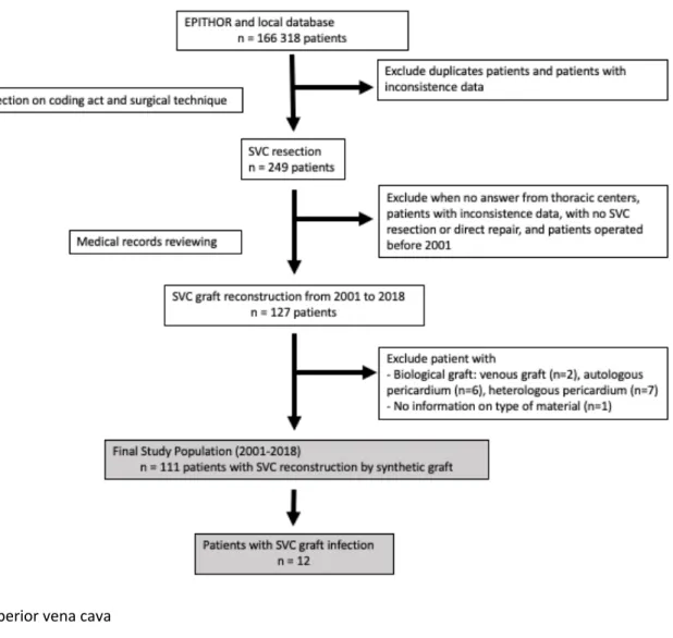
Data from 2001 to 2018, prospectively entered in EPITHOR and database from participating center, were retrospectively reviewed. Patient data were first screened on the coding act and surgical procedure. After eliminating all patients with inconsistent or missing values, patients with unknown information on mandatory variables for study, 249 patients were mentioned to have SVC resection. All participating thoracic centers were asked to review each medical record in order to check the type of SVC reconstruction (direct repair, patch enlargement or
graft reconstruction, replacement was performed with venous graft (n=2), autologous pericardium patch (n=6) or heterologous bovine (n=4) or equine pericardium patch (n=3). Patient who underwent a direct repair of SVC, autologous or heterologous pericardial patch were not included. Finally, we selected 111 patients with a synthetic SVC graft replacement (Fig.1). Ninety-eight patients were operated for malignant thoracic diseases (NSCLC, MT, neuroendocrine carcinoma, mesothelioma, metastasis of breast or rectal cancer, thyroid cancer) and thirteen for benign superior vena cava obstruction (secondary to indwelling catheter, fibrosis mediastinitis, hemangioendothelioma of SVC).
Baseline characteristics, comorbidities, treatment-related variables (neoadjuvant chemo or radiotherapy, type of lung or mediastinal resection, extended resection, lymph node dissection, partial or total SVC reconstruction, SVC clamping time, use of hemostatic or surgical glue), disease-related variables (histological finding, stage, tumors margin), and follow-up variables (adjuvant therapy, infectious complications and their management) were recorded.
The number of comorbidities were used as a categorial variable, since recent data from EPITHOR suggested the superiority of this variable in a predictive model of in-hospital mortality. The comorbidities selected for analysis were chronic bronchitis, smoking, pulmonary hypertension, pulmonary embolism, diabetes mellitus, coronary artery disease, valvular heart disease, cardiac arrhythmia, congestive heart failure, peripheral vascular
used for the diagnosis of graft infection (18,19). Diagnosis was made if two of the following three criteria were present: (i) clinical finding with either general signs (fever, sepsis, chills) or local signs (wound infection, bronchopleural fistula with pyothorax, mediastinitis); (ii) biological signs (C-reactive protein > 100mg/L, white blood count > 10 000/mm3) or
morphological signs of infection (perigraft air, abscess corresponding to pyothorax, perigraft fluid collection persisting more than 8 weeks after surgery, false aneurysm); (iii) positive bacterial culture of intraoperative samples or blood samples. SVC graft infection were classified as early-onset infection when occurring within 4 months postoperatively, and as late-onset when occurring after 4 months postoperatively (20). SVC graft in a pyothorax or mediastinitis where considered as graft infection.
Figure 1: Study flow chart of patients with SVC reconstruction after non-small cell lung cancer
resection, mediastinal tumor resection or benign superior vena cava obstruction.
SVC, superior vena cava
1.3. Outcome definition
Primary outcome was the incidence and management of SVC graft infection. Secondary outcomes were overall survival, operative and overall mortality, complications and risk factor
respiratory failure requiring invasive (acute respiratory distress syndrome) or non-invasive ventilation. Arrythmia, cardiac failure, coronary events, deep venous thrombosis, pulmonary embolism, stroke, acute limb ischemia were considered as cardiovascular complications. Surgical complications were prolonged air leak, hemothorax, bronchopleural fistula, empyema, nerve palsy (vocal cord and phrenic), wound abscess, cardiac luxation and mediastinitis. Infectious complications were urinary tract infection, septicemia or fever unrelated to pneumonia or surgical complications. Iatrogenic complications were defined as any allergy (to antibiotic therapy, low-weight heparin) or drug overdose (oral anticoagulation).
1.4. Statistical analysis
All statistical analyses were performed with R software (R-Project, version 3.6.1). The tests were two-sided with p<0.05 considered statistical significance. The assumption of normality was assessed with the Shapiro-Wilk test and homogeneity of the variance with a Bartlett test. Continuous variables are expressed as mean ± standard deviation and compared by a Student’s t-test or a Wilcoxon-Mann-Whitney test if non-compliance with normality. Categorial data were compared by a Chi-square test or a Fisher exact test if needed. Differences between the SVC graft infected patients and others were considered as significant for variables yielding a p-value £ 0.05. Preoperative and operative variables with a p<0.05 were included in the univariate analysis. A logistic regression analysis was performed to determine independent risk factors of graft infection. Odd ratios were calculated and presented with a 95% confidence interval. Analysis of actuarial survival was performed by means of Kaplan-Meier method. The log-rank test was performed to compare survival between NSCLC and MT.
2. Results:
2.1. Baseline characteristics
Characteristics of the 111 selected patients are shown in Table I.
Table I. Patient Characteristics
Characteristics All patients
(n = 111) Patients with no SVC graft infection (n = 99) Patients with SVC infection (n = 12) p-value Sex Men Females 76 (68.5) 35 (31.5) 67 (67.7) 32 (32.3) 9 (75) 3 (25) 0.75 Age 53 ± 15 53± 15 52 ± 17 0.966 BMI 25 ± 4.9 25± 4.9 23 ± 5.1 0.272 Number of comorbidities ³ 3 35 (31.5) 30 (30.3) 5 (41.7) 0.513 FEV1 % COPD 77 ± 19 37 (39.4) 79 ± 19 27 (32.9) 64 ± 17 10 (83.3) 0.014 0.001 Induction Therapy (%) 53 (47.7) 45 (45.5) 8 (66.7) 0.279 Type of resection n (%) Pneumonectomy Lobectomy
Mediastinal tumor resection Extend of resection
Atypical lung resection Lobectomy Carina Left Atrium Right Atrium Esophagus 32 (28.8) 35 (31.5) 41 (36.9) 24 (21.6) 13 (11.7) 21 (18.9) 4 (3.6) 6 (5.4) 1 (0.9) 26 (26.3) 34 (34.3) 37 (37.4) 20 (20.2) 12 (12.1) 18 (18.2) 4 (4) 6 (6.1) 0 6 (50) 1 (8.3) 4 (33.3) 4 (33.3) 1 (8.3) 3 (25) 0 0 1 (8.3) 0.101 0.099 0.99
Glue 28 (27.2) 23 (25.3) 5 (41.7) 0.3 Histology NSCLC Thymoma Thymic carcinoma NSGCT Others 48 (43.2) 17 (15.3) 8 (7.2) 13 (11.7) 22 (19.8) 41 (41.4) 16 (16.2) 7 (7.1) 12 (12.1) 20 (20.2) 7 (58.3) 1 (8.3) 1(8.3) 1 (8.3) 2 (16.7) Length of hospitalization (n) 19 ± 16 17 ± 13 36 ± 26 0.007
Numbers in parentheses are percentages (%). Others histology type included benign superior vena obstruction, metastasis of breast or rectal cancer, sarcoma, mesothelioma, lymphoma and thyroid cancer.
SVC, superior vena cava; BMI, body mass index; PTFE, polytetrafluorethylene; NSGCT, non-seminomatous germinal cell tumors
SVC reconstruction consisted in a patch enlargement in 3 patients (2.7%) and total graft replacement in 108 patients. Initial SVC replacement was made with PTFE graft in 109 patients (98.2%) and with Dacron in 2 (1.8%) patients. SVC reconstruction was truncal in 41 patients (36.9%), single left or right brachiocephalic vein in 61 patients (57%), and a Y-graft reconstruction in 3 patients (2.7%). Mean clamping time was 33 ± 19 minutes.
A total of twelve patients (10.8%) were diagnosis with SVC graft infection. Graft infected patients were more likely to be COPD (83.3% vs 32.9%; p=0.001), have a lower FEV1 (64 ± 17 vs 79 ± 19; p=0.014), a longer hospital stay (36 ± 26 days vs 17 ± 13 days; p=0.007). A significantly increased number of patients with graft infection received transfusion (100% vs 46.5%; p=0.003). There was no other statistical difference concerning induction therapy and operative characteristics (manual closure of bronchial stump, lymph node dissection, use of flap coverage and additional glue or hemostatic material) between the two groups.
Cardiovascular complications occurred in 33.3% in SVC graft infected patients and 29.3% in non-graft infected patients. There was significantly more surgical (100% vs 32.3% p < 0.001),
in patients with no-graft infection. Empyema, mediastinitis and BPF occur only in SVC graft infected patients, in 7 patients (58.3%), 5 patients (41.7%) and 4 patients (33.3%) respectively (p < 0.001).
2.2. SVC graft infection
Eleven patients underwent a single graft reconstruction and one patient a patch enlargement. Early-onset graft infection occurred in 11 patients (ten in the first month and one in the third month). Late-onset graft infection occurred in one patient at six years after initial surgery. All SVC graft infection was either due to pyothorax or mediastinitis. We report no case of graft infection secondary to hematogenous dissemination.
In early-onset graft infection, sepsis (including fever and hyperleukocytosis) was present in 6 patients (50%), purulent discharge in 4 patients (33.3%), sternal instability in one patient. Altered general status with chronic cough was present in one patient with late pyothorax. Computed tomography was available only in seven patient (58.3%) and revealed perigraft collection, retrosternal abscess, perigraft air bubbles, pleural fluid with bronchial stump dehiscence and sternal disjunction.
Identification of causative microorganism was achieved in 11 (91.6%) patients. Bacteriological finding and SVC graft infection management are listed in Table II. Staphylococcus species were identified in five patients, MRSA in two patients, Streptococcus species in two patients,
of the infection was obtained after a surgical debridement with bronchial stump closure alone (patient 7) or in association with flap coverage and surgical techniques decreasing pleural space (open window thoracotomy and/ or thoracoplasty). Among the two patients with
Candida albicans graft infection, one had later-onset graft infection. (patient 6). The patient
had several operations including epiplooplasty, open window thoracostomy, muscle flap coverage, thoracoplasty and partial removal of PTFE graft. Because of the recurrence of the sepsis, the two extremities of the PTFE graft were removed by sterno-thoracotomy (Annexe I). The patient is still alive with a chronic mild SVC syndrome without recurrence of disease and infection two years after the complete remove of the graft. The other one had early graft infection (patient 5). He was treated with BPF exclusion, omental flap wrapping around the SVC graft without infection recurrence at follow-up (Annexe II).
Five SVC graft infection were secondary to mediastinitis, all surgically managed with a large cleaning and debridement in association with adapted antibiotic therapy. In three patients, infection recurrence appeared, successfully managed either medically (long-life suppressive antibiotic) or surgically in case of virulent microorganism (Pseudomonas aeruginosa or MRSA): one omental flap and one graft excision. No SVC syndrome was present in the last patient before discharge.
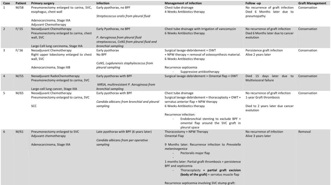
Table II: Outcomes for patients with SVC graft infection
Case Patient Primary surgery Infection Management of infection Follow -up Graft Management
1 M/58 Pneumonectomy enlarged to carina, SVC, esophagus, chest wall
Adenocarcinoma, Stage IIIA Adjuvant Chemotherapy
Early pyothorax, no BPF
Streptococcus oralis from pleural fluid
Chest tube drainage
4 Weeks Antibiotics therapy No recurrence of graft infection Died 6 Months later due to pneumopathy
Conservation
2 F/ 55 Neoadjuvant Chemotherapy
Pneumonectomy enlarged to carina, chest wall, SVC
Large Cell lung carcinoma, Stage IIIA
Early Pyothorax, no BPF P. Aeruginosa from pleural fluid
streptococcus, CoNS from pleural fluid and bronchial sampling
Chest tube drainage with Irrigation of vancomycin
6 Weeks Antibiotics therapy No recurrence of graft infection Died 6 Months later due to cancer evolution
Conservation
3 F/ 56 Neoadjuvant Chemotherapy
Right upper lobectomy enlarged to chest wall, SVC
Adenocarcinoma, Stage IIIB
Early pyothorax No BPF
CoNS, Lugdunensis staphylococcus from pleural sampling
Surgical lavage-debridement + OWT
+ NPW therapy + removal of osteosynthesis material. 6 Weeks Antibiotics therapy
Recurrence septicemia
- Suppressive antibiotherapy
Persistence graft infection
Alive 2 years later Conservation
4 M/55 Neoadjuvant RadioChemotherapy Pneumonectomy enlarged to carina, SVC Large-cell lung cancer, Stage IIIA
Early pyothorax with BPF
MRSA, multiresistant P. Aeruginosa from bronchial sampling
Surgical lavage-debridement + Omental flap + OWT Died 15 days later due to
Multivisceral failure Conservation 5 M/65 Neoadjuvant Chemotherapy
Pneumonectomy enlarged to carina, SVC SCC
Early pyothorax with BPF
Candida albicans from bronchial and pleural sampling
Chest tube drainage
Surgical lavage-debridement + thoracoplasty + OWT + serratus anterior flap + NPW therapy
6 Weeks Antibiotics therapy Recurrence infection:
- Endobronchial stenting to exclude BPF +
No recurrence of graft infection 1-year Graft thrombosis Died to 2 years later due cancer evolution
- Total excision of the graft. No
revascularization 7 M/ 64 Pneumonectomy enlarged to SVC
Adjuvant radio-chemotherapy SCC, Stage IIIA
Early pyothorax with BPF Microbiological sample negative
Surgical lavage-debridement + Closure of Bronchial stump
Recurrence infection and BPF: - OWT
Died due to respiratory failure 1
month later Conservation
8 M/ 20 Induction chemotherapy
Mediastinal germ cell mass excision enlarged to SVC, LA, ML RUL
Teratoma
Early mediastinitis
Methicillin-resistant Staphylococcus epidermidis from per operative sampling
Surgical lavage-debridement + irrigation with antiseptic solution
Persistence infection with collection in contact with SVC patch
- Patch excision
Lost to follow up at discharge Removal
9 F/ 17 Neoadjuvant chemotherapy
Thymectomy enlarged to RUL, pericardium, SVC
Thymic carcinoma, Masaoka IIIB
Early mediastinitis
MSSA from purulent discharge Peroperative sampling: negative
Surgical lavage-debridement
6 Weeks Antibiotics therapy Lost to follow-up at discharge Conservation
10 M/ 49 Thymectomy enlarged to SVC and RUL Adjuvant Radiotherapy
Thymoma B2, Masaoka IIIB
Early mediastinitis
MSSA from peroperative sampling
Surgical lavage-debridement
6 Weeks Antibiotics therapy Lost to follow-up at discharge Conservation 11 M/ 73 Tumor resection enlarged to SVC, RUL,
diaphragm
Sarcomatoid mesothelioma
Early mediastinitis
P. Aeruginosa from peroperative sampling
Surgical lavage-debridement Antibiotics therapy Persistence mediastinitis:
- Omental flap
No recurrence of infection
Alive 4 months later Conservation 12 M/ 55 SVC replacement
SVC obstruction due to indwelling catheter
Early mediastinitis MSSA from blood cultures
Surgical lavage-debridement 6 Weeks Antibiotics therapy Recurrence
- Suppressive antibiotherapy
No recurrence of infection
Alive 3 years later Conservation
BPF, bronchopleural fistula; MSSA, methicillin-sensible staphylococcus aureus; MRSA, methicillin-resistant staphyloccocus aureus; CoNS, Coagulase-negative staphylococcus;
P.Aeruginosa, Pseudomonas Aeruginosa; PTFE, polytetrafluoroethylene; OWT, open window thoracostomy; NPWT, negative pressure wound therapy; SCC, squamous cell
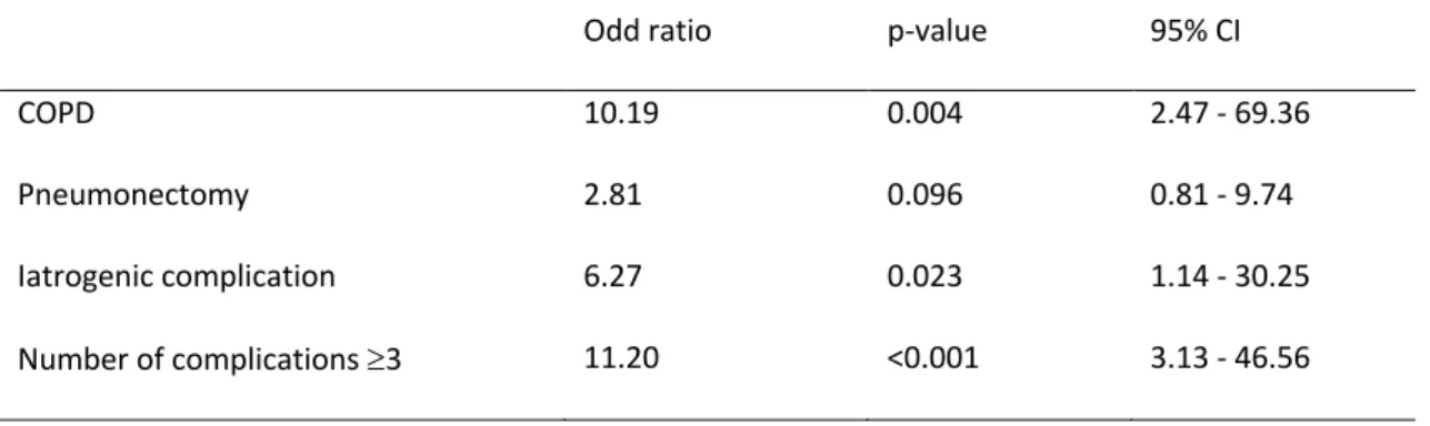
2.3. Risk factors
Risk factor of graft infection in univariate analyses are listed in Table III. The multivariate analysis demonstrates that risk factors of graft infection included COPD (OR 20.97, 95%CI 3.35-212.70; p=0.003) and iatrogenic complications (OR 9.09, 95%CI 1.12 - 88.66; p= 0.0038). The number of comorbidities ³ 3, age ³ 55 years and pneumonectomy were not significant.
Table III: Risk factors of graft infection in univariate analysis
Odd ratio p-value 95% CI
COPD 10.19 0.004 2.47 - 69.36
Pneumonectomy 2.81 0.096 0.81 - 9.74
Iatrogenic complication 6.27 0.023 1.14 - 30.25 Number of complications ³3 11.20 <0.001 3.13 - 46.56 CI, confidence interval
2.4. Long-term results
Mean follow-up was 24 ± 35 months. 61 patients (49.6%) were lost-to follow-up with a median follow-up of two months. At the completion of the study, 17 patients (14.5%) were still alive among them one having SVC graft infection. Overall survival is shown in Figure 2. The overall mortality rate was is 26.1% (n=29). The operative death rate is about 10.8% (n=12).
Figure 2: Survival curve: SVC replacement after NSCLC resection (red line) versus mediastinal
tumors (blue line). Five-year survival was 40% (n=13) after NSCLC resection and 70% after mediastinal tumors resection (NSGCT, thymoma, thymic carcinoma, sarcoma, and lymphoma). Log rank test p=0.01.
3.
Discussion
Main results
Our study is the first gathering as many patients with prosthetic SVC graft reconstruction and focusing on graft infection, its risk factors, bacteriological aspect, outcome and management. It confirms the scarcity and potential life-threatening condition of SVC graft infection as in other series mentioning this complication (7,9). Caused by contiguous
infection (empyema or mediastinitis), favored by surgical and iatrogenic complications, graft conservation was always attempted at first reoperation whatever bacteriological finding.
Primary Outcome
Different classification of prothesis vascular graft infection (PVGI) are described. Fitzgerald criteria are the most commonly used for diagnosis (18) and Samson classification for the deepness of infection (21). SVC graft infection can either be intracavitary if associated to empyema or extracavitary in case of mediastinitis. We did not used the Samson classification as all infections in our series involved at least the body of the graft with septicemia or fistula, mentioned as major graft infection(21). Recently, the MAGIC consensus proposed major and minor diagnosis criteria divided in three categories (clinical, radiological, laboratory) as for endocarditis diagnosis (22). The added value of this consensus relies on the adjunction of positron emission tomography (PET/CT), radiolabeled leukocyte imaging and the ranking of
Traditionally, PTFE is thought to be the best material for prosthetic SVC replacement. Known for its low thrombogenicity potential, long-term patency has been reported due to fast reendothelization of the inner lumen of the graft by a thin layer of neointima (7). Furthermore, rigidity associated with ringed PTFE (10) might prevent compression from surrounding structures or secondary to mediastinal fibrosis. However, our nation study confirmed a 10,8% risk of prosthetic SVC graft infection, in accordance with the 7% (1) (and 10% (7) rate reported in the literature. Meanwhile, no case of infection has been reported with bovine pericardial conduit (5,24–26) nor cryopreserved aortic allograft suggesting a better tolerance in infected condition. This latter has been used for benign pathologies (mediastinal fibrosis, chronic SVC obstruction) or malignant tumors resection without infection after a median follow-up ranging from 18 months to 4 years (27–29).
Our study shows a large majority of early-onset infection in 91.6% (n=11). All, but one, were treated with conservation of the prosthesis. Late-onset infection occurred only in 8.3% (fungal graft infection in patient 6) and need prosthesis removal. One persistence of graft infection was observed among the 7 survival patients with 19 months mean follow-up (4 - 36 months). Among the 5 other patients, 2 died postoperatively (patients 4 and 7) and 3 were lost to follow up after discharge (patients 8, 9 et 10).
Beside our series, the literature is scarce. To the best of our knowledge, six papers collecting eight cases are reported in literature. Spaggiari and colleagues reported two SVC prosthesis graft infection on 28 SVC graft reconstruction. One patient had a lung abscess and the other one a BPF (1,9) They also reported a case after an early empyema due to tracheal fistula following right upper lobectomy enlarged to carina and esophagus (30). All were treated with
recurrence. Alifano and colleagues reported a case of SVC graft infection to Gram-negative bacteria after an early empyema without BPF. Surgical debridement and latissimus dorsi flap were also performed (31). Bacha and colleagues successfully treated a mediastinitis with SVC graft infection with omentopexy and tracheostomy after a third recurrence of thyroid cancer. The association of antibiotics, omental flap and tracheostomy allowed a successful control of prosthesis infection due to mediastinitis after tracheal necrosis (7,32). Dartevelle and colleagues successfully treated a PTFE graft infection secondary to empyema with BPF. After failure of a Clagett procedure, the progressive graft clamping followed by graft removal allowed the control of infection although patient died of lung pneumonia (7). Itoda and colleagues reported a successful SVC graft excision after an early mediastinitis to MRSA following a thymectomy enlarged to the right upper lobe and SVC. That strategy was complicated by a pseudoaneurysm of the ascending aorta and redo surgery was needed (33). Okubo and colleagues reported a late chronic mediastinitis to Pseudomonas Aeruginosa complicated with mycotic aneurysm of the brachiocephalic artery and a septic SVC graft thrombosis after thymectomy in the treatment of a Masaoka stage III B3 thymoma. PTFE graft excision without reconstruction was performed, in association with brachiocephalic artery replacement using a Dacron graft and omental flap (34).
Surgical irrigation strategy (irrigation, chest tube drainage and antibiotic therapy) should be reserved to high risk patients due to a higher in-hospital mortality rate contrary to other options (36). Umminger and colleagues treated 14 patients according to this protocol. Among them 2 died postoperatively, and 2 patients were reoperated on due to graft infection recurrence (37).
The graft-sparing strategy (surgical debridement, muscle or omental flap, NPWT, and antibiotic therapy) in association with surgical technique to treat empyema (OWT, thoracoplasty) (38,39) appears to be the preferential option. In fact, this approach shows the lowest mortality and recurrence of infection rate (36,37). Furthermore, the use of muscle or omental flap to cover the graft is an independent protective factor in-hospital mortality in multivariate analysis (HR 0.24, 95% CI 0.10 - 0.56; p = 0.001) (36). In case of post pneumonectomy BPF, the control of the SVC graft infection is conditioned by the early treatment of BPF. The use of bronchial stenting should improve the outcome of these patients (40–42).
Graft removal was used in case of late-onset graft infection (37), fungal (43,44) and multiresistant microorganism graft infection (33). MRSA infection was found to be a risk factors of death with a significant lower one-year survival than non-MRSA graft infection (47.1% vs 78.6%, p= 0.016) (36). Moreover, according to last guidelines on thoracic graft infection, graft removal still remains the better option for major graft infection (Type III to V of Samson classification) (45). Revascularization may not be always mandatory and will depend on clinical signs of superior vena cava syndrome.
Suppressive antibiotic therapy is admitted when dealing with PVGI medically treated or with graft-sparing surgery in case of documented microorganism (46). Recently Oda and colleague
(96.8%) than patient without antibiotic therapy (76.2%) (36). Thus, the administration of a suppressive antibiotic therapy might be discussed after SVC graft infection treated with a graft-sparing surgery on a case-by-case basis after a multidisciplinary meeting.
Secondary outcomes
In our series, surgical complications including a majority of mediastinitis, empyema with or without BPF were found as a major local risk factor of SVC raft infection confirming the results of previous study (7). We think that early diagnosis of these complications may improve the control of infection and the prognosis.
We also found that patients with SVC graft infection had more severe COPD status and a lower FEV1 compared to non- graft infected patients. Our results differ of those from series including abdominal aortic graft or peripheral graft (19). General status of our patients is mainly dependent of COPD status while patients with aneurysmal or occlusive arterial pathologies have a higher trend to have obesity, diabetes mellitus, renal insufficiency (19,47).
Although ubiquitous, biofilm formation is a well-known trigger in the pathogenesis of prosthesis vascular graft infection (PVGI) (46,48). In fact, it contains all elements needed for bacterial growth, acts as a barrier for antibiotics decreasing their efficacity. It favored delayed infection and their chronicity. The three principal ways of PVGI are bacterial exposure during surgery, contiguous infectious process and hematogenous spread (49). In our series, no case
enteric fistula. MRSA is particularly virulent with the ability to destroy native vasculature (49). Furthermore, MRSA graft infection showed a significantly lower one-year survival contrary to no MRSA graft infection (47.1 vs 78.6%; p=0.016 respectively) (36). Gram-negative bacteria such as Pseudomonas aeruginosa are high virulence microorganism due to the production of toxins elastase and protease allowing erosion of intima and media layer of vessel (49). Recent guidelines on PVGI advice a graft excision whenever possible for major graft infection to MRSA, Pseudomonas aeruginosa or multidrug resistant microorganism (45).
Concerning thoracic vascular graft infection to candida species, few data are available. Medical treatment based on antifungal therapy, graft-sparing surgical management or graft excision have been used for thoracic aortic graft infection to Candida species. Results are controversy with failure and success with all three options (50–52). In any feasible case, authors advice surgical graft excision in association with closed long term follow up due to indolent candidemia. When excision is high risk surgery, graft-sparing strategy associated with suppressive antifungal therapy seems reasonable option (53). In previous study, no case of SVC graft infection to Candida albicans were reported. In our study, identified risk factor for candidemia in Patient 5 and 6 are neoadjuvant chemotherapy and broad-spectrum antibiotic therapy.
Limitations
This study presents some limitations. This is a retrospective study with a high number of patients lost to follow-up after discharge. Data concerning adjuvant therapy or microbiological results were not always available in medical records. Consequently, assessment of the graft infection on the adjuvant treatment were not feasible. Secondly, a small number of patients
The rate of SVC graft infection might be underestimated but it reflects the scarcity of this complication.
Conclusion
In conclusion, SVC graft infection is rare and dreaded complication. Treatment should be at least a surgical graft-sparing strategy with an extensive debridement and the use of a pedicled flap to cover the infected graft in association with an antibiotic therapy. Early-onset graft infection secondary to empyema without BPF, high risk patient or low-virulent pathogen seems appropriate criteria for a graft-sparing approach. Graft removal might be reasonable in case of late graft infection, in presence of bronchopleural fistula, virulent or resistant microorganism (Gram negative bacteria, MRSA, PA, candida species). Reconstruction in a contamined region might not be mandatory in case of graft thrombosis, the absence of superior vena cava syndrome and the presence of venous collateralization. We do not recommend medical conservative treatment due to poor results in eradicate the infection and a higher in-hospital morality rate.
REFERENCES
1. Spaggiari L, Thomas P, Magdeleinat P, Kondo H, Rollet G, Regnard JF, et al.
Superior vena cava resection with prosthetic replacement for non-small cell lung cancer: long-term results of a multicentric study. Eur J Cardiothorac Surg. juin 2002;21(6):1080‑6.
2. Dartevelle PG, Mitilian D, Fadel E. Extended surgery for T4 lung cancer: a 30 years’ experience. General Thoracic and Cardiovascular Surgery. juin 2017;65(6):321‑8.
3. Nakano T, Endo S, Kanai Y, Otani S, Tsubochi H, Yamamoto S, et al. Surgical outcomes after superior vena cava reconstruction with expanded polytetrafluoroethylene grafts. Ann Thorac Cardiovasc Surg. 2014;20(4):310‑5.
4. Sekine Y, Suzuki H, Saitoh Y, Wada H, Yoshida S. Prosthetic reconstruction of the superior vena cava for malignant disease: surgical techniques and outcomes. Ann Thorac Surg. juill 2010;90(1):223‑8.
5. Maurizi G, Poggi C, D’Andrilli A, Vanni C, Ciccone AM, Ibrahim M, et al. Superior Vena Cava Replacement for Thymic Malignancies. Ann Thorac Surg. févr
2019;107(2):386‑92.
6. Macchiarini P. Superior vena cava obstruction. In: Patterson G, Cooper J, Deslauriers J, Lerut A, Rive T, éditeurs. Pearson’s Thoracic and esophageal surgery. 3rd éd. Elsevier; 2008. p. 1684‑96.
7. Dartevelle PG, Chapelier AR, Pastorino U, Corbi P, Lenot B, Cerrina J, et al. Long-term follow-up after prosthetic replacement of the superior vena cava combined with resection of mediastinal-pulmonary malignant tumors. J Thorac Cardiovasc Surg. août 1991;102(2):259‑65.
8. Gloviczki P, Kalra M. Superior Vena Cava Obstruction: surgical treatment. In: Rutherford’s Vascular Surgery. Elsevier. 2014. p. 971‑81.
superior vena cava resection for lung cancer. Analysis of prognostic factors. Lung Cancer. juin 2004;44(3):339‑46.
10. Leo F, Bellini R, Conti B, Delledonne V, Tavecchio L, Pastorino U. Superior vena cava resection in thoracic malignancies: does prosthetic replacement pose a higher risk? Eur J Cardiothorac Surg. avr 2010;37(4):764‑9.
11. Okereke IC, Kesler KA, Rieger KM, Birdas TJ, Mi D, Turrentine MW, et al. Results of superior vena cava reconstruction with externally stented-polytetrafluoroethylene vascular prostheses. Ann Thorac Surg. août 2010;90(2):383‑7.
12. Lanuti M, De Delva PE, Gaissert HA, Wright CD, Wain JC, Allan JS, et al. Review of superior vena cava resection in the management of benign disease and pulmonary or
mediastinal malignancies. Ann Thorac Surg. août 2009;88(2):392‑7.
13. Bernard A, Bouchot O, Hagry O, Favre JP. Risk analysis and long-term survival in patients undergoing resection of T4 lung cancer. Eur J Cardiothorac Surg. août
2001;20(2):344‑9.
14. Venuta F, Rendina EA, Longo F, De Giacomo T, Anile M, Mercadante E, et al. Long-term outcome after multimodality treatment for stage III thymic tumors. Ann Thorac Surg. déc 2003;76(6):1866‑72; discussion 1872.
15. Thomas PA, Falcoz P-E, Bernard A, Le Pimpec-Barthes F, Jougon J, Brouchet L, et al. Bilobectomy for lung cancer: contemporary national early morbidity and mortality outcomes. Eur J Cardiothorac Surg. févr 2016;49(2):e38-43; discussion e43.
study for surgical management of thymectomy for non-thymomatous myasthenia gravis from the French national database EPITHOR. Eur J Cardiothorac Surg. sept 2016;50(3):418‑22. 18. FitzGerald SF, Kelly C, Humphreys H. Diagnosis and treatment of prosthetic aortic graft infections: confusion and inconsistency in the absence of evidence or consensus. J Antimicrob Chemother. déc 2005;56(6):996‑9.
19. Legout L, Sarraz-Bournet B, D’Elia PV, Devos P, Pasquet A, Caillaux M, et al.
Characteristics and prognosis in patients with prosthetic vascular graft infection: a prospective observational cohort study. Clin Microbiol Infect. avr 2012;18(4):352‑8.
20. Back MR. Local Complication: graft infection. In: Rutherford’s Vascular Surgery. Elsevier. 2014. p. 654‑72.
21. Samson RH, Veith FJ, Janko GS, Gupta SK, Scher LA. A modified classification and approach to the management of infections involving peripheral arterial prosthetic grafts. J Vasc Surg. août 1988;8(2):147‑53.
22. Habib G, Lancellotti P, Antunes MJ, Bongiorni MG, Casalta J-P, Del Zotti F, et al. 2015 ESC Guidelines for the management of infective endocarditis: The Task Force for the Management of Infective Endocarditis of the European Society of Cardiology (ESC). Endorsed by: European Association for Cardio-Thoracic Surgery (EACTS), the European Association of Nuclear Medicine (EANM). Eur Heart J. 21 nov 2015;36(44):3075‑128. 23. Lyons OTA, Baguneid M, Barwick TD, Bell RE, Foster N, Homer-Vanniasinkam S, et al. Diagnosis of Aortic Graft Infection: A Case Definition by the Management of Aortic Graft Infection Collaboration (MAGIC). Eur J Vasc Endovasc Surg. déc 2016;52(6):758‑63. 24. Ciccone AM, Venuta F, D’Andrilli A, Andreetti C, Ibrahim M, De Giacomo T, et al. Long-term patency of the stapled bovine pericardial conduit for replacement of the superior vena cava. Eur J Cardiothorac Surg. déc 2011;40(6):1487‑91; discussion 1491.
Reconstruction of the superior vena cava by biologic conduit: assessment of long-term patency by magnetic resonance imaging. Ann Thorac Surg. sept 2013;96(3):1039‑45.
26. Spaggiari L, Leo F, Veronesi G, Solli P, Galetta D, Tatani B, et al. Superior vena cava resection for lung and mediastinal malignancies: a single-center experience with 70 cases. Ann Thorac Surg. janv 2007;83(1):223‑9; discussion 229-230.
27. Spera K, Kesler KA, Syed A, Boyd JH. Human aortic allograft: an excellent conduit choice for superior vena cava reconstruction. J Cardiothorac Surg. 15 janv 2014;9:16. 28. Tyner J, Baradarian S, Armstrong B, Cutter S. Bilateral brachiocephalic vein and superior vena cava reconstruction with an aortic allograft. Ann Thorac Surg. 21 juin 2019; 29. Gómez-Caro A, Martinez E, Rodríguez A, Sanchez D, Martorell J, Gimferrer JM, et al. Cryopreserved arterial allograft reconstruction after excision of thoracic malignancies. Ann Thorac Surg. déc 2008;86(6):1753‑61; discussion 1761.
30. Spaggiari L, Solli P, Veronesi G, Pastorino U. Intrathoracic myoplasty for prosthesis infection after superior vena cava replacement for lung cancer. Ann Thorac Surg. oct
2002;74(4):1231‑3.
31. Alifano M, Puyo P, Magdeleinat P, Levasseur P, Regnard JF. Management of empyema complicating lobectomy with superior vena cava replacement. Ann Thorac Surg. nov 2000;70(5):1720‑1.
32. Bacha EA, Chapelier AR, Macchiarini P, Fadel E, Dartevelle PG. Surgery for invasive primary mediastinal tumors. Ann Thorac Surg. juill 1998;66(1):234‑9.
irradiation. J Thorac Cardiovasc Surg. sept 2005;130(3):918‑9.
35. Saleem BR, Meerwaldt R, Tielliu IFJ, Verhoeven ELG, van den Dungen JJAM, Zeebregts CJ. Conservative treatment of vascular prosthetic graft infection is associated with high mortality. Am J Surg. juill 2010;200(1):47‑52.
36. Oda T, Minatoya K, Kobayashi J, Okita Y, Akashi H, Tanaka H, et al. Prosthetic vascular graft infection through a median sternotomy: a multicentre review †. Interact Cardiovasc Thorac Surg. juin 2015;20(6):701‑6; discussion 706.
37. Umminger J, Krueger H, Beckmann E, Kaufeld T, Fleissner F, Haverich A, et al. Management of early graft infections in the ascending aorta and aortic arch: a comparison between graft replacement and graft preservation techniques. Eur J Cardiothorac Surg. oct 2016;50(4):660‑7.
38. Krassas A, Grima R, Bagan P, Badia A, Arame A, Barthes FLP, et al. Current indications and results for thoracoplasty and intrathoracic muscle transposition. Eur J Cardiothorac Surg. mai 2010;37(5):1215‑20.
39. Regnard JF, Alifano M, Puyo P, Fares E, Magdeleinat P, Levasseur P. Open window thoracostomy followed by intrathoracic flap transposition in the treatment of empyema complicating pulmonary resection. J Thorac Cardiovasc Surg. août 2000;120(2):270‑5. 40. Andreetti C, D’Andrilli A, Ibrahim M, Ciccone AM, Maurizi G, Mattia A, et al. Effective treatment of post-pneumonectomy bronchopleural fistula by conical fully covered self-expandable stent. Interact Cardiovasc Thorac Surg. avr 2012;14(4):420‑3.
41. Andreetti C, Menna C, D’Andrilli A, Ibrahim M, Maurizi G, Poggi C, et al. Multimodal Treatment for Post-Pneumonectomy Bronchopleural Fistula Associated With Empyema. Ann Thorac Surg. déc 2018;106(6):e337‑9.
43. Nicolini F, Budillon AM, Borrello B, Gherli T. Ascending aortic graft thrombosis from an endoluminal candida albicans infection. Eur J Cardiothorac Surg. mai
2015;47(5):e233-234.
44. Doscher W, Krishnasastry KV, Deckoff SL. Fungal graft infections: case report and review of the literature. J Vasc Surg. oct 1987;6(4):398‑402.
45. Wilson WR, Bower TC, Creager MA, Amin-Hanjani S, O’Gara PT, Lockhart PB, et al. Vascular Graft Infections, Mycotic Aneurysms, and Endovascular Infections: A Scientific Statement From the American Heart Association. Circulation. 15 2016;134(20):e412‑60. 46. Revest M, Camou F, Senneville E, Caillon J, Laurent F, Calvet B, et al. Medical treatment of prosthetic vascular graft infections: Review of the literature and proposals of a Working Group. Int J Antimicrob Agents. sept 2015;46(3):254‑65.
47. Mirzaie M, Schmitto JD, Tirilomis T, Fatehpur S, Liakopoulos OJ, Teucher N, et al. Surgical management of vascular graft infection in severely ill patients by partial resection of the infected prosthesis. Eur J Vasc Endovasc Surg. mai 2007;33(5):610‑3.
48. Darouiche RO. Treatment of infections associated with surgical implants. N Engl J Med. 1 avr 2004;350(14):1422‑9.
49. Bianco V, Kilic A, Gleason TG, Arnaoutakis GJ, Sultan I. Management of thoracic aortic graft infections. J Card Surg. oct 2018;33(10):658‑65.
50. Nemoto T, Tokuda Y, Hirose M, Naitoh Y, Yamasaki Y, Shimizu T, et al. Thoracic Aortic Graft Infection due to Candida Albicans with Multiple Embolism in the Left-side
labeled white blood cells. Clin Nucl Med. déc 1984;9(12):734.
53. Di Benedetto G, Citro R, Longobardi A, Mastrogiovanni G, Panza A, Iesu S, et al. Giant Candida mycetoma in an ascending aorta tubular graft. J Card Surg. sept
Annexe I
Computed tomography scanner of patient 6. a. At diagnosis of graft infection recurrence, computed tomography scanner shows perigraft air bubble. b. After graft removal, pleural space filled of pectoralis muscle and anterior serratus muscle flap.
Annexe II
Computed Tomography scanner of patient 5. At initial diagnosis, right pleural space full off liquid and air bubbles around the right bronchial stump. b. After first graft-sparing surgery consisted in thoracoplasty, thoracostomy and omental flap. c. Graft thrombosis at 1-year.
(Conseil national de l’ordre des médecins) SERMENT D'HIPPOCRATE
Au moment d’être admis(e) à exercer la médecine, je promets et je jure d’être fidèle aux lois de l’honneur et de la probité.
Mon premier souci sera de rétablir, de préserver ou de promouvoir la santé dans tous ses éléments, physiques et mentaux, individuels et sociaux.
Je respecterai toutes les personnes, leur autonomie et leur volonté, sans aucune discrimination selon leur état ou leurs convictions. J’interviendrai pour les protéger si elles sont affaiblies, vulnérables ou menacées dans leur intégrité ou leur dignité. Même sous la contrainte, je ne ferai pas usage de mes connaissances contre les lois de l’humanité.
J’informerai les patients des décisions envisagées, de leurs raisons et de leurs conséquences.
Je ne tromperai jamais leur confiance et n’exploiterai pas le pouvoir hérité des circonstances pour forcer les consciences.
Je donnerai mes soins à l’indigent et à quiconque me les demandera. Je ne me laisserai pas influencer par la soif du gain ou la recherche de la gloire.
Admis(e) dans l’intimité des personnes, je tairai les secrets qui me seront confiés. Reçu(e) à l’intérieur des maisons, je respecterai les secrets des foyers et ma conduite ne servira pas à corrompre les mœurs.
Je ferai tout pour soulager les souffrances. Je ne prolongerai pas abusivement les agonies. Je ne provoquerai jamais la mort délibérément.
Je préserverai l’indépendance nécessaire à l’accomplissement de ma mission. Je n’entreprendrai rien qui dépasse mes compétences. Je les entretiendrai et les perfectionnerai pour assurer au mieux les services qui me seront demandés.
J’apporterai mon aide à mes confrères ainsi qu’à leurs familles dans l’adversité. Que les hommes et mes confrères m’accordent leur estime si je suis fidèle à mes