Original article
Menstrual cycle and timing of breast surgery in premenopausal node-positive
breast cancer: Results of the International Breast Cancer Study Group
(IBCSG) Trial VI
A. Goldhirsch, R. D. Gelber, M. Castiglione, A. O'Neill, B. Thurlimann, C.-M. Rudenstam,
J. Lindtner, J. Collins, J. Forbes, D. Crivellari, A. Coates, F. Cavalli, E. Simoncini, M. F. Fey,
O. Pagani, K. Price, H.-J. Senn & other members of the International Breast Cancer Study Group
* Seepage 755 for list of participating institutions and authors
Summary
Purpose: It has been postulated that breast cancer surgery
performed during the follicular phase of the menstrual cycle is
associated with poorer outcome.
Patients and methods: We tested this hypothesis by
evaluat-ing disease-free survival (DFS) for 1033 premenopausal
pa-tients who received definitive surgery either during the
follicu-lar phase (n = 358) or the luteal phase (n - 675). All patients
were enrolled in a randomized trial conducted between July
1986 and April 1993. All had node positive breast cancer and
randomization was stratified by estrogen receptor (ER) status.
All patients received at least three cycles of adjuvant
cyclo-phosphamide, methotrexate, and 5-fluorouracil (CMF). The
median follow-up was 60 months.
Results: Patients who underwent definitive surgery for
breast cancer in the follicular phase had a slightly worse
disease-free survival than those operated on during the luteal
phase (five-year DFS percentage: 53% versus 58%; hazard
ratio, 1.13; 95% confidence interval (CI), 0.94-1.38; P = 0.20).
The effect was significantly greater for the subpopulation of
300 patients with ER-negative primaries (P = 0.02 interaction
effect; five-year DFS percentages 42% vs. 59%; hazard ratio
1.60; 95% CI, 1.12-2.25; P = 0.008). The effect of timing of
surgery diminished for analyses based on lesser surgical
proce-dures, e.g., excisional biopsies. In particular, no effect of
tim-ing was observed for fine needle aspiration procedures.
Conclusions: Surgical procedures which are more extensive
than a fine needle aspiration biopsy might be associated with
worse prognosis if conducted during the follicular phase of the
menstrual cycle. This phenomenon was seen predominantly for
high risk breast cancer with low levels or no estrogen receptors
in the primary tumor.
Key words: adjuvant chemotherapy, breast cancer, menstrual
cycle, premenopausal, surgery
Introduction
The timing of surgery within the various phases of the
menstrual cycle was hypothesized to influence
disease-free survival and overall survival for patients with
oper-able breast cancer. However, the data from various
retro-spective analyses of this aspect provide conflicting
re-sults [1, 2]. The first report that premenopausal women
who are operated during the follicular phase have a
significantly worse prognosis when compared with those
operated during the luteal phase was based upon a
cohort of 41 patients [3]. This observation was
con-firmed by some investigators [4-7], while others did not
find a significant difference in prognosis according to
whether the operation took place in the follicular or in
the luteal phase [8-10]. In the largest series in which a
difference was observed [7], its magnitude was greater
for patients with node-positive disease. The relationship
of the extent of a surgical procedure to the menstrual
phase [11] is also controversial. We, therefore,
systemati-cally collected data on the menstrual phase for patients
with node-positive breast cancer who entered a
random-ized trial in which all received CMF adjuvant
chemo-therapy without the addition of endocrine
manipula-tions. Furthermore, we recorded all surgical procedures
which were associated with the diagnosis and treatment
of the disease [12].
Patients and methods
Data from International Breast Cancer Study Group (IBCSG) trial VI [12], which accrued 1554 pre- and perimenopausal patients from July 1986 to April 1993 were considered for the analysis. All patients had a histologically proven, node positive unilateral breast cancer with either estrogen receptor (ER) positive or negative status. Surgery of the primary tumor was either a total mastectomy with axillary clearance or a breast conserving procedure (quadrantectomy or lumpectomy) with axillary lymph node clearance and subsequent local radiotherapy. Patients received one of the following: A) cyclophosphamide, metho-trexate, and fluorouracil for six consecutive courses on months 1 to 6 (CMF x 6); B) CMF x 6 plus three single courses of reintroduction CMF given on months 9, 12, and 15; Q CMF for three consecutive courses on months 1 to 3 (CMF x 3); or D) CMF x 3 plus three single
Table 1. Reasons for exclusion of patients from timing of surgery
analysis.
Day at surgefy ocfore after
Figure 1. Date of most recent menses prior to entry into the trial and
estimation of menstrua] phase.
courses of reintroduction CMF given on months 6, 9, and 12. Seventy-five percent of the patients were randomized to receive at least six cycles of CMF. Trial details, eligibility and evaluation, as well as results at 60 months median follow-up are described elsewhere [12]. For this analysis, the timing of definitive surgery (total mastectomy, or lumpec-tomy or quadranteclumpec-tomy) in relation to a woman's menstrual cycle was considered (definition A). When each patient enrolled in the trial we asked 'date start of most recent menstruation (prior to date of random-ization)'. We refer to this as 'menses date' in this paper. If definitive surgery was between 3 and 12 days (inclusive) following menses date, or between 16 and 25 days prior to menses date, then surgery was said to be performed during the follicular phase. If definitive surgery was between 0 and 2 or 13 and 28 days following recent menses date, or between 1 and 15 or 26 and 28 days prior to menses date, then surgery was said to be performed during the luteal phase (Figure 1). Patients who had a hysterectomy (n - 106), had surgery performed beyond 28 days of last menses (n = 233), or had an incomplete menses date, making it impossible to classify the timing of surgery (n = 103), were excluded from the analysis. Of the 1475 eligible patients from trial VI, 1033 had sufficient data to be included in this analysis (Table 1).
To evaluate whether surgeries of lesser extent have a similar influ-ence on the results, we considered two additional analyses. In the first, the date of surgical procedure was the date of definitive surgery or the date of a diagnostic procedure which was more intrusive than fine needle aspiration (trucut, incisional, or excisional biopsy), whichever was performed first (n = 1016, definition B, Table 1). In the second analysis, the date of surgery was the date of fine needle aspiration. This analysis was restricted to only those patients who had this less intrusive procedure (n - 465, definition C, Table 1). For both of these analyses, menses phase was determined as described previously for definition A. ER subgroups were also considered within each of the definitions since ER status was a stratification factor in trial VI and ER status has been an important factor in predicting response to endocrine therapies. Disease-free survival (DFS) was defined as the length of the time from the date of randomization to any relapse (including ipsilateral
Total eligible Evaluable Not evaluable
Did not receive fine needle aspiration Missing 'method of diagnosis' date Hysterectomy
Surgery beyond 28 days of menses Incomplete menses dateb
Definition* A 1475 1033 -106 233 103 B 1475 1016 -4 106 246 103 C 1475 465 727 13 40 180 50 * Definition A: tuning of definitive surgery; definition B: timing of first invasive procedure; definition C: timing of fine needle aspiration.
Includes patients with incomplete date of last menses (primarily missing day only) who could not be classified as having the target procedure (definition A, B, Q beyond 28 days of menses.
breast recurrence), the appearance of a second primary cancer (includ-ing contralateral breast cancer), or death, whichever occurred first. The Kaplan-Meier method was used to estimate survival distributions for DFS [13]. The two-sided log-rank procedure was used to assess the statistical significance of treatment differences between the survival distributions [14], Multivariate analyses were conducted using Cox proportional hazards regression models [15]. The data were analyzed at a median observation time of 60 months, and five-year DFS percen-tages are presented.
Results
The first analysis included 1033 patients out of the 1475
women randomized in trial VI. Overall there was no
significant difference in the disease-free survival between
the groups operated on in the two distinct phases of the
menstrual cycle (Figure 2a; log-rank P - 0.20).
Evaluat-ing the results separately for the two prospectively
strati-fied subgroups, there was an effect among patients with
estrogen receptor-negative tumors, while no effect was
observed for patients with estrogen receptor-positive
0 1 2 3 4
Years from Randomization
1 2 3 4
Years from Randomization
0 1 2 3 4
Years from Randomization
Figure 2. Kaplan-Meier plots for disease-free survival according to the timing of definitive surgery within the menstrual cycle (follicular or luteal
phase) for premenopausal patients with node-positive breast cancer in IBCSG Trial VI (12): all patients (n = 1033, panel A\ patients with estrogen receptor-negative tumors (n = 300, panel B), or patients with estrogen receptor-positive tumors (n = 733, panel Q .
Table 2 Estimated five-year DFS percentages and hazard ratios
according to menses phase for definition A.
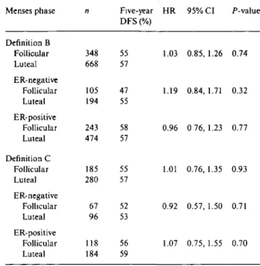
Table 3. Estimated five-year DFS percentages and hazard ratios
according to menses phase for definition B and C. Patient population n Five-year
menses phase DFS (%)
HR 95% CI /"-value Menses phase Five-year DFS (%)
HR 95% CI P-value
All patients (definition A) Folhcular Luteal ER-negative Follicular Luteal ER-positive Follicular Luteal 358 675 108 192 250 483 Mastectomy (definition A) Follicular 245 Luteal 470 ER-negative Follicular Luteal ER-positive Follicular Luteal 72 137 173 333 53 58 42 59 57 58 49 53 38 58 53 51 Less than mastectomy (definition A)
Follicular 113 62 Luteal 205 70 ER-negative Follicular Luteal ER-positive Follicular Luteal 36 55 77 150 49 60 68 73 1.13 1.60 0.98 1.07 1.62 0.93 1.41 1.57 1 24 0.94, 1.38 1.12,2.25 0.77, 1.24 0.86, 1.35 1.08,2.43 0.70, 1.19 0.93,2.12 0.80, 3.08 0.73,2.11 0.20 0.008 0.86 0.52 0.02 0.51 0.11 0.18 0.43
Abbreviations: n - number of patients; DFS - disease-free survival; HR - hazard ratio, follicular: luteal; 95% CI - 95% confidence interval; ER - estrogen receptor.
tumors (Figures 2b and 2c, respectively; test for
inter-action P = 0.02). Patients with estrogen
receptor-nega-tive tumors whose operation was performed during the
follicular phase had a five-year disease-free survival of
42% compared with 59% for those operated on during
the luteal phase (hazard ratio 1.6; 95% CI 1.12-2.25;
P = 0.008). In contrast, patients with estrogen
receptor-positive tumors had five-year disease-free survival of 57%
and 58% if operated on during the follicular or luteal
phase, respectively.
To investigate whether the results were influenced by
the extent of definitive surgery, we evaluated the
out-come separately for patients who received a mastectomy
or less than mastectomy. For the patients who received a
mastectomy (n = 715), the results were similar. Overall
there was no significant difference in disease-free survival
between the two menses categories (log-rank P = 0.52).
Again, an effect between menses categories was only
found for the patients with estrogen receptor-negative
tumors (test for interaction P = 0.02). For the patients
who received less than mastectomy (n = 318), no effect
was found overall or within estrogen receptor subgroups
(test for interaction P - 0.56). Table 2 presents the
Definition B Follicular Luteal ER-negative Follicular Luteal ER-positive Follicular Luteal Definition C Follicular Luteal ER-negative Folhcular Luteal ER-positive Follicular Luteal 348 668 105 194 243 474 185 280 67 96 118 184 55 57 47 55 58 57 55 57 52 53 56 59 1.03 0.S5, 1.26 0.74 1.19 0.84,1.71 0.32 0.96 0 76,1.23 0.77 1.01 0.76, 1.35 0.93 0.92 0.57, 1.50 0.71 1.07 0.75,1.55 0.70
Abbreviations: see Table 2.
estimated five-year disease-free survival percentages,
hazard ratios, and P-values for the results according to
timing of definitive surgery.
The second analysis included 1016 patients and was
based on date of first surgical procedure including any
type of surgery defined as more intrusive than a fine
needle aspiration. Of the women 217 (22%) received
either a trucut, incisional, or excisional biopsy. Again
there was no significant difference in the disease-free
survival between the groups operated on in the two
distinct phases of the menstrual cycle (log-rank P = 0.74).
Evaluating the results separately for the two prospectively
stratified subgroups, the difference in effects between the
estrogen receptor-negative and estrogen
receptor-posi-tive cohorts was no longer statistically significant (test
for interaction P - 0.31). Table 3 presents the estimated
five-year disease-free survival percentages, hazard ratios,
and P-values for these analyses.
The third analysis included 465 patients for whom a
fine needle biopsy was performed for cytology. There
was no difference in disease-free survival between the
groups operated on in the two distinct phases of the
menstrual cycle either overall (log-rank P — 0.93), or for
subpopulations defined by estrogen receptor content of
the primary tumor. Table 3 presents the results of these
analyses.
Multivariate analyses using proportional hazards
re-gression models were conducted to adjust for effects of
estrogen receptor status (positive vs. negative), number
of positive nodes ( ^ 4 vs. 1 -3), age ( ^ 40 vs. < 40 years),
tumor size ( > 2 vs. < 2 cm), tumor grade (III vs. other;
unknown vs. other), vessel invasion (yes vs. unknown;
no vs. unknown), and treatment (CMF x 3 vs. other).
The conclusions based on these models are the same as
those based on the univariate analyses shown in Tables 2
and 3. The effect of timing of surgery remained
stat-istically significant for the patients with ER negative
primaries.
Discussion
The timing of surgery within various phases of the
menstrual cycle was hypothesized to influence prognosis
of premenopausal patients with breast cancer. Surgery
during the follicular phase was thought to be
unfavor-able when compared with surgery during the luteal
phase [1]. The tissue trauma due to surgery is known to
enhance biological processes that may stimulate tumor
growth [16]. In studies conducted on the production of
growth factors by surgically traumatized tissues, an
increased production of TGF-a was observed at the
wound site [17]. It is known that estrogens may lead to
an increased production of TGF-a by the stroma and by
estrogen receptor containing tumor cells [18]. Estrogens
may cause a greater susceptibility to the effects of
growth factors on tumor cells which are rapidly
prolifer-ating; for example, those which do not contain estrogen
receptors [19]. The presence of progesterone, naturally
available during the luteal phase, might slow this tumor
cell proliferation. Increased availability of progesterone
was hypothesized to be associated with improved
out-come for women operated during the luteal phase [20].
Mechanisms related to invasion, metastatic potential
and angiogenesis might also be affected differently in
the absence or presence of progesterone [21, 22].
Several features distinguish our study population
from other series that addressed the timing of surgery
and menstrual cycle. All patients had node-positive
dis-ease, all received CMF adjuvant chemotherapy, and all
had estrogen receptor data available prior to study
entry. We also conducted three different analyses defined
according to the extent of surgery used. The magnitude
of the decrease in disease-free survival associated with
surgery during the follicular phase was reduced as the
extent of the surgical intervention decreased. We also
observed that the effect of the timing of surgery was
most striking for patients with estrogen receptor
nega-tive tumors.
For the premenopausal patient the definition of an
estrogen receptor-negative tumor is confounded by the
presence of circulating estrogens and by changes in
ex-pression of steroid hormone receptors during the
men-strual cycle [23]. It should also be recognized that there
is a lack of precision in determining the phase of the
cycle. We did not measure hormone levels at the time of
surgery. Nevertheless, this is the first time that we have
observed an effect of the timing of surgery within the
phase of the menstrual cycle. Although the results of
the subgroup analyses should be treated with caution,
there is some biologic rational for observing the effect
in the subpopulation of patients with estrogen
receptor-negative tumors. Such tumors have a more rapid cell
proliferation and are associated with a higher risk of
relapse despite adjuvant chemotherapy. An association
between timing of surgery in the menstrual cycle and
outcome among patients with estrogen
receptor-nega-tive tumors was also observed by Saad et al. [24].
Before analysis we recorded the hypothesis that the
largest effect would be seen in patients with estrogen
receptor-negative tumors. Our data supported this
hy-pothesis.
We identified the patients having tumors with the
highest proliferative potential and the worst prognosis
as those whose clinical course was most strongly
asso-ciated with the timing of the surgical procedure.
How-ever, this is the very population of patients whose
out-come might be adversely affected by delaying surgical
intervention to await the luteal phase of the cycle.
Alter-native interventions should therefore be considered to
reduce tumor cell proliferation and/or alter the hormonal
status of the host in such patients. Patient selection for
these procedures requires pre-operative evaluation. A
potential advantage of pre-operative chemotherapy,
cur-rently being evaluated in randomized clinical trials,
might be seen particularly in those patients with rapidly
proliferating tumors who undergo invasive surgical
pro-cedures during the follicular phase.
Although the IBCSG has not yet investigated
pre-operative chemotherapy, our trial of peripre-operative
chemo-therapy [25] supports the idea that such treatment may
be more effective in patients with estrogen
receptor-negative tumors. Thus, among 168 premenopausal
pa-tients with estrogen receptor-negative tumors, there was
a trend toward superior five-year disease-free survival
for patients commencing therapy in the perioperative
period (55% ± 5%) compared with those receiving only
conventionally timed therapy (44% ± 4%; P - 0.39). No
such trend was seen among patients with estrogen
re-ceptor-positive tumors.
These results highlight the importance of
consider-ing tumor and host factors when developconsider-ing optimal
integrated strategies for management of early breast
cancer.
Acknowledgements
We thank the patients, physicians, nurses, and data
man-agers who participate in the International Breast Cancer
Study Group trials. We also acknowledge Mary Isley for
her special contributions as Coordinating Data Manager
for the group. We also gratefully acknowledge the initial
support provided by the Ludwig Institute for Cancer
Research and the continuing support for central
coordi-nation, data management, and statistics provided by
The Swiss Cancer League, The Cancer League of Ticino,
The Swedish Cancer League, The Australia-New
Zea-land Breast Cancer Trials Group, The National Health
and Medical Research Council of Australia (grant
num-bers 880513 and 910420), The Australian Cancer Society,
The Frontier Science and Technology Research
Founda-tion, and The Swiss Group for Clinical Cancer Research.
* Appendix
International Breast Cancer Study Croup: Participants and authors International Breast Cancer Study Group, Bern, Switzerland: M. Cas-tiglione-Gertsch, A Goldhirsch (study coordinators), B. Gusterson, R. Bettelheim, R. Reed (study pathologists), H. Gusset, K. Geiser (data management), Ch. Hurny, J. Bernhard (quality of life office), A. Hangartner, R. Maibach, R. Pedowski; Dana-Farber Cancer In-stitute and Harvard Medical School, Boston, MA, USA: R. Gelber (study statistician), K. Price, H. Peterson, A. O'Neill, M. Zelen; Frontier Science & Techology Research Foundation, Amherst, NY, USA: M. Isley, R. Hinkle; Auckland Breast Cancer Study Group, Auckland, New Zealand: R. G. Kay, I. M. Holdaway, V. J. Harvey, M. F. Jagusch, L. Neave, B. M. Mason, B. Evans, C. S. Benjamin, J. F. Carter, J. C. Gillman, D. Mack, D. Benson-Cooper; Centro di Riferi-mento Oncologico, Aviano, Italy: S. Monfardini, E. Galhgioni, D. Crivellari, A. Buonadonna, S. Massarut, C. Rossi, E. Candiani, A. Carbone, R. Volpe, M. G. Trovo, M. Roncadin, G. F. Santini, D. Villalta, F. Coran, S. Morassut; Spedali Civili & Fondazione Beretta, Brescia, Italy: G. Marini, E. Simoncini, P. Marpicati, A. Zaniboni, U. Sarton, A. Barni, L. Ceratti, A. Alghisi, E. Raffaglio, M. P. Garattini, A. Albertini, P. Grigolato, L. Morassi, R. Bergonzini, C. Lauriola; Groote Schuur Hospital, Cape Town, Rep. of South Africa: A. Gudgeon, D. M. Dent, A. Tiltman, A. Hacking, E. Dowdle, P. Steynor, J. Toop; University of Dusseldorf, Dusseldorf, Germany: H. G. Schnurch, D. Mosny, H. G. Bender, H. Bojar; Presidio Ospeda-liero, Gorizia, Italy: A. Veronesi, S. Foladore, G. Pamich, G. Bianchi, A. Torretta; West Swedish Breast Cancer Study Group, Goteborg, Sweden: C. M. Rudenstam, A. Wallgren, S. Persson, J. Mattsson, E. Cahlin, L. O. Hafstrom, S. Holmberg, R. Hultborn, G. Colldahl-Jaderstrdm, B. Gustavsson, G. Carlsson, L. Ivarsson, O. Thoren, O. Ruusvik, L. G. Nicklasson, S. Dahlin, G. Karlsson, B. Lindberg, A. Sundback, S. Bergegardh, H. Salander, C. Andersson, Y. Hessman, O. Nelzen, M. Heideman, T. Ramhult, J. H. Svensson, P. Lindberg, S. BjSrk; The Institute of Oncology, Ljubljana, Slovenia: J. Lindtner, D. Erzen, O. Cerar, B. Stabuc, R. Golouh, J. Lamovec, S. Sebek, M. Kramberger, I. Vrhovec; Madrid Breast Cancer Group, Madrid, Spain: H. Cortes-Funes, F. Martinez-Tello, C. Mendiola, F. Cruz-Vigo, M. L. Larrodera, A. Sierra, P. Miranda, S. Alonso; Anti-Cancer Counsil of Victoria, Melbourne, Australia: J. Collins, R. Snyder, P. Gregory, W. I. Burns, M. Green, T. Gale, M. Henderson, S. Hart, S. Neil, P. Kitchen, R. Lovell, R. McLennan, R. Reed, I. Russell, M. Schwarz, R. Basser; Royal Adelaide Hospital, Adelaide, Australia: A. Robertson, P. Gill, M. L. Carter, P. Malycha, E. Yeoh, G. Ward, A. S. Y. Leong, J. Lommax-Smith, D. Hoosfall, R. D'Angelo; Sir Charles Gairdner Hospital, Nedlands, Western Autralia: M. Byrne, G. van Hazel, J. Dewar, M. Buck, H. J. Sheiner, D. Ingram, G. Sterrett, R. Hahnel; University of Newcastle, Waratah, Australia: J. F. Forbes, J. Steward, S. W. Darbar, J. M. Bishop, B. Simms, V. Ziogas; University of Sydney and Royal Prince Alfred Hospital, Sydney, Australia: M. H. N. Tattersall, A. Coates, F. Niesche, R. West, S. Renwick, J. Donovan, P. Duval, R. J. Simes, A. Ng, D. Glenn, R. A. North, J. Beith, R. G. O'Connor, M. Rice, G. Stevens; SAKK (Swiss Group for Clinical Cancer Research): Inselspital, Bern: M. Fey, A. Barth, E. Dreher, M. Isenschmid, H. Schneider, K. Buser, J. Ludin, J. M. Ltlthi, H. J. Altermatt, J. A. Laissue, R. Markwalder, H. Burgi; Kantonsspi-tal, St. Gallen: H. J. Senn, B. Thulimann, W. F. Jungi, R. Morant, Ch. Oehlschlegel, Th. Hardmeier, K. Lflscher, G. Ries, M. Topfer, U. Lorez, D. Benz, O. Schiltknecht, B. Spati, L. Schmid; Ospedale San Giovanni, Bellinzona: F. Cavalli, C. Sessa, L. Bronz, G. Martinelli, W. MOller, P. Luscieti, E. Passega, E. Pedrinis, P. Rey, S. Martinoli, A. Spinelli, M. A. Galfetti, A. Lombardi, A. Pedrazzini, G. Losa, M. Varini, M. Ginier; Kantonsspital, Basel: R. Herrmann, J. F. Harder, U. Laffer, A. C. Almendral, U. Eppenberger, J. Torhorst; Hopital des
Cadolles, Neuchatel: P. Siegenthaler, D. Piguet, V. Barrelet, R. P. Baumann; Kantonsspital, Luzern: R. Joss; Kantonsspital, Zurich: Ch. Sauter, U. Metzger.V. Engeler, U. Haller, O. Kochli; Centre Hopitalier Universitaire, Lausanne: S. Leyvraz, L. Perey, P. Anani, F. Gomez, R. O. Mirimanoff, G. Chapuis, P. De Grandi, P. Reymond; Hopital Cantonal, Gene\'a: P. Alberto, P. Schafer, F. Krauer, M. Forni, M. Aapro, R. Egeli, R. Megevand, E. Jacot-des-Combes, A. Schindler; Swiss Cancer League, Bern, Switzerland: G. Noseda, W. Weber, W. Lehmann.
References
1. Hrushesky WJM. Menstrual cycle timing of breast cancer, in Adjuvant Therapy of Breast Cancer V. In Senn H-J, Gelber RD, Goldhirsch A, Thurlimann B (eds): Recent Results in Cancer Research. Berlin. Springer-Verlag 1996; 273-5.
2. Fentiman IS, Gregory WM, Richards MA. Effect of menstrual phase on surgical treatment of breast cancer. Lancet 1994; 344: 402 (Letter).
3. Hrushesky WJM, Bluming AZ, Gruber SA et al. Menstrual influence on surgical cure of breast cancer. Lancet 1989, ii: 949-52. 4. Senie R, Rosen P, Rhodes P et al. Timing of breast cancer excision during the menstrual cycle influences duration of disease-free survival Ann Intern Med 1991; 115: 337^2.
5. Badwe R, Gregory W, Chaudary et al. Timing of surgery during menstrual cycle and survival of premenopausal women with operable breast cancer. Lancet 1991; 337. 1261-4.
6. Ville Y, Lasry S, Spyratos F. Menstrual status and breast cancer surgery. Breast Cancer Res Treat 1990; 16: 119-21.
7. Veronesi U, Luini A, Mariani L et al. Effect of menstrua] phase on surgical treatment of breast cancer. Lancet 1994; 343: 1545-7. 8. Powles T, Jones A, Ashley S et al. Menstrual effect on surgical
cure of breast cancer. Lancet 1989; ii: 1343-4.
9. Gelber RD, Goldhirsch A. Menstrual effect on surgical cure of breast cancer. Lancet 1989; 1344 (Letter).
10. KromanN, Hojgaard A, Andersen KWet al. Timing of surgery in relation to menstrual cycle does not predict the prognosis in primary breast cancer. Eur J Surg Oncol 1994; 20: 430-5. 11. Von Minckwitz G, Kaufmann M, Dobberstein S et al. Surgical
procedure can explain varying influence of menstrual cycle on prognosis of premenopausal breast cancer patients. Breast 1995; 4: 29-32.
12. The International Breast Cancer Study Group. Duration and reintroduction of adjuvant chemotherapy for node-positive pre-menopausal breast cancer patients. J Gin Oncol 1996; 14: 1885— 94.
13. Kaplan EL, Meier P. Nonparametric estimation from incomplete observations. J Am Stat Assoc 1958; 53: 457-81.
14. Mantel N. Evaluation of survival data and two new rank order statistics arising in its consideration. Cancer Chemother Rep 1966; 50: 163-70.
15. Cox DR. Regression models and life tables (with discussion). JR Stat See B (Methodology) 1972; 34: 187-220.
16. Eggermont AMM, Steller EP, Sugarbaker PH. Laparotomy en-hances intraperitoneal tumor growth and abrogates the antitu-mor effects of interleukin 2 and lymphokine-activated killer cells. Surgery 1987; 102: 71-8.
17. Ono I, Gunji H, Suda K et al. Evaluation of cytokines in donor site wound fluid. Scand J Plast Reconstr Surg Hand Surg 1994; 28: 269-73.
18. Liu SC, Sanfilippo B, Perroteau I et al. Expression of transform-ing growth factor alpha (TGF-ot) in differentiated rat mammary tumors: Estrogen induction of TGF-oc production. Mol Endocri-nol 1987; 1: 683-92.
19. Dickson RB, Lippman ME. Control of human breast cancer by estrogens, growth factors, and oncogenes. In Lippman ME, Dick-son RB (eds): Breast Cancer: Cellular and Molecular Biology. Boston, MA. Kluwer Academic Publishers 1988; 119-65.
20. Badwe RA, Wang DY, Gregory WM et al. Serum progesterone at the time of surgery and survival in women with premenopausal operable breast cancer. Eur J Cancer 1994; 30: 445-8.
21. Weidner N, Folkman J, Pozza F et al. Tumor angiogenesis: A new significant and independent prognostic indicator in early-stage breast carcinoma. J Natl Cancer Inst 1992; 84. 1875-7.
22. Thompson EW, Paik S, Brunner N et al. Association of increased basement membrane-invasiveness with absence of estrogen recep-tor and expression of vimentin in human breast cancer cell lines. J Cell Physiol 1992; 150: 534.
23. Smyth CM, Benn DE, Reeve TS. Influence of the menstrual cycle on the concentrations of estrogen and progesterone receptors in primary breast cancer biopsies. Breast Cancer Res Treat 1988; 11: 45-50.
24. Saad Z, Vincent M, Bramwell Vet al. Timing of surgery influences
survival in receptor-negative as well as receptor-positive breast cancer. Eur J Cancer 1994; 30: 1348-52.
25. Ludwig Breast Cancer Study Group. Combination adjuvant chemotherapy for node-positive breast cancer: Inadequacy of a single perioperative cycle. N Engl J Med 1988; 319: 677-83. Received 18 April 1997; accepted 23 June 1997.
Correspondence to.
Prof. A. Goldhirsch
International Breast Cancer Study Group European Institute of Oncology
Via Ripamonti 435 20141 Milan Italy
![Figure 1. Date of most recent menses prior to entry into the trial and estimation of menstrua] phase.](https://thumb-eu.123doks.com/thumbv2/123doknet/14924483.663702/2.882.473.830.151.346/figure-date-recent-menses-prior-entry-estimation-menstrua.webp)