HAL Id: dumas-02075879
https://dumas.ccsd.cnrs.fr/dumas-02075879
Submitted on 21 Mar 2019HAL is a multi-disciplinary open access archive for the deposit and dissemination of sci-entific research documents, whether they are pub-lished or not. The documents may come from teaching and research institutions in France or abroad, or from public or private research centers.
L’archive ouverte pluridisciplinaire HAL, est destinée au dépôt et à la diffusion de documents scientifiques de niveau recherche, publiés ou non, émanant des établissements d’enseignement et de recherche français ou étrangers, des laboratoires publics ou privés.
Profil évolutif des hémangiomes congénitaux : suivi à
long terme de 57 cas
Victoire Braun
To cite this version:
Victoire Braun. Profil évolutif des hémangiomes congénitaux : suivi à long terme de 57 cas. Médecine humaine et pathologie. 2018. �dumas-02075879�
HAL Id: dumas-02075879
https://dumas.ccsd.cnrs.fr/dumas-02075879
Submitted on 21 Mar 2019HAL is a multi-disciplinary open access archive for the deposit and dissemination of sci-entific research documents, whether they are pub-lished or not. The documents may come from teaching and research institutions in France or abroad, or from public or private research centers.
L’archive ouverte pluridisciplinaire HAL, est destinée au dépôt et à la diffusion de documents scientifiques de niveau recherche, publiés ou non, émanant des établissements d’enseignement et de recherche français ou étrangers, des laboratoires publics ou privés.
Profil évolutif des hémangiomes congénitaux : suivi a
long terme de 57 cas
Victoire Braun
To cite this version:
Victoire Braun. Profil évolutif des hémangiomes congénitaux : suivi a long terme de 57 cas. Médecine humaine et pathologie. 2018. <dumas-02075879>
Université de Bordeaux
U.F.R des sciences médicales
Année 2018
Thèse n° 3103
Thèse pour l’obtention du
DIPLOME D’ETAT de DOCTEUR EN MEDECINE
Discipline : Dermatologie et Vénéréologie
Présentée et soutenue publiquement à Bordeaux le 10 octobre 2018
par
Victoire BRAUN
Née le 23 Mars 1990 à Nancy (54)
PROFIL EVOLUTIF DES HEMANGIOMES CONGENITAUX : SUIVI A
LONG TERME DE 57 CAS
Directrice de thèse et juge : Madame le Docteur Christine Léauté-Labrèze Rapporteur externe : Monsieur le Professeur Pierre Vabres
Membres du jury :
Monsieur le Professeur Franck Boralevi Président Monsieur le Professeur Alain Taieb Juge Monsieur le Professeur Nicolas Grenier Juge Madame le Docteur Marie-Laure Jullié Juge
TABLE DES MATIERES
INTRODUCTION ... 5
I- CLASSIFICATION DES ANOMALIES VASCULAIRES ... 6
II – HEMANGIOMES CONGENITAUX (HC). ... 7
1) Généralités ... 7
2) Données épidémiologiques ... 8
3) Description clinique / morphologique ... 9
4) Formes atypiques ... 11 5) Complications ... 11 6) Imagerie ... 12 7) Histologie ... 13 8) Physiopathologie ... 14 9) Traitement ... 15
III- AUTRES TUMEURS VASCULAIRES – DIAGNOSTICS DIFFERENTIELS ... 16
1) L’hémangiome infantile (HI) ... 16
2) Autres tumeurs vasculaires ... 17
ETUDE ... 19
RATIONNEL DE l’ETUDE ... 20
I- ARTICLE ... 21
II- RESULTATS COMPLEMENTAIRES : ANALYSE HISTOLOGIQUE ... 42
III- DISCUSSION ... 46 IV- LIMITES ... 49 CONCLUSION ... 50 BIBLIOGRAPHIE ... 52 ANNEXE ... 57
LISTE DES ABREVIATIONS
AT : angiome en touffe
GLUT-1 : glucose transporter-1 HC : hémangiome congénital
HEK : hémangioendothéliome kaposiforme HI : hémangiome infantile
ISSVA : International Society for the Study of Vascular Anomalies NICH : Non Involuting Congenital Hemangioma
PICH : Partially Involuting Congenital Hemangioma PS100 : protéine S100
RICH : Rapidly Involuting Congenital Hemangioma SKM : syndrome de Kasabach-Merritt
FIGURES
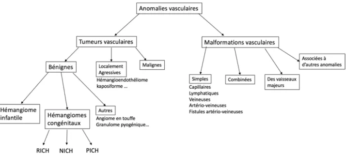
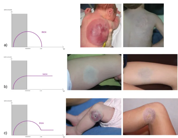
Figure 1 : Classification des anomalies vasculaires. Figure 2 : Schéma évolutif des RICH, NICH et PICH. Figure 3 : Aspect clinique initial d’un RICH et d’un NICH.
Figure 4 : Evolution de 2 RICH avec séquelles : atrophie sous-cutanée et chalazodermie. Figure 5 : Hémangiomes infantiles superficiel, profond, et mixte.
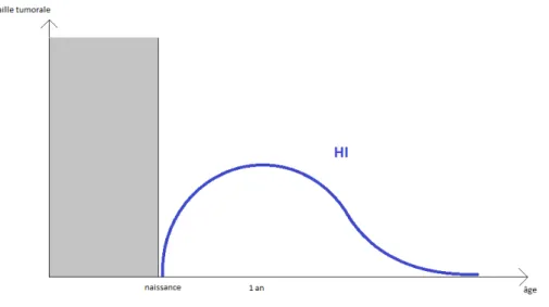
Figure 6 : Schéma évolutif de l’HI.
Figure 7 : Photo histologique. Gradient de maturation des capillaires lobulaires.
Figure 8 : Photo clinique et histologique d’un RICH avec séquelle atrophique majeure. Figure 9 : Photo histologique. Immunomarquage PS100 montrant la présence de filets nerveux intra-lésionnels.
Figure 10 : Photo histologique. Immunomarquage PS100. Dissection des filets nerveux par les petits capillaires.
I- CLASSIFICATION DES ANOMALIES VASCULAIRES
Les anomalies vasculaires superficielles, souvent nommées à tord « angiomes », représentent en fait un large spectre de lésions dont le pronostic est variable, certaines étant relativement communes et bénignes (c’est le cas de l’hémangiome infantile (HI)), alors que d’autres sont beaucoup plus rares ou peuvent mettre en jeu le pronostic vital.
La classification actuelle de la Société Internationale d’étude des Anomalies Vasculaires (ISSVA) (1), mise à jour en 2015, stratifie les lésions vasculaires en 2 groupes : les malformations vasculaires, et les tumeurs vasculaires (Figure 1). Cette classification repose sur des critères histologiques, biologiques, radiologiques et hémodynamiques.
Les tumeurs vasculaires sont caractérisées par une prolifération cellulaire (essentiellement faite de cellules endothéliales), alors que les malformations vasculaires résultent d’anomalies de la morphogénèse vasculaire et sont composées de cellules matures non prolifératives.
Figure 1 : Classification des anomalies vasculaires.
RICH = Rapidly Involuting Congenital Hemangioma ; NICH = Non Involuting Congenital Hemangioma , PICH = Partially Involuting Congenital Hemangioma
L’hémangiome infantile (HI) est la tumeur vasculaire la plus fréquente du nourrisson.
Longtemps, d’autres tumeurs vasculaires infantiles bien plus rares, comme les hémangiomes congénitaux (HC) qui font l’objet de ce travail, ont été confondues avec l’HI. Elles en sont aujourd’hui clairement différenciées, et bien individualisées sur le plan clinique et histologique.
II – HEMANGIOMES CONGENITAUX (HC).
1) Généralités
Le terme « hémangiome congénital » (HC) a été cité pour la première fois en 1996 (2), désignant une tumeur bénigne entièrement développée à la naissance, après une phase de prolifération in utéro. Les HC ne présentent donc pas de croissance post-natale, à la différence des HI.
Le profil évolutif des HC est variable (Figure 2). Sur la base de leur évolution naturelle, 2 sous-types d’HC ont été décrits initialement (désignés par leurs acronymes en langue anglaise) : le RICH (Rapidly Involuting Congenital Hemangioma) (3) qui ne subit pas de poussée post-natale et régresse complètement en quelques mois après la naissance (moins d’un an en général), et le NICH (Non Involuting Congenital Hemangioma) (4) qui persiste après la naissance sans aucune tendance à l’involution, et grandit proportionnellement à la taille de l’enfant. Récemment, une 3ème
entité a été décrite : le PICH (Partially Involuting Congenital Hemangioma) (5). Il s’agit d’hémangiomes congénitaux dont la régression débute comme celle d’un RICH, puis s’arrête après une involution incomplète laissant une lésion résiduelle ressemblant finalement à un NICH.
Figure 2 : Schéma évolutif des RICH (a), NICH (b) et PICH (c).
2) Données épidémiologiques
Les HC sont des tumeurs cutanées rares, dont l’incidence est difficile à déterminer, du fait de leur description récente, et du faible nombre d’études de ces lésions. Dans une étude prospective américaine évaluant les anomalies cutanées chez 594 nouveau-nés dans les 48 premières heures de vie, l’incidence des RICH était de 0,3% (6). Leur répartition semble égale entre les 2 sexes (2), contrairement aux HI qui surviennent majoritairement chez la petite fille.
3) Description clinique / morphologique
L’HC siège préférentiellement sur les membres, volontiers à proximité des articulations, ou en région céphalique.
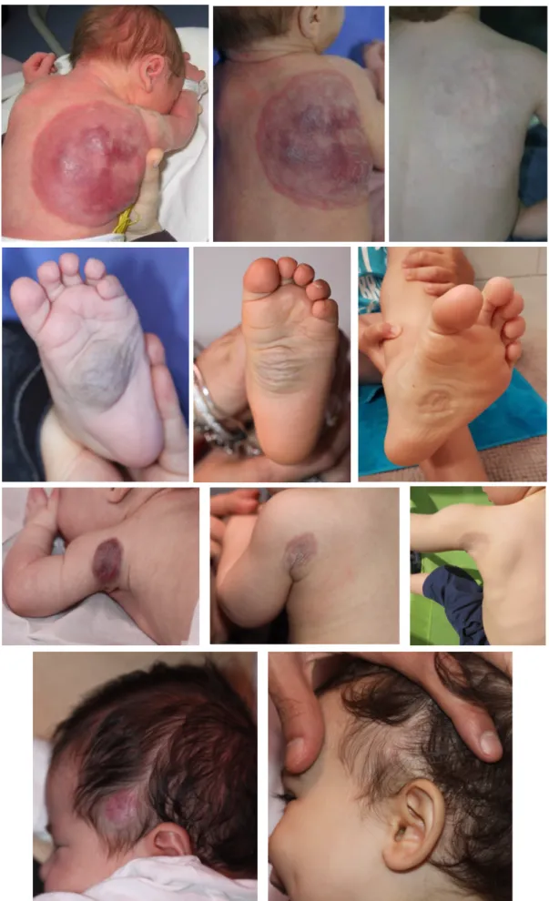
Il se présente sous forme d’une tumeur cutanée le plus souvent unique, arrondie, de couleur érythémato-violacée à bleutée, souvent recouverte de télangiectasies et entourée d’un halo pâle périphérique (dit halo anémique). Parfois, un réseau veineux de drainage est visualisé au pourtour de la lésion. A la palpation, l’HC est de consistance ferme initialement, non pulsatile, et l’auscultation ne met pas en évidence de souffle. Le RICH apparaît souvent comme une volumineuse masse saillante (Figure 3.a), alors que le NICH est fréquemment moins volumineux, sous forme d’une macule ou plaque infiltrée (Figure 3.b) Initialement, le PICH a un aspect similaire au RICH, mais son involution est incomplète.
Figure 3 : Aspect clinique initial d’un RICH à gauche sous forme d’une volumineuse masse saillante (a), et d’un NICH à droite sous forme d’une lésion plane (b).
Après son involution, le RICH peut laisser place à des séquelles à type de peau chalazodermique, d’atrophie sous-cutanée (Figure 4) ou de persistance de veines de drainage bien visibles dans la zone.
Figure 4 : Evolution de 2 RICH avec séquelles à type d’atrophie sous-cutanée et chalazodermie.
Le diagnostic d’HC est souvent évident cliniquement en raison de ses caractéristiques sémiologiques et évolutives stéréotypées, et ne nécessite donc pas de réaliser des examens complémentaires dans la plupart des cas.
En cas de présentation atypique, la réalisation d’une biopsie cutanée et d’un bilan d’imagerie doit être discutée.
4) Formes atypiques
La majeure partie des HC sont de localisation cutanée superficielle. Exceptionnellement, ils peuvent avoir une localisation profonde, intra-musculaire (7) ou viscérale. Plusieurs cas d’HC hépatiques ont été décrits (8). Des cas d’HC intra-crâniens ont également été rapportés (9– 11). Récemment, un cas d’hémangiomatose congénitale a été décrit chez un nouveau-né qui présentait un volumineux HC du dos, associé à une centaine de papules dont les biopsies révélaient qu’il s’agissaient d’HC multifocaux généralisés (12).
5) Complications
Bien qu’étant des tumeurs bénignes, les HC peuvent, rarement, présenter des complications potentiellement graves.
L’ulcération se voit essentiellement dans les RICH, à la naissance ou dans les premières
semaines de vie. Elle peut être spontanée ou induite par un traumatisme, et est souvent douloureuse. Il faut être vigilant devant ces HC car quelques cas d’hémorragies massives mettant en jeu le pronostic vital ont été rapportés avec des HC compliqués d’ulcération (13– 15). Ces hémorragies à haut débit sont liées à l’érosion de vaisseaux superficiels de gros calibre au niveau de l’ulcération. Elles nécessitent souvent un support transfusionnel, et peuvent conduire à une embolisation ou une chirurgie d’exérèse de la tumeur en cas d’échec de la compression. Un cas d’hémorragie massive secondaire à la rupture d’un HC au cours d’un accouchement par voie basse a également été rapporté (16). Par ailleurs, l’ulcération peut être la porte d’entrée à une surinfection locale. Enfin, elle peut laisser des cicatrices atrophiques et/ou hypochromiques.
Les RICH peuvent parfois être associés à une thrombopénie transitoire (17) et/ou une coagulopathie de consommation dans les premiers jours de vie. La thrombopénie est souvent
modérée et d’amélioration rapide en quelques jours, spontanément ou après exérèse de l’HC. Elle nécessite rarement une transfusion plaquettaire. Ces anomalies hématologiques pourraient être liées à un piégeage tumoral plaquettaire et à une consommation de facteurs de la coagulation par des micro-thromboses intra-tumorales.La thrombopénie associée aux HC semble distincte du syndrome de Kasabach-Meritt (SKM), au cours duquel la thrombopénie est plus profonde, prolongée durant plusieurs semaines à mois, et contemporaine d’une augmentation de la taille tumorale.
Les HC de grande taille peuvent également avoir un retentissement hémodynamique, provoquant une insuffisance cardiaque à haut débit, qui peut être diagnostiquée en anténatal ou dans les premiers jours de vie. Cette cardiopathie est le plus souvent transitoire, résolutive après involution, embolisation ou exérèse de l’HC, mais l’évolution peut parfois être défavorable avec un taux de mortalité rapporté jusqu’à 30 % (18).
Les HC ne présentent pas d’association syndromique ou à d’autres malformations (sauf fortuite).
6) Imagerie
Les aspects d’imagerie sont relativement similaires entre les RICH, NICH et PICH.
En écho-doppler, il s’agit de lésions généralement confinées au tissu graisseux sous-cutané,
d’échostructure le plus souvent hétérogène, avec une densité vasculaire élevée. Elles sont constituées de multiples vaisseaux artériels à flux rapides, et de nombreuses veines. On observe parfois des calcifications, et des micro-fistules artério-veineuses qui sont plus fréquentes dans le NICH (19).
L’écho-doppler est un examen de réalisation simple, non invasif, qui ne nécessite pas d’anesthésie générale chez l’enfant, qui a donc son intérêt en cas de présentation clinique atypique de l’HC.
Par ailleurs, la mesure des index de résistance peut être un paramètre de suivi utile et fiable pour les RICH et les PICH, puisqu’il augmente à mesure que l’hémangiome involue (13). A l’IRM, les HC ont un aspect très bien limité et homogène, en isosignal T1 et en hypersignal T2, avec rehaussement tumoral homogène et intense après injection de gadolinium, et zones d’absences de signal (« flow-void ») liées aux vaisseaux à flux rapide.
L’angiographie des HC montre un parenchyme homogène et désorganisé, avec des artères
nourricières larges et irrégulières, des anévrismes de taille variable, des fistules artério-veineuses, et parfois des microthrombi intravasculaires (20). L’absence d’opacification veineuse précoce permet de faire la différence avec les malformations artério-veineuses (19).
Etant donné la croissance intra-utérine des HC, il est théoriquement possible d’en faire le diagnostic prénatal par échographique, ce qui est de plus en plus fréquent. Ce dépistage anténatal est rare (mais possible) au premier trimestre (6), plus fréquent à partir du second trimestre (21). Parfois, l’involution débute même in utero, avec une taille tumorale à la naissance inférieure à celle évaluée par l’échographie prénatale, ou un aspect de séquelle chalazodermique qui témoigne de la régression de la lésion.
7) Histologie
La biopsie cutanée est parfois indispensable pour éliminer les diagnostics différentiels en cas de présentation clinique atypique de la lésion (fibrosarcome congénital (22), hémangioendothéliome kaposiforme, angiome en touffe…).
Ils sont organisés en lobules composés de capillaires bordés de cellules endothéliales turgescentes (« en clous de tapissier ») entourées d’une fine membrane basale, au sein d’un stroma fibreux interlobulaire dense contenant des vaisseaux anormaux : artères, veines dystrophiques et canaux lymphatiques (23). En immunohistochimie, les cellules endothéliales n’expriment pas le glucose transporter-1 (GLUT-1), contrairement aux hémangiomes infantiles.
Il existe cependant quelques différences notables entre les sous-types d’HC :
Dans les NICH, les capillaires lobulaires sont généralement de plus grande taille et plus dilatés. Le stroma inter-lobulaire des NICH contient fréquemment des microfistules artério-lobulaires et artério-veineuses, alors qu’elles sont exceptionnelles dans les RICH.
Les RICH, eux, présentent fréquemment moins de capillaires lobulaires en leur centre, là où débute généralement la régression tumorale. Aux derniers stades d’involution du RICH, on peut voir des calcifications dystrophiques, des micro-thromboses et des dépôts d’hémosidérine (3,24).
8) Physiopathologie
La physiopathologie des HC demeure incomprise. Aucun facteur de prédisposition au développement des HC n’est à ce jour connu.
Récemment, des mutations somatiques activatrices des gènes GNAQ et GNA11 ont été détectées dans les HC par séquençage de l’exome entier sur tissu frais et congelé (25). Ces deux gènes codent pour des sous-unités régulatrices des protéines G (utilisant le GTP), et la modification de structure induite par la mutation entraine une activation constitutive de la voie des MAP kinases et de la protéine kinase C (26). Il s’agirait de mutations somatiques post-zygotiques survenant sur un territoire cutané limité, touchant une cellule progénitrice se différenciant ensuite en lignée vasculaire. Le profil des mutations est similaire dans les RICH
et les NICH, ce qui suggère que d’autres facteurs génétiques, épigénétiques et/ou environnementaux influencent l’évolution des HC. Des mutations somatiques différentes touchant les mêmes gènes GNAQ et GNA11 ont été mises en évidence dans d’autres anomalies vasculaires (angiome plan, syndrome de Sturge-Weber (27), phacomatose pigmento-vasculaire et mélanose étendue (28)).
9) Traitement
La plupart des HC sont non compliqués et ne nécessitent pas de traitement particulier.
Chez les enfants ayant un RICH, il est licite de proposer une abstention-surveillance lors de la première année de vie, du fait de la régression attendue. Les séquelles résiduelles (lipoatrophie, excédant cutané anétodermique, télangiectasies) sont souvent cosmétiquement acceptables, ou peuvent faire l’objet d’une correction ultérieure lorsque l’enfant est en âge d’en faire la demande. Cependant il arrive que les RICH soient excisés d’emblée en période néonatale, probablement du fait de leur caractère volumineux qui peut paraître inquiétant et conduire à un traitement immédiat.
En cas de RICH compliqué (ulcération persistante malgré les soins locaux, hémorragie ne cédant pas à la compression, instabilité hémodynamique non contrôlée par le traitement médical), l’exérèse chirurgicale est indiquée en urgence, d’emblée ou après embolisation artérielle pour contrôler le saignement.
En cas d’hémangiome d’involution incomplète (PICH) ou non involutif (NICH), l’exérèse chirurgicale est discutée au cas par cas, selon la localisation de la lésion qui peut avoir un préjudice esthétique ou fonctionnel (par exemple aux mains : difficultés à l’apprentissage de l’écriture ; aux pieds : difficultés à l’apprentissage de la marche…)
La corticothérapie systémique, souvent prescrite en cas d’HC compliqué et notamment en cas d’insuffisance cardiaque, semble n’avoir que peu d’impact sur l’évolution de la lésion. En
considérant que les corticoïdes inhibent la vasculogénèse dans des cellules souches dérivées d’HI via la suppression du VEGF-A (Vascular Endothelial Growth Factor A) (29), on peut supposer que ce traitement a un rôle limité dans le traitement des HC étant donné leur absence de prolifération post-natale (18).
De la même façon, les bétabloquants et notamment le Propranolol sont inefficaces dans le traitement des HC, alors qu’ils sont le traitement de premier choix des HI.
III- AUTRES TUMEURS VASCULAIRES – DIAGNOSTICS
DIFFERENTIELS
1) L’hémangiome infantile (HI)
L’HI est la tumeur vasculaire la plus fréquente, touchant 4 à 10 % des nourrissons et enfants (30), avec une nette prédominance pour le sexe féminin et plus fréquente chez le prématuré. Cliniquement, on distingue 3 types d’HI : l’HI superficiel ou tubéreux, l’HI profond ou sous-cutané, et l’HI mixte (Figure 5).
Figure 5 : Hémangiomes infantiles de type superficiel (à gauche), profond (au centre), et mixte (à droite).
Histologiquement, il est caractérisé par une positivité du marqueur GLUT-1.
Son évolution est stéréotypée (31) : il apparaît dans les premières semaines de vie, suit une croissance rapide dans les 3 à 6 premiers mois de vie, se stabilise, puis entame une phase de régression lente vers l’âge de 12 mois, qui peut durer plusieurs années (Figure 6).
Figure 6 : Schéma évolutif de l’HI.
De ce fait, la plupart des HI ne nécessitent pas de traitement. Les principales indications thérapeutiques concernent les HI menaçant le pronostic vital (causant détresse respiratoire et insuffisance cardiaque), ceux entraînant un risque fonctionnel (obstruction visuelle, amblyopie, difficultés à l’alimentation), les HI ulcérés ou entraînant une importante déformation anatomique, principalement au niveau du visage (32). Le traitement de première ligne repose alors sur le Propranolol oral, à débuter le plus précocement possible (à partir de l’âge de 5 semaines) et pour une durée d’au moins 6 mois.
2) Autres tumeurs vasculaires
L’angiome en touffe (AT) apparaît comme une plaque ou macule érythémateuse ou marron, et survient chez l’enfant ou le jeune adulte (33). Quelques cas sont présents à la naissance. Certaines lésions régressent spontanément, en particulier dans les cas congénitaux. Histologiquement, les AT sont composés de petites touffes de capillaires dermo-hypodermiques ayant une distribution caractéristique « en boulet de canon » (en amas séparés) (1).
L’hémangioendothéliome kaposiforme (HEK), lui, implique souvent les tissus profonds, se présentant comme une tumeur localement agressive. Histologiquement, il ressemble à l’angiome en touffe avec de grands lobules capillaires disposés en fentes et confluents, et un pattern infiltrant (1).
L’AT et l’HEK expriment les marqueurs lymphatiques (34) mais n’expriment pas GLUT-1. Ils peuvent être associées au syndrome de Kasabach-Merritt (SKM), caractérisé par une thrombocytopénie profonde et une coagulopathie de consommation, survenant de manière concomittante à une modification de la tumeur qui devient ecchymotique et inflammatoire. Le pronostic vital peut alors être engagé.
ETUDE
RATIONNEL DE l’ETUDE
Les hémangiomes congénitaux sont peu décrits dans la littérature du fait de leur faible prévalence. Il n’existe que peu d’études de suivi à long terme, et les effectifs sont souvent réduits.
Il est souvent rapporté que les RICH sont de bon pronostic et régressent très bien. Or il semblerait qu’une certaine proportion d’hémangiomes de type RICH à la naissance ne régressent finalement que de façon incomplète (PICH). Ces PICH ont été décrits récemment, et leur proportion par rapport aux autres hémangiomes congénitaux est inconnue.
Les mécanismes physiopathologiques de ces lésions vasculaires restent non élucidés, et les facteurs qui déterminent leur profil évolutif (stabilité, régression complète ou régression partielle) sont inconnus.
Par ailleurs, la douleur n’est pas un symptôme fréquemment décrit dans les études portant sur les hémangiomes congénitaux. Pourtant, il semblerait que bon nombre de ces tumeurs sont ou deviennent douloureux au cours de leur évolution.
L’objectif principal de ce travail était de décrire le profil évolutif, en corrélation avec leurs caractéristiques cliniques et paracliniques, des hémangiomes congénitaux vus dans l’unité de dermatologie pédiatrique du CHU de Bordeaux de 2004 à 2016. Nous avons également effectué une relecture histologique des cas biopsiés ou opérés.
I- ARTICLE
Evolutive pattern of congenital hemangiomas:
a long-term follow-up of fifty-seven cases
Victoire Braun a, Carlotta Gurioli MD b, Franck Boralevi a, MD PhD, Alain Taieb a, MD PhD, Nicolas Grenier c MD PhD, Maya Loot d MD, Marie-Laure Jullié e, MD, Christine Léauté-Labrèze a, MD.
Affiliations: a Department of Pediatric Dermatology at Bordeaux University Hospital ; b Department of Specialised, Experimental and Diagnostic Medicine, Dermatology, University of Bologna, Italy,c Department of Radiology at Bordeaux University Hospital, d Department
of Pediatric Surgery at Bordeaux University Hospital, e Department of pathology at Bordeaux University Hospital.
Address Correspondence to: Victoire Braun, service de dermatologie du CHU de Bordeaux,
1 rue Jean Burguet, 33000 Bordeaux
Short Title: Long term follow-up of congenital hemangiomas. Funding Source: No external funding for this manuscript.
Financial Disclosure: The authors have no financial relationships relevant to this article to
disclose.
Conflict of Interest: The authors have no potential conflicts of interest to disclose.
Abbreviations: CH: congenital hemangioma ; GLUT-1: glucose transporter-1 ; IH: infantile hemangioma ; NICH non involuting congenital hemangioma ; PICH: partially involuting congenital hemangioma ; PS100: S100 protein ; RICH: rapidly involuting congenital hemangioma.
Table of Contents Summary: In this study, we describe epidemiological, clinical features,
evolutive data, complications, imaging and histologic results of the three subtypes of congenital hemangiomas.
What’s Known on This Subject: Congenital hemangiomas are fully developed at birth.
Three subtypes of congenital hemangiomas have been described based on their clinical behavior: rapidly involuting (RICH), non involuting (NICH) or partially involuting (PICH) congenital hemangiomas.
What This Study Adds: RICH, NICH and PICH have many overlapping characteristics,
suggesting they are part of same spectrum. A long term follow-up shows that one third of congenital hemangiomas looking like RICH at birth undergo only partial involution (PICH). Ulceration is particularly frequent in PICH.
Contributors' Statement:
Dr Braun collected data, carried out the initial analyses, and drafted the initial manuscript. Dr Gurioli collected data.
Pr Boralevi and Pr Taieb collected data and critically reviewed the manuscript for important intellectual content.
Pr Grenier collected data and performed imaging. Dr Loot collected data and performed surgery. Dr Jullie proofread all histological samples
Dr Léauté-Labrèze conceptualized and designed the study, coordinated and supervised data collection, and revised the manuscript.
All authors approved the final manuscript as submitted and agreed to be accountable for all aspects of the work.
Abstract
Background: Congenital hemangiomas (CHs) are rare benign vascular tumors fully
developed at birth. Three subtypes of CHs have been described based on their clinical behaviour: rapidly involuting congenital hemangioma (RICH), non involuting congenital hemangioma (NICH) or partially involuting congenital hemangioma (PICH).
The objective was to define the clinical, evolutive and paraclinical characteristics of the three subtypes of CHs.
Methods: Children with CHs attending department of pediatric dermatology at Bordeaux
University Hospital over a 13-year period were retrospectively included. Epidemiological, clinical, evolution data, photographs, and imaging results were reviewed. Histological examination of available samples was performed.
Results: 57 cases were included: 22 RICH, 22 NICH, and 13 PICH. There was a male
predominance and the most common location was the limbs. RICH, NICH and PICH had overlapping characteristics: a single telangiectatic lesion with peripheral pale halo. NICH were flat, whereas RICH and PICH were bulky at birth. Mean age at complete involution of RICH was 13.6 months. 1/3 of CHs having RICH aspect at birth finally had incomplete involution and became PICH. Heart failure and thrombocytopenia were rare complications. PICH were frequently ulcerated. Pain was common for NICH and PICH. Imaging and histological results were relatively similar between the 3 subtypes of CHs.
Conclusions: Our case series describe the characteristics and the evolution pattern of the
three subtypes of CHs with some overlapping features, reinforcing the hypothesis that RICH, NICH and PICH belong to the same pathological spectrum.
INTRODUCTION
Congenital hemangiomas (CHs) are rare vascular benign tumors that proliferate in utero and are fully developed at birth (1), unlike infantile hemangiomas (IHs) which appear within the first weeks after birth, rapidly enlarge and then involute slowly over the upcoming years (2) . On the basis of their natural history and clinical characteristics, 2 types of CHs were first described: Rapidly Involuting Congenital Hemangioma (RICH) (3) which generally regresses by first year of age, and Non Involuting Congenital Hemangioma (NICH) (4) which remains stable and proportionately grows with the child. A third sub-group has recently been described: Partially Involuting Congenital Hemangioma (PICH) (5) that initially regresses like a RICH, and then stops after an incomplete involution.
CH usually appears as a unique well limited lesion with a red-purple color, covered with fine or coarse telangiectasia and frequently surrounded by a peripheral pale halo. RICH often look like a raised tumor, whereas NICH forms a flat plaque or macule. PICH initially looks similar to RICH, but undergoes only partial involution, with a residual lesion finally resembling a NICH.
The pathophysiology of CHs remains unclear, and the factors determinating their evolutive pattern after birth are unknown. The aim of this study was to define the natural history of CHs, in correlation with their clinical and evolutive characteristics, over a 13 year period.
PATIENTS AND METHODS
This study has been approved by our local ethical committee. Patients were retrieved from the photographic database of the department of pediatric dermatology at Bordeaux University Hospital between the years of 2004 and 2016, to identify patients with CHs searching for « congenital hemangioma », « RICH » « NICH » and « PICH ».
Epidemiological, clinical features and evolution data, as well as complications and treatment were collected from each patients' medical record. Imaging results were also collected if existing. Photographs were reviewed by the author and the director of the study.
For those who had biopsy or excision, histopathological examination was performed with immunohistochemical analysis of Glucose Transporter 1 (GLUT-1) and S100 protein (PS100).
Cases were excluded if the clinical characteristics were inconsistent with the diagnosis of CH, or if the duration of follow-up was too short to determine the sub-group of the CH (RICH, NICH or PICH).
RESULTS Patients
69 patients were identified from the photographic database. 12 were excluded, due to uncertain diagnosis (n=5) or inadequate duration of follow-up to support the diagnosis of CH (n=7). 57 patients were included: 22 with RICH, 22 with NICH and 13 with PICH. Detailed characteristics are found in Table I.
For 5 patients, the lesion had been antenatally detected by ultrasonography and/or MRI.
There was 3 RICH and 2 PICH, localized on the head and neck (n=4) or upper limb (n=1). The tumor size was greater than 2 centimeters (between 2 and 5 cm, n=3 ; > 5 cm, n=2) and the diagnosis was made during the second (n=3) or third (n=1) trimester of pregnancy (in pertinence to the last case, the date is unknown).
The following obstetrical complications were reported: 1 premature delivery threat, 1 group B streptococcal maternal infection, 1 gestational diabetes, 1 peripartum hemorrhage. 2 patients
had fetal heart failure (1 RICH and 1 PICH), and one patient required an extraction by caesarean section.
Regarding comorbidities, we noted: 2 macrocrania cases (with a family history of macrocephaly for the first case, and associated developmental delay and polydactyly for the second), 1 septal ventricular defect and 1 atrial septal defect without clinical impact, and 1 hypogammaglobulinemia with recurrent infections.
As per the parent’s medical history, there was: 1 therapeutic abortion for left ventricular hypoplasia, 1 Sjögren's syndrome associated with systemic lupus erythematosus, 1 Basedow's
disease, 1 epilepsy, 1 Willebrand’s disease, and 1 renal cancer.
Median age at the first consultation was 2.7 weeks for RICH, 4.1 weeks for PICH, whereas it was 58 months for NICH.
Clinical Characteristics of CHs
All patients had a single lesion, whose clinical and semiological characteristics are detailed in
Table II.
Evolution
Taking into account the clinical and evolutive characteristics of the 57 CHs, we distinguished 22 RICH (Figure 1), 22 NICH (Figure 2) and 13 PICH (Figure 3).
For the 22 patients with RICH, involution started at the average age of 5.2 weeks. Complete involution was objectified within less than a year for 13 patients, and more than a year for 8 patients, with an average age of 13.6 months. The shortest delay for complete regression was 4 months, and the longest was 30 months. One patient had an excision surgery of his CH at 3 days of life ; such patient had a bulky raised lesion resembling a RICH.
Concerning the 13 children with PICH, they all had a diagnosis of RICH initially, because of their similar presentation. For 12 patients, their hemangioma presented an initial regression which started at the average age of 7.4 weeks, but then stabilized at the average age of 21.9
months. One patient had a prenatal diagnosis of CH, and the ultrasound follow-up revealed a partial involution in utero. One child had a scrotal bulky CH strongly resembling a RICH, even if his lesion remained stable in size between the age of 29 and 38 months. It was considered as a PICH which could have involuted very early, or in utero too. All had a residual telangiectatic lesion.
In regards to the 22 patients with NICH, their hemangioma remained stable and proportionally grew with them.
The following sequelae were observed after regression for RICH: anetoderma (n=6), subcutaneous atrophy (n=11), residual telangiectasia (n=4), pale or bluish area (n=8). Residual dysplastic draining veins were seen for 10 NICH, 5 PICH and 2 RICH.
Complications
Complications of the CHs are listed in table III.
2 children had heart failure in utero. The first child had a large cervical PICH whose regression began in utero, and birth was induced at 33 weeks of amenorrhea for fetal rhythm abnormalities. His cardiac function normalized few hours after birth. The other child had a large RICH at the elbow. He presented in utero a moderate cardiac decompensation with right heart dilatation and small pericardial effusion, which necessitated intensive care at birth. Excision surgery was performed at 3 days of life with a favorable outcome.
Transient thrombocytopenia was observed in two patients in the first few days of life, with a platelet count of 102 and 118 G/L, spontaneously normalized on the 2nd (first child) and 9th
day (second child) without any hemorrhagic complication.
Ulceration was observed in 6 CHs, 5 of which were PICH (38.5%). Massive acute hemorrhage occurred in one patient with a large ulcerated PICH of the thigh, at the age of 4 weeks. He had anemia with a hemoglobin level of 6 g/dl, requiring several transfusions. After
failure of corticosteroid therapy, surgical excision was successfully performed at the age of 8 weeks.
Other reported complications were: swelling with heat or presence of fever (RICH n=1, NICH n=1, PICH n=1), transient alopecia (RICH of the scalp n=1), infection (PICH n=1), and functional discomfort walking on all fours (PICH of the leg n=1).
During follow-up, pain was reported for 16 CHs, mainly NICH (40.9%) and PICH (38.5%). It was often subjective symptoms with abnormal sensations evoking neuropathic pain.
Treatment
A surgical excision was performed for 12 cases (21%): 1 RICH at 3 days because of heart failure, 6 PICH with an average age of 29.5 months and 5 NICH with an average age of 81 months. CHs were located at the head or neck for 5 patients, trunk for 3 patients and limbs for 4 patients, and 2 children needed expansion prosthesis for surgery. The main indication of surgery was aesthetic outcome or because the lesion was painful or was bleeding. 1 patient with NICH and 1 with PICH have been treated by beta blockers (oral and topical) without any effect. 1 patient with a NICH of the face had sclerosis (2 sessions) with an increased size of his lesion after this treatment.
Imaging
33 patients had 1 or more doppler ultrasound imaging. Results were available for 29 of them (RICH n=11, NICH n=9, PICH n=9). Imaging was performed early (before the age of 3 months) for all patients with RICH, only 1 with NICH and 7 with PICH. It always showed a highly vascularized tumor (except for one patient with a non-contributory image), often well limited, made of multiples enlarged veins and arteries with high-velocity bloodflow. CHs tended to be hypoechoic (except for 1 RICH and 1 PICH with hyperechoic lesions), homogenous as well as heterogeneous. Calcifications were described for 2 RICH and 1 PICH. An arteriovenous shunt was reported for 1 PICH.
Magnetic resonance imaging was performed in 2 patients (1 NICH and 1 PICH), showing well circumscribed tumors with hyperintensity on T2-weighted sequences, and homogeneous T1 gadolinium enhancement.
Histology
Histopathological examination of the lesion (biopsy or excisional specimen) was performed for 13 patients: it concerned 3 RICH, 5 NICH and 6 PICH. 1 NICH could not have been analyzed because the histological material was unavailable.
CHs were composed of variably sized capillary lobules with draining arteries and normal or dysplastic veins, surrounded by fibrous tissue. There was a maturation gradient with some lobules made of small compact capillaries, and some larger lobules having more mature appearance with dilated and larger draining channels with prominent fibrosis. Tumor architecture could not be analyzed for the 2 RICH with biopsy because the sample size was small. GLUT-1 staining was negative for all 13 cases. PS100 immunostaining showed variable quantity of intralesional nerve bundles for all cases.
DISCUSSION
Our series of 57 cases over a 13-year period confirms the rare prevalence of CHs. We can approximately estimate the prevalence at 1 per 1000 births (we viewed 5 CHs annually, compared to 200 IHs with a prevalence of 3 to 10 %). We observed a male predominance (sex ratio 1.7) whereas sex ratio in general is equally balanced in literature (1), in contrast to IHs which have predilection for female (6). Contrary to IHs, CHs did not occur more frequently in premature infants and the rate of caesarean sections was similar to the general population in France (7).
The first consultation was much earlier for infants with RICH than NICH. It may be explained by the alarming bulky aspect of RICH and PICH at birth. As described in the literature (8,9),
the most common locations were the limbs, followed by head or neck and trunk. Most CHs were larger than 2 cm and even 5 cm for one third of them, especially NICH, which can be explained by the older age presentation of patients at the first consultation in this group, as NICH grows proportionally with the child.
The diagnosis of CH is often clinically evident, and therefore does not require complementary investigations. However, in case of an atypical presentation, skin biopsy and radiological examination must be brought to attention to eliminate other vascular tumors such as Kaposiform Hemangioenthelioma or Tufted Angioma which can have some similar clinical features (8,10) but a different prognosis.
NICH were essentially flat lesions, whereas RICH and PICH had an exophytic presentation. However, the three subtypes of CHs have some clinical overlapping characteristics. All the lesions were solitary, telangiectasias were almost constant and more than half of CHs presented a peripheral halo of vasoconstriction. In addition, the three subtypes of CHs have many similar imaging characteristics. Color Doppler ultrasonography is the imaging technique of choice in children, as it is a non-invasive procedure. CHs are usually localized in the subcutaneous fat, they are heterogeneous, hypoechoic (11) with multiples visible arteries and veins exhibiting high-velocity blood flow (5), and sometimes calcifications (12), as seen for 3 cases. Vascular microshunts are usually more frequent in NICH (12), whereas it was only described for 1 PICH in our study. The resistance index is a reliable maturation parameter, since it increases as the tumor is in its involutive phase (13). On magnetic resonance imaging, CHs demonstrate well-defined limits with homogeneous or patchy enhancement, hyperintensity on T2-weighted sequences, flow voids and fat standing (5,12,14). Vascular anomalies are diagnosed prenatally with increasing frequency (15), and among CHs RICH are the most frequently detected because of their large sizes (16).
Histologically, RICH, NICH and PICH also had overlapping features, as they are composed of variably sized capillary lobules with draining arteries and associated dysplastic veins and lymphatic vessels, surrounded by fibrous tissue. They are different from classical infantile hemangiomas and GLUT-1 is always negative. In this series, we observed the presence of numerous intralesional nerve bundles in capillary lobules and/or fibrous tissue that could contribute to the pain frequently reported during follow-up, especially in NICH and PICH. Presence of such neural component had been previously reported in congenital vascular malformations (17), but not in congenital hemangiomas.
Most CHs are asymptomatic, but some early complications may be seen, like prenatal heart failure. Indeed, large CHs may have hemodynamic consequences due to their high blood flow. Most of the time, the cardiopathy is transient, resolving after involution. However, Weitz et al. reported a mortality rate of 30% in their literature review (18), prompting close monitoring for large CHs. Two cases had transient mild thrombocytopenia at birth. The retrospective series of Baselga and al. reported 7 RICH, larger than 5 cm, with thrombocytopenia (19) ; the thrombocytopenia was also moderate, and the nadir was reached in the first week of life, with fast biological improvement at 2 weeks. It appears to be distinct from Kasabach-Meritt syndrome, in which the haematological anomaly is deeper, lasting several weeks to months in parallel to an increase tumour size.
Almost half of PICH cases had central ulceration. Thus, ulceration could be a factor predicting partial involution of the CH. For those cases, extra care should be given because several cases of life-threatening massive hemorrhages have been reported (13,20,21), like one patient involved in our study. Those high-flow bleedings could be due to a wall erosion of large vessels just under the ulceration.
When a patient presents a RICH type CH at birth, it is difficult to assess if that lesion will totally regress or if it will evolve as a PICH. In our study, 37 % of CHs looking like RICH at
birth finally regressed only partially and became PICH. In some cases, the residual tumor was indistinguishable from NICH, as previously reported (5,22). We observed that RICH and PICH started their regression at the average age of 5 and 7 weeks respectively, which does not differentiate them initially. However, their evolution behaviour is different: RICH achieved a complete regression at the average age of 13.6 months, whereas for PICH we observed a partial involution during the first months of life, before the lesion stabilized around the age of 2.
During follow-up, we observed that pain is frequently reported in NICH and PICH. In their series of NICH, Lee and al. also reported pain for 43 % of cases and hypothesized that vasoconstriction may cause local tissue ischemia resulting in pain(23). However, dysesthesia was frequently reported, rather evoking neuropathic pain. In this series, we observed a relative hyperplasia of nerve bundles in the lesions that may explain the symptoms.
For most patients with RICH, except for those with life-threatening complications, close follow-up is the first-line treatment. As there is no efficient medical treatment reported, surgical excision with or without preoperative embolization to control bleeding is the treatment of choice for CHs, depending on the location of the lesion which may have an aesthetic or functional prejudice.
CONCLUSION
In this series describing the evolutive pattern of the three subtypes of congenital hemangiomas we confirm that they have many overlapping features, suggesting that they are part of the same spectrum, reinforcing the hypothesis proposed by Mulliken et al. (9) proposing that RICH is the precursor of NICH. Involution phase could occur in utero for NICH, whereas regression begins after birth for RICH. Thus, PICH represent the missing link between these two subtypes of CHs. In case of CH looking like RICH at birth, caution must
be given when discussing the prognosis, as involution does not come to its end for one third of patients, which are then called PICH. In this study, ulceration appears to be a predictive marker of incomplete involution of the lesion, suggesting PICH. In addition, neuropathic pain is frequently reported in PICH and NICH, which may be due to abundant nerve bundles in the tumor.
References
1. Boon LM, Enjolras O, Mulliken JB. Congenital hemangioma: evidence of accelerated involution. J Pediatr. mars 1996;128(3):329‑35.
2. Bruckner AL, Frieden IJ. Hemangiomas of infancy. J Am Acad Dermatol. avr 2003;48(4):477-493; quiz 494-496.
3. Berenguer B, Mulliken JB, Enjolras O, Boon LM, Wassef M, Josset P, et al. Rapidly involuting congenital hemangioma: clinical and histopathologic features. Pediatr Dev Pathol Off J Soc Pediatr Pathol Paediatr Pathol Soc. déc 2003;6(6):495‑510.
4. Enjolras O, Mulliken JB, Boon LM, Wassef M, Kozakewich HP, Burrows PE. Noninvoluting congenital hemangioma: a rare cutaneous vascular anomaly. Plast Reconstr Surg. juin 2001;107(7):1647‑54.
5. Nasseri E, Piram M, McCuaig CC, Kokta V, Dubois J, Powell J. Partially involuting congenital hemangiomas: a report of 8 cases and review of the literature. J Am Acad Dermatol. janv 2014;70(1):75‑9.
6. Léauté-Labrèze C, Harper JI, Hoeger PH. Infantile haemangioma. Lancet Lond Engl. 01 2017;390(10089):85‑94.
7. Lafitte A-S, Dolley P, Le Coutour X, Benoist G, Prime L, Thibon P, et al. Rate of caesarean sections according to the Robson classification: Analysis in a French perinatal network - Interest and limitations of the French medico-administrative data (PMSI). J Gynecol Obstet Hum Reprod. févr 2018;47(2):39‑44.
8. Enjolras O, Picard A, Soupre V. [Congenital haemangiomas and other rare infantile vascular tumours]. Ann Chir Plast Esthet. oct 2006;51(4‑5):339‑46.
9. Mulliken JB, Enjolras O. Congenital hemangiomas and infantile hemangioma: missing links. J Am Acad Dermatol. juin 2004;50(6):875‑82.
10. Putra J, Gupta A. Kaposiform haemangioendothelioma: a review with emphasis on histological differential diagnosis. Pathology (Phila). juin 2017;49(4):356‑62.
11. Rogers M, Lam A, Fischer G. Sonographic Findings in a Series of Rapidly Involuting Congenital Hemangiomas (RICH). Pediatr Dermatol. 1 janv 2002;19(1):5‑11.
12. Gorincour G, Kokta V, Rypens F, Garel L, Powell J, Dubois J. Imaging characteristics of two subtypes of congenital hemangiomas: rapidly involuting congenital hemangiomas and non-involuting congenital hemangiomas. Pediatr Radiol. déc 2005;35(12):1178‑85. 13. Agesta N, Boralevi F, Sarlangue J, Vergnes P, Grenier N, Léauté-Labrèze C. Life-threatening haemorrhage as a complication of a congenital haemangioma. Acta Paediatr Oslo Nor 1992. oct 2003;92(10):1216‑8.
14. Krol A, MacArthur CJ. Congenital Hemangiomas: Rapidly Involuting and Noninvoluting Congenital Hemangiomas. Arch Facial Plast Surg. 1 sept 2005;7(5):307‑11.
15. Marler JJ, Fishman SJ, Upton J, Burrows PE, Paltiel HJ, Jennings RW, et al. Prenatal diagnosis of vascular anomalies. J Pediatr Surg. mars 2002;37(3):318‑26.
16. Brix M, Soupre V, Enjolras O, Vazquez M-P. [Antenatal diagnosis of rapidly involuting congenital hemangiomas (RICH)]. Rev Stomatol Chir Maxillofac. avr 2007;108(2):109‑14.
17. Meijer-Jorna LB, Breugem CC, de Boer OJ, Ploegmakers JPM, van der Horst CMAM, van der Wal AC. Presence of a distinct neural component in congenital vascular malformations relates to the histological type and location of the lesion. Hum Pathol. oct 2009;40(10):1467‑73.
18. Weitz NA, Lauren CT, Starc TJ, Kandel JJ, Bateman DA, Morel KD, et al. Congenital cutaneous hemangioma causing cardiac failure: a case report and review of the literature. Pediatr Dermatol. déc 2013;30(6):e180-190.
19. Baselga E, Cordisco MR, Garzon M, Lee MT, Alomar A, Blei F. Rapidly involuting congenital haemangioma associated with transient thrombocytopenia and coagulopathy: a case series. Br J Dermatol. juin 2008;158(6):1363‑70.
20. Vildy S, Macher J, Abasq-Thomas C, Le Rouzic-Dartoy C, Brunelle F, Hamel-Teillac D, et al. Life-threatening hemorrhaging in neonatal ulcerated congenital hemangioma: two case reports. JAMA Dermatol. avr 2015;151(4):422‑5.
21. Al Malki A, Al Bluwi S, Malloizel-Delaunay J, Mazereeuw-Hautier J. Massive hemorrhage: A rare complication of rapidly involuting congenital hemangioma. Pediatr Dermatol. mai 2018;35(3):e159‑60.
22. Acebo E, Gardeazábal J, González-Hermosa R, Pérez-Barrio S, Díaz-Pérez JL. Congenital hemangioma: a report of evolution from rapidly involuting to noninvoluting congenital hemangioma with aberrant Mongolian spots. Pediatr Dermatol. avr 2009;26(2):225‑6.
23. Lee PW, Frieden IJ, Streicher JL, McCalmont T, Haggstrom AN. Characteristics of noninvoluting congenital hemangioma: a retrospective review. J Am Acad Dermatol. mai 2014;70(5):899‑903.
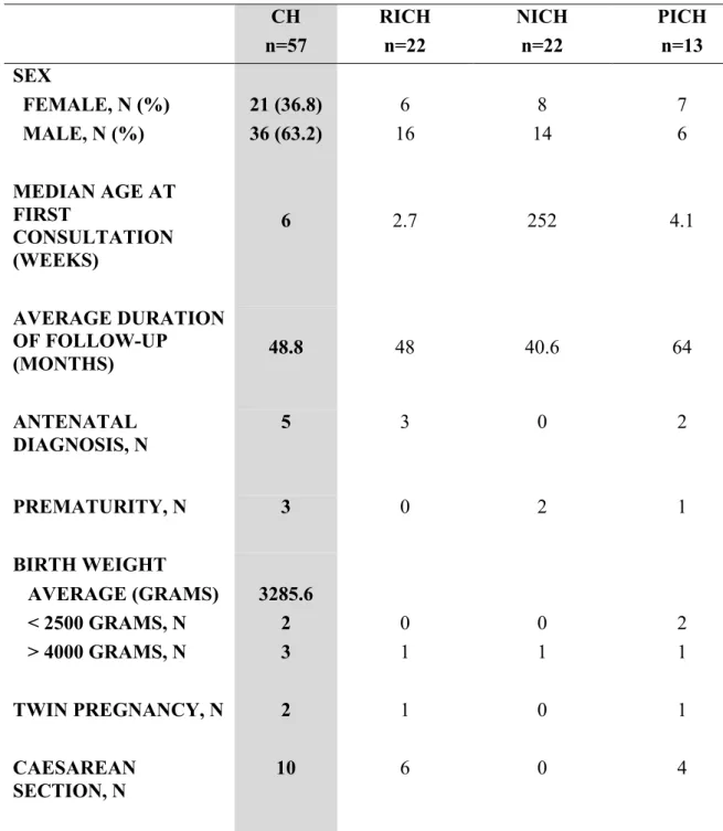
CH n=57 RICH n=22 NICH n=22 PICH n=13 SEX FEMALE, N (%) MALE, N (%) 21 (36.8) 36 (63.2) 6 16 8 14 7 6 MEDIAN AGE AT FIRST CONSULTATION (WEEKS) 6 2.7 252 4.1 AVERAGE DURATION OF FOLLOW-UP (MONTHS) 48.8 48 40.6 64 ANTENATAL DIAGNOSIS, N 5 3 0 2 PREMATURITY, N 3 0 2 1 BIRTH WEIGHT AVERAGE (GRAMS) < 2500 GRAMS, N > 4000 GRAMS, N 3285.6 2 3 0 1 0 1 2 1 TWIN PREGNANCY, N 2 1 0 1 CAESAREAN SECTION, N 10 6 0 4
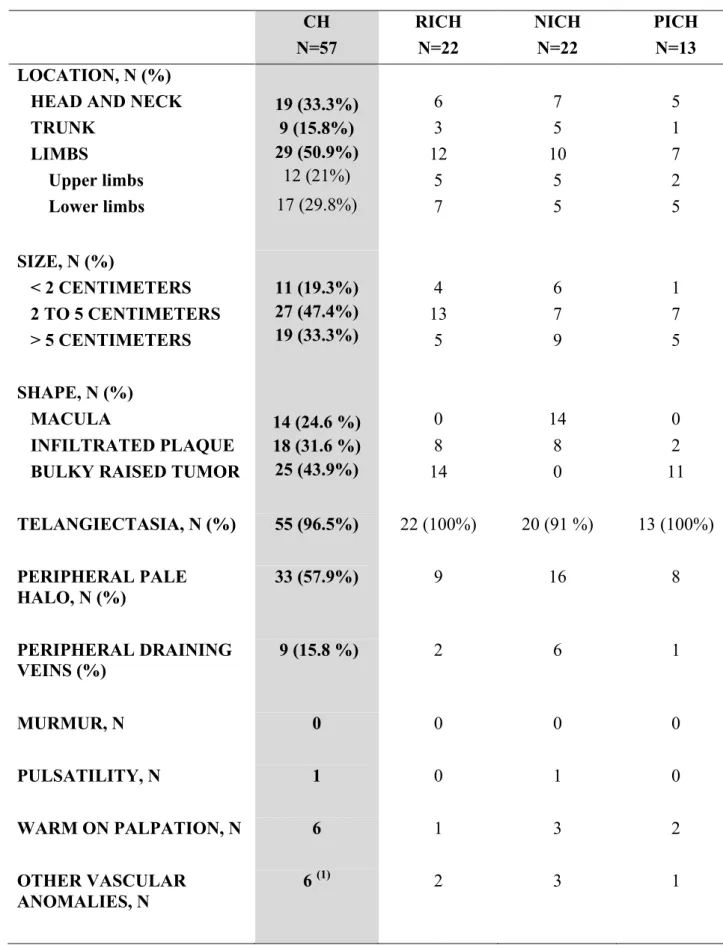
CH N=57 RICH N=22 NICH N=22 PICH N=13 LOCATION, N (%)
HEAD AND NECK TRUNK LIMBS Upper limbs Lower limbs 19 (33.3%) 9 (15.8%) 29 (50.9%) 12 (21%) 17 (29.8%) 6 3 12 5 7 7 5 10 5 5 5 1 7 2 5 SIZE, N (%) < 2 CENTIMETERS 2 TO 5 CENTIMETERS > 5 CENTIMETERS 11 (19.3%) 27 (47.4%) 19 (33.3%) 4 13 5 6 7 9 1 7 5 SHAPE, N (%) MACULA INFILTRATED PLAQUE BULKY RAISED TUMOR
14 (24.6 %) 18 (31.6 %) 25 (43.9%) 0 8 14 14 8 0 0 2 11 TELANGIECTASIA, N (%) 55 (96.5%) 22 (100%) 20 (91 %) 13 (100%) PERIPHERAL PALE HALO, N (%) 33 (57.9%) 9 16 8 PERIPHERAL DRAINING VEINS (%) 9 (15.8 %) 2 6 1 MURMUR, N 0 0 0 0 PULSATILITY, N 1 0 1 0 WARM ON PALPATION, N 6 1 3 2 OTHER VASCULAR ANOMALIES, N 6 (1) 2 3 1
Table II. Clinical and semiological characteristics of the 57 CHs.
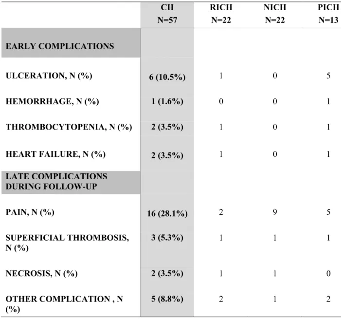
CH N=57 RICH N=22 NICH N=22 PICH N=13 EARLY COMPLICATIONS ULCERATION, N (%) 6 (10.5%) 1 0 5 HEMORRHAGE, N (%) 1 (1.6%) 0 0 1 THROMBOCYTOPENIA, N (%) 2 (3.5%) 1 0 1 HEART FAILURE, N (%) 2 (3.5%) 1 0 1 LATE COMPLICATIONS DURING FOLLOW-UP PAIN, N (%) 16 (28.1%) 2 9 5 SUPERFICIAL THROMBOSIS, N (%) 3 (5.3%) 1 1 1 NECROSIS, N (%) 2 (3.5%) 1 1 0 OTHER COMPLICATION , N (%) 5 (8.8%) 2 1 2
II-
RESULTATS
COMPLEMENTAIRES :
ANALYSE
HISTOLOGIQUE
12 enfants ont eu une exérèse de leur hémangiome, et 2 une biopsie. Nous avons effectué une relecture histologique de 13 cas : 3 RICH, 4 NICH et 6 PICH. Un cas (NICH) n’a pas pu être analysé (matériel tumoral non disponible).
Ils siégeaient dans le derme et/ou l’hypoderme. Ils étaient formés de capillaires de taille variable, organisés en lobules au sein d’un stroma fibreux, associés à des vaisseaux de type veineux, artériel et lymphatique. La présence de gros vaisseaux d’aspect malformatif était notée dans tous les cas. Il existait un gradient de maturation au niveau des capillaires lobulaires, avec des lésions d’allure précoce faites de lobules capillaires très compacts, et des lésions caractérisées par de plus grands lobules à vaisseaux matures dilatés et parfois dysplasiques, avec une fibrose centrale et péri-lobulaire beaucoup plus marquée (Figure 7). Ce dernier aspect de lésion mature a été noté pour l’ensemble des NICH étudiés. Pour les 2 cas biopsiés (2 RICH), il n’a pas été possible d’analyser leur architecture du fait de la petite taille du prélèvement.
Figure 7 : Gradient de maturation des capillaires lobulaires (3 lésions). En haut à gauche : capillaires très compacts ; en haut à droite : capillaires d’aspect plus mature, dilatés, fibrose modérée ; en bas au centre : grands lobules à vaisseaux matures dilatés et dysplasiques, avec fibrose dense (HES x 100).
On notait la présence de calcifications chez 2 cas (1 RICH et 1 PICH).
Des thromboses vasculaires étaient visualisées chez 2 cas (1 NICH et 1 PICH).
Chez un enfant ayant un RICH du bras d’évolution très atrophique, la biopsie réalisée à l’âge de 21 mois objectivait une disparition complète du tissu adipeux hypodermique entre le derme et le muscle (Figure 8).
Figure 8 : A gauche, photo de RICH d’évolution particulièrement atrophique. A droite, biopsie cutanée de la lésion montrant une disparition complète de l’hypoderme entre le derme et le muscle (HES x 25).
En immunohistochimie, le marqueur GLUT-1 était négatif pour les 13 cas étudiés.
L’anticorps PS100 a mis en évidence la présence de filets nerveux intra-lésionnels, souvent hyperplasiques, situés en intra-lobulaire et en inter-lobulaire (Figure 9).
Figure 9 : Immunomarquage PS100 : mise en évidence de multiples filets nerveux intra-lésionnels, prédominants en zone inter-lobulaire (grossissement x 70).
Au sein d’un RICH dont l’exérèse avait été réalisée à l’âge de 4 jours, on notait une dislocation des filets nerveux par les structures capillaires et lymphatiques (Figure 10). Chez 2 PICH de localisation occipitale, les filets nerveux étaient bien plus volumineux, hypertrophiques en profondeur.
Figure 10 : Immunomarquage PS100 : Dislocation des filets nerveux d’un RICH par les petits vaisseaux (capillaires et lymphatiques) (grossissement x 150).
III- DISCUSSION
Notre étude rapporte les caractéristiques épidémiologiques, cliniques, évolutives avec les complications, et paracliniques de 57 hémangiomes congénitaux : 22 RICH, 22 NICH et 13 PICH. A notre connaissance, il s’agit de la plus grande série s’intéressant aux 3 sous-types d’HC.
RICH et PICH sont indistinguables à la naissance, se présentant tous deux sous forme d’une volumineuse lésion d’allure tumorale (contrairement aux NICH qui eux sont plans). C’est leur évolution au cours du temps qui permet de les différencier : le RICH a une régression complète en un peu plus d’un an, alors que le PICH n’a qu’une régression partielle, qui se stoppe vers l’âge de 2 ans, laissant une lésion résiduelle ressemblant fortement à un NICH. Ainsi, en cas d’HC ressemblant à un RICH à la naissance, il faut rester prudent sur le pronostic, puisque dans notre série plus d’un tiers ont eu une régression seulement partielle, répondant à la définition du PICH. Ici, l’ulcération précoce semblait être un marqueur d’évolution ultérieure vers un PICH, puisqu’elle était retrouvée chez 5 des 13 cas d’HC ayant eu une involution partielle (PICH).
Leurs caractéristiques cliniques, histologiques et radiologiques communes font penser que RICH, NICH et PICH sont en fait 3 lésions appartement au même spectre tumoral, mais à des stades évolutifs différents : Les RICH seraient les précurseurs des NICH, et le PICH représenterait le lien entre ces 2 types d’HC.
Nous retrouvions les complications précoces des HC classiquement décrites dans la littérature : insuffisance cardiaque, thrombopénie, ulcération. En revanche, peu de séries rapportent l’apparition de douleurs au niveau de l’hémangiome au cours du suivi. Dans notre
série, le caractère douloureux de la lésion était décrit chez 40,9 % des NICH et 38,5 % des PICH. Dans la grande majorité des cas, les symptômes rapportés faisaient évoquer une composante neuropathique : douleur à l’effleurement, au contact, sensation de lancements, picotements. Ces douleurs étaient souvent décrites après l’âge de 4 ans, peut-être parce que les enfants n’étaient pas en mesure de l’exprimer avant cet âge. Un enfant rapportait d’importantes douleurs résiduelles au site de son hémangiomes (au bras) alors qu’il avait entièrement régressé. Le mécanisme de ces douleurs n’est pas clair. Les thromboses parfois constatées, ou la présence d’une ulcération/nécrose, peuvent expliquer les douleurs nociceptives. Dans leur série de NICH, Lee et al. (35) émettaient l’hypothèse que la vasoconstriction (cliniquement évidente dans le NICH qui présente souvent un halo anémique périphérique) pourrait entraîner une ischémie tissulaire locale, se traduisant par la survenue de douleurs.
Notre relecture histologique avec l’immunomarquage PS100 a mis en évidence l’existence de filets nerveux intra-lésionnels de taille variable chez les 13 patients. La présence de ces filets nerveux intra-lésionnels, parfois nombreux, pourrait également être impliquée dans la survenue des douleurs. Elle n’a jamais été rapportée dans la littérature concernant les HC. En revanche, l’existence de nombreux nerfs matures intra-lésionnels a déjà été rapportée dans les malformations vasculaires par Meijer-Jorna et al., qui considéraient cette composante nerveuse comme faisant partie intégrante du développement de l’anomalie vasculaire (36). Dans cette même étude, la composante nerveuse était également décrite comme plus marquée pour les cas intéressant la région céphalique et cervicale, comme pour 2 de nos cas.
La présence séquellaire d’un réseau périphérique de drainage veineux d’allure dysplasique (veines dilatées, tortueuses) était notée chez 17 cas au cours du suivi pour les NICH et après
régression pour les RICH et PICH. En histologie, nous avons mis en évidence la présence de gros vaisseaux d’aspect malformatif chez tous les cas relus. Ces aspects cliniques et histologiques soulèvent la question de phénomènes malformatifs associés aux hémangiomes congénitaux, bien que ceux-ci soient classés dans le groupe des tumeurs vasculaires.
La possible association de phénomènes tumoraux et malformatifs a déjà été rapportée par Garzon et al. pour quelques lésions vasculaires dont les caractéristiques cliniques faisaient évoquer une intrication des 2 mécanismes (37).
IV- LIMITES
Notre étude est limitée par son caractère rétrospectif, et le faible effectif de patient sur 13 ans inhérent à la prévalence rare des hémangiomes congénitaux.
Le recrutement hospitalier exclusif, dans un centre de référence des maladies rares, a pu induire un biais de sélection avec inclusion de cas potentiellement plus sévères.
L’analyse histologique n’a porté que sur 13 cas, et les RICH étaient peu représentés, du fait de leur caractère régressif qui permet de surseoir à l’exérèse de la lésion.
CONCLUSION
Nous rapportons une série rétrospective de 57 cas s’intéressant au profil évolutif des 3 sous-types d’hémangiomes congénitaux sur une période de 13 ans.
RICH, NICH et PICH présentent de nombreuses caractéristiques cliniques et paracliniques communes, suggérant leur appartenance à un même spectre tumoral.
En cas d’hémangiome congénital de type RICH à la naissance, il faut rester prudent sur le pronostic puisque l’involution ne semble complète que dans deux tiers des cas. Dans un tiers des cas, l’involution ne va pas jusqu’à son terme, et on parle alors de PICH.
Les complications ne sont pas si rares, en particularité l’ulcération centrale précoce qui semble être un marqueur d’involution incomplète de type PICH, et les douleurs qui sont très fréquemment rapportées lors du suivi, et qui pourraient être liées à la présence d’abondants filets nerveux intra-lésionnels.
1. Wassef M, Blei F, Adams D, Alomari A, Baselga E, Berenstein A, et al. Vascular Anomalies Classification: Recommendations From the International Society for the Study of Vascular Anomalies. Pediatrics. juill 2015;136(1):e203-214.
2. Boon LM, Enjolras O, Mulliken JB. Congenital hemangioma: Evidence of accelerated involution. J Pediatr. 1 mars 1996;128(3):329‑35.
3. Berenguer B, Mulliken JB, Enjolras O, Boon LM, Wassef M, Josset P, et al. Rapidly involuting congenital hemangioma: clinical and histopathologic features. Pediatr Dev Pathol Off J Soc Pediatr Pathol Paediatr Pathol Soc. déc 2003;6(6):495‑510.
4. Enjolras O, Mulliken JB, Boon LM, Wassef M, Kozakewich HP, Burrows PE. Noninvoluting congenital hemangioma: a rare cutaneous vascular anomaly. Plast Reconstr Surg. juin 2001;107(7):1647‑54.
5. Nasseri E, Piram M, McCuaig CC, Kokta V, Dubois J, Powell J. Partially involuting congenital hemangiomas: a report of 8 cases and review of the literature. J Am Acad Dermatol. janv 2014;70(1):75‑9.
6. Kanada KN, Merin MR, Munden A, Friedlander SF. A prospective study of cutaneous findings in newborns in the United States: correlation with race, ethnicity, and gestational status using updated classification and nomenclature. J Pediatr. août 2012;161(2):240‑5. 7. Kucuk U, Pala EE, Bayol U, Cakir E, Cukurova I, Gumussoy M. Huge congenital haemangioma of the tongue. JPMA J Pak Med Assoc. déc 2014;64(12):1415‑6.
8. Roebuck D, Sebire N, Lehmann E, Barnacle A. Rapidly involuting congenital haemangioma (RICH) of the liver. Pediatr Radiol. 1 mars 2012;42(3):308‑14.
9. Fadell MF, Jones BV, Adams DM. Prenatal diagnosis and postnatal follow-up of rapidly involuting congenital hemangioma (RICH). Pediatr Radiol. 1 août 2011;41(8):1057‑60.
10. Richard F, Garel C, Cynober E, Soupre V, Bénifla JL, Jouannic JM. Prenatal diagnosis of a rapidly involuting congenital hemangioma (RICH) of the skull. Prenat Diagn. 1 mai 2009;29(5):533‑5.
11. Elia D, Garel C, Enjolras O, Vermouneix L, Soupre V, Oury J-F, et al. Prenatal imaging findings in rapidly involuting congenital hemangioma of the skull. Ultrasound Obstet Gynecol. 1 mai 2008;31(5):572‑5.