Université de Montréal
Asthma During Pregnancy
par Faranak Firoozi
Faculté de Pharmacie
Thèse présentée à la Faculté des études supérieures en vue de l’obtention du grade de Ph.D.
en Sciences Pharmaceutiques
Université de Montréal Faculté des études supérieures
Cette thèse intitulée :
Asthma during Pregnancy
présentée par : Faranak Firoozi
a été évaluée par un jury composé des personnes suivantes :
Jean Lachaine, président-rapporteur Lucie Blais, directeur de recherche
Sylvie Perreault, co-directeur Denyse Gautrin, membre du jury Wendy Ungar, examinateur externe
Résumé
L’asthme est connu comme l’une des maladies chroniques les plus fréquentes chez la femme enceinte avec une prévalence de 4 à 8%. La prévalence élevée de l’asthme fait en sorte qu’on se préoccupe de l’impact de la grossesse sur l’asthme et de l’impact de l’asthme sur les issus de la grossesse. La littérature présente des résultats conflictuels concernant l’impact de l’asthme maternel sur les issus périnatales comme les naissances prématurées, les bébés de petit poids et les bébés de petit poids pour l’âge gestationnel (PPGA). De plus, les données scientifiques sont rares concernant l’impact de la sévérité et de la maîtrise de l’asthme durant la grossesse sur les issus périnatales. Donc, nous avons mené cinq études pour réaliser les objectifs suivants: 1. Le développement et la validation de deux indexes pour mesurer la sévérité et la maîtrise de l’asthme. 2. L’évaluation de l’impact du sexe du fœtus sur le risque d’exacerbation de l’asthme maternel et l’utilisation de médicaments antiasthmatiques durant la grossesse; 3. L’évaluation de l’impact de l’asthme maternel sur les issus périnatales; 4. L’évaluation de l’impact de la sévérité de l’asthme maternel durant la grossesse sur les issus périnatales; 5. L’évaluation de l’impact de la maîtrise de l’asthme maternel durant la grossesse sur les issus périnatales. Pour réaliser ces projets de recherche, nous avons travaillé avec une large cohorte de grossesse reconstruite à partir du croisement de trois banques de données administratives du Québec recouvrant la période entre 1990 et 2002. Pour les trois dernières études, nous avons utilisé un devis de cohorte à deux phases d’échantillonnage pour obtenir, à l’aide d’un questionnaire postal, des informations complémentaires qui ne se trouvaient pas dans les banques de données, comme la consommation de cigarettes et
plus élevés chez les femmes asthmatiques que chez les femmes non asthmatiques. De plus, nous avons démontré que le risque d’un bébé PPAG était significativement plus élevé chez les femmes avec un asthme sévère (OR:1.48, IC 95%: 1.15-1.91) et modéré (OR: 1.30, IC 95%:1.10-1.55) que chez les femmes qui avaient un asthme léger. Nous avons aussi observé que les femmes qui avaient un asthme bien maîtrisé durant la grossesse étaient significativement plus à risque d’avoir un bébé PPAG (OR:1.28, IC 95%: 1.15-1.43), un bébé de petit poids (OR: 1.42, IC 95%:1.22-1.66), et un bébé prématuré (OR: 1.63, IC 95%:1.46-1.83) que les femmes non asthmatiques. D’après nos résultats, toutes les femmes asthmatiques même celles qui ont un asthme bien maîtrisé doivent être suivies de près durant la grossesse car elles courent un risque plus élevé d’avoir des issus de grossesses défavorables pour leur nouveau-né.
Mots-clés : Asthme, grossesse, bébé de petit poids pour son âge gestationnel, bébé de
Abstract
Asthma is known as one of the most frequent chronic diseases encountered during pregnancy with prevalence estimated between 4 and 8%. The high prevalence of asthma during pregnancy results in some concerns about the impact of pregnancy on maternal asthma and also the impact of maternal asthma on perinatal outcomes. The literature presents conflicting results concerning the impact of maternal asthma during pregnancy on perinatal outcomes, such as preterm birth, low-birth-weight (LBW) infant and small-for-gestational-age (SGA) infant. Also, scientific evidence is scarce regarding the impact of asthma severity and control during pregnancy on these perinatal outcomes. We thus conducted a research project composed of five studies to achieve the following objectives: 1. to develop and validate two database indexes, one to measure the control of asthma and the other to measure asthma severity; 2. to evaluate the effect of fetal gender on maternal asthma exacerbations and the use of asthma medications during pregnancy; 3. to evaluate the impact of maternal asthma on adverse perinatal outcomes; 4. to evaluate the impact of the severity of asthma during pregnancy on adverse perinatal outcomes; 5. to evaluate the impact of adequately controlled maternal asthma during pregnancy on adverse perinatal outcomes. A large population-based cohort was reconstructed through the linking of three of Quebec’s (Canada) administrative databases covering the period between 1990 and 2002. A two-stage sampling cohort design was used to collect additional information on the women’s life-style habits by way of a mailed questionnaire for the three last studies.
asthmatic women. Also, mothers with adequately controlled asthma during pregnancy were found to be at higher risk of adverse perinatal outcomes than non-asthmatic women (SGA (OR:1.28, 95%CI: 1.15-1.43), LBW (OR: 1.42, 95%CI:1.22-1.66), and preterm deliveries (OR: 1.63, 95%CI:1.46-1.83)). According to our results, all asthmatic women even those with adequately controlled asthma should be closely monitored during pregnancy because they are at increased risk of adverse perinatal outcomes.
Keywords : Asthma, Pregnancy, Small for gestational age, Low birth weight, Preterm
Table of Contents
1. Introduction ... 3 2. Objectives ... 6 3. Literature review ... 8 3.1. Asthma ... 8 3.1.1. Prevalence of asthma ... 8 3.1.2. Pathology of asthma ... 9 3.1.3. Pathophysiology of asthma ... 10 Airway remodeling ... 12 3.1.4. Diagnosis of asthma ... 12 Asthma symptoms ... 12 Objective measurements ... 13 3.1.5. Management of asthma ... 14 Lifestyle management ... 14Pharmacological management of asthma ... 15
Pharmacologic agents (39, 45) ... 15
3.2. Asthma in pregnancy ... 18
3.2.1. Pathophysiology of asthma during pregnancy ... 18
3.2.2. Management of asthma during pregnancy ... 20
3.3. Impact of pregnancy on asthma ... 23
3.3.1. Impact of fetal gender on maternal asthma exacerbation ... 23
3.7. Impact of adequately controlled asthma during pregnancy on adverse perinatal
outcomes ... 40
3.8. Impact of maternal asthma severity on adverse perinatal outcomes ... 43
3.8.1. Small for gestational age ... 43
3.8.2. Preterm birth ... 44
3.8.3. Low birth weight ... 45
3.9. Risk factors for SGA, LBW infants or preterm birth ... 50
3.9.1. SGA Babies ... 50
3.9.2. Preterm Birth ... 50
3.9.3. Low Birth Weight ... 51
4. Method ... 53 4.1. Study Design ... 53 4.2. Sources of data ... 56 4.2.1. Administrative databases ... 56 4.2.2. Questionnaire ... 58 4.3. Study Population ... 58
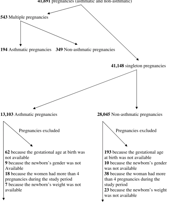
4.3.1. Cohort of pregnant women and newborns, first stage of sampling ... 58
Inclusion criteria ... 59
Exclusion criteria ... 59
Final cohort details ... 60
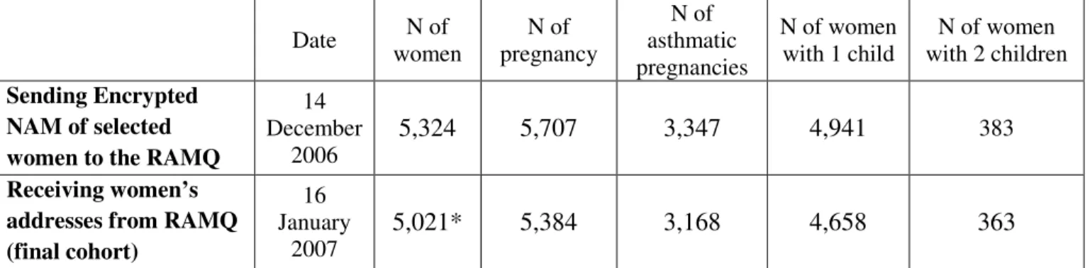
4.4. Two-Stage Sampling ... 62
4.4.1. Sub-cohort of selected pregnant women, second stage of sampling ... 62
4.4.2. Sample constructed based on RAMQ data ... 64
Sending Encrypted NAM of selected women to the RAMQ ... 64
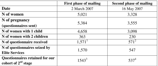
4.4.3. Mailing procedure ... 64
4.5. Outcomes definition ... 66
4.5.1. Small for gestational age ... 66
4.5.4. Moderate to severe maternal asthma exacerbations ... 67
4.5.5. ICS or SABA use during pregnancy ... 68
4.6. Exposures definition ... 68
4.6.1. Fetal Gender ... 68
4.6.2. Asthma during pregnancy ... 69
4.6.3. Asthma severity during pregnancy ... 69
4.6.4. Asthma control during pregnancy ... 69
4.7. Confounding variables ... 70
4.7.1. Risk factors for maternal asthma exacerbation during pregnancy ... 70
Variables retrieved from the administrative databases ... 70
Asthma-related variables ... 70
4.7.2. Risk factors for adverse perinatal outcomes ... 70
Variables retrieved from the administrative databases ... 70
Maternal characteristics ... 70
Pregnancy-related variables ... 71
Maternal co-morbidities ... 71
Variables retrieved from the questionnaire ... 71
Maternal characteristics ... 71
Pregnancy-related variables ... 72
Life style habits ... 72
4.8. Statistical analysis ... 72
4.8.1. GEE for Logistic Regression ... 73
Fifth article ... 163
General discussion ... 187
5.5. Contribution of our results to the literature in the field of asthma in pregnancy 189 5.5.1. Impact of feminine sex hormones on asthma ... 189
5.5.2. Impact of asthma on adverse perinatal outcomes ... 190
5.5.3. Smoking during pregnancy ... 190
5.6. Strengths of the study ... 191
5.6.1. Databases ... 191
5.6.2. Questionnaire ... 192
5.7. Limits of the study ... 192
5.7.1. Biases in the study design ... 192
Selection Bias ... 193
Participation Bias ... 193
Confounding Bias ... 194
Information Bias ... 195
Recall Bias ... 195
Other information bias ... 196
5.7.2. External Validity ... 198
5.8. Clinical implication of our results ... 198
5.9. Further research ... 199
List of tables
Table 1. ………29 Table 2. ………38 Table 3. ………39 Table 4. ………46 Table 5. ………52 Table 6. ………54 Table 7. ………63 Table 8. ………64 Table 9 ………65List of figures
Figure 1. ………11 Figure 2. ………61 Figure 3. ………75
To my loved ones: Madjid and Melody
Remerciements
Je tiens tout d’abord à remercier ma directrice de recherche Dre Lucie Blais pour son encadrement scientifique, son soutien moral et son encouragement tout au long de ce projet. Ce projet a été réalisé grâce à sa présence généreuse et sa patience. Je tiens à remercier ma co-directrice de recherche, Dre Sylvie Perreault pour ses conseils ponctuels et rigoureux. Merci à tous les membres de Jury qui ont accepté d’évaluer ce travail.
Merci à Mme Amélie Forget, notre assistante de recherche pour son exactitude et sa disponibilité très appréciée tout au long de la réalisation de mon projet de recherche. Merci à Mme Karine Chouinard pour s’en charger très rigoureusement de logistique des envoies des questionnaires. Son grand sens de l’organisation m’a beaucoup aidé.
Je tiens à remercier de façon particulière mon conjoint, Madjid qui m’a accompagné toute au long de cet exercice précieux d’acquérir le savoir. Et merci à ma petite Meldoy pour sa présence douce et joyeuse qui m’a donné l’énergie pour finir ce chapitre de ma vie.
Mes pensés vont à mon père qui m’a ouvert les yeux sur la science. Tout au long de sa présence généreuse dans ma vie, il m’a donné le goût d’aller chercher plus loin sur le chemin de savoir. Je pense aussi à ma mère qui m’a donné tant d’amour et m’a encouragé jusqu’à dernier moment de sa vie mais hélas, elle n’a pas pu attendre de voir le résultat final. Je remercie sincèrement mon frère pour ses conseils, son soutient moral et son encouragement.
Abbreviations
RR: Relative riskaRR: Adjusted Relative risk OR: Odds Ratio
CI: Confidence Interval aRR: Adjusted relative risk LBW: Low birth weight SGA: Small for gestational age
RAMQ: Régie de l’Assurance Maladie du Quebec SABA: Short-acting beta2-agonists
ICS: Inhaled corticosteroids IgE: Immunoglobulin E ASA: Acetylsalicylic acid
NSAIDs: Nonsteroidal anti-inflammatory drugs FEV1: Forced expiratory volume in the first second PEF: Peak expiratory flow
LTRAs: Leukotriene receptor antagonists US: United States
IUGR: Intrauterine growth retardation NAEP: National Asthma Education Program ED: Emergency department
GLM: Generalized linear models
CAI: Commission d’accès à l’information du Québec COPD : Chronic obstructive disease
FVC : Forced expiratory vital capacity AMD : Adjusted mean difference LABA : Long-acting beta2 agonist
1. Introduction
Asthma is one of the most prevalent chronic diseases affecting Canadians. According to 2000-2001 Statistics Canada survey, 8.4% of the adult population (aged 12 and more) had physician-diagnosed asthma (over 2.5 million Canadians) (1). Despite several advances in the treatment of asthma, there has been an increase in the prevalence of asthma among adults in the past 20 years in Canada and in many other industrialized countries (2). This increase over the past decades has made asthma the most common chronic disease during pregnancy affecting approximately 8% of women (3-7). The high prevalence of asthma during pregnancy results in some concerns about the impact of pregnancy on maternal asthma and also the impact of maternal asthma on perinatal outcomes.
It has been reported that fetus gender could influence the course of maternal asthma. A few studies have suggested that a pregnant woman’s asthma may worsen when carrying a female fetus (8-11). It has also been reported that asthma during pregnancy could increase the risk of pregnancy induced hypertension, caesarean delivery, prematurity, low birth weight (LBW) infant and perinatal/neonatal mortality (12-16). In addition, it is reported that women with poorly controlled asthma during pregnancy are more likely to deliver LBW, small for gestational age (SGA) and preterm infants than non asthmatic pregnant women (14, 17-19). Also, it has been concluded by a few authors
conflicting results, methodological differences between studies, questionable clinical significance of some of the results as well as lack of statistical power in several studies, make it difficult to conclude with a reasonable degree of certainty on these associations. To overcome these methodological issues and in order to further investigate the association between fetal gender and maternal asthma, we conducted a large population-based cohort study. To study the association between maternal asthma and perinatal outcomes including low birth weight (LBW), preterm birth and small for gestational age (SGA) baby, we added a second stage of sampling to the cohort in order to obtain additional essential confounding variables.
This thesis is presented by articles including one methodological article and four articles presenting the results of the epidemiologic studies in which we investigated the associations described in the previous paragraph. The methodological paper presents the results of a study related to the development and validation of database indexes of asthma severity and control. These indexes measure the control and the severity of asthma in currently treated asthmatic patients using the information obtained from the administrative healthcare databases of the Canadian province of Quebec; Régie de
l’Assurance Maladie du Quebec (RAMQ) and MED-ECHO over a period of 12 months.
These two indexes were used to measure asthma severity and control in two of the four epidemiologic studies.
This thesis includes six other chapters. The objectives of our studies are presented in the second chapter. The third chapter is devoted to the literature review, wherein, a summary of existing knowledge in the field of asthma during pregnancy and its consequences on the health of the mother and the newborn is presented. The
chapter, the methodology employed to conduct the studies is explained. In this section, more details regarding the two-stage sampling design and the data collection via questionnaires which were not presented in the articles will be presented. Chapter five includes five articles which form the result section of my thesis. The last two chapters are devoted to the general discussion and conclusion, respectively.
2. Objectives
This thesis includes five scientific articles which describe the five studies that were undertaken. The research objectives pursued within each of the five studies are described in this chapter.
Study 1. Development and Validation of Database Indexes of Asthma Severity and Control
To develop and validate two database indexes, one to measure the control of asthma and the other to measure the severity of asthma in currently treated asthma patients using information related to dispensed asthma medications and medical services.
Study 2. Effect of Fetal Gender on Maternal Asthma Exacerbations in Pregnant Asthmatic Women
To evaluate the effect of fetal gender on the risk of uncontrolled maternal asthma through the study of exacerbations, use of inhaled short-acting beta2-agonists
(SABA) and inhaled corticosteroids (ICS) during pregnancy.
Study 3. Impact of maternal asthma on perinatal outcomes
To evaluate the potential effect of asthma during pregnancy on adverse perinatal outcomes including SGA infant, LBW infant, and preterm birth.
Study 4. Effect of maternal moderate to severe asthma on perinatal outcomes
To evaluate the effect of the severity of asthma during pregnancy on the risk of having a SGA infant, a LBW infant, and a preterm birth.
Study 5. Are controlled asthmatic pregnant women more at risk of perinatal outcomes than non-asthmatic women?
To investigate whether or not asthmatic women with controlled asthma are at increased risk of having a SGA infant, a LBW infant, or a preterm birth over non asthmatic women.
3. Literature review
3.1. Asthma
The Greek physician Hippocrates used the word asthma for the first time to describe an illness. In Greek it means ‘labour breathing’ (23). Asthma is a chronic inflammatory disease which affects the respiratory tract and is characterized by intermittent or persistent episodes of reversible bronchoconstriction due to increased responsiveness of airways to various stimuli (2, 23-26). Both genetic and environmental factors are believed to contribute to the initiation and progression of the disease (27). Clinically, asthma is manifested by wheezing, dyspnea, chest tightness, and cough (25-27). Although the first manifestation of the disease can occur at any age, half of the patients have asthma onset prior to age 10 years, occurring twice often in boys than in girls, and by the age 30, the prevalence of asthma has become equal between sexes (28, 29). In addition, half of the children suffering from asthma have a substantial or complete remission of symptoms during adolescence, but in many cases, the patients may suffer from recurrence of asthma symptoms in adult life (27, 28, 30).
3.1.1. Prevalence of asthma
Asthma is one of the most prevalent chronic diseases affecting Canadians. According to the 2000-2001 Statistics Canada survey, about 2.2 million Canadians have been diagnosed with asthma by a physician (8.4 % of the population aged 12 years or more) (31). In Canada, an estimated 12% of children and 6% of adults have active asthma (taking medications for asthma or experiencing some symptoms in the past twelve months) (32, 33). There has been an increase in the prevalence of asthma in the past 15 years and mostly in the westernized countries (2, 34). Prevalence rates tend to be higher
Although there is no clear explanation for the observed increasing incidence of asthma, it has been proposed that it may be the result of some alterations in the everyday life-styles (35). The early exposure to various allergens during pregnancy and childhood may influence the development of the immune system (2). In genetically predisposed individuals, the altered immune system may result in an increased allergic response to foreign substances and in this way predispose the child to asthma (2). Possible factors include changes in nowadays housing conditions with greater exposure to indoor aeroallergens, such as cats, house dust mites, cockroaches, and moulds, changes in diet, environmental changes such as outdoor pollution and indoor poor air quality as a result of more insulated home constructions (2, 35). The known risk factors related to developing asthma (incidence) are (23, 30, 36-40):
• A family history of asthma or allergic reaction (eczema, allergic rhinitis), • Exposure to high levels of aeroallergens during infancy,
• Exposure to environmental tobacco smoke both prenatally and postnatally • Exposure to chemical irritants in the workplace
• Extensive vaccination programmes • Changes in diet
o Food preservation
o Adding antibiotic to the food
• Inappropriate use of broad-spectrum antibiotics • Changes in life style;
o Better insulated and more energy efficient homes which result in a warm and humid environment with low ventilation rate
identified including hypertrophy of smooth muscles, thickening of the basement membrane due to collagen deposition, filling up of the airways by mucus and inflammatory cells, and engorgement of the vessels and microvascular leakage (23, 27, 30). The swelling and the mucus plugging inside the airways lead to chronic airways narrowing which makes it hard for the air to pass through resulting in distressed breathing (23, 30).
3.1.3. Pathophysiology of asthma
The aetiology of asthma is not completely known, but it is suggested that bronchial inflammation or its consequences plays an important role in the pathogenesis and persistence of asthma (25).Generally, asthma is classified into two major categories based on the presence or absence of an underlying immune reaction (23, 25, 28, 30).
The Extrinsic asthma (allergic) occurs in atopic individuals who show allergic reaction to foreign bodies such as house dust mites, grass pollen, and cat and dog dander (history of allergic disease) (23, 28). The term ‘extrinsic’ implies that an inhaled allergen is the cause of the initiation of the broncho-spasm. Allergic or atopic asthma is the most common type of asthma and type I hypersensitivity reaction explains the cause of the bronchial inflammation in this group of patients (25). The type I hypersensitivity or immunoglobulin E (IgE) mediated hypersensitivity is caused by inappropriate production of IgE to specific allergens (23, 30, 41). In allergic asthma, IgE binds tightly on the surface of the mast cells, which result in mast cell degranulation and rapid releasing of histamine and other inflammatory mediators (Figure 1) (28, 30). The inflammatory mediators induce bronchial smooth muscle contraction, mucus hyper secretion and increased vascular permeability (2, 23, 27, 28).
The Intrinsic asthma often exhibits in middle-age individuals and the cause of bronchial inflammation is much less clear in this type of asthma (23, 25, 28). Generally, the non-immunological mechanisms contribute to initiate bronchospasm responsible for other symptoms (23, 25, 27, 28). The common non-allergic triggers are occupational exposures to chemical irritants, respiratory tract viral infection, cold weather, air pollution, tobacco smoke, strong odours, drugs such as Acetylsalicylic acid (ASA) and other nonsteroidal anti-inflammatory drugs (NSAIDs), antiadrenergic and cholinergic drugs (e.g. beta blockers and bethanechol), physical exercise, and psychological stress (23, 25, 28, 30). In clinical practice, making a clear distinction between extrinsic and intrinsic asthma is often difficult.
Rubin’s Pathology. Wolters Kluwer, Lippincott Williams & Wilkins; 2008, with
permission from the editors (28).
Airway remodeling
When asthma is poorly controlled, the persistent inflammation can result in the airway remodeling. Damage to the protective endothelial layer allows infiltration of inflammatory mediators into the mucosa and can yield permanent airway abnormalities due to subbasement membrane collagen deposition and fibrosis (23, 27, 30). These changes provide the grounds for proposing prompt and continuous use of corticosteroid in the treatment of asthma (23, 27).
3.1.4. Diagnosis of asthma
The clinical diagnosis of asthma is based on a complete medical history, physical examination and lung function tests (35). A family history of asthma or atopic disease, the presence of typical asthma symptoms that improve with asthma medication, objective evidences of variability in lung function over time and evidences of hyper-responsiveness of the airways using a provocation challenge test can help to make an accurate asthma diagnosis (2, 27, 39). Generally, an increase of 15% or more in FEV1 (an absolute
improvement of at least 200 ml) after inhaling a bronchodilator (short-acting beta-2 agonist) is considered as a significant response and is a confirmation of the diagnosis of asthma (23, 41, 42).
patient and can change over time (41). Symptoms are frequently nocturnal or occur in early morning and from one acute asthma exacerbation to another one, patients may be asymptomatic (30). Significant sputum production is present in about 30% to 50% of patients with asthma (41). The microscopic examination of the sputum manifests large amount of eosinophils (41).
Objective measurements
Lung function measurements reflect the degree of airway obstruction and may be normal between exacerbations. Performing these measurements are required to confirm the diagnosis of asthma and the severity of the disease (30, 44). A spirometer is used to objectively measure the volume of air inhaled and exhaled, and to determine how effectively and how quickly the lungs can be emptied and filled again (23, 30). In clinical practice, the following tests are usually carried out to measure the lung function, the forced expiratory volume in the first second (FEV1), the peak expiratory flow (PEF) and
the provocation test (24, 30).
FEV1 is the maximum volume of air expired in the first second of maximal expiration after a full inspiration and is a useful measure of how quickly full lungs can be emptied (23). PEF is the maximal flow rate achieved during expiration and this occurs very early in the forced expiratory manoeuvre (23). The PEF and FEV1 are both
Broncho-provocation challenge testing with methacoline or histamine is conducted in patients whose pulmonary function tests are normal, but the diagnosis of asthma is not completely ruled out (30, 44). Changes in patient’s lung function (usually FEV1) are measured after inhalation of incremental doses of stimulant (methacoline or
histamine) (23). The concentration of stimulant that causes a 20% decrease in the patient’s FEV1 is known as the Pc20 and a Pc20 of less than 4 mg/ml methacoline is
highly suggestive of diagnosis of asthma (23).
3.1.5. Management of asthma
According to Canadian guidelines, treatment should be determined on the basis of frequency and severity of symptoms, occurrence of acute exacerbations, activity limitation, degree of airway obstruction and response to medication (2, 26, 39). The main objectives in the treatment of chronic asthma are to prevent irreversible airway damage and to control asthma (23). The control of asthma is usually defined as reducing airway inflammation to achieve minimal symptoms during the day and night, to achieve normal lung function (PEF or FEV1 greater than 80% of the predicted value or personal best
value), and normal daily activity including sports (23, 39, 43). ICSs are very effective at suppressing inflammation in asthma; however, symptoms and airway obstruction usually recur when the drugs are discontinued (27).
Lifestyle management
Medication is not the only way to control asthma. Non-pharmacological options such as environmental control approaches are also important to avoid or eliminate known exacerbating allergens like pollens that induce or trigger asthma (24, 41). In addition,
Pharmacological management of asthma
According to the Canadian Asthma Consensus Conference Guidelines for asthma management, there are two types of medication to treat persistent asthma; controllers and relievers (39). The controller medications or preventers (anti-inflammatory and some bronchodilators) should be used on a regular basis to control the underlying inflammation and prevent bronchospasm symptoms and attacks. The relievers (short-acting bronchodilators) are used to relieve airway constriction and its accompanying acute symptoms, only on demand and at the minimum required dose and frequency. (2).
The choice of medication is based on the severity of the disease and may vary over the time as symptoms change (2, 41). Since persistent asthma is a chronic condition, patients have to take long-term anti-inflammatory medication daily to control the underlying inflammation and to prevent symptoms and attacks (23, 41). Most asthma guidelines propose a stepwise approach, which ranges from administrating short-acting inhaled beta agonists for very mild intermittent asthma to oral corticosteroids for severe asthma (23, 24, 27, 41). Therapy is preferably given by inhalation to deliver the drugs to the desired site of action with a much smaller dose and lower systemic drug concentrations which reduces systemic adverse effects (24, 27, 39, 41).
• Anticholinergic agents such as Ipratropium bromide are recommended for patients who present tremor or tachycardia with SABA, or are unresponsive to these agents or suffer from bronchospasm induced by a beta-blocker (24, 44). Ipratropium bromide is administrated by inhalation and has a delayed onset of action comparing to inhaled beta2 agonists, but its bronchodilator effect lasts
longer (23, 30).
2. Controllers
a. Anti-inflammatory agents
• Corticosteroids decrease the airway hyper responsiveness via their anti-inflammatory properties (24, 44). They should be used regularly to achieve maximum effect (30).
o Inhaled corticosteroids have less systemic and side effects than oral steroids (24, 44). Inhaled budesonide, beclomethasone dipropionate, fluticasone propionate and ciclesonide block the late phase of asthma and are recommended for chronic asthma prophylaxis (24).
o Oral corticosteroids are helpful in managing acute exacerbations and their regular use may be necessary in severe asthma (30, 39). Improvement in pulmonary function may begin within 1-3 hours after their administration; however, the maximum effect could achieve about 6-9 hours later (23, 44). To avoid their significant side effects, the treatment should be administered on short periods (one to two weeks) (23).
• Inhaled Nonsteroidal agents such as Cromolyn sodium and Nedocromil
sodium prevent both the early and late phase of asthma exacerbation (24, 44).
They could be used for prophylaxis of exercise-induced asthma or be used regularly in conjunction with other asthma therapy or as an alternative to ICS in less severe asthma cases (30, 44). However, ICSs are more effective and used more commonly than these medications.
• Leukotriene receptor antagonists (LTRAs) have anit-inflammatory and bronchodilator properties and are equivalent to low dose of ICS (30, 46). Zafirlukast and montelukast, currently available in Canada are usually used as an add-on therapy in patients who are inadequately controlled by ICSs (23, 44, 46).
b. Bronchodilators
• Inhaled long-acting beta2 agonists are used regularly twice a day and
considered as an add-on therapy for patients who already taking ICSs without achieving the desired control of asthma (24, 44). Salmeterol and formoterol are available in Canada (24, 44).
• Theophylline compounds such as theophylline, aminophylline and oxtriphylline are the third-line therapy (24, 30, 44). Due to their systemic toxicity and their mild clinical effect, these medications are only
3.2. Asthma in pregnancy
The prevalence of asthma among pregnant women is estimated between 4 to 7% and it is known as one of the most frequent chronic diseases encountered during pregnancy (3-5, 7, 47). In a recent study, Known et al investigated the trends in asthma prevalence during pregnancy in the United States (US) over the past decades and they concluded that the prevalence of self-reported asthma in the US was between 8.4% and 8.8% (48). In the same study the authors assessed international reports of asthma in pregnancy using standardized definitions of asthma within a shared time frame. They found significant differences in asthma prevalence during pregnancy worldwide, and an overall increasing prevalence of asthma during pregnancy over time (48). In addition, these percentages are probably underestimated because, in many cases, either the women do not report their previously diagnosed asthma or they are simply undiagnosed beforehand (49). The increasing prevalence of asthma during pregnancy necessitates a better understanding of the pathophysiology of asthma during pregnancy, and the reciprocal effect of asthma and pregnancy.
3.2.1. Pathophysiology of asthma during pregnancy
Several physiological respiratory alterations occur normally in pregnant women (50). Estrogen changes in pregnancy affect the upper respiratory tract and the airway mucosa resulting in mucosal edema, hypersecretion and capillary congestion (23, 50). Also, the increasing abdominal growth during pregnancy induces an elevation of the diaphragm which is associated with pulmonary function changes (23, 50). As a result, the expiratory reserve volume is reduced, however, the total lung capacity and FEV1 remains
unchanged (50, 51). Increased circulating levels of progesterone and its stimulatory impact on the respiratory center induce an increase in minute ventilation (the total amount
compensation through renal loss of bicarbonate (50, 52). Thus, during pregnancy, normal blood gases reveal a higher pO2 (100 to 106 mmHg) and a lower pCO2 (28 to 30 mmHg) than in the non-pregnant state and pCO2 > 35 mmHg or pO2<70 mmHg associated with bronchial obstruction represent more severe respiratory failure during pregnancy compared to the same blood gas measurements in the nongravid state (50-52).
Normal fetal oxygen pressure of placental blood flow is 30 to 37 mmHg (51, 52). To compensate this low level of pO2 in the fetus comparing to the adult (about one third), the fetus shows some adaptations: high fetal cardiac output, high affinity of fetal haemoglobin for oxygen, high fetal haemoglobin concentration (about 15 to 20 g per L) and a system of vascular shunts which directs available blood oxygen to high priority organs (liver, heart and brain) (50-52).
Asthma during pregnancy can induce hypoxia combined with acute or compensated respiratory acidosis, as well as potentially an acute respiratory alkalosis that decreases the placental blood flow , increases systemic and pulmonary vascular resistance and decreases cardiac output (52-54). Asthmatic pregnant women who suffer from acute asthma exacerbations during their pregnancy can experience hypoxia and hypercapnia (28). Consequently, to obtain a reduction in oxygen consumption, the fetus may reduce breathing and body movements to redistribute oxygen to high priority organs (50, 51). In fetus suffering from lack of oxygen, the rate of oxygen extraction by fetal tissues may
3.2.2. Management of asthma during pregnancy
The goal of asthma therapy in pregnancy is to provide adequate oxygenation to the mother and baby. Concerns about teratogenicity of asthma medications during pregnancy might lead women to stop or reduce their use, but this fear must be balanced against the risk of asthma exacerbations and their potential adverse effects on the mother and the developing fetus.
The National Asthma Education Program (NAEP) in the US has issued guidelines for the treatment of asthma during pregnancy which recommend to treat asthma as aggressively in pregnant women as in non pregnant women (57). ICS are recommended as the first line maintenance therapy in women with persistent asthma during pregnancy and different studies have shown that ICS can be used with relative safety with minimal systemic absorption and few side effects (39, 57, 58). Inhalation has advantages as means of giving drugs during pregnancy because the therapeutic action can be achieved with minimum pharmacological effect to the fetus (24). The relatively low molecular weight and high lipidosolubility of budesonide predicts its substantial placental transfer. (59). However, the actual amount of active budesonide reaching the fetus may be small because of the low systemic bioavailability after inhalation and extensive placental metabolism to inactive compounds (59). In addition, it has been reported that ICS taken at recommended doses during pregnancy were associated with a reduction in the risk of congenital malformations (60). Systemic treatment should not be withheld if indicated, because the risk of asthma exacerbations and their potential adverse effects on the mother and the developing fetus is important and they should be treated rapidly. Prednisolone is an appropriate corticosteroid for oral use since very little of the drug reaches the fetus (24).
Indeed, none of the drugs usually used to treat asthma has been shown to cause congenital malformations (61-67). However, using the systemic corticosteroids should be kept only for severe cases of asthma exacerbation during pregnancy because it has been reported that the risk of cleft lip and palate may increase up to 3 folds by these medications (62, 65). A significant higher incidence of congenital abnormalities in the children of asthmatic women comparing to non-asthmatic ones has been reported by a few studies (aOR ranging from 1.10 to 1.37) (14, 68, 69) however, the results of some preceding studies were not the same (21, 22, 70). Therefore, it is important that concerns about teratogenicity of asthma medications be balanced against the risk of asthma exacerbations and their potential adverse effects on the developing fetus.
Leukotriene modifiers should be avoided during pregnancy because limited safety information is available for this situation (27, 65, 71). The pharmacologic and toxicologic profiles of inhaled long-acting beta2 agonist are similar to the short-acting beta2 agonists, with the exception of their prolonged retention in the lungs (71). However,
they should be used only if there is no other alternative because limited data are available on their use during pregnancy (71-73). ICSs are the cornerstone of therapy in asthma during pregnancy and in fact, different studies have shown that ICS can be used with relative safety and minimal side effects during pregnancy (39, 57, 58).
pregnancy (76). Of this subset, a third did so without first discussing it with their prescribing physician or obstetrician.
3.3. Impact of pregnancy on asthma
Pregnancy could influence the course of asthma. A few studies have shown that asthma worsened during pregnancy in about 33% of women, improved in about 33% and stay unchanged in about 33% (77, 78). Both the variable nature of the disease as well as the asthma variability due to pregnancy could explain in part the change in the course of asthma during pregnancy (79). However, the mechanisms of these changes have not been well clarified and in general the course of asthma during pregnancy is not predictable (79). It was shown that the first trimester and the last month of pregnancy are relatively free of exacerbation and the second and third trimester have more potential for increased symptoms and the need for medications (77, 78, 80, 81). The course of asthma during pregnancy is influenced by the severity of the pre-existing asthma and severe asthma is more likely to worsen during pregnancy than mild asthma (77, 78, 82). The majority of women who experience increased severity of asthma during one pregnancy will have increased severity during subsequent pregnancies (83).
3.3.1. Impact of fetal gender on maternal asthma exacerbation
A few studies have suggested that fetal gender could influence the course of asthma during pregnancy (8-10). These studies have concluded that asthmatic mothers pregnant with female fetus reported more symptoms and had slightly lower lung function than mothers pregnant with male fetus (8-11).
In a blind-controlled prospective study (n=34), Beecroft et al. have found that asthmatic women pregnant with a female fetus reported significantly more shortness of breath (72% vs. 31%), nocturnal awakening (55% vs. 37%), and general asthma symptoms (50% vs. 31%) than women pregnant with a male fetus (8).
Dodds et al. have evaluated steroids use during pregnancy among a sample of 817 pregnant asthmatic women without having specific data on asthma severity or symptoms and found an increased usage of steroids during pregnancy among mothers of a female fetus as compared to mothers of a male fetus (20% vs. 14%) (9). This outcome is difficult to interpret since it is unclear whether or not it includes only oral corticosteroids or both inhaled and oral formulations, which in the later case would not necessarily reflect uncontrolled asthma. Moreover, no conclusion on the statistical significance of this difference can be made since no statistical inference was reported in the article.
In a recent study, Kwon et al. used a prospective cohort design to assess the association between fetal gender and maternal airway lability among pregnant asthmatic women (10). Among 702 pregnant women with asthma, they measured an objective outcome i.e. peak expiratory flow (PEF). The PEF was assessed at enrolment and at 21, 29, and 37 weeks of gestation. The 10% reported difference in log diurnal variation of the PEF between pregnancies of male and female fetuses reached statistical significance, but the clinical significance of the observed difference is questionable (10).
Conversely, Baibergenova et al. did not find any significant association between fetal gender and visits to an emergency department (ED) for asthma during pregnancy between pregnancies of male and female fetuses (84). This study was based on a large
Information (CIHI). From this cohort, Baibergenova et al. first identified all patients who visited an ED during pregnancy and then found that 0.49% and 0.48% of those ED visits were for asthma among women pregnant with a female and a male fetus, respectively (p-value > 0.05).
Among the hypotheses put forward to explain the mechanisms behind the association between fetal gender and maternal asthma control during pregnancy, the one related to the regulation of placental glucocorticoid and immune response in asthmatic pregnancies seems the most plausible (8, 10, 84). Indeed, Clifton and Murphy and their research teams have reported that female fetus alters maternal asthma during pregnancy by upregulating maternal inflammatory pathways (85-88) and thus if asthma-associated inflammatory pathways are not treated with inhaled steroids during pregnancy, the mother could suffer asthma exacerbation.
3.4. Impact of asthma on pregnancy
Asthma could affect pregnancy outcomes. Asthma in pregnancy has been associated with maternal and fetal morbidity (16, 21, 89). It has been reported that pregnant asthmatic women have an increased risk of vaginal bleeding (16), pregnancy-induced hypertension (16, 89), cesarean section (90, 91) and complicated labor (90) comparing to non-asthmatic women . However, it seems that the magnitude of these adverse outcomes is related to the degree of control and severity of maternal asthma (49). The association between maternal asthma and adverse perinatal outcomes has been evaluated by several studies; however, the literature reports inconsistent results.
3.5. Impact of asthma on adverse perinatal outcomes
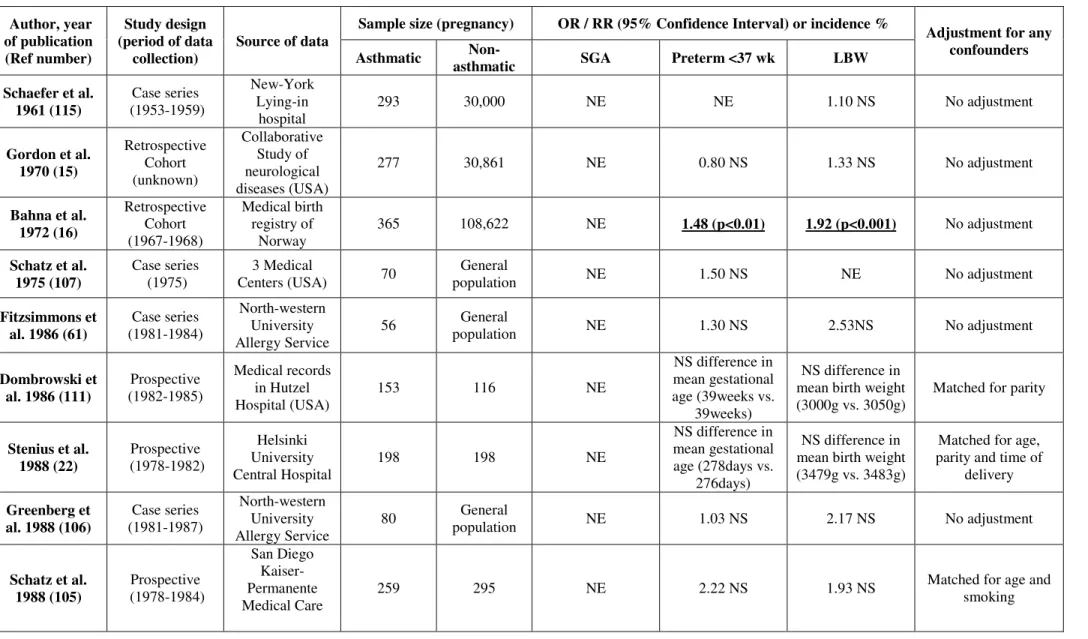
Studies comparing adverse perinatal outcomes such as preterm birth, LBW infant or SGA infant between pregnancies of asthmatic women and non-asthmatic women are summarized in Table1. Different study designs were used in 36 studies presented in Table 1. There are 14 prospective studies, 5 case series, 14 retrospective studies, 2 case-control studies, and one cross-sectional survey. The results of one other systematic review (meta-analysis) are also presented in this table. The sample size of asthmatic group ranged from 32 (92) to 36,985 (93) pregnancies and the sample size of non-asthmatic group ranged from 77 (94) to 1,320,000 (93) pregnancies. Among these studies, 27 adjusted for diverse potential confounding variables, but only 14 studies adjusted for smoking.
In Table 1, for each study, general information regarding the study design, the period of data collection, the sample size of the asthmatic and non-asthmatic groups and the list of confounding variables are presented. The magnitude of the risk for the adverse
3.5.1. Small for gestational age
Most of the older studies evaluated the impact of maternal asthma on adverse perinatal outcomes did not assess the risk of delivering SGA infant in asthmatic women compared to non-asthmatic women. Among 13 studies that evaluated the risk of this adverse perinatal outcome among asthmatic women, 5 large studies reported a significant association between maternal asthma and the risk of SGA infants (adjusted relative risk (aRR) ranged from 1.16 to1.50) (14, 17, 95-97). However, 8 other studies found no significant increased risk of SGA associated with asthma (21, 63, 91, 94, 98-101). Lack of adjustment for several potential confounders and lack of power due to small sample sizes probably explain the differences in results.
3.5.2. Preterm birth
Preterm birth, occurring prior to 37 weeks of gestation was evaluated in 29 studies and 10 of them have reported a significant increase of risk in asthmatic women that ranged from 1.15 to 4.00. Risk of preterm birth has been reported to be significantly increased in asthmatic women as compared to non-asthmatic women in 5 large studies (asthmatic population size > 2000) that adjusted for several potential confounders (aOR: 1.11-1.78) (14, 17, 19, 93, 97). In addition, 4 other smaller studies (asthmatic population size < 500) reported a significant increased risk of preterm birth with ORs ranging from
with and without asthma exacerbation during pregnancy were compared to non asthmatic women (113). The authors found no significant increased risk of preterm delivery in women who had (RR: 1.46, 95%CI: 0.77-2.78) and those who did not have an asthma exacerbation during pregnancy (RR: 0.93, 95% CI: 0.74-1.17).
3.5.3. Low birth weight
Among 22 studies which evaluated the impact of maternal asthma on the risk of LBW infant (weight<2500 g at birth), 7 reported a statistical significant 1.15 to 9.36 increase in the incidence of LBW. Bahna et al were the first authors who reported a significant increased risk of LBW (OR: 1.92) in asthmatic women (16). This increased risk was later confirmed in 3 large studies (asthmatic population size > 2000) conducted retrospectively and adjusted for some potential confounders including smoking (OR: 1.15 to 1.32) (14, 93, 97). Two other smaller studies also reported a significant increased risk of LBW ranging from 2.95 to 9.36 (90, 114). No significant differences in the risk of LBW was observed among asthmatic women as compared to non-asthmatic women in 15 other studies (7, 15, 20, 21, 61, 63, 91, 92, 94, 95, 105, 106, 109, 115, 116). In addition, Murphy et al investigated the effect of asthma and asthma exacerbation on LBW through a meta-analysis using data from three studies (113). They observed a significantly increased risk in women who had (RR: 2.54, 95% CI:1.52-4.25) but no increased risk in those who did not have an asthma exacerbation during pregnancy (RR: 1.12, 95% CI: 0.89-1.40) (113).
Table 1. Description of studies that assessed the impact of maternal Asthma on SGA, preterm birth and LBW Author, year of publication (Ref number) Study design (period of data collection)
Source of data Sample size (pregnancy) OR / RR (95% Confidence Interval) or incidence % Adjustment for any confounders Asthmatic asthmatic Non- SGA Preterm <37 wk LBW
Schaefer et al. 1961 (115) (1953-1959) Case series New-York Lying-in hospital 293 30,000 NE NE 1.10 NS No adjustment Gordon et al. 1970 (15) Retrospective Cohort (unknown) Collaborative Study of neurological diseases (USA) 277 30,861 NE 0.80 NS 1.33 NS No adjustment Bahna et al. 1972 (16) Retrospective Cohort (1967-1968) Medical birth registry of Norway 365 108,622 NE 1.48 (p<0.01) 1.92 (p<0.001) No adjustment Schatz et al.
1975 (107) Case series (1975) Centers (USA) 3 Medical 70 population General NE 1.50 NS NE No adjustment
Fitzsimmons et
al. 1986 (61) (1981-1984) Case series
North-western University Allergy Service 56 General population NE 1.30 NS 2.53NS No adjustment NS difference in
Author, year of publication (Ref number)
Study design (period of data
collection) Source of data
Sample size (pregnancy) OR / RR (95% Confidence Interval) or incidence % Adjustment for any confounders Asthmatic asthmatic Non- SGA Preterm <37 wk LBW
Lao et al. 1990
(90) Retrospective (1984-1987) Hospital, Hong Kong 87 87 NE 3.09 NS 9.36 (p<0.01) Matched for age and parity
Mabie et al.
1992 (98) (1986-1989)Case series Tennessee Hospital, 200 22,651 0.87 NS 0.90 NS NE No adjustment
Perlow et al. 1992 (91) Retrospective (1985-1990) Long Beach Memorial Medical Center Hospital 150 130 5.6 (0.8-40.2) 4.0 (1.1-15.5) 3.4 (0.9-12.1) No adjustment Doucette et al.
1993 (92) (1980-1982) Prospective Haven Hospital Yale-New 32 3,850 NE 1.78 (0.53-6.02) 0.73 (0.1-5.29)
Adjusted for education, race, vaginal bleeding, smoking in 2nd month Schatz et al. 1995 (21) Prospective Cohort (1978-1990) San Diego Kaiser-Permanente Medical Care 486 486 1.33 (p=0.33) 1.65 (p=0.14) 1.64 (p=0.16)
Matched for age, smoking, parity, year
of delivery Stenius et al. 1995 (108) (1982-1990) Prospective Helsinki University Central Hospital and Helsinki Maternity Hospital
504 237 NE 1.15 NS NE Matched for age and parity
Jana et al. 1995
(20) (1983-1992) Prospective Nehru hospital, India 182 364 NE p>0.05 p>0.05 Matched for age and parity
Corchia et al. 1995 (114)
Cross-Sectional
Survey 3 areas of Lazio Region, Italy 55 2,871 NE NE
Smoking
6.62 (1.75-25.07)
No smoking
Adjusted for infant gender, and maternal education within each
Author, year of publication (Ref number)
Study design (period of data
collection) Source of data
Sample size (pregnancy) OR / RR (95% Confidence Interval) or incidence % Adjustment for any confounders Asthmatic asthmatic Non- SGA Preterm <37 wk LBW
Kramer et al. 1995 (102) Case-Control (1990-1992) 3 McGill University-affiliated hospitals among cases 244 341 among cases 200 NE 2 2.32 (1.38-3.89) 3 2.42 (1.44-4.08) NE
Matched for race, and smoking prior and
during pregnancy Stenious et al. 1996 (104) Prospective Cohort (1982-1992) Helsinki University Central Hospital
4457 237 NE 1.0 NS NE Matched for age and
parity Alexander et 1998 al. (7) Retrospective Cohort (1991-1993) Nova Scotia Perinatal Database 5N: 375 6B: 303 7S: 139 13,709 NE 5N: 1.0 (0.5-1.7) 6B: 1.0 (0.5-1.8) 7S: 1.4 (0.6-3.0) 5N: 0.9 (0.5-1.5) 6B: 1.4 (0.8-2.2) 7S: 1.0 (0.4-2.5)
Adjusted for age, previous delivery of
LBW, parity, pre-delivery weight, and
smoking Demissie et 1998 al. (14) Retrospective Cohort (1989-1992) Administrative databases of New Jersey Hospitals 2,289 9,156 Crude 1.33 (1.17-1.51) Adjusted 1.26 (1.10-1.45) Crude 1.59 (1.40-1.80) Adjusted 1.36 (1.18-1.55) Crude 1.57 (1.34-1.86) Adjusted 1.32 (1.10-1.58)
Adjusted for age, education, marital status, parity, race, chronic &gestational
diabetes, chronic HTA, smoking, alcohol and drug use
Author, year of publication (Ref number)
Study design (period of data
collection) Source of data
Sample size (pregnancy) OR / RR (95% Confidence Interval) or incidence % Adjustment for any confounders Asthmatic asthmatic Non- SGA Preterm <37 wk LBW
Minerbi-Codish et al. 1998 (94) Prospective Cohort (1993-1994) Medical Center (Israel) 101 77 No statistically significant difference No statistically significant difference No statistically significant difference
Matched for age and ethnic origin Liu et al. 2001 (17) Retrospective Cohort (1991-1996) Med-Echo database (Quebec) 2,193 8,772 Crude 1.19 (1.02-1.37) Adjusted 1.16 (1.00-1.35) Crude 1.59 (1.35-1.87) Adjusted 1.40 (1.18-1.66) NE
Adjusted for age, chronic and gestational diabetes,
chronic HTA, and caesarean delivery Olesen et al. 2001 (63) Retrospective Cohort (1991-1996) Hospital Discharge Registry and North Jutland Prescription Database (Denmark) 9303 108,717 higher among
exposed one NE higher among exposed one
Adjusted for age, co-habitation status, smoking and child
gender Sobande et al. 2002 (112) Prospective Cohort (1997-2000) Maternity Hospital (Saudi Arabia) 88 106 NE 39.41 vs. 39.43 (p>0.05) 2,855g vs. 3,051g (p=0.006) No adjustment Sorensen et al. 2003 (103) Nested case-control (1994-1995) Healthcare network of Swedish Medical center, (Seattle, USA) among cases 20 among cases 292 NE Crude 2.03 (1.01-4.09) Adjusted 2.37 (1.15-4.88) NE
Adjusted for age, race, parity, Medicaid
status and smoking
Bracken et al. 2003(99) Prospective Cohort (1996-2001) 56 obstetric practices and 15 clinics (Connecticut, 873 1,333 Crude 1.22 (0.89-1.68) Adjusted 1.15 (0.79-1.67) Crude 1.49 (1.07-2.08) Adjusted 1.36 (0.92-2.00) NE
Adjusted for age, marital status, race,
education, pre-pregnancy weight,
Author, year of publication (Ref number)
Study design (period of data
collection) Source of data
Sample size (pregnancy) OR / RR (95% Confidence Interval) or incidence % Adjustment for any confounders Asthmatic asthmatic Non- SGA Preterm <37 wk LBW
Mihrshahi et al. 2003 (109) Prospective data collection (2003) Questionnaires to women identified in antenatal clinics of 6 Sydney hospitals 340 271 NE 2.13 NS 1.47 NS
Adjusted for age, parity, nulliparous, socioeconomic factors, exposure to smoking Dombrowski et al. 2004 (100) Prospective Cohort (1994-1999) 16 centers of Maternal-Fetal Medicine Unit Network (USA) Mild: 873 Moderate-Severe:866 Severe: 52 881 MS:1.2 (0.8-1.8) Mi:1.2 (0.8-1.7) Sev: 1.6 (0.6-4.4) Mi: 1.0 (0.6-1.8) MS: 0.9 (0.5-1.6) Sev: 2.2 (1.2-4.2) NE
Adjusted only for analyses of severe patients for previous
preterm, smoking, race, BMI ACS et al. 2005 (116) Retrospective (1980-1996) Hungarian Congenital Abnormality Registry 757 37,394 NE 1.56 statistical significance not reported 1.61 Statistical significance not reported
Adjusted for age, birth order, employment status. Anti-asthmatic drugs Sheiner et al. 2005 (95) Retrospective Cohort (1988-2002) Soroka University Medical center (Israel) 1,963 137,205 1.5 (1.1-1.9) No difference in mean 1.10 NS
Adjusted for failure to progress in labour,
Author, year of publication (Ref number)
Study design (period of data
collection) Source of data
Sample size (pregnancy) OR / RR (95% Confidence Interval) or incidence % Adjustment for any confounders Asthmatic asthmatic Non- SGA Preterm <37 wk LBW
Murphy 2006
(113) Meta analyse, (2006)
3 studies for evaluating LBW and 4 studies for
evaluating preterm birth No- exacerbation 855 Exacerbation 79 No- exacerbation 1,312 Exacerbation 126 For No- exacerbation 31,662 For Exacerbation 31,285 For No-exacerbation 31,662 For Exacerbation 31,899 NE No-exacerbation vs. non-asthmatic 0.93 (0.74-1.17) Exacerbation vs. non-asthmatic 1.46 (0.77-2.78) No-exacerbation vs. non-asthmatic 1.12 (0.89-1.40) Exacerbation vs. non-asthmatic 2.54 (1.52-4.25) LBW: (15), (20), (21) Preterm birth: (15), (20), (21), (104) Enriquez et al. 2007 (96) Retrospective cohort (1995-2003) Tennessee Medical Program Asthmatic 9,154 Exacerbation 2,105 No- Exacerbation 7,049 131,145 Asthmatic vs. Non-asthmatic 1.19 NS very SGA 1.20 (p< .0001) Asthmatic: Ns Exacerbation: NS No-exacerbation: NS Asthmatic: Mean 3,131g vs. 3,173g (p< .0001) Exacerbation 1.22 (< .0002) No-exacerbation 1.16 (< .0002)
Adjusted for race, age, smoking, education, comorbidity and adequacy of prenatal care Clark et al. 2007 (101) Prospective; Clinic (2001-2003) Antenatal clinics of Manchester Children’s University 370 No med. 170 ICS& β2 178 β2 only 718 ICS& β2 Boys: 1.56 (p=.011) Girls: 0.95 (p=.56) NE ICS& β2 ↓ in mean birth weigh -112 (-193, -30.7) NS for other Adjusted for smoking, race, parity,
Author, year of publication (Ref number)
Study design (period of data
collection) Source of data
Sample size (pregnancy) OR / RR (95% Confidence Interval) or incidence % Adjustment for any confounders Asthmatic asthmatic Non- SGA Preterm <37 wk LBW
Kallen et al. 2007 (97) Retrospective cohort (1995-2004) Swedish Medical Birth Registry 23,988 All birth registered 846,635 1.16 (1.07-1.26) 1.11 (1.05-1.18) 1.15 (1.07-1.23) No adjustment mentioned NS: statistically non significant
NE: not evaluated HTA: hypertension
1Receiving chronic medication but non-steroid dependent 2history of Asthma
3Physician diagnosed asthma
4No acute attack of asthma during the study period 5N:No asthma medication use
6B: beta agonist use only 7S: Steroid use
8All birth during study period
9Receiving at least one prescription for asthma during pregnancy 10Buying no drug prescription during pregnancy
The differences between these results could be partly explained by important differences between studies in:
• The study sample sizes, • Study designs,
• Asthmatic definition, • Non-asthmatic definition, • Data collections,
• Control for confounders,
• Asthma severity during pregnancy,
3.6. Asthma severity and asthma control
Control and severity of asthma are two different but complementary concepts (117). In fact, one can have severe asthma but adequately controlled and another one can have mild asthma but poorly controlled. The severity of asthma could influence the control over time. Canadian experts have recommended that the dose of ICS necessary to obtain good control of asthma should be included when evaluating severity (39). The accurate classification of asthma severity and control is a definite challenge both in clinical practice and in research since they are conceptually related and some of the criteria used in their assessment overlap.
3.6.1. Asthma control
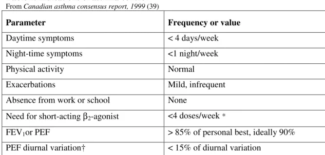
Current series of criteria in the assessment of the control of asthma were established by the Global Initiative for Asthma (GINA) and the Canadian Asthma Consensus Guidelines (39, 43). They include daytime and nocturnal symptoms, physical activity limitations, the occurrence of asthma exacerbations, the need for inhaled SABA, school or work absenteeism, and forced expiratory volume in the first one second (FEV1)
or peak respiratory flow (PEF) values. The optimal control of asthma based on Canadian Asthma Consensus Guidelines has been defined by the presence of minimal respiratory symptoms, no physical activity limitation, normal respiratory function, and absence of the need for rescue bronchodilator more than recommended (see table 2) (39).
Table 2. Indications of asthma control From Canadian asthma consensus report, 1999 (39)
Parameter Frequency or value
Daytime symptoms < 4 days/week
Night-time symptoms <1 night/week
Physical activity Normal
Exacerbations Mild, infrequent
Absence from work or school None
Need for short-acting β2-agonist <4 doses/week *
FEV1or PEF > 85% of personal best, ideally 90%
PEF diurnal variation† < 15% of diurnal variation
FEV1= forced expiratory volume in 1 second; PEF = peak expiratory flow obtained with a portable peak flow meter.
*May use 1 dose/day for prevention of exercise-induced symptoms.
† Diurnal variation is calculated by subtracting the lowest PEF from the highest and dividing by the highest PEF multiplied by 100.
3.6.2. Asthma severity
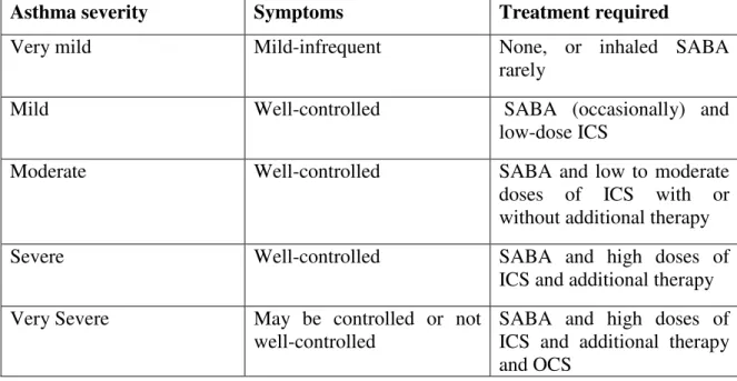
Different methods are advocated by various guidelines for the assessment of asthma severity (39, 43, 118). The GINA guidelines as well as the US National Asthma Education and Prevention Program Consensus guidelines relative to the assessment of severity rely upon the evaluation of the disease’s clinical features (asthma symptoms, occurrence of asthma exacerbation and respiratory function) prior to the initiation of any anti-asthmatic treatment (43, 118). However, the Canadian Asthma Consensus Guidelines assess asthma severity once the treatment has been instigated and rely upon a combination of factors, many of which overlap with measures of asthma control. These include pulmonary function tests, the treatment required to obtain asthma control, the history of hospital admissions, and life-threatening asthma attacks (see table 3) (39).
Table 3: Levels of asthma severity based on treatment needed to obtain control From Canadian asthma consensus report, 1999 (39)
Asthma severity Symptoms Treatment required
Very mild Mild-infrequent None, or inhaled SABA
rarely
Mild Well-controlled SABA (occasionally) and
low-dose ICS
Moderate Well-controlled SABA and low to moderate
doses of ICS with or without additional therapy
Severe Well-controlled SABA and high doses of
ICS and additional therapy Very Severe May be controlled or not
well-controlled
SABA and high doses of ICS and additional therapy and OCS
3.7. Impact of adequately controlled asthma during
pregnancy on adverse perinatal outcomes
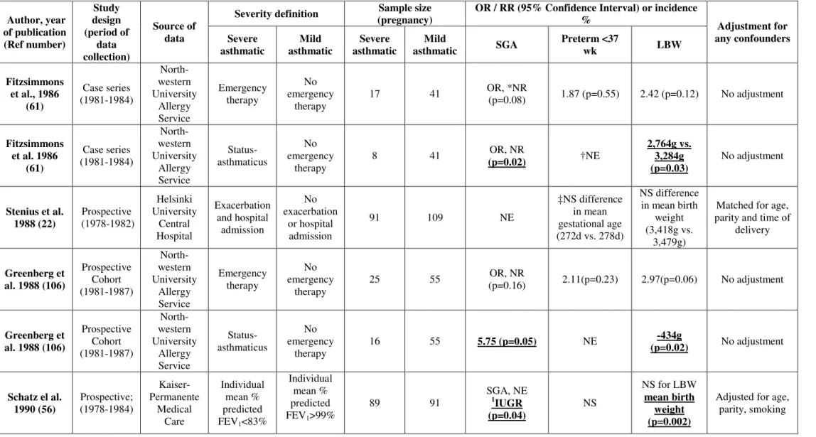
Poorly controlled asthma is potentially dangerous to the fetus since hypoxia combined with respiratory alkalosis decrease placental blood flow and potentially impaired fetal oxygenation (50, 52, 57, 119). Decreased fetal blood oxygen could result in abnormal growth and development of the fetus (120). Jana et al. found that maternal uncontrolled severe asthma (ED visit and systemic corticosteroids use during pregnancy) leads to fetal growth retardation (mean birth weight: 2469g vs 2842g; p<0.05) and low birth weight (53.3% vs 20.5%; p<0.01) more often than women with adequately controlled asthma during pregnancy (20). These results have also been confirmed in other studies (56, 61, 106).
The question is whether better controlled asthma lead to improve fetal growth. To our knowledge, only one study evaluated directly whether or not women with adequately controlled asthma are at higher risk of perinatal outcomes than non-asthmatic women. In a prospective controlled study comparing women with actively managed asthma during pregnancy and non-asthmatic women, Schatz et al observed relative risks as large as 1.65 for perinatal outcomes, but concluded that there was no difference between the groups since the relative risks were not statistically significant (21). In two other studies, Stenius-Aarniala et al and Jana et al. came to the same conclusion, although the association between the control of asthma and perinatal outcomes was only indirectly evaluated (20, 22).
In a prospective study conducted between 1978-1990, Schatz et al assessed perinatal outcomes in 486 actively managed pregnant asthmatic women as compared with
reconfirmation of their diagnostic of asthma at the entry to the study and they were managed in the allergy clinic to prevent acute asthmatic episodes and asthma symptoms that interfere with sleep or normal activity. Moreover, all women received routine obstetric care. The authors reported a RR of 1.33 for SGA (p value =0.33), a RR of 1.64 for LBW (p=0.16), and a RR of 1.65 (p=0.14) for preterm births. However, they concluded that there was no difference between the groups since these RRs were not found to be statistically significant (21).
Jana et al. compared the perinatal outcomes in 182 pregnancies from asthmatic women with those of 364 non-asthmatic women matched for age and parity between 1983-1992 (20). The asthmatic women were followed in an Obstetric-Medical Disorder Clinic and there were close cooperation between the obstetrician and chest physician in the patient’s management. The asthmatic women were advised to continue anti-asthmatic medication throughout pregnancy and they were provided with instructions in the case of acute exacerbation of asthma.
In this study, the authors investigated the risk of adverse perinatal outcomes in women who required emergency hospitalization and was managed with high flow of oxygen, high doses of inhaled β2-agonists, intravenous corticosteroids and aminophylline
infusion during the study period (severe asthmatic patients) as compared with non-asthmatic controls (20). Moreover, they compared women who had used oral and/or