Amphiphilic Linear-Dendritic Block Copolymers
For Drug Delivery
by
Phuong Nguyen
B.S., University of California San Diego (2001) M.S.CEP, Massachusetts Institute of Technology (2004)
Submitted to the Department of Chemical Engineering in Partial Fulfillment of the Requirements for the Degree of
Doctor of Philosophy in Chemical Engineering at the
MASSACHUSETTS INSTITUTE OF TECHNOLOGY
JUNE 2007
© Massachusetts Institute of Technology 2007. All rights reserved.
,.J • - .
Author:
1
%.
Department of Chemical EngineeringJune 27, 2007
Certified by:
Paula T. Hammond, Professor Thesis Supervisor Certified by:.
Robert Langer, Institute Professor Thesis Supervisor Accepted by: c -OF TEOHNOLOGY
MAR 0 6 2008
LIBRARARCHIES
LIBRARIES
William Deen, Professor Chairman, Departmental Committee on Graduate Studies
Amphiphilic Linear-Dendritic Block Copolymers
For Drug Delivery
by
Phuong Nguyen
B.S., University of California San Diego (2001)
M.S.CEP, Massachusetts Institute of Technology (2004)
Submitted to the Department of Chemical Engineering on
June 27, 2007 in Partial Fulfillment of the Requirements for the Degree of Doctor of Philosophy in Chemical Engineering
ABSTRACT
Polymeric drug delivery systems have been widely used in the pharmaceutical industry. Such systems can solubilize and sequester hydrophobic drugs from degradation, thereby increasing circulation half-life and efficacy. However, there are still challenges in the design of drug delivery vehicles to achieve efficient drug delivery in a site-specific manner. In this thesis, an amphiphilic linear-dendritic block copolymer was designed, synthesized, and applied as a new polymeric drug delivery platform.
First, to develop the drug delivery vehicle, an ABA dendritic-linear-dendritic block copolymer consisting of poly(amidoamine) (PAMAM) and poly(propylene oxide) (PPO) was synthesized. In order to determine the viability of the linear-dendritic block copolymer as a drug delivery vehicle, the solution-phase self-assembly behavior and the self-assembled structures were characterized experimentally and through molecular dynamics simulations. The triblock self-assembles in aqueous media to form stable micelles with low CMC values. Dynamic light scattering results and TEM indicate the formation of particles ranging from 9 to 18 nm in diameter, with smaller diameters exhibited at higher generations. Static light scattering also confirmed the trend where the aggregation number decreased with higher generations. The experimental characterization results indicated that the physical characteristics of the PPO-PAMAM micelles were desirable and within the design specifications necessary for drug delivery. The experimental results were utilized to set up simulations where further knowledge of the microstructure of the micelles formed could be gained. It was found that the block copolymers simulated formed micelles in the same size range that was seen experimentally. However, the simulations indicated that the micelles displayed greater asphericity than dendrimers. Backfolding of the terminal amine ends was encountered, which would have implications for the configuration and spacing of any additional
targeting ligand attached to the dendritic ends. Further analysis revealed that with increasing generation, the porosity of the micelles increased, which could affect the diffusion rate of drugs released out of the system. Another important finding detailed the preferential localization of a model hydrophobid drug, triclosan, in an equilibrated micelle structure.
Additional experiments were performed to assess the feasibility of the nanoparticles for drug delivery applications. Drug loading studies were performed with a model hydrophobic drug, triclosan, resulting in high loading efficiencies. In comparison, linear block copolymers were half as efficient in loading triclosan. It was determined that the dendritic block synergistically increased the drug loading due to either acting as an additional block capable of encapsulating drug or sterically favoring the seclusion of the drug in the core. The linear-dendritic block copolymer synthesized was found to be a promising candidate for drug delivery due to its relative stability in aqueous solution and
its drug encapsulation and release properties.
Overall, the linear-dendritic block copolymer displayed physical characteristics and self-assembly behavior that satisfied the design criteria for a viable drug delivery vehicle. As a further step, the potential benefits of the novel linear-dendritic architecture were explored in two different drug delivery applications. First, PPO-PAMAM was explored as a circulating nanoparticle with the capability of multivalently targeting to specific cells, due to the presence of the dense functional groups on the dendritic block forming the corona of the micelles. PPO-PAMAM was functionalized with galactose and targeted to hepatocellular carcinoma cells. It was found that the polymer was not cytotoxic and could bind to the asialoglycoprotein receptor. The galactose-functionalized micelles were loaded with a chemotherapeutic, doxorubicin, and delivered to the carcinoma cells more efficiently than non-functionalized micelles and bare doxorubicin. The results from
in vitro testing showed that PPO-PAMAM micelles with targeting capability are
promising circulating drug delivery vehicles.
In order to ensure success of subsequent testing in vivo of the targeted linear-dendritic block copolymer system, some improvements to the system were explored. First, PPO-PAMAM micelles were stabilized by physical entrapment of the hydrophobic core. An emulsion polymerization of hydrophobic methacrylate monomers created an interpenetrating polymer keeping the micelles intact at concentrations below the CMC and in a solubilizing solvent, methanol. This improvement would ensure that once injected into the bloodstream, the micelles would not destabilize and release high concentrations of drug. Another improvement that was explored was the synthesis of a new linear-dendritic block copolymer composed of a hydrophobic poly(amino acid) and a polyester dendron. Additionally, poly(ethyleneglycol) (PEG) groups were attached to the outer surface of the polyester dendron. The new system synthesized has a low CMC and is thermodynamically slow to break apart in the bloodstream. Furthermore, the micelles formed would be able to circulate for longer times with PEG aiding in evading the reticuloendothelial system.
The second drug delivery application explored, which advantageously utilized the dendritic blocks on the outer surface of the block copolymer micelles was as a localized drug delivery coating created by the layer-by-layer (LbL) assembly approach. The electrostatic LbL assembly approach offers large potential in the area of drug delivery from thin films and surfaces; however, because the processing technique is aqueous-based, there have been few strategies proposed to incorporate hydrophobic molecules into these films. Here we created an LbL film that is capable of incorporating hydrophobic drug at high loadings via encapsulation with linear-dendritic block copolymer micelles and demonstrate for the first time release times of a hydrophobic antibacterial agent over a period of several weeks--a significant improvement over reports of other micelle-encapsulated thin films with release times of several minutes. The PAMAM block, which is polycationic, enabled LbL deposition with negatively charged poly(acrylic acid) (PAA). The stable PPO-PAMAM micelles incorporated into the LbL films encapsulated a hydrophobic bactericide, triclosan. Film thickness and UV-vis measurements confirm the formation of the LbL film and incorporation of triclosan into the film. Fluorescence measurements of PPO-PAMAM/PAA films with pyrene indicated the presence of hydrophobic domains in the film. GISAXS revealed regular spacing of approximately 10.5 nm in the direction parallel to the film substrate, which is approximately the same size as the PPO-PAMAM micelles in aqueous solution. Volume fraction measurements based on elemental analysis and TGA confirm the GISAXS data. An in vitro release study revealed long release times of triclosan on the order of weeks, and a Kirby Bauer test was performed on Staphylococcus Aureus demonstrating that the drug released was
still active to inhibit the growth of bacteria.
Linear-dendritic block copolymer micelles were successfully used in two different drug delivery applications where the dendritic block could be fully utilized. It is hoped that with the research and results presented in this thesis further development of this drug
delivery platform can result in a product successfully treating a serious disease.
Thesis Supervisor: Paula T. Hammond
Title: Bayer Chair Professor of Chemical Engineering
Thesis Supervisor: Robert Langer Title: Institute Professor
Acknowledgements
Many individuals have contributed to the success and completion of the research presented in this thesis. I would first like to thank my family for their continued support over my formative educational years. My parents have contributed in many ways to the completion of my thesis including encouraging and supporting my educational goals and helping me through the ups and downs of research. Additionally, my brother and sister, Peter and Kathy, have helped to remind me that there is life outside of graduate school.
I would also like to extend my appreciation to my thesis committee who has contributed greatly to the work in this thesis. Foremost, my advisor, Professor Paula Hammond has introduced me to the world of polymer chemistry and has helped me towards becoming a resourceful and independent scientist. The rest of my thesis committee members, Professor Bob Langer, Professor Daniel Blankschtein, and Professor Bruce Zetter have given me much encouragement in my thesis research and my
career development.
Each of the collaborators I have worked with has contributed greatly to my thesis work, and I would sincerely express gratitude to each of them. Dr. Nicole Zacharia and Eric Verploegen added to and rounded out the layer-by-layer work. Dr. Brian Stephenson played a large role in the simulations work, performing most of the simulations and supplying a great deal of expertise.
I would also like to thank the past and present members of the Hammond group, who provided an enjoyful and collaborative work environment. I am most grateful to Dr. Kris Stokes and Dr. Lu Tian for always being available for valuable discussions and great insights.
Last, I am grateful for the NSF Graduate Research Fellowship, the Robert T. Haslem Presidential Fellowship, and the MIT Chemical Engineering department for financial support for this thesis work.
Table of Contents
List of Figures... ... ... 13
List of Tables ... 18
CHAPTER 1: INTRODUCTION ... 19
1.1 M otivation... 19
1.2 Background: Colloidal Drug Delivery Systems ... .... 22
1.2.1 Factors for Passive Targeting... 22
1.2.2 Active Targeting ... ... 24
1.2.2.1 Carbohydrates ... 25
1.2.2.2 Folate... 26
1.2.2.3 Targeting Challenges ... 27
1.2.3 N anoparticles for Drug Delivery: Liposom es ... .. 29
1.2.4 Nanoparticles for Drug Delivery: Dendrimers ... 32
1.2.5 Nanoparticles for Drug Delivery: Block Copolymer Micelles ... 36
1.3 Thesis Objectives ... 38
1.4 Thesis Overview ... ... 41
1.5 References ... 43
CHAPTER 2: Synthesis and Characterization of Poly(propylene oxide)-b-Poly(am idoam ine) ... ... 53
2.1 Introduction... 53
2.2 Experim ental ... ... 55
2.2.1 M aterials ... ... 55
2.2.2 Synthesis of PPO-PAM AM ... 55
2.2.3 Characterization of PPO-PAM AM ... 61
2.3 Results and Discussion ... 65
2.3.1 Synthesis of PPO-PAM AM ... 65
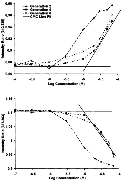
2.3.2 Fluorescence Studies... 67
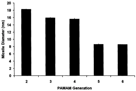
2.3.3 Dynam ic Light Scattering ... 73
2.3.4 TEM ... 75
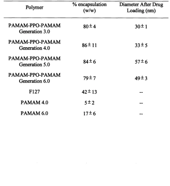
2.3.5 Drug Loading Studies ... 76
2.3.6 Drug Release Studies ... 80
2.4 Conclusions... 82
2.5 References... 84
CHAPTER 3: Experiments and Molecular Dynamics Simulations of PPO-PAMAM
Linear-Dendritic Block Copolymer Unimers and Micelles ... 91
3.1 Introduction ... 9 1 3.2 Experim ental Section... ... 93
3.2.1 M aterials ... ... 93
3.2.2 Synthesis of PPO-PAMAM ... 93
3.2.3 Aggregation Number Determination ... ... 94
3.2.4 Simulation Methodology and Parameters ... ... 95
3.2.5 System Preparation and Equilibration ... ... 96
3.3 Results and Discussion ... 100
3.3.1 Micelle Aggregation Number ... 101
3.3.2 Computer Simulation: Asphericity ... 103
3.3.3 Computer Simulation: Radius of Gyration ... 105
3.3.4 Computer Simulation: Monomer Density Distribution ... 111
3.3.5 Computer Simulation: Terminal Group Distribution... 113
3.3.6 Computer Simulation: Dendrimer/Water Interface ... 114
3.3.7 Computer Simulation: Locus of Hydrophobic Drug Solubilization ... 117
3.3.8 Computer Simulation: Solvent Accessible Surface Area ... 119
3.4 Conclusion ... 122
3.5 R eferences ... ... 124
3.5 R eferences ... ... 124
CHAPTER 4: PPO-PAMAM IN- VITRO TESTING... 129
4.1 Introduction ... 129
4.2 Experimental ... 131
4.2.1 Materials ... 131
4.2.2 Synthesis of Galactose Functionalized PPO-PAMAM... 131
4.2.3 Polymer Characterization... 133
4.2.4 In Vitro Testing ... 134
4.3 R esults and D iscussion ... ... 136
4.3.1 Synthesis of Galactose Functionalized PPO-PAMAM... 137
4.3.2 In Vitro Testing... 141
4.4 C onclusions... 152
4.5 R eferences... 153
CHAPTER 5: Stabilization of PPO-PAMAM Micelles... 157
5.1 Introduction ... 157
5.2 Experimental ... 159
5.2.1 Materials ... 159
5.2.2 Stabilization of Generation 4.5 PPO-PAMAM-COOH Micelles ... . 159
5.3 Results and Discussion ... ... 162
5.3.1 Stabilization of Generation 4.5 PPO-PAMAM-COOH Micelles ... 162
5.3.2 Characterization of Stabilized Generation 4.0 PPO-PAMAM Micelles ... 164
5.4 C onclusions... 171
5.5 R eferences... 172
CHAPTER 6: Synthesis and Characterization of Poly(P-Benzyl-L-Aspartate)-b-Polyester Dendron... 175 6.1 Introduction ... 175 6.2 Experimental ... 176 6.2.1 Materials ... 176 6.2.2 Synthesis ... 176 6.2.3 Polymer Characterization ... 183 6.2.4 In Vitro Testin ... 185
6.3 Results and Discussion ... 6.3.1 Synthesis of PBLA-B 16-PEG-Gal... 6.3.2 Characterization of PBLA-B 1 6-PEG... ... 6.3.3 In Vitro Testing ... 6.4 C onclusions... ... 6.5 R eferences ... ... 187 188 191 193 195 197 CHAPTER 7: Incorporation of PPO-PAMAM Linear-Dendritic Block Copolymer Micelles Into Extended Release Antibacterial Layer-by-Layer Films... 201
7.1 Introduction ... 201 7.2 Experim ental ... 204 7.2.1 M aterials ... 204 7.2.2 Synthesis of PPO-PAMAM ... 204 7.2.3 LbL Film Formation ... 205 7.2.4 LbL Film Characterization... 206 7.2.5 In Vitro Testing ... 207
7.3 Results and D iscussion ... ... 208
7.3.1 Formation and Characterization of Micelle-Containing LbL Films... 208
7.3.2 Drug Release and Efficacy ... 220
7.4 C onclusions... 225
7.5 R eferences... 226
CHAPTER 8: Conclusions and Future Work ... 231
8.1 Thesis Summary... 231
8.2 Future Work ... 236
8.2.1 Improvements to the PPO-PAMAM System... 236
8.2.2 Improvements to the PBLA-B16-PEG System... 238
8.2.3 Multivalent Targeting ... 241
8.2.4 PPO-PAMAM Micelles in LbL Films ... 244
8.3 Concluding Remarks... 246
8.4 R eferences ... ... 247
List of Figures
Figure 1-1. Linear-dendritic block copolymers self-assembling into a micelle ... 21
Figure 1-2. Liposomes are composed of lipid bilayers that are able to encapsulate hydrophilic drug in its aqueous core and hydrophobic drug in the lipid region ... 29
Figure 1-3. A PAMAM dendrimer with conjugated PEG ... 33
Figure 1-4. The formation of micelles from a diblock copolymer... 36
Figure 1-5. Formation of micelles with clustered or multifunctional ligands ... 39
Figure 2-1. Synthesis of PPO-PAMAM by reaction with excess i) methyl acrylate, CH2CHCOOCH3 and ii) ethylenediamine, NH2CH2CH2NH2 ... . . . .. .... 66
Figure 2-2. a) (top) Ratios of the fluorescence intensity at 340 to 335 nm obtained from the excitation spectra plotted against concentration for PAMAM generations 2, 4, and 6. b) (bottom) Intensity ratios at 273 to 293 nm from emission spectra plotted against concentration ... 68
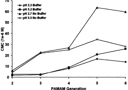
Figure 2-3. CMC of PPO-PAMAM Generations 2 through 6 in low (pH 2.2-2.7) or medium pH (pH 5.2-5.5) with or without buffer... 71
Figure 2-4. Hydrodynamic diameter of PPO-PAMAM micelles as a function of PAMAM generation tested in 0.15 M PBS solution... 74
Figure 2-5. TEM micrographs of PPO-PAMAM generation 3.0 micelles air-dried on a 400 mesh carbon grid from phosphate buffered aqueous solutions ... 75
Figure 2-6. Drug encapsulation capacity of PPO-PAMAM generations 3.0 through 6 .0 ... 7 8 Figure 2-7. Release profile of triclosan from PPO-PAMAM generation 3.0 micelles at pH 7.4 and pH 5.0... ... 81
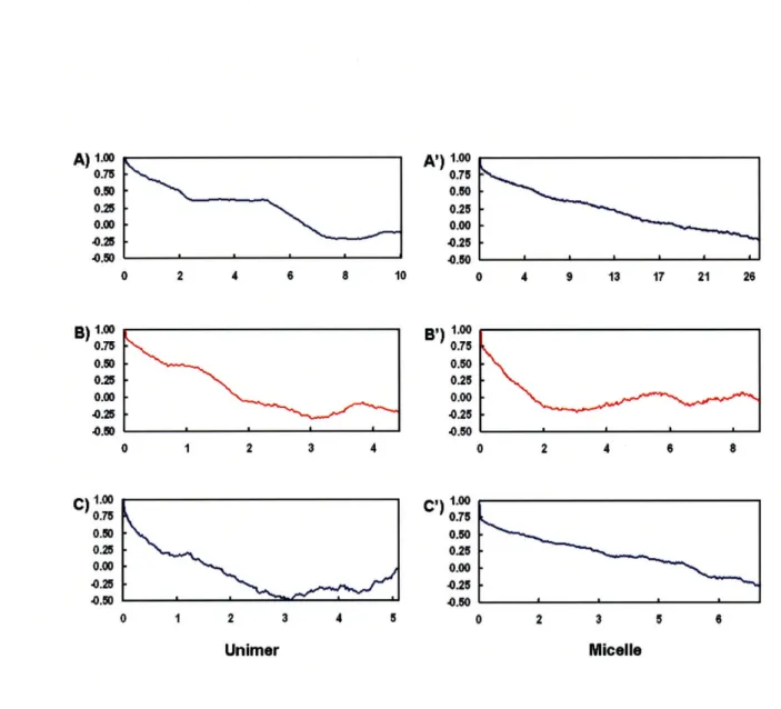
Figure 3-1. Autocorrelation profiles for unimers of PPO-PAMAM generations 4,
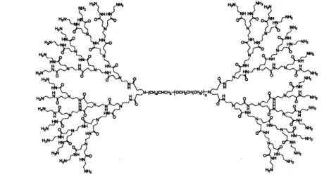
5, and 6 (A, B, C, respectively) and for micelles of PPO-PAMAM generations 4, 5, and 6 (A, B, C, respectively) ... 99 Figure 3-2. Chemical structure of generation 4.0 PPO-PAMAM ... 100 Figure 3-3. Zimm plots of micellar solutions of PPO-PAMAM generations 4.0
(A), 5.0 (B), and 6.0 (C)... 102
Figure 3-4. Snapshots of PPO-PAMAM unimers and micelles post-equilibration. Hydrogen atoms are depicted in white, carbon atoms cyan,
nitrogen atoms blue, and oxygen atoms red... 104
Figure 3-5. The time evolution of Rg for generations 4, 5, and 6 PPO-PAMAM
unimers (A, B, and C, respectively) and micelles (A', B', and C',
respectively) ... ... 109
Figure 3-6. PAMAM monomer radial density distribution of PPO-PAMAM micelles generations 4 (blue circles), 5 (green diamonds), and 6
(red triangles) ... 112
Figure 3-7. The radial density distribution of the terminal groups. Generation 4 is shown as blue circles, generation 5 as green diamonds, and
generation 6 as red triangles ... 115
Figure 3-8. The density of atoms comprising each micelle (the solid line) and
water atoms as a function of distance from the micelle center of mass for the PPO-PAMAM generation 4 system (A), generation 5 system (B), and generation 6 system (C) ... 116 Figure 3-9. Radial distribution density profiles are shown for PPO-PAMAM
dendrimer atoms (blue circles), the atoms in 10 triclosan
molecules (green diamonds), and water atoms (red triangles) ... 118
Figure 3-10. The square root of the total solvent accessible surface area, SASA1/2,
as a function of the probe sphere radius for PPO-PAMAM micelles generation 4 (blue circles), generation 5 (green
diamonds), and generation 6 (red triangles) ... 121
Figure 4-1. Model of PPO-PAMAM generation 4.0 generated in Materials Studio
Figure 4-2. Figure 4-3. Figure 4-4. Figure 4-5. Figure 4-6. Figure 4-7. Figure 4-8. Figure 5-1. Figure 5-2. Figure 5-3. Figure 5-4. Syi FT
nthesis of PAMAM-Galactose from a half-generation PPO-PAMAM deprotected through base hydrolysis. Galactose is added through traditional amidation chemistry ... 139
IR of PPO-PAMAM generation 2.5 before and after methyl ester
deprotection... 140 A) Primary rat hepatocytes incubated with ASF-AF488 (25 ug/ml).
B) Primary rat hepatocytes incubated with ASF-AF488 (25 ug/ml) and excess ASF (1 mg/ml). C) Primary rat hepatocytes incubated with ASF-AF488 (25 ug/ml) and excess PPO-PAMAM-Gal gen. 3.5 (0.5 mg/ml). D) Primary rat hepatocytes incubated with ASF-AF488 (25 ug/ml) and excess PPO-PAMAM-Gal gen. 4.5 (0.5 m g/m l)... Drug release of doxorubicin from PPO-PAMAM generation 4.0
micelles in PBS at 37 C ... 143
147 Cell viability as a function of doxorubicin concentrations via different
drug delivery modalities ... 149
IC50 values of doxorubicin for either bare doxorubicin delivery or
doxorubicin delivery via unmodified or galactose-functionalized
m icelles ... 150 Inhibition of PPO-PAMAM generation 4.5 galactose modified
micelles containing doxorubicin with various concentrations of
A SF ... 151
Amphiphilic linear-dendritic block copolymer micelles may break apart upon dilution at concentrations below the CMC or in
solvents that solubilize both polymeric blocks ... 162
Methacrylate monomers are solubilized in the core of PPO-PAMAM micelles through an emulsion. The monomers are polymerized with a UV-initiated polymerization reaction, which forms
polymers that interpenetrate the PPO core... 163 Fluorescence measurements of DPH incubated with either
uncrosslinked or crosslinked PPO-PAMAM micelles... Drug release of doxorubicin from crosslinked and uncrosslinked
micelles in PBS at 37 C ... ...
166
Figure 5-5. Cytotoxocity of crosslinked PPO-PAMAM micelles with HepG2 cells ... 170
Figure 6-1. Synthesis of PBLA-B16-PEG composed of a hydrophobic linear block
of poly(P-benzyl-L-aspartate), a polyester dendron, and PEG on
the outer periphery of the dendron... 189
Figure 6-2. Functionalization of PBLA-B16-PEG with galactosylamine. First, the
PEG group was activated with NHS, and then the polymer was
further reacted with galactosylamine ... 190
Figure 6-3.
Figure 6-4.
Figure 6-5.
Figure 7-1.
Ratio of the emission intensity of pyrene at 373 nm and 393 nm at various concentrations of PBLA-B 16-PEG ... Release profile of doxorubicin from PBLA-B 16-PEG micelles in PBS
at 37 oC ...
192
194
Cytotoxicity of PBLA-B 16-PEG on HepG2 hepatocellular carcinoma
cells. The polymer is non-toxic over a large concentration range ... 195
Schematic illustrating the formation of LbL films with positively charged PPO-PAMAM micelles encapsulating either hydrophobic drug or pyrene and negatively charged PAA ... 209
Figure 7-2. Chemical structure of PPO-PAMAM generation 4.0... 210 Figure 7-3.
Figure 7-4.
Figure 7-5.
Figure 7-6.
Figure 7-7.
Growth curve of PAA and PPO-PAMAM encapsulating triclosan
indicating linear growth from 4 to 25 bilayers... 213
UV-vis measurements of triclosan at a characteristic wavelength of 281 nm at varying number of bilayers in a
PPO-PAMAM-triclosan/PAA LbL film showing linear incorporation of triclosan into the film ... 214
Fluorescence measurements of PPO-PAMAM-pyrene/PAA LbL films as a function of the number of bilayers in the film ... 215 Pyrene emission spectra of LPEI/PAA films and PPO-PAMAM
G4/PAA films ... 217 GISAXS scattering data of a 10 bilayer LbL film composed of
Figure 7-8. Illustration of the possible configuration of PPO-PAMAM polymer
micelles within the LbL film with the spacing determined by
GISAXS of 10 nm ... 219
Figure 7-9. Drug release profile of triclosan in an LbL film composed of
PPO-PAMAM-triclosan/PAA. The release was performed at 37 'C in
0.1 M PBS ... 222
Figure 7-10. FTIR spectra of PPO-PAMAM/PAA films after triclosan release or
PPO-PAMAM/PAA films fabricated with no drug ... 223 Figure 7-11. Agar plate of S. Aureus growth inhibited by release of triclosan from
a 10 bilayer LbL film ofPPO-PAMAM micelles encapsulating
triclosan and PAA ... 224
Figure 8-1.
Figure 8-2.
Synthetic scheme to create a triblock copolymer of PPO, polycysteine, and PAMAM, where the polycysteine block adds a smart response component to the drug delivery vehicle ... 239 Synthetic scheme for the synthesis of a triblock linear-dendritic block
copolymer with a polyester dendron, a linear PBLA end block, and a poly(amino acid) middle block that could be used as either a proton sponge, biologically responsive crosslinking system, or a
covalently crosslinking system ... 240
Figure 8-3. Activation of folate through conventional carbodiimide chemistry... 242 Figure 8-4. Synthetic scheme to introduce folate targeting ligand to the dendritic
List of Tables
Drug encapsulation characteristics of PPO-PAMAM for generations 3
through 6 and control polymers ... ... 77
Simulation condition parameters for unimers and micelles of
PPO-PAMAM generations 4 through 6... ... 98
Zimm plot results of PPO-PAMAM generations 4, 5, and 6 micelles in D I w ater ... 101
Table 3-3. Moments of inertia values for PPO-PAMAM unimers and micelles... 106
Rg values obtained from simulation and calculated through
experimental data for PPO-PAMAM unimers and micelles... 110 Average SASA values for PPO-PAMAM generations 4, 5, and 6
m icelles ... Percent galactose functionalization of various generations of
PPO-PA M A M ... ... .... ... IC50 and CMC values for various generations of PPO-PAMAM
modified with galactose and unmodified... Formulation of methacrylate monomers by varying the monomer
concentration...
120
142
146
168
Table 5-2. Various formulations of methacrylate monomers ... 168
Composition of PAA and PPO-PAMAM with or without triclosan LbL
films as determined by two different methods... 221
Table 2-1. Table 3-1. Table 3-2. Table 3-4. Table 3-5. Table 4-1. Table 4-2. Table 5-1. Table 7-1.
CHAPTER 1: INTRODUCTION
1.1 Motivation
Systemic drug delivery is a nonspecific treatment that can cause harmful side effects. For example, chemotherapy kills cancer cells but also causes unwanted side effects by destroying other healthy rapidly growing cells within the body. New developments in localized or targeted drug delivery methods can be a promising alternative. Currently, colloidal particles such as liposomes or block copolymers have been designed and studied for use as drug carriers for delivering drugs to localized sites. These colloidal particles can encapsulate a wide variety of drugs and isolate them from the physiological environment where degradation can occur. Furthermore, they can solubilize hydrophobic drugs at concentrations above the solubility limit of the drug in aqueous solution. As a result of employing a nanoparticle, the half-life of the drug is increased as it is sequestered from degradation reactions within the body, and the distribution of drug within the body can be altered.' Primarily, drug delivery nanoparticles are delivered parenterally, and the circulation times of these nanoparticles can be increased by the use of polyethylene glycol (PEG) grafted to the exterior of the nanoparticles. PEG creates a hydrodynamic barrier that prevents recognition, uptake, and removal by the reticuloendothelial system (RES).2-4 Increased circulation times allow for
greater passive targeting to targeted sites through the enhanced permeability and retention (EPR) effect.5 The EPR effect occurs in tumor tissue that have increased permeability of blood vessels in tumor tissue and decreased lymphatic drainage.6
Although these studies have contributed to the development of promising drug delivery vehicles targeting drugs to solid tumors, there are still challenges in the use of colloidal particles for site-specific drug delivery. The first challenge is to develop a nanoparticle that is stable in aqueous solution and does not break apart when it is
administered into the body.7 The second challenge is to control the biodistribution of the drug delivery vehicle and have effective extravasation of the nanoparticles from the vascular system into the treatment site.8 Once the particle enters the tumor, it must then be taken up by the cancer cells and undergo endocytosis. The next challenge is to effectively release the drug in a controlled manner while within the endosome or after escaping the endosome.9 Other challenges include the ability to increase drug-loading capacity, to vary
the size of the particle while maintaining stability, to prevent the aggregation of particles, to introduce cell-specific targeting capability through the addition of targeting ligand, and to vary ligand valency on the surface of the particle.
To address these issues, the scope of this thesis includes the design and creation of a novel amphiphilic linear-dendritic block copolymer. As shown in Figure 1-1, the linear-dendritic block copolymer consists of a hydrophobic linear block attached to a hydrophilic dendron. These copolymers will undergo self-assembly in solution due to thermodynamic driving forces that strive to isolate the hydrophobic linear block from the surrounding media and to expose the hydrophilic dendron. In addition to synthesizing the polymer, the aqueous phase self-assembly behavior of the linear-dendritic block copolymer has also been studied to determine its feasibility as a drug delivery vehicle. Molecular dynamics simulations of some of the linear-dendritic block copolymer micelle
The linear-dendritic block copolymer micelles were then used in two different drug delivery applications. The main application that was studied was exploring the use of the linear-dendritic block copolymers as circulating nanoparticles for drug delivery. In
vitro studies were undertaken to evaluate the role of multivalency and the effectiveness of
the targeting ligand to deliver the drug-encapsulated colloidal particles to carcinoma cells. Additional improvements were investigated including stabilizing the linear-dendritic block copolymers through physical entrapment of the hydrophobic cores and also developing a different linear-dendritic block copolymer system for future use.
The other application that was explored with the linear-dendritic block copolymer micelles was to incorporate these micelles into a thin film as a coating for biomedical implants. The block copolymer micelles were used to provide a hydrophobic environment available as a hydrophobic drug depot in a film formed through the layer-by-layer dipping process.
1.2 Background: Colloidal Drug Delivery Systems
Colloidal drug delivery systems have shown a lot of promise in the effective targeting of cytotoxic drugs to cancer tumors and areas of infarction by protecting the drugs from degradation and prolonging circulation within the body. By encapsulating drugs in a nanoparticle, the distribution of the drug is less determined by the properties of the drug and more dependent on the size and surface properties of the drug carrier. There are two different mechanisms that can be exploited to alter the biodistribution of the nanoparticles circulating in the body. First, the size of the nanoparticles can be manipulated to affect their passive accumulation at sites with arterial damage. Specific targeting of the drug-encapsulating nanoparticles can be introduced through the attachment of cell-specific targeting ligand to the outer surface of the particles.
1.2.1 Factors for Passive Targeting
The size of the circulating nanoparticles is a critical factor in controlling the biodistribution of the drug. First, there are size restrictions that have to be met in order for the nanoparticles to avoid removal from circulation. The nanoparticle must be larger than 2-3 nm to avoid being filtered out by the kidney but smaller than 150 nm in order to avoid the RES.10 The RES is mainly composed of monocytes and macrophages in the liver and spleen that have the ability to recognize microparticulate systems and remove them from circulation. Recognition by the RES is aided by the adsorption of proteins, called opsonins, onto the surface of the nanoparticles which promote phagocytosis. 1' , 12 With an increase in size of the drug delivery vehicle, there is an increased rate of uptake by the RES.13
The size of the nanoparticle is important as it will determine the rate of extravasation and uptake into the cells. Although macroparticles have a tendency to accumulate in tumor tissue through the EPR effect, it has been found that as the size of the particle increases, the time for extravasation exponentially increases.14' 15 Once in the tumor tissue, the size of the particle may also impede transport of the particle through the solid tumor. There is high interstitial fluid pressure that is often present in tumor masses which can decrease the ability of particles to penetrate the tumor as its size gets larger.16 Another consideration is that it is more difficult to internalize bulky particles by nonphagocytic cells.1 7 Typically, endocytosis can only occur for particles that are smaller than 150 nm in diameter. Overall, an intermediate sized particle is necessary in order to avoid the RES and facilitate extravasation and transport through the tumor.
The surface properties of the drug carrier are also crucial in determining the biocompatibility, biodistribution, and retention within the circulatory system. It has been found that neutrally charged liposomes have a lower tendency to be cleared by the RES in comparison to positively or negatively charged liposomes. However, they have a higher tendency to aggregate, which could also induce uptake by the RES.'8 Cationic
particles are quickly removed from systemic circulation due to electrostatic interactions with negatively charged cell membranes and the extracellular matrix.' Because of this interaction, it has been found that particles with primary amine groups exposed can cause haemolysis'9 and are cytotoxic.20 Receptors found on macrophages can recognize negative surface charges; thus, anionic particles are rapidly cleared from the body.2
To improve retention times and reduce immunogenecity, the surfaces of the drug carriers can be modified with hydrophilic moieties that confer a hydration barrier around
the particle. The most common modification is the conjugation of short chains of PEG. PEG has been well-characterized for biological use and parenteral applications.18 Other polymers that have been used to form steric barriers include poly(acryloyl morpholine), poly(acrylamide), poly(vinyl pyrrolidone), and polyvinyl alcohol.9' 18 PEG is usually the polymer of choice due to its high water solubility, low toxicity, low immunogenicity, and high chain mobility.21 With the addition of PEG, the drug carriers have a reduced rate of uptake by the RES, thus prolonging the circulation half-life.4' 22 Water molecules form a sheath around the PEG chains, creating a particle with a large hydrodynamic radius that prevents the adsorption of opsonins to the surface.2 3 The addition of the PEG palisade
also reduces the toxicity of the nanoparticle by decreasing interactions with the physiological environment20, 24 and alters the distribution of nanoparticles within the
body. For example, after conjugation of PEG to a cationic liposome, accumulation dramatically decreased in the lung.2 5
By designing the drug delivery vehicle with an optimal size and the correct surface properties, the blood circulation time of the drug can be dramatically improved. With the increased circulation half-life and the EPR effect, more of the nanoparticles can passively accumulate within leaky vasculature seen in tumors, inflammations, and
infarcted areas.
1.2.2 Active Targeting
To increase the retention of drug delivery nanoparticles in the affected sites beyond what is offered by passive targeting, the attachment of various cell-specific targeting ligands has been explored. In order for active targeting to be effective, there are three key issues that have to be addressed. First, the drug delivery vehicle must circulate
within the system long enough for localization to the site of the tumor and for interaction with the targeted cells. This is ensured through designing the nanoparticle with the ability to passively accumulate at the tumor site in high concentrations as discussed previously. The next consideration is that the ligand or the receptor to the targeted cells should be highly specific. For example, rapidly growing tumor cells upregulate certain receptors to allow for greater uptake of nutrients; therefore, tumor cells have a greater density of specific receptors in comparison to healthy cells. The last consideration is that the targeting molecule on the surface of the drug carrier should be stable in vivo and its potential for opsonization is minimal.18
There are several classes of targeting ligands that have been used for drug targeting. This includes antibodies, carbohydrates, peptides, cell surface receptor ligands, and oligonucleotide aptamers.26 For the treatment of tumor cells, ligands that are recognized by cell surface receptors have been the most popular, including transferrin and Apo E/Apo B.'8 Transferrin receptors are overexpressed on the surface of many
tumor cells, and transferrin has been attached to liposomes to deliver drug to tumors and into tumor cells.2 7' 28
1.2.2.1 Carbohydrates
Binding between carbohydrates and sugar binding proteins called lectins can be as specific as enzyme-substrate or antigen-antibody interactions. Lectins have multiple binding domains that bind to specific carbohydrate moieties.29 Through multivalent
interactions, the strength of binding is increased dramatically from a Kd in the 10-3 M
range for monovalent interactions down to a Kd of 10-6 M and 10-9 M for divalent and trivalent oligosaccharides, respectively.2 9 There are two broad classes of lectins.30
C-type lectins require calcium for binding, and this includes the hepatic asialoglycoprotein receptor. S-type lectins, also known as galectins, are calcium independent. It has been
found that galectins-1 and -3 are overexpressed on the surface of colon cancer cells.30 Targeting of carbohydrates to lectins have been applied in multiple applications including gene therapy31 and drug delivery.32 Drug delivery systems containing
asialofetuin, galactose, or N-acetyl-galactosamine have been able to bind and be endocytosed by the asialoglycoprotein receptor present on hepatocytes. 33 Derivatives of
N-(2-hydroxypropyl)methacrylamide covalently bound to sugar ligands have been able to deliver doxorubicin to colon adenocarcinoma and hepatic carcinoma cells.34' 35 The
higher the sugar content in the HPMA polymer, there was a higher extent of cell binding due to the multivalent binding required with lectins.34
1.2.2.2 Folate
Another popular targeting ligand is folate, a vitamin required in several metabolic pathways and is essential for the biosynthesis of nucleotide bases. It is consumed in elevated quantities by proliferating cancer cells. Folate is transported across the cell by either of two membrane-associated proteins, a reduced folate carrier or the folate receptor (FR). The reduced folate carrier is the primary pathway for uptake of folate while the folate receptor is found mainly on polarized epithelial cells and activated macrophages. FR has been found to be overexpressed in cancer cells. Furthermore, FR is only accessible after the cells have been transformed into malignant cells. Normally, FR is selectively expressed on the apical membrane surface (surface facing the body cavity) of epithelial cells. After transformation into a malignant cell, the polarity is lost and FR on the cells is accessible to drugs in circulation.36 Because of these mechanisms for tumor
specificity, folic acid is a natural choice to be used as a molecule for targeting drugs to cancer cells. The advantages to using folic acid is that it is nonimmunogenic, has a high binding affinity (Kd -. 1 nM), has good stability, has simple and defined conjugation chemistry, is available in large quantities, and is compatible with the use of organic solvents.37
Folic acid is taken into cells by a process called potocytosis. Folate receptors occur in dense clusters on the cell surface associated to caveolae, which are invaginated pits that have a diameter of approximately 50 nm. Each cluster is composed of 700 receptor molecules. Once the caveolae are closed, the pH decreases to approximately 5.0 and the folate is released from the receptor.38 Due to the mechanism of folate uptake, it has been found that folate binding is multivalent. The presence of multiple folates on a single dendrimer decreases the Kd dramatically from about 2500 to 170,000-fold.39
1.2.2.3 Targeting Challenges
Although there are several promising ligands that are being explored to target to specific cells for drug treatment, extravasation to the tumor site has been found to be the rate-limiting step for liposomes. For example, folate targeting has not increased the concentration of liposomes in subcutaneously growing tumor implants.4 0 However,
targeting could be used for improved delivery and retention of therapeutics within tumor tissue. Retrograde movement of drug-carrying nanoparticles may be hindered due to binding of the nanoparticles to target cells, and retention of the drug is enhanced through receptor-mediated endocytosis or potocytosis. The therapeutic index of the drug could increase as the drug distribution shifts from the extracellular space of the tumor into the intracellular compartments of the tumor cells-although, the relative contributions of
drug released in the extraceullular space and the intracellular space are unknown. In a study of HER2-targeted immunoliposomes containing doxorubicin (DOX), there was better tumor penetration, tumor cell uptake, and therapeutic efficacy.4 1
In several studies, the role of multivalency has shown to be dramatic. Conjugation of a large number of ligand on the surface of the drug particle has increased the avidity of the particle to the target cells. For polymeric systems, as the ligand density on the surface of the particle was increased, binding increased exponentially.34 Similarly,
the addition of 1000-fold greater concentration in free folate was necessary in order to compete with the binding of folate-targeted liposomes to a folate receptor.42 Therefore
another advantage for creating a targeted nanoparticle is that with greater affinity and efficacy dosage could be lowered.
Although the use of a ligand may confer a greater affinity to targeted cell types, the improved avidity and affinity may also negatively impact tumor penetration. It has been demonstrated that antibody-free vesicles more readily permeate tumors than immunoliposomes. This may be due to the high affinity of the antibody to the antigen where the rate of transport into the tumor is smaller than the rate of binding to tumor cells, thus retarding penetration into the tumor and binding occurs only at the periphery.43 Modeling of antibodies percolating through solid tumors shows that lower affinity antibodies are able to penetrate into tumors more than higher affinity antibodies.44 Thus,
in the creation of a targeted nanoparticle, there is an optimal ligand density that will balance the need for affinity to target cells and greater penetration into solid tumors.
1.2.3 Nanoparticles for Drug Delivery: Liposomes
Liposomes are phospholipid bilayer vesicles which can entrap hydrophilic drug molecules in its aqueous interior or can incorporate hydrophobic molecules into its bilayer as shown in Figure 1-2. The most common formulation of liposomes utilizes phosphatidylcholine, which is neutrally charged, with fatty acyl chains of varying length and degree of saturation. Cholesterol can also be added to modulate the rigidity and stability of the liposome. In the mid-1990s, liposomes were approved by the FDA for use as therapeutics in humans. Liposomal drug delivery systems have mainly been developed for antifungal and anticancer therapies.18
Figure 1-2. Liposomes are composed of lipid bilayers that are able to encapsulate hydrophilic drug in its aqueous core and hydrophobic drug in the lipid region.
Liposomes are characterized with long systemic circulation times in comparison to the free drug. However, they are also characterized by efficient uptake by the RES. In order to evade the RES and increase circulation times, liposomes have been modified by adding PEG to their outer surface.4 5' 46 Liposomes modified with PEG have also been approved by the FDA.
As an advancement, targeted liposomes have been developed. A lot of research has been done to couple antibodies to liposomes primarily for cancer targeting. For example, anti-HER2 liposomes to target HER2-overexpressing tumors47 and CC52
liposomes to target rat colon adenocarcinomas48 have been efficacious. Liposomes have
also been developed with carbohydrates conjugated to the surface. For example, mannosylated liposomes were developed to target macrophages49 and the brain.50 Asialofetuin, a protein that presents multiple galactose on its surface, has been conjugated to liposomes for drug delivery and gene delivery to hepatocytes. Increased liver uptake was seen in one study,51 while the addition of asialofetuin specifically delivered to
HepG2 cells and increased transfection in another study.52
Liposomes attached with folate molecules have been successfully targeted to cells
in vitro. In one experiment, folate was attached to a liposome via a PEG spacer of
molecular weight approximately equal to 3350, while smaller PEGs were attached to act as the polymer brush to evade the RES. The PEG spacer was long enough to allow the attached folate to extend away from the liposome surface and to avoid steric hindrance by the PEG brush. These liposomes contained a fluorescent dye, calcein, as their cargo and were incubated with KB cell monolayers, which are known to overexpress FR. It was found that approximately 2.5x 10⁵ liposomes were bound by each KB cell at saturation. The authors also found that the folate-PEG liposomes displayed a much higher affinity for the cells than free folate. Most likely, due to the length of the PEG spacer, more than one folate on a liposome could bind to the cell.4 6
In another study, folate-PEG liposomes with doxorubicin as cargo were incubated with cultured KB cells. It was found that cellular doxorubicin uptake with
folate-conjugated liposomes was 45-fold higher than that of non-targeted liposomes and 1.6 times higher than free doxorubicin. Furthermore, the cytotoxicity of these liposomes was 86 times and 2.7 times higher, respectively.53 These results indicate that the folate-PEG liposomes are more effective towards targeting cells that overexpress FR than non-targeted liposomes or free doxorubicin.
Liposomes conjugated to PEG-folate have also been tested in vivo. Biodistribution studies have indicated that folate-targeted liposomes showed similar patterns of distribution to non-targeted liposomes and did not improve uptake in tumors.4 1 Although in an ascitic tumor model, where the distribution between the tumor cell compartment and extracellular fluid could be discerned, liposome levels that were associated to the cells were significantly higher for folate-targeted liposomes.40 Other in
vivo studies examining the efficacy of folate-targeted liposomes have shown mixed
results. Studies were done in various tumor models, including M109-HiFR, M109R-HiFR, and J6456-HiFR cells, indicating that active targeting improved treatment. In a KB carcinoma model, folate-targeted liposomes showed significantly greater tumor inhibition.54
Although there are promising results for ligand-targeted liposomes, there are still some challenges associated with the use of liposomes. If hydrophobic drug is encapsulated in the bilayer region, the liposome could become destabilized.55
Furthermore, as seen in the previous examples, liposomes need to be modified with PEG in order to be stable in the circulation system. However, with the modifications there is a trade-off. Receptor-ligand interactions can be blocked by the attached PEG. Lee was able to avoid this problem by adding a PEG spacer, but other problems exist due to
stabilizing PEG modifications. The largest difficulty is liposome unloading efficiency following endocytosis by a target cell.9' 36 Because of the need to balance stability and
solubility of the liposome, the density of PEG on liposomes is not closely packed. This could lead to liposome aggregation or protein adsorption causing instability and cargo release or elimination by the RES. Furthermore, high levels of PEG-liposomes can often be found in the liver and the spleen or in tissues that are not the desired targets.56
Another disadvantage of liposomes is their size. Smaller liposomes have been found to be more unstable, allowing more leakage of drug,13 while larger liposomes can
not be internalized well.40 It has also been found that the permeation rates of liposomes (which are approximately 100 nm up to 500 nm in diameter) into the targeted cellular areas are lower than those of smaller nanoparticles due to the liposomes' large size.57 Finally, liposomes are not a flexible system. For example, modifications to improve interactions, such as hydrophobic, hydrogen bonding, and electrostatic interactions of the drug molecules to the interior of the liposome can not be easily altered.
1.2.4 Nanoparticles for Drug Delivery: Dendrimers
Another class of nanoparticles that are being explored for targeted drug delivery is dendrimers.56 Dendrimers have an architecture that is globular and highly branched, with regular molecular repeats created from stepwise synthesis as shown in Figure 1-3. They have a dense exterior with porous interior regions that have the potential to store drug molecules. Some of the advantages that dendrimers may have over linear polymers are that dendrimers are monodisperse with respect to molecular weight, their size and structure are highly controllable, and they have a high degree of functionality due to their branched architecture.1 5 A variety of dendrimers have been created for drug delivery
applications, including polyamidoamine, poly(propyleneimine), ss polyether,5 9 and polyester' dendrimers.
M-PEG-attached PAMAM dendrimer
Figure 1-3. A PAMAM dendrimer with conjugated PEG.20
The most widely studied and characterized dendrimer is the polyamidoamine (PAMAM) dendrimer. However, in a toxicity study of dendrimers, it was found that PAMAM dendrimers with terminal primary amine groups were quite cytotoxic and were cleared rapidly from the circulation when administered intravenously.5 8 In order to remediate this problem, PAMAM dendrimers are synthesized with PEG attached to the outer surface to reduce the adsorption of proteins and to increase circulation times. Additionally, with PEGylation, there is an increase in drug-loading capacity and a decrease in hemolytic toxicity and drug release rate.24 In work done by Kojima, PAMAM dendrimers with PEG grafted on were shown to be capable of encapsulating two types of anticancer drugs, methotrexate and adriamycin. It was found that the amount that could be encapsulated increased with dendrimer generation and PEG chain
length. The maximum amount of molecules encapsulated was 6.5 adriamycin molecules or 26 methotrexate molecules per dendrimer. In drug release studies, the methotrexate loaded dendrimers released the drug slowly in an aqueous solution of low ionic strength. However, in isotonic solutions, the methotrexate and adriamycin were readily released, indicating the need for more control in the drug release mechanism.2 0
Dendrimers conjugated to targeting ligand have been synthesized and tested. Extensive studies have been performed with carbohydrate ligands. For example, one group has added lactose and maltose to the outer surface of PAMAM generation 2-4 dendrimers and have observed binding of a lectin, concavalin A, to the dendrimers. Binding was reversible with the addition of excess glucose.6 0 Another group studied the multivalent binding behavior of mannose-conjugated PAMAM dendrimers with concavalin A. With the increase in the number of mannose ligand attached to the PAMAM dendrimer, binding strength increased due to multivalent interactions. However, a maximum was reached at 50% functionalization. Higher functionalization values resulted in less binding strength due to greater steric hindrance of excess mannose on the PAMAM dendrimer surface.61,62
Folate-targeted dendrimers have also been designed. One group has been able to produce ester terminated polyether dendrimers, which are modified for the attachment of folate. This group was able to attach 12.6 folates to a Generation 2 dendrimer with 16 available attachment sites.59 Another group has gone one step further and has tested their folate-targeted dendrimers in vitro. The dendrimer used was a PAMAM dendrimer of generation 5. They attached folic acid and loaded methotrexate into the dendrimer. The
result of the in vitro study was that the targeted delivery improved the cytotoxic response of the cells to methotrexate 100 fold over the free drug.63
Although the results for carbohydrate and folate-targeted dendrimers are promising, there are some challenges and disadvantages associated with the use of dendrimers. It has been observed that dendrimers seem to move out of tumor tissue rather quickly, which prevents the drug from concentrating in the tumor.56 A similar result has been shown in a polyester dendritic system where doxorubicin was covalently bound to the dendrimer. It was found that two of their polyester dendrimer formulations were excreted in 4-5 hours after intravenous injection of mice, while another compound had significant accumulation in the liver. Their longest circulating compound had a half-life of 72 minutes.1 In order for a macromolecular delivery system to be successful, its circulation time must be long enough so that accumulation in tumor tissue can occur through the EPR effect. The circulation times reported for the polyester dendrimers are considerably shorter than those seen for block copolymer drug systems, which have been reported as high as 98.3 hours.64 Another disadvantage to using dendrimers is that the
size of the system is limited to the size of the interior of the dendrimer, which is approximately 10 nm in diameter.15 The number of generations that the dendrimer can reach is limited by the degree of crowding at the exterior, where the maximum number of generations can range from 6 to 10 depending on the structure. Drug capacity is therefore limited by steric constraints. Furthermore, the chemistry required to create dendrimers at high generations can be a disadvantage, where the chemistry can often be extensive, costly, and labor-intensive. Last, as seen in the example above, there have been issues concerning the controllability of drug release from dendrimers, and improvements in
dendrimer chemistry and structure must be made in order to create an effective controlled drug release system.
1.2.5 Nanoparticles for Drug Delivery: Block Copolymer Micelles
The last major group of polymers that have been explored as nanoparticle drug delivery systems is block copolymer micelles. Block copolymer micelles are created from copolymers composed of a hydrophilic segment and a hydrophobic segment, as seen in Figure 1-4. When placed in an aqueous environment, micellization occurs where hydrophobic segregation from the environment is the main thermodynamic driving force.21
formation of
micelles
-U irhI hWfili
diblock block copolymer
copolymer mloelle
Figure 1-4. The formation of micelles from a diblock copolymer.55
The advantages of a block copolymer system is that with the hydrophobic core, the micelles have the ability to solubilize hydrophobic drugs while at the same time have a hydrophilic surface to evade the RES.4 Micelles that are formed from the block copolymers are also small, thus facilitating evasion from the RES. Additionally, there is a lot of flexibility with a block copolymer system, where modifications in size,
;3 ;" Il-r
functionality, and stability can be made. Several block copolymer micelle systems have been synthesized and reported such as poly(aspartic acid) and poly(f3-benzyl-L-aspartate)
as the hydrophobic block and PEG as the hydrophilic block.2 1' 65 Other systems include
poly(D,L-lactic acid-co-glycolic acid)-PEG,66 poly(y-benzyl-L-glutamate)-PEG, 67 and poly(E-caprolactone)-PEG.68 All of the systems mentioned have used PEG as the hydrophilic exterior in order to create a hydrodynamic barrier against the adsorption of proteins and to evade the RES.
Block copolymer micelle systems have demonstrated to be stable and capable of encapsulating hydrophobic drugs. For example, a poly(ethylene oxide)-poly(aspartic acid) block copolymer with adriamycin, an anticancer drug, conjugated to aspartic acid have been shown to form stable micelles.69 Another system composed of poly(ethylene glycol) and conjugated doxorubicin-poly(aspartic acid), has been able to encapsulate free doxorubicin (DOX) in particles approximately 40 nm in diameter. These micelles have been tested against tumors inoculated subcutaneously and tumors implanted intravenously in mice. Three out of six mice were cured of mouse colon 26 carcinoma while two out of six mice were cured of human Lu-24 lung cancer. In comparison, free doxorubicin injected into mice was not able to cure any of them. The authors were also able to show that the block copolymer micelle accumulated in the tumor more effectively than free doxorubicin. The results of this study were promising enough to warrant clinical trials in Japan.64
In a further step towards creating micelles that can target specific areas of the body, some groups have explored the functionalization of the outer surface of block copolymer micelles. The functional ligands that have been added have included folate,
biotin, monosaccharides, and peptides.70 Specifically, one group has been able to create galactose-, lactose-, and mannose-installed PEG-Poly(D,L-lactide) block copolymer micelles averaging approximately 100 ligands on the surface. They found that the galactose and lactose functionalized micelles specifically bound to RCA-1 lectin immobilized beads while glucose-PEG-PLA micelles were eluted (RCA-1 lectin is a plant lectin which specifically recognizes f-D-galactose residues). Also, the mannose functionalized micelles specifically bound to concavalin A. Furthermore, they found that there was a "cluster effect" in binding, where multiple sugar residues from one micelle would bind to the beads. A 30-fold concentration of free galactose compared to the micelle-conjugated galactose was required to elute the micelles from the column.65, 70 Mixed micelles of poly(L-histidine)-PEG and poly(L-lactic acid)-PEG conjugated to folate have been tested in vitro, demonstrating that the targeted micelles were more effective against tumor cells than non-targeted micelles.66' 71
1.3 Thesis Objectives
Block copolymer micelles have shown to be promising alternatives to liposomes due to its flexibility. However, there is still much left to explore in this area, where many design parameters can be investigated or optimized. For this thesis, a novel linear-dendritic block copolymer has been designed. The linear-linear-dendritic block copolymer is composed of a hydrophobic linear block and a hydrophilic dendritic block, which will self-assemble in aqueous solution into nanoparticles. The proposed drug delivery system hopes to combine the advantages conferred from dendrimers, including its dense surface functionality, with the increased capacity and chemical flexibility of a block copolymer micelle. The system will be similar in size to block copolymer micelles, which are
approximately 20-60 nm. This system will be an improvement over liposome nanoparticles with its smaller size--therefore the particle will have increased circulation times and more facile extravasation into tumors. Also in contrast to liposomes, the proposed system is easily fine-tuned to increase interactions between drug and drug delivery vehicle. Additionally, because it is a polymer system, it will have the ability to include a mechanism that will induce degradation of the nanoparticle system once it is in the endosome. With the addition of the dendritic outer structure, the density of ligand can be explored while maintaining approximately the same particle size. In comparison to dendrimers, the proposed drug delivery vehicle will have one to two orders of magnitude greater functionality with a large aggregation number (PEG-PBLA micelles have shown aggregation numbers ranging from 200-400),72, 73 while for block copolymer micelles, the functionality of the proposed system will be several times higher depending on the dendron generation. An additional advantage of using the proposed system over dendrimers is that mixed micelles with different ligands could be used to create multi-functional systems or to manipulate the clustering density of ligands on the surface as shown in Figure 1-5.
Figure 1-5. Formation of micelles with clustered or multifunctional ligands.
P·9-As an improvement over diblock copolymer micelles, the linear-dendritic block copolymers can be stabilized. It has been pointed out that after micelles are injected into the bloodstream, thermodynamic fluctuations could occur causing the breakdown of the micelle (because of the dilution effect leading to concentrations lower than the critical micelle concentration) and the precipitation of hydrophobic drugs.4 Therefore, the
crosslinking of the dendritic micelles will prevent this from occurring. Overall, the design of a new dendritic copolymer micelle will be able to address the issues that previously studied nanoparticle systems have had and hopefully will exceed expectations
in performance.
Few linear-dendritic block copolymers have been synthesized and studied for drug delivery.74 Additionally, those systems that have been explored self-assemble with a hydrophobic dendritic block forming the core and a PEG linear block forming the outer surface of the micelles. In this work, the first objective was to synthesize a linear-dendritic block copolymer with a hydrophilic dendron and a hydrophobic linear block. The second objective was to characterize and obtain a better understanding of the aqueous phase self-assembly behavior of the linear-dendritic block copolymer to ultimately determine its feasibility as a drug delivery vehicle. The third objective was to develop drug delivery approaches that could determine whether the dendritic block could confer advantages to the block copolymer micelles over conventional linear block copolymer micelles. This includes using the linear-dendritic block copolymer micelles in two different applications. The first application explored utilized the linear-dendritic block copolymer micelles as circulating targeted drug delivery vehicles. The other
application studied was to incorporate these linear-dendritic block copolymers into films as potential coatings for biomedical implants.
1.4 Thesis Overview
The remainder of this thesis is divided into 7 additional chapters. Chapter 2 discusses the synthesis of a linear-dendritic block copolymer. The solution-phase characterization of the block copolymer was performed including critical micelle concentration measurements, light scattering measurements, and TEM. Furthermore, encapsulation of a model hydrophobic drug was investigated.
In Chapter 3, results of molecular dynamics simulations of the linear-dendritic block copolymer micelles are detailed. Various micelle configurations are explored based on experimental data obtained through static light scattering experiments used to determine the aggregation number of the micelles. The simulations were performed to get a better understanding of the microstructure of the micelles formed and the thermodynamics of the micelle formation. Furthermore, locus of solubilization of a model hydrophobic drug is studied.
Chapter 4 explores the use of the linear-dendritic block copolymer micelles as circulating colloidal nanoparticles for targeted drug delivery. The linear-dendritic block copolymer was functionalized with a carbohydrate targeting ligand. Its binding behavior and drug delivery efficacy was determined in vitro.
Chapter 5 and Chapter 6 put forward methods to improve the linear-dendritic block copolymer micelles for future testing in vivo. First, a stabilization technique is explored in Chapter 5 to ensure that during administration into the body, the micelles
would not break apart. In Chapter 6, the synthesis of an improved linear-dendritic block copolymer is detailed, and its solution-phase and encapsulation behavior is investigated.
Chapter 7 discusses the second application for the linear-dendritic block copolymer micelles. The micelles are incorporated into a film created through a layer-by-layer dipping method. The micelles create hydrophobic domains for encapsulation of hydrophobic drugs. The films formed were characterized to ensure that the micelles incorporated were not disrupted. Additionally, in vitro testing of the films were done as a proof-of-concept for potential use as an antibacterial film on various biomedical implants. Last, Chapter 8 includes the overall conclusions to the thesis project and suggests future directions for the project.