FORENSIC MEDICINE
Pulmonary thrombembolism as cause of death
on unenhanced postmortem 3T MRI
Christian Jackowski&Silke Grabherr&Nicole Schwendener
Received: 14 August 2012 / Revised: 15 October 2012 / Accepted: 1 November 2012 / Published online: 16 December 2012 # European Society of Radiology 2012
Abstract
Objectives To investigate unenhanced postmortem 3-T MR imaging (pmMRI) for the detection of pulmonary throm-bembolism (PTE) as cause of death.
Methods In eight forensic cases dying from a possible car-diac cause but with homogeneous myocardium at carcar-diac pmMRI, additional T2w imaging of the pulmonary artery was performed before forensic autopsy. Imaging was carried out on a 3-T MR system in the axial and main pulmonary artery adapted oblique orientation in situ. In three cases axial T2w pmMRI of the lower legs was added. Validation of imaging findings was performed during forensic autopsy. Results All eight cases showed homogeneous material of intermediate signal intensity within the main pulmonary artery and/or pulmonary artery branches. Autopsy con-firmed the MR findings as pulmonary artery thrombembo-lism. At lower leg imaging unilateral dilated veins and subcutaneous oedema with or without homogeneous mate-rial of intermediate signal intensity within the popliteal vein were found.
Conclusions Unenhanced pmMRI demonstrates pulmonary thrombembolism in situ. PmMR may serve as an alternative to clinical autopsy, especially when consent cannot be obtained.
Key Points
• Postmortem MRI (pmMRI) provides an alternative to clinical autopsy
• Fatal pulmonary thrombembolism (PTE) can now be diagnosed using postmortem MRI (pmMRI).
• Special attention has to be drawn to the differentiation of postmortem clots.
Keywords Postmortem magnetic resonance imaging . Pulmonary thrombembolism . Forensic imaging . Postmortem imaging . Natural death
Introduction
The value of postmortem magnetic resonance imaging (pmMRI) for forensic examinations has been investigated for several years now. It has proved to be supportive in foetal or neonatal autopsy, traumatic causes of death [1] and assessing clinical forensic cases [2] as well. However, also in natural causes of death it provides additional infor-mation [3]. Especially in cases of cardiac failure due to myocardial infarction, pmMRI can demonstrate the myocar-dial regions affected even in cases that are difficult to assess at autopsy [4–6].
In the clinical setting, acute pulmonary embolism is mainly diagnosed using angiography (usually CT angiogra-phy). Unenhanced MR imaging has been investigated for the visualisation of pulmonary thrombembolism (PTE) us-ing time of flight or MR perfusion techniques but has not reached widespread clinical application [7,8].
Pulmonary thromboembolism, being a common natural cause of death, has already been demonstrated by postmor-tem angiography using CT [9]. However, it has not yet been shown whether any form of unenhanced postmortem cross-sectional imaging can demonstrate PTE. In an ongoing study investigating myocardial infarction on 3-T MRI, eight of the study cases presenting with a cardiac anamnesis turned out to have no myocardial alteration at cardiac
C. Jackowski (*)
:
N. SchwendenerForensic Imaging Center Bern, Institute of Forensic Medicine, University of Bern, Buehlstr. 20,
3012 Bern, Switzerland
e-mail: [email protected] S. Grabherr
Centre universitaire de medicine legale, CHUV—University of Lausanne, Rue du Bugnon 21,
1010 Lausanne, Switzerland DOI 10.1007/s00330-012-2728-3
pmMRI. In these cases an axial and pulmonary artery ori-entated oblique T2w slice stack was supplemented to visu-alise the main pulmonary artery as well as the right and left pulmonary artery. We present the typical 3-T MRI findings of a lethal PTE as obtained in our eight examined postmor-tem PTE cases.
The postmortem MR appearance of blood was already investigated in 2006 [10]. Contrary to the vital appearance of blood, which is known to be homogeneous in signal, the postmortem appearance is dominated by two different pro-cesses. After cessation of the circulation, the physiology of the blood and the vascular system completely changes. The mean circulatory pressure decreases to the vital venous pressure after the blood flow has stopped, and without further circulation of the blood, all components are sub-jected to gravitation only. The cellular components initially sink and sediment within the vascular system of the body. This leads to the formation of two different levels within the blood-filled cavities, a hyperintense (T2w) upper serum layer and a hypointense (T2w) lower erythrocyte layer.
Parallel to sedimentation postmortem clotting occurs. The amount of postmortem clotting is dependent on differ-ent factors such as functionality of the coagulation system, possible anticoagulation therapy such as warfarin, cause of death (fatal haemorrhage, sepsis, etc.) and time frame of agony. Fast clotting in relation to the speed of sedimentation leads to more erythrocytes included within the clot while slow clotting includes fewer erythrocytes but more fibrin. The T2w appearance of postmortem clots is of heteroge-neous nature with brighter components (fibrin) and darker areas (erythrocytes). Postmortem clotting occurs mainly within the great blood-filled cavities such as the cardiac chambers, the aorta and the main pulmonary artery. Post-mortem clots do not stick to the vessel wall or appear to have been pushed into the periphery of vessels but arrange themselves around the boundary between serum and eryth-rocytes. Therefore, a homogeneous clot completely occlud-ing a vessel is indicative of a vital thromboembolism.
Materials and methods
Study population
Eight subjects (4 female, 4 male, age at death 58.5±18 years) were examined before forensic autopsy (Table1). The study cases were sudden deaths or found death with no known medical history so that a forensic autopsy was ordered by the local authorities. Cardiac failure was suggested a possi-ble cardiac cause of death and therefore all subjects under-went a cardiac pmMRI examination. As the myocardium showed no alteration, an additional T2w imaging of the pulmonary artery was added. MRI examination was
performed within 48 h of death. Autopsy was performed on the same or the following day by board-certified forensic pathologists. The study was approved by the local authori-ties as well as the local ethics committee.
PmMRI
The bodies were placed supine in a 3-T system (Achieva 3.0 TX, Philips Medical Systems, Best, The Netherlands) and imaged using a 16-channel SENSE torso XL coil. Based on axial thoracic T2w images an individual three-point plan-ning along the main, the right as well as the left pulmonary artery was performed, and a 2-mm T2w (axial – TSE, TE 90, TR 4,050 ms, 3-mm thickness and oblique–TSE, TE 100 ms, TR 3,482 ms, 1.5-mm thickness) acquisition was carried out. Examination time was 25 min. In three of the cases the lower legs were imaged in coronal and axial orientation with T2w (TSE, TE 90, TR 4,702 ms, 3-mm thickness) images using the same coil to search for a possi-ble source of the embolism. Image interpretation was per-formed according to Jackowski et al. [6,10].
Results
Table 1 gives the collection of MR and autopsy findings. Generally PTE appeared on T2w images within the hypoin-tense layer of sedimented erythrocytes as a homogeneous structure of intermediate signal intensity. Contrary to postmor-tem clots, which mostly accumulate in a free-floating manner near the boundary between serum and erythrocytes within the greater blood-filled cavities (Fig.1), PTE was present within the branches of the pulmonary artery always completely oc-cluding the vessels in which it got stuck (Fig.2). In all cases PTE occurred bilaterally and in two cases was only bilateral and paracentric (affecting the lobar arteries only).
In the three subjects who underwent an MR examination of the lower leg homogenous contents could also be visual-ised within the venous system (Fig. 3) and/or unilateral dilatation of the venous system combined with distinctive subcutaneous oedema was present.
Discussion
This study demonstrates the cross-sectional appearance of PTE in postmortem unenhanced 3-T MRI. All included cases showed similar findings in terms of signal behaviour and general appearance of PTE. The location of PTE dif-fered slightly.
The main challenge in assessing clots postmortem is the differentiation between postmortem clotting and vitally embolised clots. The present study presents typical
appearances of both so that now criteria exist that determine the vital or postmortem nature of postmortem visualised clots.
In all the cases included imaging findings interpreted as PTE were confirmed as PTE at autopsy. There were no false-positive findings on 3-T MRI in our rather small study group. However, a validation in a larger study should be obtained especially to investigate the false negatives.
To the best of the authors’ knowledge, PTE was visual-ised for the first time postmortem using unenhanced 3-T MRI. As advantage over postmortem angiography techni-ques that have been shown to detect PTE too [11, 12], unenhanced imaging does not interfere with toxicological
investigations and does not change the histological appear-ances of soft and organ tissues. The second advantage of pmMR to be mentioned in that context is that visualising the thrombus without the injection of any contrast agent volume decreases the risk that a clot developed postmortem will be pushed into the pulmonary artery by the postmortem injec-tion. Thus it is the unenhanced visualisation that clearly has to be favoured. Recently, it has been shown that very tiny thrombotic findings such as a coronary thrombosis can also be visualised using unenhanced 3-T MRI [6].
Examination of the lower legs revealed venous altera-tions in all three of the cases examined. Either a homoge-neous structure occluding the vessels was found or unilateral dilatation of the veins could be observed. As we know from autopsy, is it not possible to find the originating thrombus within the leg veins in every case as sometimes the entire thrombus embolises. Therefore, the pmMRI find-ings of the leg veins are in line with general pathological experiences.
Natural causes of death are decreasingly autopsied in clinical pathology and the non-forensic clinical autopsy numbers keep declining. The effects on the quality of na-tional mortality statistics, quality control in medicine and medical education are discussed manifold [13–15]. Efforts to counteract this unfavourable trend have remained unsuc-cessful so far and clinical autopsy is vanishing. In the present study, the cause of death was established before autopsy as already described for cardiac causes of death [4,5]. The authors would like to recommend the usage of pmMRI in non-forensic circumstances especially when myocardial infarctions, pulmonary thrombembolism, intra-cerebral haemorrhages or comparable findings are of inter-est. Particularly hospital deaths without subsequent autopsy could be understood better by performing postmortem radi-ology, especially pmMRI. This would help to reduce the
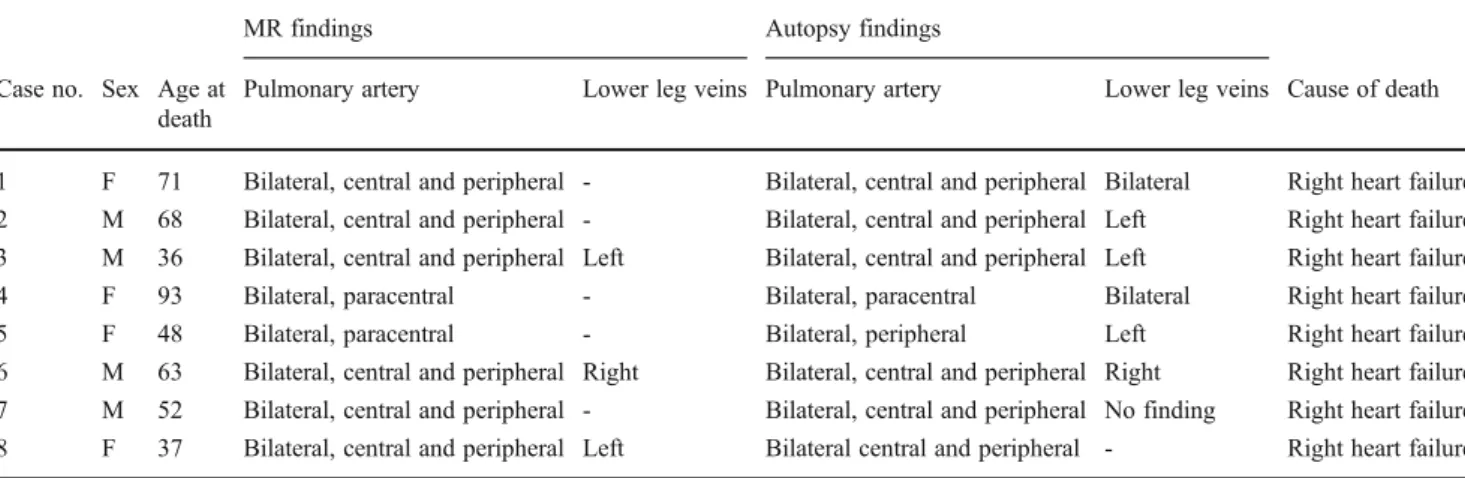
Table 1 PTE findings in eight study cases
MR findings Autopsy findings Case no. Sex Age at
death
Pulmonary artery Lower leg veins Pulmonary artery Lower leg veins Cause of death
1 F 71 Bilateral, central and peripheral - Bilateral, central and peripheral Bilateral Right heart failure 2 M 68 Bilateral, central and peripheral - Bilateral, central and peripheral Left Right heart failure 3 M 36 Bilateral, central and peripheral Left Bilateral, central and peripheral Left Right heart failure 4 F 93 Bilateral, paracentral - Bilateral, paracentral Bilateral Right heart failure 5 F 48 Bilateral, paracentral - Bilateral, peripheral Left Right heart failure 6 M 63 Bilateral, central and peripheral Right Bilateral, central and peripheral Right Right heart failure 7 M 52 Bilateral, central and peripheral - Bilateral, central and peripheral No finding Right heart failure 8 F 37 Bilateral, central and peripheral Left Bilateral central and peripheral - Right heart failure Location of the thrombus within the pulmonary artery and within the lower leg veins as obtained by unenhanced postmortem MRI and at autopsy. MR reading was performed according to Jackowski et al. 2006 and 2011 [6, 7]. Lower leg imaging was only performed in three cases. Lower leg dissection was performed in seven of the eight cases. In all cases right heart failure due to PTE was given as cause of death
Fig. 1 Postmortem clotting. T2w cardiac short axis image presenting distended postmortem clotting within the right ventricle (solid arrow). Postmortem clots are heterogeneous in signal and appear free-floating within the greater blood-filled cavities at the sedimentation level be-tween hyperintense serum (dashed arrow) and hypointense erythro-cytes (doubled arrow)
negative effects of the declining clinical autopsy rates. In the forensic environment, postmortem imaging is mainly a sup-portive tool to substantiate the quality of postmortem exami-nations in general and to increase the quality of the autopsy itself. However, it would be the widespread non-forensic application from which our community would further ben-efit. One possible application could be within the
non-forensic clinical–pathological settings that suffer from de-clining clinical autopsy rates. Treating physicians may ob-tain consent for a pmMRI examination more easily than for a clinical autopsy.
This study had a number of limitations. First of all, the small study population has to be mentioned as with eight cases no reasonable statistical validation could be performed. The
a
a
b
c
d
Fig. 2 PmMR findings within the pulmonary artery in four of the study cases. a Case 8: Oblique T2w image at the main pulmonary artery. Within the right and left pulmonary artery homogeneous material of intermediate signal intensity is seen (arrows). b Case 5: Axial T2w image at the level of the right pulmonary artery. Within the right and left pulmonary artery homogeneous material of intermediate signal intensity is seen (arrows). c Case 4: Axial T2w image at the level of the right pulmonary artery. Within the left lower lobe artery a homogeneous structure of intermediate signal intensity occludes the vessel. Note the atelectatic left lower lobe (arrow). d Case 3: Axial T2w image at the level of the left pulmonary artery. Within the vessel a homogeneous structure of intermediate signal intensity completely occludes the vessel (arrows)
a
b
Fig. 3 PmMR findings within the lower legs of two PTE cases. a Case 6: Axial T2w images at the level of the fibular head and slightly below. A homogeneous structure with intermediate signal intensity is demon-strated within the vena poplitea (arrow). Contrary to the left side an oedematous thickening of the subcutis is obvious (dashed arrows). The lower image shows a dilated vein without thrombus in a more distal
cross section (arrow). b Case 8: Axial T2w images at three different levels. A dilated vena poplitea (arrow upper image) and dilated intra-muscular veins (arrows middle image) are present in comparison to the right side. The lower image demonstrates intense subcutaneous oede-ma within the left lower leg (dashed arrows)
study remains a morphological descriptive collection of eight cases and was not performed in a blinded manner. Secondly, no values of absolute or relative signal intensities are given. As in all postmortem studies the influence of the temperature on the absolute or relative signal intensities has to be taken into consideration. The body temperatures at imaging ranged from 4° to 35 °C. As both T1- and T2 relaxation times are temperature dependant, the absolute and relative values are not comparable between the study cases themselves or to further studies. The temperature dependence of absolute val-ues needs to be addressed in a further study.
In conclusion, the pmMRI appearance of PTE was de-scribed in eight cases. Structures of homogeneous interme-diate signal intensity in T2w images occluding branches of the pulmonary artery are highly indicative of PTE and allow the differentiation with post-mortem clots. Although foren-sically used as an autopsy supporting tool, pmMRI may provide an alternative postmortem examination approach for clinical pathology that suffers severely from declining autopsy rates.
Acknowledgements The authors would like to thank the team of forensic examiners around Dr. Morten Keller-Sutter (Institute of Fo-rensic Medicine Zürich) for the reliable support at case handling and autopsy validation. Funding: The study was supported in part by Philips Medical Switzerland.
References
1. Yen K, Sonnenschein M, Thali MJ et al (2005) Postmortem multi-slice computed tomography and magnetic resonance imaging of odontoid fractures, atlantoaxial distractions and ascending medul-lary edema. Int J Legal Med 119:129–136
2. Yen K, Vock P, Christe A et al (2007) Clinical forensic radiology in strangulation victims: forensic expertise based on magnetic reso-nance imaging (MRI) findings. Int J Legal Med 121:115–123
3. Aghayev E, Yen K, Sonnenschein M et al (2004) Virtopsy post-mortem multi-slice computed tomography (MSCT) and magnetic resonance imaging (MRI) demonstrating descending tonsillar her-niation: comparison to clinical studies. Neuroradiology 46:559– 564
4. Jackowski C, Christe A, Sonnenschein M, Aghayev E, Thali MJ (2006) Postmortem unenhanced magnetic resonance imaging of myocardial infarction in correlation to histological infarction age characterization. Eur Heart J 27:2459–2467
5. Jackowski C, Warntjes MJ, Berge J, Bar W, Persson A (2011) Magnetic resonance imaging goes postmortem: noninvasive detec-tion and assessment of myocardial infarcdetec-tion by postmortem MRI. Eur Radiol 21:70–78
6. Jackowski C, Hofmann K, Schwendener N, Schweitzer W, Keller-Sutter M (2012) Coronary thrombus and peracute myocardial infarction visualized by unenhanced postmortem MRI prior to autopsy. Forensic Sci Int 214:e19
7. van Beek EJ, Wild JM, Fink C, Moody AR, Kauczor HU, Oudkerk M (2003) MRI for the diagnosis of pulmonary embolism. J Magn Reson Imaging 18:627–640
8. Ohno Y, Koyama H, Matsumoto K et al (2010) Dynamic MR perfusion imaging: capability for quantitative assessment of dis-ease extent and prediction of outcome for patients with acute pulmonary thromboembolism. J Magn Reson Imaging 31:1081– 1090
9. Ross S, Spendlove D, Bolliger S (2008) Postmortem whole-body CT angiography: evaluation of two contrast media solutions. AJR Am J Roentgenol 190:1380–1389
10. Jackowski C, Thali M, Aghayev E et al (2006) Postmortem imag-ing of blood and its characteristics usimag-ing MSCT and MRI. Int J Legal Med 120:233–240
11. Jackowski C, Persson A, Thali MJ (2008) Whole body postmortem angiography with a high viscosity contrast agent solution using poly ethylene glycol as contrast agent dissolver. J Forensic Sci 53:465–468
12. Grabherr S, Doenz F, Steger B et al (2011) Multi-phase post-mortem CT angiography: development of a standardized protocol. Int J Legal Med 125:791–802
13. Shojania KG, Burton EC (2008) The vanishing nonforensic autop-sy. N Engl J Med 358:873–875
14. Shojania KG, Burton EC, McDonald KM, Goldman L (2005) Overestimation of clinical diagnostic performance caused by low necropsy rates. Qual Saf Health Care 14:408–413
15. Shojania KG, Burton EC (2004) The persistent value of the autop-sy. Am Fam Physician 69:2540–2542