SCIENTIFIC ARTICLE
Comparison of the critical shoulder angle in radiographs
and computed tomography
Samy Bouaicha&Christine Ehrmann&
Ksenija Slankamenac&William D. Regan&Beat K. Moor
Received: 26 January 2014 / Revised: 26 March 2014 / Accepted: 30 March 2014 / Published online: 18 April 2014 # ISS 2014
Abstract
Objective The critical shoulder angle (CSA) is an indicator of degenerative shoulder pathologies. CSAs above 35° are associ-ated with degenerative rotator cuff disease, whereas values be-low 30° are common in osteoarthritis of the glenohumeral joint. Measurements are usually performed on radiographs; however, no data have been reported regarding the reliability of CT scan measurements between different readers or the reproducibility of measurements from radiographs to CT scans. The purpose of our study was to clarify whether CSA measurements on radiographs and CT scans of the same patients show similar values. Materials and methods CSA measurements of 60 shoulders (59 patients) were performed on radiographs and multiplanar reconstructions of corresponding CT scans. Inter-reader reli-ability and inter-method correlation were calculated.
Results The mean discrepancy between readers was only 0.2° (SD ±1.0°) on radiographs. CT scan measurements showed a mean discrepancy of 0.3° (SD ±1.2°). The inter-reader reli-ability was 0.993 for radiographs and 0.989 for CT scans. There was a very strong inter-method correlation between the CSA measured on radiographs and CT scans (Spearman’s
rho=0.974). The mean differences between angles on ra-diographs and CT measurements were −0.05° (SD ±1.2°) and 0.1° (SD ±1.2°), respectively.
Conclusion Measurements of the CSA on anterior-posterior radiographs and CT scans are highly correlated, and inter-modality differences are negligible.
Keywords Critical shoulder angle . Radiographs . Computed tomography
Introduction
The critical shoulder angle (CSA) is a new radiographic mea-surement tool that may indicate the presence of degenerative shoulder pathologies. This parameter revealed not only an association with degenerative rotator cuff tears but also with the presence of glenohumeral osteoarthritis. While a CSA greater than 35° was correlated with degenerative rotator cuff tears, a CSA below 30° was common in osteoarthritis of the glenohumeral joint [1, 2]. The CSA measurement is usually performed on true anterior-posterior radiographs of the shoul-der to determine the angle between the glenoid plane (superior glenoid rim to inferior glenoid rim) and the gleno-acromial plane (inferior glenoid rim to the most lateral extent of the acromion) [1]. Although the reliability of CSA measurements on radiographs has been proven (even on projections of a scapular position altered by up to 20° [1]) there is no informa-tion regarding the measurement of the CSA on cross-secinforma-tional imaging, such as CT scans. Because CSA measurements are based on defined bony landmarks, it remains unclear if the measured angles differ between cumulative radiographs and uniplanar CT scans. In the present study, we aimed to determine the degree of correlation between CSA measurements on CT scans and radiographs and to determine if CSA measurements on CT scans are reliable between different readers.
S. Bouaicha
:
W. D. ReganDivison of Arthroscopic, Reconstructive Surgery and Joint Preservation, Department of Orthopaedics, University of British Columbia, Vancouver, Canada
S. Bouaicha (*)
:
K. SlankamenacDivision of Trauma, Zurich University Hospital, Raemistrasse 100, 8091 Zurich, Switzerland
e-mail: [email protected] C. Ehrmann
Department of Radiology, University of British Columbia, Vancouver, Canada
B. K. Moor
Department of Orthopaedics, Insel University Hospital, Berne, Switzerland
Skeletal Radiol (2014) 43:1053–1056 DOI 10.1007/s00256-014-1888-4
Materials and methods Patients
A consecutive selection of anterior-posterior radiographs and corresponding CT scans of 60 shoulders (59 patients) were extracted from the hospital radiological database and retro-spectively analysed. Patients with different clinical shoulder pathologies including instability, osteoarthritis and rotator cuff pathologies were included. Only complete data sets with sufficient image quality were eligible for the study. Radio-graphs and CT scans were obtained within a period of 12 months in every patient. Patients with bony deformities or significant wear (loss of glenoid contour) as well as patients with fractures of the glenoid, scapula spine or acromion were excluded. Data sets were anonymized after selection for fur-ther analysis.
All radiographs and CT scans were analysed by two inde-pendent readers, one orthopaedic surgeon (S.B.) and one fellowship-trained musculoskeletal radiologist (C.E.). Mea-surements of both radiographs and CT scans were performed on PACS stations in the radiology department. This study was conducted according to the medical-ethical guidelines of our institution for retrospective data analysis.
Measurements on radiographs
As previously described, the CSA was measured on true anterior-posterior radiographs (upright position) by first draw-ing a line connectdraw-ing the superior and inferior bony margins of the glenoid and then drawing a second line from the inferior bony margin of the glenoid to the most lateral border of the acromion [1] (Fig.1). Slight malrotation in the coronal and sagittal plane was accepted since previous cadaver studies [1] showed constant CSA measurements in malrotation up to 20°.
Measurements on CT scans
All scans were made using a 64-slice CT unit (Toshiba, Torrance, CA, USA). Imaging was obtained in supine position with both arms in neutral rotation. Three-dimensional recon-structions of the scapula were computed using multiplanar reconstruction (MPR). The plane of measurement was sys-tematically defined by the cut with the largest visible distance between the superior and inferior border of the glenoid on the coronal plane and the most lateral point on the cut with the most lateral extension of the acromion on the axial plane. In most of the cases, therefore, a new slight oblique coronal plane was defined by both readers independently since the standard coronal set did not include the most lateral extension of the acromion (Figs.2 and 3). Clearly identifiable osteophytes were not included to angle measurements.
Statistical analysis
We expressed the distribution of variables using means and standard deviations for normally distributed data and medians and interquartile ranges for non-normally distributed data. We tested data for normality using the Kolmogorov-Smirnov test and performed quantile-quantile plots of dependent variables [3,4].
We calculated inter-reader correlation coefficients (ICC) to assess the reproducibility of the ratings. We considered ICCs of 0.7 or higher to be sufficient [5]. We also calculated Spearman’s correlation coefficient to assess the relationship between the CSA measured in the radiographs and CT scans. All analyses were conducted using the STATA software (version 12, StataCorp., College Station, TX, USA).
Results
Of the 59 patients (60 shoulders) included in the study, 34 were male and 25 were female (median age, 60 years; inter-quartile range, 41.5–71 years). Glenohumeral osteoarthritis (n=29) and shoulder instability (n=16) were the most
Fig. 1 Measurement of the critical shoulder angle on a conventional anterior-posterior radiograph of a right shoulder: One line is drawn to connect the superior and inferior bony margins of the glenoid, and a second line is drawn from the inferior bony margin of the glenoid to the most lateral border of the acromion
frequent pathologies. Other diagnostic findings included unspecific shoulder pain (n=5), rotator cuff tears (n=4), rheumatoid arthritis (n=3), one case with arthritis of the acromioclavicular joint, one subcapital humeral fracture and one fracture of the scapula without extension to the glenoid. A total of 30 left and 30 right shoulders were assessed.
The mean CSA measured by reader 1 was 33.2° (SD ±5.9°) on radiographs and 33.3°(SD ±5.7°) on CT scans, while the mean CSA measured by reader 2 was 33.1° (SD ±5.9°) on radiographs and 33.0° (SD ±5.6°) on CT scans. The mean discrepancy between reader 1 and reader 2 on radiographs was 0.2° (SD ±1.0°), and the mean discrepancy on CT scan mea-surements was 0.3° (SD ±1.2°).
The mean differences between radiographs and CT mea-surements were−0.05° (SD ±1.2°) for reader 1 and 0.1° (SD ±1.2°) for reader 2. The calculated inter-reader reliability was 0.993 for radiographs and 0.989 for CT scans. The inter-method correlation between the CSA measured on the radio-graphs and the CSA measured on CT scans was very strong (Spearman’s rho=0.974) (Fig.4).
Discussion
Combining the glenoid inclination and the lateral extension of the acromion in a single radiographic parameter, the CSA has proven to be reliable for the assessment of the scapular geom-etry associated with various degenerative shoulder patholo-gies [1,2]. Independent correlations between these two factors and shoulder impingement or degenerative cuff disease have previously been reported [6–9]. With the introduction of the CSA, a correlation between scapular geometry and glenohumeral osteoarthritis was demonstrated for the first
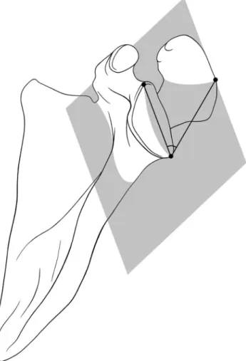
Fig. 2 Schematic view of the defined plane for the multiplanar recon-struction (MPR) of the CT scan. The plane is defined by the three points: superior border of the glenoid, inferior border of the glenoid and most lateral point of the acromial extension
Fig. 3 Measurement of the critical shoulder angle on the defined plane with MPR on the corresponding CT scan of the same right shoulder. The angle is measured on the plane defined by the cut with the largest visible distance between the superior and inferior border of the glenoid on the coronal plane and the most lateral point on the cut with the most lateral extension of the acromion on the axial plane
Fig. 4 Relationship between the CSAs measured on CT scans and radiographs
time. While angles above 35° are highly associated with degenerative cuff tears, measurements below 30° are common in osteoarthritic shoulders [1]. Researchers have yet to prove the biomechanical concept of the“balanced shoulder” where mechanical loading by the deltoid muscle and its force vector promotes an increased load on the glenoid cartilage (low CSA) or on the supraspinatus tendon (high CSA) in unbal-anced shoulders.
However, despite the reliability and reproducibility of the CSA measurement technique on radiographs, it is possible that angle measurements on cumulative images, such as ra-diographs, are not as precise as measurements on tomographic cross-sectional images where the exact landmarks can be defined. Because MRI images tend to display heterogenic definition of cartilage contours and bony edges, computed tomography, with its high resolution of bony structures, seems to be the most appropriate modality for precise CSA measure-ments. The option of computed reconstruction of the plane with the maximal superior-inferior distance of the glenoid combined with the exact determination of the coronal plane with the maximal lateral extent of the acromion is theoretically superior to measurements on three-dimensional, cumulative radiographs in terms of reproducibility and reliability.
The results of our study not only prove that measurements on CT scans are highly reliable but also indicate that the differences between measurements on anterior-posterior ra-diographs and CT scans are negligible. However, our data do not provide any explanation for the finding of radiographic and CT scan measurements being very similar despite the implementation of the third dimension with computed tomog-raphy. The relative position of the acromion to the glenoid in space has not been investigated in a large number of individ-uals yet. Further anatomical studies to clarify this issue are needed.
According to our results, CT-based assessments of the CSA are not necessary to improve measurement accuracy. In-creased radiation exposure and costs can be avoided by using only radiographs.
The small number of patients (and shoulders) included is one of the study’s limitations. However, the strong correlation of the measurements between radiographs and CT scans (r=0.974) indicates sufficient cohort size. Furthermore,
one of the two readers was a board-certified orthopaedic surgeon; therefore, potential differences in measurement tech-nique compared to the fellowship-trained musculoskeletal radiologist may be postulated. This fact, we believe, has a minor impact only, since the orthopaedic surgeon is used to read CT scans of joints in his daily routine and was taught how to use the MPR software for this study.
Conclusion
The present data demonstrate that measurements of the CSA on anterior-posterior radiographs and CT scans are highly correlated and that inter-modality differences are negligible.
Conflict of interest The authors declare that they have no conflict of interest.
References
1. Moor BK, Bouaicha S, Rothenfluh DA, Sukthankar A, Gerber C. Is there an association between the individual anatomy of the scapula and the development of rotator cuff tears or osteoarthritis of the glenohumeral joint?: a radiological study of the critical shoulder angle. Bone Joint J. 2013;95-B(7):935–41.
2. Moor BK, Wieser K, Slankamenac K, Gerber C, Bouaicha S. Relationship of individual scapular anatomy and degenerative rotator cuff tears. J Shoulder Elbow Surg. 2014;23(4):536–41.
3. Solari M, Chakravarti I. Handbook of methods of applied statistics. Nature. 1967;216(5118):901–908.
4. Vardeman S. Graphical methods for data analysis. J Qual Technol. 1984;16(3):177–8.
5. Fleiss J. The design and analysis of clinical experiments. New York: Wiley; 1986.
6. Wong AS, Gallo L, Kuhn JE, Carpenter JE, Hughes RE. The effect of glenoid inclination on superior humeral head migration. J Shoulder Elbow Surg. 2003;12(4):360–4.
7. Hughes RE, Bryant CR, Hall JM, Wening J, Huston LJ, Kuhn JE, et al. Glenoid inclination is associated with full-thickness rotator cuff tears. Clin Orthop Relat Res. 2003;407:86–91.
8. Tetreault P, Krueger A, Zurakowski D, Gerber C. Glenoid version and rotator cuff tears. J Orthop Res. 2004;22(1):202–7.
9. Nyffeler RW, Werner CM, Sukthankar A, Schmid MR, Gerber C. Association of a large lateral extension of the acromion with rotator cuff tears. J Bone Joint Surg Am. 2006;88(4):800–5.