HAL Id: hal-02445188
https://hal.archives-ouvertes.fr/hal-02445188
Submitted on 17 Nov 2020HAL is a multi-disciplinary open access archive for the deposit and dissemination of sci-entific research documents, whether they are pub-lished or not. The documents may come from teaching and research institutions in France or abroad, or from public or private research centers.
L’archive ouverte pluridisciplinaire HAL, est destinée au dépôt et à la diffusion de documents scientifiques de niveau recherche, publiés ou non, émanant des établissements d’enseignement et de recherche français ou étrangers, des laboratoires publics ou privés.
Design, Synthesis and Efficacy of Hybrid Triclosan-gold
Based Molecules on Artemisinin-resistant Plasmodium
falciparum and Leishmania infantum Parasites
Manel Ouji, Sandra Bourgeade-Delmas, Álvaro Fernández Álvarez,
Jean-Michel Augereau, Alexis Valentin, Catherine Hemmert, Heinz Gornitzka,
Françoise Benoit-Vical
To cite this version:
Manel Ouji, Sandra Bourgeade-Delmas, Álvaro Fernández Álvarez, Jean-Michel Augereau, Alexis Valentin, et al.. Design, Synthesis and Efficacy of Hybrid Triclosan-gold Based Molecules on Artemisinin-resistant Plasmodium falciparum and Leishmania infantum Parasites. ChemistrySelect, Wiley, 2020, 5 (2), pp.619-625. �10.1002/slct.201904345�. �hal-02445188�
1
Design, synthesis and efficacy of hybrid triclosan-gold based molecules on
artemisinin-resistant Plasmodium falciparum and Leishmania infantum parasites
Manel Ouji,a, $ Sandra Bourgeade Delmas,b,$ Álvaro Fernández Álvarez,a,$ Jean-Michel Augereau,aAlexis Valentin,b,* Catherine Hemmert,a,*Heinz Gornitzka,c,* Françoise Benoit-Vical, a,d *
a
LCC-CNRS, Université de Toulouse, CNRS, Toulouse, France
b
UMR 152 PharmaDev, Université de Toulouse, IRD, UPS, Toulouse, France
c
LCC-CNRS, Université de Toulouse, CNRS, UPS, Toulouse, France
d
INSERM, Institut National de la Santé et de la Recherche Médicale, France
$
Participated equally to this work.
*
Corresponding authors: AV (alexis.valentin@univ-tlse3.fr); CH ( catherine.hemmert@lcc-toulouse.fr); HG (heinz.gornitzka@lcc-toulouse.fr); FBV (francoise.vical@inserm.fr)
GRAPHICAL ABSTRACT
One molecule to target two pathologies. In the fight against major parasite diseases such as malaria and leishmaniasis, novel drugs active against drug-resistant parasites are needed. Hybrid molecules combining triclosan and gold(I) complexes appear as a promising avenue.
2
ABSTRACTThe emergence of resistant parasites, Plasmodium and Leishmania, responsible of malaria and leishmaniasis diseases, to the currently available drugs, remains one of the major threats to public health. Therefore, there is an urgent need to develop new molecules. Several hybrid molecules combining triclosan and gold(I) complexes have been synthesized and evaluated, in vitro, for their activities on P. falciparum and L. infantum parasites. The obtained results showed a good activity of the compounds. However, the cytotoxicity assays suggest a structure modulation to improve the selectivity. The synthesized molecules were also tested in a context of Plasmodium falciparum artemisinin-resistance and the results showed no cross-resistance induced between one gold(I) complex and artemisinin. This highlights that these compounds appear as a strong basis for further pharmacomodulation work to propose molecules targeting resistant parasites.
3
INTRODUCTIONMalaria and leishmaniasis are among the most important parasitic diseases through the world.[1] Because they are co-endemic in many countries, develop drugs targeting these two diseases appears as a promising goal.
Indeed, in on one hand, malaria is the parasitic disease with the largest global epidemic impact with 3.2 billion people who are at risk of infection. Around half of the world’s population can contract this disease leading to about 438,000 deaths in 2017.[1] Plasmodium falciparum, widespread in tropical and sub-tropical regions, is the main causative agent of malaria mortality and morbidity mostly affecting children under five years.
The introduction of artemisinin and its derivatives contributed to the notable decrease of malaria mortality for the 20 last years. To reduce the risk of falciparum malaria drug resistance, the WHO recommended in 2001 to use ACTs (Artemisinin-based Combination Therapies) combining artemisinin derivatives with one or two other antimalarial drugs having different mode of action and pharmacokinetic properties.[2] However, the efficacy of these drug combinations declined in South-East Asia since 2006, due to the emergence and rapid spread of P. falciparum resistance to artemisinins and partner drugs.[3] This situation is increasingly alarming because, today, the most commonly used ACT in Asia, dihydropiperaquine, is no longer active on artemisinin-resistant parasites.[4,5] According to the countries, the ACTs failure rate is now up to 10% for one ACT, for two ACTs and even for the 5 ACTs.[6]
Usually, in malaria resistances, drug resistant parasites are able to proliferate and multiply during treatment but in the case of artemisinins resistance, the involved mechanism is quiescence-based.[7–9] When treating parasites with artemisinin, most of them die, except a sub-set able to escape the treatment by a cell cycle arrest and then to resume their development when the drug is removed leading to clinical failures in the field.[8,10] Focus on artemisinin-resistant parasites is thus essential to fight malaria.
A previous work demonstrated the involvement of lipid metabolism and respiratory chain in artemisinin resistance quiescence-based.[11] Therefore, targeting these two biochemical pathways appeared as a great avenue to develop new antimalarials. Indeed, the bacterial-like type II fatty acid synthesis (FASII) of the P. falciparum apicoplast,[12] is a pretty interesting drug target because of its difference from the mammalian type I system. FASI pathway is composed of a single enzyme complex; conversely, FASII pathway uses several mono-functional proteins.[13] Interestingly, Chen et al. have shown, in vitro, that among the genes remaining actively transcribed in dihydroartemisinin-induced quiescent parasites, those coding for the FASII pathway were found.[11] In addition, they demonstrated that treating quiescent parasites with FASII inhibitors, triclosan (TC), an antibacterial and anti-fungal agent, and haloxyfop delayed their recrudescence. Moreover, mitochondria seems also to remain active during quiescence, and could contribute to the survival and the recovery of the quiescent parasites after treatment.[11,14]
That is why this work focused on cationic gold(I) N-heterocyclic carbene (NHC) complexes because they act by inhibiting specifically the mitochondrial enzyme thioredoxin reductase,[15] which plays a key role in the defense against oxidative stress. These gold(I) complexes exhibited very promising activities against several cancer cell lines.[16] Moreover, interesting activities have been also highlighted in infectious agents including Mycobacterium,[17,18] Plasmodium falciparum [19,20] and
Leishmania infantum [21,22].
On the other hand, leishmaniasis have a worldwide distribution in large areas of the tropics, subtropics and Mediterranean basin, involving more than 98 countries.[23] The burden of leishmaniasis increased over the last decades and worldwide, the population of 350 million of people is at risk. An estimated 2 million new cases occur annually with about 12 million people currently infected. [24] Leishmaniasis due to the protozoa Leishmania, is transmitted by the bite of infected female phlebotomine sandflies that inject the infective stage, metacyclic promastigotes, during blood meals. Parasites at the puncture wound are phagocytized by macrophages and transform into amastigotes. Amastigotes multiply in infected cells and affect different tissues, depending in part on which Leishmania species is involved.
4
These differing tissue specificities cause the differing clinical manifestations of the various forms of leishmaniasis such as cutaneous
,
[25]mucocutaneous, or visceral [26].
Chemotherapy is the major strategy to treat leishmaniasis including liposomal amphotericin B, a combination of pentavalent antimonials, paromomycin, pentamidine and miltefosine and orally administrated “azoles” (ketoconazole, itraconazole, fluconazole). For cutaneous disease, paromomycin, fluconazole, or pentamidine may be effective (CDC, Ressources for Health professionals, 2018). However, their drawbacks such as limited efficacy and safety, high costs and the emergence of drug-resistant parasites encourage the urgent search for new drugs alternatives. In this way, metal-based drugs are also promising antileishmanial agents.[27]
The use of metals and their salts as antimicrobial or pathogenic agents declined sharply in the middle of the last century upon the introduction of penicillin-derived antibiotics. In the last century the use of gold-based drugs experienced an extraordinary increase with the development of new gold-based compounds such as auranofin, solganol and myochrisine for the treatment of rheumatoid arthritis. The successful application of gold(I) compounds in the treatment of rheumatoid arthritis led to the synthesis of new gold(I) and gold(III) compounds for the treatment of several diseases. It has been described that the cytotoxic effects of gold compounds are focused in a specific mitochondrial mechanism.[15,28] Gold compounds have also been reported as antibacterial and antileishmanial agents.[29]
The purpose is first to evaluate the activity of the homemade hybrid molecules against P. falciparum and L. infantum strains in vitro and secondly to focus the efficacy of the most active hybrids in a context of artemisinin resistance requiring a dedicated assay based on the malaria parasite recrudescence.
RESULTS AND DISCUSSION
Chemistry
A series of hybrid molecules gold(I)-NHC-TC was synthesized easily according to a three steps strategy (Scheme 1). First a Williamson etherification of TC with 1,3-dibromopropane was performed in dry acetonitrile at 80°C using a microwave reactor. TC-C3 was isolated after purification by column chromatography with a yield of 52%. The proligands were then prepared by quaternization of a substituted imidazole, namely 1-methylimidazole, 1-mesitylimidazole,[30] benzylimidazole, 1-quinolinimidazole,[22] 1-thioanisolimidazole,[21] in acetonitrile at 80°C for 24 to 62 h. The imidazolium salts 1a-1e were obtained as white or beige powders with yields ranging from 43 to 67%. Finally, direct metalation of the proligands with one half equivalent of Au(SMe2)Cl and K2CO3 (1.4 equiv) in
acetonitrile at 60°C for 16 h lead to the desired complexes 2a-2e with yields from 76 to 99%. All the imidazolium salts 1a-1e and the complexes 2a-2e were characterized by 1H and 13C NMR spectra, HRMS and elemental analysis. The most characteristic feature in the 1H NMR spectra are the resonances for imidazolium protons (H2) located at 10.73, 10.35, 10.96, 12.31, and 11.32 ppm, for
1a-1e, respectively. The 13C NMR spectra unequivocally demonstrates the formation of the gold(I) complexes showing the resonance for the carbenic carbons at 184.03, 183.94, 183.99, 182.17, and 182.38 ppm, for 2a-2e, respectively. The elemental analysis of the gold(I) complexes correspond to the general formula [AuL2][Br] and the high resolution mass spectra (ES) exhibited the classical peaks
5
Scheme 1. Synthesis of triclosan based proligands 1a-1e and gold(I)-NHC complexes 2a-2e.Reference complex 3.
Crystals of complex 2a suitable for X-ray diffraction analysis were obtained by slow gas-phase diffusion of diethyl ether into a saturated dichloromethane solution of complex 2a. The X-ray structure of 2a (Figure 1) shows the classic linear geometry for gold(I) with a C-Au-C angle of 178.5°. The gold carbon distances of 2.018 and 2.029 Å are in the normal range for NHC-Au-NHC compounds. The two NHC planes are nearly coplanar with a torsion angle of ≈2°. The TC derivatives are placed in trans position looking on the NHC-Au-NHC plane and the two bulky substituents are on same side of this plane, which can be explained by the arrangement of the cations in the solid state (Figure 2). Two cations forming a dimer connected by an aurophilic interaction reflected by an Au-Au distance of 3.503 Å.
6
Figure 1. Crystal structure of 2a depicted at the 50% level. Hydrogen atoms, the anion and a non-coordinating ether molecule have been omitted for clarity. Selected bond lengths [Å] and angles [°]: Au-C1 2.029(6), Au-C(20) 2.018(6), C1-Au-C(20) 178.5(2).Figure 2. Dimeric arrangement of 2a in the solid state.
Antiplasmodial activity of the compounds
Five hybrid molecules corresponding to gold(I) complexes 2a-2e, their carbene precursors 1a-1e, and a gold(I) reference complex 3 (Scheme 1) were first screened in vitro using the P. falciparum strain F32-TEM (Table 1) in order to determine the IC50 and to compare to the values obtained with the
antimalarial reference drug artemether and the FASII inhibitor reference molecule triclosan.
Triclosan showed a weak antiplasmodial activity with an IC50 value of 6 µM. These results are in
accordance with a previous study which firstly studied the antiplasmodial activity of this compound 41. Interestingly, when triclosan is coupled with a substituted imidazole derivative, the resulting imidazolium salts showed 6 (1c) to 18 (1e) fold higher activities against P. falciparum. Moreover, the corresponding gold(I) complexes showed also 4 (2b) to 50 (2a) fold better activities than triclosan. Proligand (1a) and the corresponding gold complex (2a) showed better activities with IC50 values of
343 nM and 120 nM, respectively.
Globally, the presence of gold(I) can have opposite effects with an improvement of the efficacy comparatively to the corresponding proligand for 1a (343 nM) vs 2a (120 nM) and 1c (958 nM) vs 2c (386 nM) or a deleterious effect like for 1b (382 nM) vs 2b (1465 nM), and 1e (328 nM) vs 2e (875 nM). In the system1d/2d, the IC50 values are very close, 833 and 654 nM, respectively.
Au1 C1 C20 N N N N O O O O Cl Cl Cl Cl Cl Cl
7
The complex 3 was synthesized as a gold(I)-NHC reference, involving a quinoline function and a benzyl group on the two nitrogen atoms of the NHCs. 3 displayed a high antiplasmodial activity with IC50 of 170 nM, a ten-fold better activity than an analog complex containing a quinoline moiety and a
methyl group on the carbene (IC50: 1µM) previously published[20]. Unfortunately, replacement of a
benzyl substituent by a triclosan did not allow improvement of the activity of the resulting gold(I) complex 2d (IC50: 654 nM).
Table 1. Antimalarial and cytotoxic activities of the hybrid molecules
Compounds Antiplasmodial activity on P. falciparum IC50 ± SEM (nM) [a] Cytotoxicity on Vero Cells IC50 ± SEM (nM) [b] Selectivity index: Vero cells / P. falciparum
activities 3 170 ± 9 1.1. 103 ± 0.2. 103 6.5 1a 343 ± 40 - - 2a 120 ± 18 0.9. 103 ± 0.03. 103 7.5 1b 382 ± 78 - - 2b 1465 ± 265 - - 1c 958 ± 245 - - 2c 386 ± 70 - - 1d 833 ± 136 - - 2d 654 ± 67 - - 1e 328 ± 55 - - 2e 875 ± 51 - - Artemether[c] 6.1 ± 1 214.103± 29.103 35000 Triclosan 6000 ± 103 36. 103 6
[a] Values of IC50, 50% inhibitory concentration, on Plasmodium falciparum where obtained using both SYBR Green and radioactivity assays, and correspond to the mean of at least 4 independent experiments.
[b] Cytotoxic activities of the compounds were evaluated against Vero cell line. [c] Artemether, the antiplasmodial control drug, was routinely tested.
Antileishmanial activity of the compounds
The cationic Au(I)bis(NHC-TC) 2a-2e and their carbene precursors 1a-1e complexes were screened in
vitro on both the promastigote and the axenic amastigote stages of L. infantum, by determining their
8
pentadimine and miltefosine as well as the pharmacophore triclosan and the gold drug reference auranofin. The results are summarized in table 2.
All compounds were firstly tested against promastigotes of L. infantum showing moderate or no activity with IC50 values from 5.51 µM for 2a to 63.87 µM for 2b. Looking at the activities against
promastigotes of the proligand/complex couples, there was just one clear enhanced activity by addition of gold(I) in the case of the methyl substituted couple 1a-2a, complex 2a being the most active complex. Then all compounds were tested against the relevant axenic amastigote form of L.
infantum. Except 1a, all proligands displayed high activities in the low micro- and submicromolar
range (IC50 values from 0.57 to 1.10 µM). The same tendency could be observed for the gold
complexes with IC50 values ranging from 0.21 to 1.54 µM, excluding the mesityl substituted gold(I)
complex 2b with an IC50 of 11.62 µM. Comparing to their respective proligands against axenic
amastigotes, two complexes were more active (2a and 2e), two were less active (2b and 2c), while one showed the same activity (2d). Regarding these data, the role of gold(I) could not be totally established in relation with the amastigote activity. Nevertheless, the two most active gold(I) complexes of this series towards the clinically relevant axenic amastigote form of L. infantum are 2a (IC50 = 0.21 µM) and 2e (IC50 = 0.39 µM) and these TC derivates complexes (except 2b) are globally
as efficient as the two commercial drugs pentadimine and miltefosine.
These Au(I)bis(NHC-TC) series had similar activity to previously reported cationic Au(I)bis(NHC) and neutral Au(I)(NHC)Cl complexes including aliphatic and aromatic substituents.[21,22] This suggests that no enhanced activity was found by addition of the triclosan moiety.
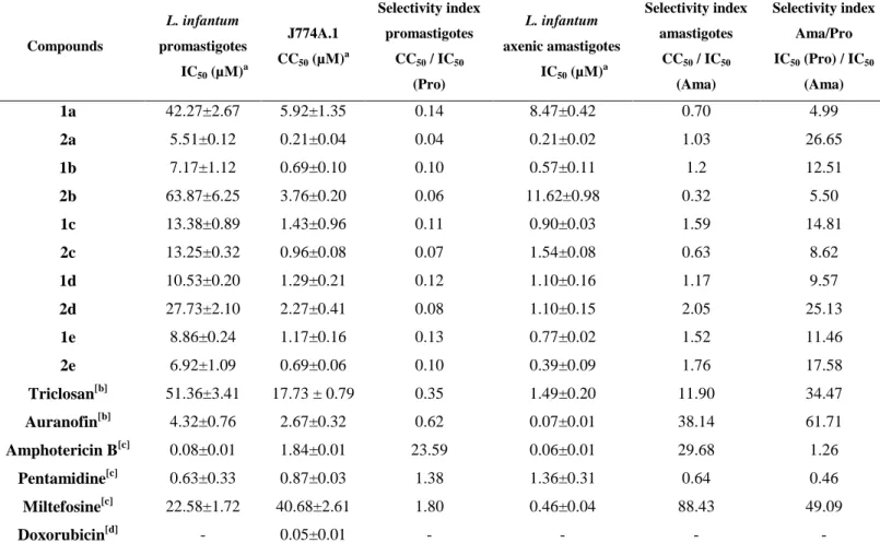
Table 2. In vitro antileishmanial activity and cytotoxicity of compounds 1a-2e and 2a-2e
[a] Mean of three independent experiments.
Compounds L. infantum promastigotes IC50 (µM)a J774A.1 CC50 (µM)a Selectivity index promastigotes CC50 / IC50 (Pro) L. infantum axenic amastigotes IC50 (µM)a Selectivity index amastigotes CC50 / IC50 (Ama) Selectivity index Ama/Pro IC50 (Pro) / IC50 (Ama) 1a 2a 1b 2b 1c 2c 1d 2d 1e 2e Triclosan[b] Auranofin[b] Amphotericin B[c] Pentamidine[c] Miltefosine[c] Doxorubicin[d] 42.27±2.67 5.51±0.12 7.17±1.12 63.87±6.25 13.38±0.89 13.25±0.32 10.53±0.20 27.73±2.10 8.86±0.24 6.92±1.09 51.36±3.41 4.32±0.76 0.08±0.01 0.63±0.33 22.58±1.72 - 5.92±1.35 0.21±0.04 0.69±0.10 3.76±0.20 1.43±0.96 0.96±0.08 1.29±0.21 2.27±0.41 1.17±0.16 0.69±0.06 17.73 ± 0.79 2.67±0.32 1.84±0.01 0.87±0.03 40.68±2.61 0.05±0.01 0.14 0.04 0.10 0.06 0.11 0.07 0.12 0.08 0.13 0.10 0.35 0.62 23.59 1.38 1.80 - 8.47±0.42 0.21±0.02 0.57±0.11 11.62±0.98 0.90±0.03 1.54±0.08 1.10±0.16 1.10±0.15 0.77±0.02 0.39±0.09 1.49±0.20 0.07±0.01 0.06±0.01 1.36±0.31 0.46±0.04 - 0.70 1.03 1.2 0.32 1.59 0.63 1.17 2.05 1.52 1.76 11.90 38.14 29.68 0.64 88.43 - 4.99 26.65 12.51 5.50 14.81 8.62 9.57 25.13 11.46 17.58 34.47 61.71 1.26 0.46 49.09 -
9
[b]Triclosan and Auranofin were used as pharmacophore and gold-based drug references, respectively. [c] Amphotericin B, miltefosine and pentamidine were used as antileishmanial compounds of reference. [d] Doxorubicin was used as positive control of cytotoxicity.
Selectivity index of the compounds regarding their antiplasmodial and antileishmanial activities
Concerning Plasmodium, as 2a was the most active molecule among the gold(I)-triclosan hybrids tested, its cytotoxicity was assessed on mammalian cells in order to determine the specificity of its activity (Table 1). The selectivity index (S.I.) was 7.5 for 2a which is comparable to 6.5 for the gold(I) complex (3) and 6 for triclosan. These low S.I. highlighted that 2a has a same range of activity against normal mammalian cells and P. falciparum.
Concerning Leishmania, complexes were tested in vitro for their cytotoxicity, measured by the CC50
on the murine J774A.1 macrophages cell line (positive control = doxorubicin), giving access to the corresponding selectivity indexes (CC50/IC50). The selectivity indexes for promastigotes (CC50/IC50
Pro) ranged from 0.10 to 0.14 for the proligands and from 0.04 to 0.10 for the gold(I) complexes, indicating a toxicity of the compounds and in particular the complexes. Likewise, the S.I. for amastigotes (CC50/IC50 Ama) were from 0.32 to 2.05, meaning that both proligands and complexes are
rather toxic and not selective. The S.I. amastigote/ promastigote ranged from 4.99 to 14.81 for the proligands, and from 5.50 to 26.65 for the complexes, with increased selectivity in most cases by coordination of the gold(I) on the NHCs.
Finally, the gold reference auranofin exhibited higher activity and selectivity (IC50 (Pro) = 4.32 µM, SI
= 2.67 and IC50 (Ama) = 0.07 µM, SI = 38.14) on the two stages of L. infantum than all complexes,
with values on the amastigote form very close to the reference drug amphotericin B. Related to the pharmacophore triclosan (IC50 (Pro) = 51.36 µM, IC50 (Ama) = 1.49 µM), most of the complexes and
ligands tested in this study showed more potency on promastigotes and most of them displayed equal (2c) or better activity (1b-1e, 2a, 2d, 2e) on axenic amastigotes. For comparison, the selectivity of the simple gold(I) complexes previously reported was higher than those of these complexes including a triclosan moiety, suggesting that the incorporation of the triclosan derivative could be related with loss in selectivity, without increasing the activity.
In vitro cross-resistance between hybrid molecules and artemisinin
In a context of artemisinins resistance, including dihydroartemisinin (DHA) the active metabolite of artemisinin derivatives used in ACTs, the standard in vitro susceptibility assay based on the inhibition of parasite proliferation is not appropriated to evaluate drug resistance.[7,39,42] Indeed, there is no significant difference between the IC50 values from a resistant-artemisinin and a sensitive-artemisinin
strain. An entry into dormancy (quiescence) of a small sub-population of the parasites in presence of the drug is responsible for artemisinins resistance, parasites restarting their proliferation after elimination of this drug. That explains that classical chemo-sensitivity assays are unsuitable and the necessity for an assay evaluating the parasite abilities to proliferate after the drug removal. That is why, a relevant recrudescence assay was developed and was used here to explore the activity of the molecules against F32-ART, a highly resistant strain and comparatively to the artemisinin-sensitive line F32-TEM.[7,39] In this test, the cross-resistance between artemisinin and another compound is demonstrated by a faster resumption of proliferation for the F32-ART strain compared to the sensitive F32-TEM strain after exposure of these strains to the molecule tested.
The 9.7 days of difference between both strains after artemisinin treatment confirmed the cross-resistance between all molecules from artemisinin class. It is to note that no cross-cross-resistance was evidenced for triclosan with similar recrudescence days (0.8 day of difference) for ART and F32-TEM (Table 3). However, this result needs to be moderated by the low activity of triclosan on P.
cross-10
resistance with artemisinin, so neither the triclosan part, nor the gold(I) permitted to avoid this cross-resistance especially highlighted at 1 and 2 µM (Table 3).
Nevertheless, gold(I) complex 3 (table 3) didn’t show cross-resistance with artemisinin. This is consistent with previous results which revealed that atovaquone, a mitochondrial electron transfer inhibitor, didn’t induce any cross-resistance with artemisinins,[39]
confirming mitochondria as potential drug target to eliminate artemisinins-resistant parasites. In this way, further complexes containing gold(I) will be developed.
Table 3. Recrudescence capacity of Plasmodium falciparum F32-ART and F32-TEM strains after 48h-drug exposure [a]
Compounds Doses Number of
experiments
Median (range) recrudescence days
Mean ± SEM difference of
recrudescence days p-value
F32-TEM F32-ART Artemisinin 18µM 6 9.5 (6 – 17) 18.5 (17 - >30) 9.7 ± 0.3 0.006 Triclosan 7µM 5 5 (2 – 7) 6 (2 – 9) 0.8 ± 0.3 0.42 3 1µM 3 10 (7-15) 11 (8-17) 1.3 ± 0.5 0.49 3 2µM 1 12 13 1 2a 500nM 1 15 17 2 2a 1µM 2 25->30 >30 >5 2a 2µM 1 16 >30 >14 2a 10µM 1 >30 >30 -
[a]Synchronized ring-stage parasites have undergone 48 h of drugtreatment. After that, cultures were washed and parasitemia was monitored during 30 days or until reaching the initial parasitemia, defined as the recrudescence day. If no parasites were observed at the end of the experiment, the culture was classified as showing no recrudescence, and the recrudescence day was noted as >30.
Data distribution was Gaussian. A log-rank (Mantel-Cox) test was used for statistical analysis of recrudescence days; p< 0.05 = significant difference between F32-ART and F32-TEM.
CONCLUSION
A family of triclosan derivatives containing imidazolium salts and corresponding bisNHC gold(I) complexes have been synthesized, characterized and tested on P. falciparum and L. infantum parasites. Our work permitted to design new compounds for the needy portfolio of the antiplasmodial and antileishmanial drug-candidates. Very high antiplasmodial and antileishmanial activities are reported here with bisNHC gold(I); for L. infantum the level of activity was similar for some bisNHC gold(I) complexes to those of the reference compounds. However, the next structure-activity relationships should be oriented towards a better selectivity. Moreover, in a context of widespread malaria
11
resistance, the upcoming new gold(I) complexes should combine pharmacophores targeting specifically the biochemistry pathways of quiescent resistant Plasmodium parasites in order to avoid cross-resistance with artemisinins. Pharmaco-modulation studies are thus in progress based on these solid and promising results in order to propose novel drugs with the maintain of high-level activities, the specificity increase and to consider the risk of cross-resistance with drugs already used in a context of worldwide resistance.
MATERIALS AND METHODS
General information for synthesis
All manipulations were performed under an inert atmosphere of dry nitrogen by using standard vacuum line and Schlenk tube techniques. 1-Mesitylimidazole,[30] 1-(quinoline-2-yl)-1H-imidazole 22, 1-(4-methylthiophenyl)-1H-imidazole,[21] and 2-(3-bromopropoxy)-4-chloro-1-(2,4-dichlorophenoxy)-benzene (TC-C3) [31] were prepared as described in the literature. All other reagents were used as received from commercial suppliers. 1H (300 or 400 MHz), 13C (75 or 101 MHz) NMR spectra were recorded at 298 K on Bruker AV300, Bruker AV400, Bruker HD400 spectrometers in CDCl3, CD3CN
or DMSO-d6 as solvents. Elemental analyses were carried out by the “Service de Micro-analyse du Laboratoire de Chimie de Coordination (Toulouse)”. High Resolution Mass Spectrometry (HRMS) analysis were performed with a Xevo G2 QTOF Waters spectrometer using electrospray ionization (ESI) by the “Service de Spectrométrie de Masse de Chimie UPS-CNRS” (Toulouse).
Preparation of Imidazolium salts
1-(3-(5-Chloro-2-(2,4-dichlorophenoxy)phenoxy)propyl)-3-methyl-2,3-dihydro-1H-imidazol-3-ium bromide (1a). To a stirred solution of TC-C3 (131.4 mg, 0.32 mmol) in CH3CN (10 mL),
1-methylimidazole (25.5 L, 0.32 mmol) was added and the reaction mixture was stirred 1 day at 70 °C. Then, the solvent was evaporated under reduced pressure and the viscous residue was purified by washing with diethylether to afford a yellow powder (67.4 mg, 43% yield). Anal. Calcd. For C19H18BrCl3N2O2 C, 46.33; H, 3.68; N, 5.69. Found C, 46.35; H, 3.75; N, 5.53. 1 H NMR (300 MHz, CDCl3) δ 10.73 (d, J = 1.7 Hz, 1H), 7.47 (d, J = 2.5 Hz, 1H), 7.19-7.15 (3H), 7.06-6.97 (3H), 6.71 (d, J = 8.8 Hz, 1H), 4.40 (t, J = 6.8 Hz, 2H,), 4.11-4.06 (m, 2H), 4.05 (s, 3H), 2.44 (tt, J = 6.8 Hz, 2H).13C NMR (101 MHz, CDCl3) δ 152.21 (1C), 149.84 (1C), 142.68 (1C), 138.36 (1C), 131.06 (1C), 130.20 (1C), 128.31 (1C), 128.15 (1C), 123.98 (1C), 122.60 (1C), 122.45 (1C), 121.98 (2C), 118.12 (1C), 115.21 (1C), 65.22 (1C), 46.91(1C), 36.80 (1C), 29.65 (1C). HRMS (ES+): calcd. for C19H18Cl3N2O2
411.0434 found 411.0433 [M – Br-]+.
3-(3-(5-Chloro-2-(2,4-dichlorophenoxy)phenoxy)propyl)-1-mesityl-2,3-dihydro-1H-imidazol-3-ium bromide (1b). To a stirred solution of TC-C3 (344.8 mg, 0.84 mmol) in CH3CN (10 mL),
1-mesitylimidazole (139.7 mg, 0.75 mmol) was added and the reaction mixture was stirred 2 days at 70 °C. Then, the solvent was evaporated under reduced pressure and the residue was purified by washing with diethylether to afford a white powder (284.9 mg, 56% yield). Anal. Calcd. For C27H26BrCl3N2O2
C, 54.34; H, 4.39; N, 4.69. Found C, 54.24; H, 4.45; N, 4.76. 1H NMR (400 MHz, CDCl3) δ 10.35 (t, J = 1.6 Hz, 1H), 7.74 (t, J = 1.8 Hz, 1H), 7.44 (d, J = 2.5 Hz, 1H), 7.16 (dd, J = 8.8, 2.5 Hz, 1H), 7.09 (t, J = 1.8 Hz, 1H), 7.04 (d, J = 2.3 Hz, 1H), 7.01-6.97 (m, 3H), 6.93 (d, J = 8.5 Hz, 1H), 6.71 (d, J = 8.8 Hz, 1H), 4.79 (t, J = 6.9 Hz, 2H), 4.14 (t, J = 5.5 Hz, 2H), 2.55 (m, 2H), 2.36 (s, 3H), 2.02 (s, 6H). 13C NMR (101 MHz, CDCl3) δ 152.19 (1C), 149.80 (1C), 141.50 (1C), 138.97 (1C), 134.52 (2C), 134.40 (1C), 130.86 (1C), 130.20 (1C), 129.23 (2C), 128.12 (1C), 127.96 (1C), 123.87 (1C), 123.56 (1C), 122.37 (1C), 122.15 (1C), 122.03 (1C), 121.65 (1C), 117.98 (1C), 115.20 (1C), 65.18 (1C), 45.58 (1C), 30.97 (1C), 21.16 (1C), 18.32 (2C). HRMS (ES+): calcd. for C27H26Cl3N2O2 515.1060 [M – Br-]+
found 515.1064 [M – Br-]+.
1-Benzyl-3-(3-(5-chloro-2-(2,4-dichlorophenoxy)phenoxy)propyl)-1H-imidazol-3-ium bromide (1c). To a stirred solution of TC-C3 (307.9 mg, 0.75 mmol) in CH3CN (10 mL), 1-benzylimidazole
12
(86.9 L, 0.67 mmol) was added and the reaction mixture was stirred 3 days at 70 °C. Then, the solvent was evaporated under reduced pressure and the residue was purified by washing with diethylether to afford a white powder (160.3 mg, 42% yield). Anal. Calcd. For C25H22BrCl3N2O2 C,
52.80; H, 3.90; N, 4.93. Found C, 52.75; H, 3.85; N, 4.72. 1H NMR (300 MHz, CDCl3) δ 10.86 (d, J = 1.6 Hz, 1H), 7.51-7.37 (m, 6H), 7.17 (t, J = 1.8 Hz 1H), 7.15 (dd, J = 8.8, 2.5 Hz, 1H), 7.10 (t, J = 1.8 Hz, 1H), 7.03-6.91 (m, 3H), 6.68 (d, J = 8.8 Hz, 1H), 5.51 (s, 2H), 4.38 (t, J = 6.9 Hz, 2H), 4.05 (t, J = 5.5 Hz, 2H), 2.45 (m, 2H). 13C NMR (75 MHz, CDCl3) δ 152.20 (1C), 149.83 (1C), 142.64 (1C), 137.75 (1C), 131.08 (1C), 131.05 (1C), 130.16 (1C), 130.15 (1C), 129.57 (2C), 129.01 (2C), 128.23 (1C), 128.09 (1C), 123.91 (1C), 122.50 (1C), 122.01 (2C), 121.02 (1C), 117.99 (1C), 115.29 (1C), 65.20 (1C), 53.69 (1C), 46.92 (1C), 29.56 (1C). HRMS (ES+): calcd. for C25H22Cl3N2O2 487.0747 [M
– Br
-]+ found 487.0745 [M – Br-]+.
2-(3-(3-(5-Chloro-2-(2,4-dichlorophenoxy)phenoxy)propyl)-2,3-dihydro-1H-imidazol-3-ium)quinoline bromide (1d). To a stirred solution of TC-C3 (340.7 mg, 0.82 mmol) in CH3CN (10
mL), 1-(quinoline-2-yl)-1H-imidazole (113.2 mg, 0.58 mmol) was added and the reaction mixture was stirred 1 day at 70 °C. Then, the solvent was evaporated under reduced pressure and the viscous residue was purified by washing with diethylether to afford a yellow powder (170.3 mg, 48% yield). Anal. Calcd. For C27H21BrCl3N3O2 C, 53.54; H, 3.49; N, 6.94. Found C, 53.47; H, 3.58; N, 6.79.
1 H NMR (300 MHz, CDCl3) δ 12.06 (s, 1H), 8.58 (d, J = 8.8 Hz, 1H), 8.51 (d, J = 8.5 Hz, 1H), 8.42 (t, J = 1.8 Hz, 1H), 8.06-8.03 (m, 1H), 7.97-7.94 (m, 1H), 7.85 (ddd, J = 8.5, 6.9, 1.5 Hz, 1H), 7.68 (ddd, J = 8.2, 6.9, 1.2 Hz, 1H), 7.39 (d, J = 2.5 Hz, 1H), 7.36 (t, J = 1.8 Hz, 1H), 7.11 (dd, J = 8.8, 2.5 Hz, 1H), 7.02 (d, J = 2.3 Hz, 1H), 6.95 (dd, J = 8.6, 2.3 Hz, 1H), 6.87 (d, J = 8.5 Hz, 1H), 6.70 (d, J = 8.8 Hz, 1H), 4.65 (t, J = 6.5 Hz, 2H), 4.18 (t, J = 5.5 Hz, 2H), 2.65 (m, 2H). 13C NMR (101 MHz, CDCl3) δ 152.04 (1C), 149.71 (1C), 145.96 (1C), 142.90 (1C), 141.63 (2C), 137.01 (1C), 131.51 (1C), 130.78 (1C), 130.19 (1C), 128.82, (1C), 128.54 (1C), 128.29 (1C), 128.14 (3C), 124.34 (1C), 122.63 (1C), 121.87 (1C), 121.35 (1C), 118.64 (1C), 118.34 (1C), 115.09 (1C), 112.76 (1C), 65.66 (1C), 47.89 (1C), 29.69 (1C). HRMS (ES+): calcd. for C27H21Cl3N3O2 524.0669 [M – Br
-]+ found 524.0667 [M – Br-]+.
1-(3-(5-chloro-2-(2,4-dichlorophenoxy)phenoxy)propyl)-3-(4-(methylthio)phenyl)-2,3-dihydro-1H-imidazol-3-ium, bromide (1e). To a stirred solution of TC-C3 (328.4 mg, 0.80 mmol) in CH3CN
(10 mL), 1-(4-methylthiophenyl)-1H-imidazole (121.6 mg, 0.64 mmol) was added and the reaction mixture was stirred 2 days at 70 °C. Then, the solvent was evaporated under reduced pressure and the residue was purified by washing with diethylether to afford a beige powder (257.0 mg, 67% yield). Anal. Calcd. For C25H22BrCl3N2O2S C, 49.98; H, 3.69; N, 4.66. Found C, 49.85; H, 3.75; N, 4.79.
1 H NMR (300 MHz, CDCl3) δ 11.32 (d, J = 1.7 Hz, 1H), 7.66-7.62 (m, 2H), 7.45 (d, J = 2.5 Hz, 1H), 7.43 (t, J = 1.9 Hz, 1H), 7.40-7.36 (m, 3H), 7.16 (dd, J = 8.8, 2.5 Hz, 1H), 7.03-6.91 (m, 3H), 6.71 (d, J = 8.5 Hz, 1H), 4.63 (t, J = 6.8 Hz, 2H), 4.15 (t, J = 5.5 Hz, 2H), 2.64-2.56 (m, 2H), 2.54 (s, 3H). 13C NMR (101 MHz, CDCl3) δ 152.07 (1C), 149.79 (1C), 142.83 (1C), 142.75 (1C), 136.65 (1C), 131.02 (1C), 130.89 (1C), 130.20 (1C), 128.44 (1C), 128.21 (1C), 127.33 (2C), 124.08 (1C), 123.13 (1C), 122.03 (2C), 121.95 (1C), 121.68 (1C), 119.84 (1C), 118.39 (1C), 115.15 (1C), 65.64 (1C), 47.69 (1C), 29.73 (1C), 15.29 (1C). HRMS (ES+): calcd. for C25H22Cl3N2O2S 519.0462 [M – Br
-]+ found 519.0468 [M – Br-]+.
Synthesis of gold(I) complexes
Complex 2a. Under a nitrogen atmosphere, 1a (50.0 mg, 0.10 mmol), K2CO3 (23.8 mg, 0.17 mmol)
and Au(SMe2)Cl (18.0 mg, 0.06 mmol) were dissolved in CH3CN (3 mL) and stirred overnight at 60
°C. The solution was filtered through a syringe filter (0.2 µm) and the solvent removed under reduced pressure to afford a white powder (42.0 mg, 76% yield). Anal. Calcd. For C38H34AuBrCl6N4O4 C,
41.48; H, 3.11; N, 5.09. Found C, 41.24; H, 3.28; N, 4.89. 1H NMR (400 MHz, DMSO-d6) δ 7.64 (d, J = 2.6 Hz, 1H), 7.45 (d, J = 1.8 Hz, 1H), 7.35 (d, J = 1.8 Hz, 1H), 7.26 (dd, J = 8.9, 2.6 Hz, 1H), 7.22 (d, J = 2.3 Hz, 1H), 7.06 (d, J = 8.6 Hz, 1H), 7.01 (dd, J = 8.6, 2.3 Hz, 1H), 6.75 (d, J = 8.8 Hz, 1H), 4.08 (t, J = 6.9 Hz, 2H), 4.01 (t, J = 5.8 Hz, 2H), 3.76 (s, 3H), 2.07 (m, 2H). 13C NMR (101 MHz, CDCl3) δ 184.03 (1C), 152.40 (1C), 150.32 (1C), 142.68 (1C), 131.02 (1C), 130.12 (1C), 128.06 (1C), 127.95 (1C), 123.90 (1C), 122.66 (1C), 122.16 (1C), 122.07 (1C), 121.60 (1C), 117.81 (1C), 115.16
13
(1C), 60.40 (1C), 47.56 (1C), 31.00 (1C), 14.21 (1C). HRMS (ES+): calcd. for C38H34AuCl6N4O4 +
1017.0376 found 1017.0377 [M – Br-]+.
Complex 2b. Under a nitrogen atmosphere, 1b (100.0 mg, 0.17 mmol), K2CO3 (35.9 mg, 0.26 mmol)
and Au(SMe2)Cl (27.1 mg, 0.09 mmol) were dissolved in CH3CN (5 mL) and stirred overnight at 60
°C. The solution was filtered through a syringe filter (0.2 µm) and the solvent removed under reduced pressure to afford a white powder (101.3 mg, 94% yield). Anal. Calcd. For C54H50AuBrCl6N4O4 C,
49.56; H, 3.85; N, 4.28. Found C, 49.58; H, 3.86; N, 4.42. 1H NMR (400 MHz, CDCl3) δ 7.58 (d, J = 1.9 Hz, 1H), 7.43 (d, J = 2.5 Hz, 1H), 7.17 (dd, J = 8.8, 2.5 Hz, 1H), 6.99-6.94 (m, 3H), 6.90-6.89 (m, 2H), 6.82 (d, J = 1.8 Hz, 1H), 6.69 (d, J = 8.8 Hz, 1H), 4.23 (t, J = 6.9 Hz, 2H), 3.92 (t, J = 5.9 Hz, 2H), 2.37 (s, 3H), 2.21-2.14 (m, 2H), 1.80 (s, 6H). 13C NMR (101 MHz, CDCl3) δ 183.94 (1C), 152.26 (1C), 150.32 (1C), 142.76 (1C), 139.44 (1C), 134.87 (2C), 134.77 (1C), 130.97 (1C), 130.13 (1C), 129.08 (2C), 128.13 (1C), 128.06 (1C), 123.99 (1C), 123.19 (1C), 122.07(1C), 122.04 (1C), 121.69 (1C), 118.04 (1C), 115.18 (1C), 65.05 (1C), 47.80 (1C), 30.84 (1C), 21.16 (1C), 17.55 (2C). HRMS (ES+): calcd. for C54H50Cl6N4O4 1225.1623 [M – Br
-]+ found 1225.1659 [M – Br]+.
Complex 2c. Under a nitrogen atmosphere, 1c (110.0 mg, 0.19 mmol), K2CO3 (37.4 mg, 0.27 mmol)
and Au(SMe2)Cl (29.5 mg, 0.10 mmol) were dissolved in CH3CN (6 mL) and stirred overnight at 60
°C. The solution was filtered through a syringe filter (0.2 µm) and the solvent removed under reduced pressure to afford a yellow powder (118.5 mg, 95% yield). Anal. Calcd. For C50H42AuBrCl6N4O4 C,
47.95; H, 3.38; N, 4.47. Found C, 47.85; H, 3.45; N, 4.34. 1H NMR (300 MHz, CDCl3) δ 7.40 (d, J = 2.5 Hz, 1H), 7.33-7.28 (m, 3H), 7.20-7.15 (m, 3H),7.12 (dd, J = 8.9, 2.5 Hz, 1H), 7.01 (d, J = 1.9 Hz, 1H), 6.96-6.92 (m, 3H), 6.64 (d, J = 8.8 Hz, 1H), 5.35 (s, 2H), 4.25 (t, J = 6.9 Hz, 2H), 3.94 (t, J = 5.5 Hz, 2H), 2.24 (m, 2H). 13C NMR (101 MHz, CDCl3) δ 183.99 (1C), 152.37 (1C), 150.31 (1C), 142.67 (1C), 135.66 (1C), 131.01 (1C), 130.11 (1C), 129.03 (2C), 128.50 (1C), 128.05 (1C), 127.97 (1C), 127.33 (2C), 123.92 (1C), 122.54 (1C), 122.12 (2C), 121.61 (1C), 117.86 (1C), 115.20 (1C), 65.44 (1C), 54.84 (1C), 47.91 (1C), 30.91 (1C). HRMS (ES+): calcd. for C50H42AuCl6N4O4 1169.0997 [M –
Br-]+ found 1169.1025 [M – Br-]+.
Complex 2d. Under a nitrogen atmosphere and protection of the light, 1d (63.0 mg, 0.104 mmol), K2CO3 (20.2 mg, 0.15 mmol) and Au(SMe2)Cl (18.4 mg, 0.06 mmol) were dissolved in CH3CN (3
mL) and stirred overnight at 60 °C. The solution was filtered through a syringe filter (0.2 µm) and the solvent removed under reduced pressure to afford a white powder (65.4 mg, 95% yield). Anal. Calcd. For C54H40AuBrCl6N6O4 C, 51.09; H, 3.23; N, 5.15; Found C, 48.95; H, 3.25; N, 6.42.
1 H NMR (400 MHz, CDCl3) δ8.36 (d, J = 8.6 Hz, 1H), 8.06 (d, J = 8.4 Hz, 1H), 7.89 (d, J = 8.8 Hz, 1H), 7.87 (d, J = 1.9 Hz, 1H), 7.72-7.68 (m, 2H), 7.56-7.50 (m, 1H), 7.39 (d, J = 2.1 Hz, 1H), 7.38 (d, J = 2.5 Hz, 1H), 7.11 (dd, J = 8.8, 2.5 Hz, 1H), 7.02-6.91 (m, 3H), 6.64 (d, J = 8.8 Hz, 1H), 4.43 (t, J = 6.9 Hz, 2H), 3.97 (t, J = 5.5 Hz, 2H), 2.39-2.32 (m, 2H). 13C NMR (101 MHz, CDCl3) δ 182.17 (1C), 152.32 (1C), 150.26 (1C), 149.29 (1C), 146.28 (1C), 142.67 (1C), 139.56 (1C), 131.00 (1C), 130.87 (1C), 130.13 (1C), 128.60 (1C), 128.07 (1C), 127.95 (1C), 127.79 (1C), 127.63 (1C), 127.42 (1C), 123.99 (1C), 122.98 (1C), 122.02 (1C), 121.61 (1C), 121.00 (1C), 117.90 (1C), 116.36 (1C), 115.20 (1C), 65.46 (1C), 48.86 (1C), 30.77 (1C). HRMS (ES+): calcd. for C54H40AuCl6N6O4 1243.0920 found 1243.0908
[M – Br]+.
Complex 2e. Under a nitrogen atmosphere and protection of the light, 1e (100.0 mg, 0.17 mmol), K2CO3 (32.2 mg, 0.24 mmol) and Au(SMe2)Cl (23.6 mg, 0.085 mmol) were dissolved in CH3CN (6
mL) and stirred overnight at 60 °C. The solution was filtered through a syringe filter (0.2 µm) and the solvent removed under reduced pressure to afford a yellow powder (104.0 mg, 99% yield). Anal. Calcd. For C50H42AuBrCl6N4O4S2 C, 45.61; H, 3.22; N, 4.26. Found C, 45.56; H, 3.52; N, 4.36.
1 H NMR (400 MHz, CDCl3) δ 7.46 (dt, J= 7.0, 2.3 Hz, 2H), 7.42 (d, J = 1.9 Hz, 1H), 7.39 (d, J = 2.5 Hz, 1H), 7.26-7.21 (m, 2H), 7.16 (d, J = 1.9 Hz, 1H), 7.13 (dd, J = 8.8, 2.5 Hz, 1H), 7.02-6.95 (m, 3H), 6.66 (d, J = 8.8 Hz, 1H), 4.29 (t, J = 7.0 Hz, 2H), 4.00 (t, J = 5.5 Hz, 2H), 2.53 (s, 3H), 2.27 (m, 2H). 13 C NMR (101 MHz, CDCl3) δ 182.38 (1C), 152.35 (1C), 150.37 (1C), 142.55 (1C), 140.69 (1C), 135.82 (1C), 131.10 (1C), 130.10 (1C), 128.20 (1C), 128.00 (1C), 126.36 (2C), 125.39 (2C), 123.84 (1C), 123.13 (1C), 122.18 (1C), 121.87 (1C), 121.62 (1C), 117.79 (1C), 115.18 (1C), 65.71 (1C), 48.33 (1C), 30.82 (1C), 15.39(1C). HRMS (ES+): calcd. for C50H42AuCl6N4O4S2 1233.0439 found
1233.0455 [M – Br]+.
Complex 3. In a pressure flask, imidazole (3.32 g, 48.8 mmol), 2-chloroquinoline (4 g, 24.4 mmol, 0.5 eq.), K2CO3 (3.37 g, 24.4 mmol, 0.5 eq.) and a catalytic amount of CuSO4 were heated at 200 °C and
14
stirred for 2.5 h. After cooling to room temperature, the brown precipitate formed was dissolved in a mixture of CHCl3 and CH2Cl2 1:1. After filtration through a pad of celite, the solution was evaporated
and dried under vacuum to give 2-(1H-imidazol-1-yl)quinoline as a beige powder (3.95 g, 83% yield). 250 mg (1.281 mmol) of 2-(1H-imidazol-1-yl)quinoline and 0.162 mL (1.409 mmol, 1.1 eq.) of benzyl chloride were dissolved in dry CH3CN (10 mL) and stirred at 80 °C for 12 h. The solution was
evaporated and dried under vacuum to give 3-benzyl-1-(quinolin-2-yl)-1H-imidazol-3-ium chloride as a yellow powder (400 mg, 97% yield). Anal. Calcd. For C19H16ClN3 C, 70.91; H, 5.01; N, 13.06.
Found C, 70.83, H, 5.11, N, 13.12. 1H NMR (400 MHz, DMSO-d6): δ 10.57 (d, J = 1.6 Hz, 1H), 8.84 (d, J = 8.8 Hz, 1H), 8.72 (dd, J = 1.9 Hz, 1H), 8.21 (d, J = 8.8 Hz, 1H), 8.19-8.15 (m, 1H), 8.12 (dd, J = 1.9 Hz, 1H), 8.09 (d, J = 8.5 Hz, 1H), 7.95 (ddd, J = 8.5, 6.9, 1.5 Hz, 1H), 7.77 (ddd, J = 8.2, 6.9, 1.2 Hz, 1H), 7.58 (d, J = 6.0 Hz, 2H), 7.48-7.42 (m, 3H), 5.62 (s, 2H). 13C NMR (75 MHz, CDCl3): δ 146.03 (1C), 144.55 (1C), 141.58 (1C), 137.18 (1C), 132.73 (1C), 131.40 (1C), 129.68 (1C), 129.54 (2C), 129.28 (2C), 128.77 (1C), 128.25 (1C), 128.09 (1C), 128.02 (1C), 121.67 (1C), 118.91 (1C), 112.76 (1C), 54.07 (1C). HRMS (ES+): calcd. for C19H16N3 286.1344 found 286.1344 [M – Cl]
+
. Under a nitrogen atmosphere and protection of light, 214 mg (0.67 mmol) of the carbene precursor and 77.1 mg (0.33 mmol, 0.5 eq.) of Ag2O were dissolved in 12 mL of dry acetonitrile and stirred at
room temperature for 12 hours. After that, 98 mg (0.33 mmol, 0.5 eq.) of Au(SMe2)Cl were added and
stirred in the same conditions for 2 hours. The suspension obtained was filtered through a pad of celite and the solution was evaporated. The crude product was solubilized in 1 mL of dry CH2Cl2 and
precipitated with 8 mL of diethyl ether. After filtration, complex 3 was obtained as a beige powder (217 mg, 81% yield). Anal. Calcd. For C38H30AuN6Cl C, 56.83; H, 3.77; N, 10.46; Found C, 56.69; H,
3.80; N, 10.41. 1H NMR (400 MHz, CD3CN) δ 8.04 (d, J = 2.5 Hz, 2H), 7.96 (d, J = 2.0 Hz, 1H), 7.81
(d, J = 8.9 Hz, 1H), 7.78 (d, J = 7.4 Hz, 1H), 7.68 (ddd, J = 8.4, 6.9, 1.5 Hz, 1H), 7.55 (ddd, J = 8.1, 7.0, 1.2 Hz, 1H), 7.48 (d, J = 2.0 Hz, 1H), 7.29 (m, 5H), 5.51 (s, 2H). 13C NMR (101 MHz, CD3CN) δ
182.66 (1C), 149.26 (1C), 146.13 (1C), 139.53 (1C), 136.06 (1C), 130.98 (1C), 128.88 (2C), 128.39 (1C), 128.30 (1C), 127.89 (1C), 127.57 (1C), 127.45 (2C), 127.42 (1C), 122.89 (1C), 121.68 (1C), 115.66 (1C), 55.23 (1C). HRMS (ES+): calcd. for C38H30AuN6 767.2197 found 767.2199 [M – Cl]
+
.
Crystallographic data for 2a
All data were collected at low temperature using oil-coated shock-cooled crystals on a Bruker-AXS APEX II diffractometer with MoK radiation ( = 0.71073 Å). The structures were solved by direct methods [32] and all non hydrogen atoms were refined anisotropically using the least-squares method
on F.2[33]
Crystal data for 2a at 100(2) K: C42H44AuBrCl6N4O5, M = 1174.39, monoclinic, P21/c, a = 19.069(4),
b = 12.200(3), c = 19.853(5) Å, b = 102.513(8), V = 4508(2) Å3, Z = 4, D
calcd = 1.730 g/cm3, R =
0.0486 (Io > 2σ(Io) (wR2 = 0.1190 for all data), GOF = 1.030. CCDC-1960111 (2a) contain the supplementary crystallographic data. These data can be obtained free of charge from The Cambridge Crystallographic Data Centre via www.ccdc.cam.ac.uk/data_request/cif.
Plasmodium falciparum culture
The Plasmodium falciparum strains, F32-ART resistant to artemisinins and its twin sensitive to artemisinins F32-TEM [7] from the parental strain F32-Tanzania, were cultivated in vitro as described by Trager and Jensen [34] with minor modifications.[35] Briefly, parasites were grown in type O human erythrocytes (Etablissement Français de sang (EFS), Toulouse, France), diluted to 2% hematocrit and suspended in RPMI-1640 medium (GIBCO, France) with 5% Human Serum AB (EFS, Toulouse, France) at 37°C and 5% CO2.
Parasite cultures were synchronized at the ring stage using 5% sorbitol solution [36] every other day and before each experiment.
15
P. falciparum drug susceptibility assays were performed using 2 different method. Firstly, the
chemosensitivity assays were carried out by SYBR Green in 96-well flat bottom sterile plates by the addition of 100µL of the aforementioned parasites solution then 100µL of RPMI 5% SH-diluted drugs (triplicate wells). Negative controls such as parasites treated with solvents (DMSO or RPMI), and positive controls of parasites treated by artemether, were added to each set of experiments.
DNA in treated and control cultures of parasites were quantified using the SYBR Green (Thermo-Fischer) fluorescence-based method after 48 h of incubation with the drugs (Smilkstein, Sriwilaijaroen, Kelly, Wilairat, & Riscoe, 2004). Briefly, pellets were washed three times with PBS and frozen at -20°C until use. Plates were then thawed at room temperature, and 100 μl of each homogenized culture were transferred to a 96-well black plate that contained 100 μl of the SYBR Green lysis buffer (20 mm Tris base pH 7.5, 5 mm EDTA, 0.008% w/v saponin, 0.08% w/v Triton X-100). Plates were incubated for 2 h at room temperature and then read on a fluorescence plate reader (FLx800, BioTek) using excitation and emission wavelengths of 485 and 535 nm, respectively. To confirm the results obtained with the SYBR Green method, the standard chemosensitivity assay recommended by the WHO and based on the incorporation of [3H] hypoxanthine by the parasites was then performed at least one time for each molecule. The radioactivity assay was carried out according to the description of Desjardins et al [37] with some modifications.[35] After 24 h of incubation with the drug dilutions, [3H] hypoxanthine (50 μl/well/ 0.25 μCi; Perkin-Elmer) was added. The plates were incubated under the same conditions for another 24 h. After that, plates were frozen at -20°C and thawed at room temperature. This step leads to the release of the nucleic acids which were collected on filters. The filters were dried and placed with 5 ml of scintillation fluid (Perkin-Elmer). Tritium incorporation was then determined with a β-counter (Perkin-Elmer).
Concentrations inhibiting 50% of the parasite’s growth (half maximal inhibitory concentration or IC50
values) were then calculated from the obtained experimental results using GraphPad Prism software 7 (GraphPad Software, San Diego, CA, USA). Data reported in Table 1 correspond to the mean of at least 4 values of IC50 from independent experiments, obtained both by SYBR Green and by
radioactivity.
Cytotoxicity assays regarding antimalarial activity
Cytotoxicity of the selected compounds according their antiplasmodial activity, was determined thanks to a previously described method with some modifications.[38] The Vero cell line (monkey epithelial cell line) was cultured in MEM medium (Dutscher, France) supplemented with 10 % fetal bovine serum (Dutscher, France), 0.7 mM glutamine and 100μg/ml gentamicin (Sigma).
The cells were plated onto 96-well plates in 100 μL and seeded at 104 cells per well. Following 24 h of incubation at 37 °C and 5 % CO2, cells were incubated for an additional 48 h with various
concentrations of the compounds to be tested (100 µL per well). The cell proliferation was measured with MTT (1-(4,5-dimethylthiazol-2-yl)-3,5-diphenylformazan, Sigma). Briefly, 100 µL of MTT solution (0.5 mg / mL in MEM) was added to each well and the plate was further incubated for 1 h at 37 °C and 5 % CO2. Subsequently, the cells and MTT crystals were dissolved in 100 µL of DMSO
with the help of a plate shaker. The absorbance was measured at 540 nm (µQuant, BioTek). Cell proliferation was calculated from at least 3 independent experiments using GraphPad Prism software 7 (GraphPad Software, San Diego, CA, USA). Selectivity index was then calculated and corresponds to the ratio of cytotoxicity IC50 value / activity IC50 value.
Recrudescence assay
D-sorbitol synchronized ring-form cultures of F32-ART and F32-TEM at 3 % parasitemia and 2 % hematocrit were exposed to drugs to be tested for 48 h then washed with RPMI-1640 medium and finally placed in new wells in drug-free culture conditions with 10 % human serum. The parasitemia
16
was then monitored daily to determine the time required for each parasite culture to recover the initial parasitemia (3 %). If there was no recrudescence up to 30 days, the cultures were discarded and the parasites were considered as “not recrudescent”.[7,39]
A preliminary drug screening (range from 1-fold to > 100-fold the antiplasmodial IC50 value) enabled determination of the most appropriate dose of
drugs to discriminate the phenotype response of both lines in the recrudescence assay in order to evaluate possible cross-resistance of the drugs tested with artemisinins.
Statistical analysis
Statistical tests were performed using GraphPad Prism 7 software (GraphPad Software, San Diego, CA, USA). Results of recrudescence assay were subjected to normality analysis with Shapiro-Wilk. Statistical significance was determined by a log-rank (Mantel-Cox) test. P-value <0.05 was considered as the level of statistical significance.
Antileishmanial evaluation
Leishmania species used in this study is Leishmania infantum strain MHOM/MA/67/ITMAP-263
(CNR Leishmania, Montpellier, France) expressing luciferase activity.[40] Antileishmanial activity on promastigotes
The effects of the tested compounds on the growth of L. infantum promastigotes were evaluated by Luciferase Assay®. Promastigotes in log-phase in RPMI 1640 medium supplemented with 10% fetal calf serum, 2 mM l-glutamine and antibiotics (100U/mL penicillin, 100 μg/mL streptomycin and 50 μg/mL geneticin), were incubated at a density of 106 parasites/mL in sterile 96-well plates with various concentrations of compounds dissolved in DMSO (final concentration less than 0.5% v/v), in duplicate. Appropriate controls DMSO, amphotericin B, miltefosine and pentamidine (reference drugs purchased from Sigma Aldrich) were added to each set of experiments. After a 72 h incubation period at 24 °C, each plate-well was then microscope-examined for detecting possible precipitate formation. To estimate the luciferase activity of promastigotes, 80 μl of each well are transferred in white 96-well plates after mild resuspension, Steady Glow® reagent (Promega) was added according to manufacturer's instructions, and plates were incubated for 2 min at room temperature. The luminescence was measured in Microbeta Luminescence Counter (PerkinElmer). Inhibitory concentration 50% (IC50) was defined as the concentration of drug required to inhibit by 50% the
metabolic activity of L. infantum promastigotes compared to the control. IC50 were calculated by
non-linear regression analysis processed on dose–response curves, using TableCurve 2D V5 software. IC50
values represent the mean value calculated from three independent experiments.
Antileishmanial activity on axenic amastigotes
L. Infantum promastigotes in logarithmic phase were centrifuged at 900 g for 10 min; the supernatant
was removed carefully and was replaced by the same volume of RPMI 1640 complete medium at pH 5.4 and incubated for 24 h at 24 °C. The acidified promastigotes were incubated for 24 h at 37 °C in a ventilated flask. Promastigotes were then transformed into amastigotes. The effects of the tested compounds on the growth of L. infantum axenic amastigotes were assessed as follows. L. Infantum amastigotes were incubated at a density of 2.106 parasites/mL in sterile 96-well plates with various concentrations of compounds dissolved in DMSO (final concentration less than 0.5% v/v), in duplicate. Appropriate controls DMSO, amphotericin B, miltefosine and pentamidine were added to each set of experiments. After a 48 h incubation period at 37 °C, each plate-well was then microscope-examined for detecting any precipitate formation. The luciferase activity of axenic amastigotes was estimated as aforementioned for promastigotes.
17
Cytotoxicity assays regarding antileishmanial activityThe cytotoxicity of the molecules was evaluated by MTT assay on the J774A.1 cell line (mouse macrophage cell line, Sigma–Aldrich). Briefly, cells (5 × 10⁴ cells/mL) in 100 μL of complete medium [DMEM high Glucose supplemented with 10% fetal calf serum, 2 mM l-glutamine and antibiotics (100 U/mL penicillin and 100 μg/mL streptomycin)] were seeded into each well of 96-well plates and incubated at 37 °C and 5% CO2. After a 24 h of incubation, 100 μL of medium with various product
concentrations and appropriate controls treated by DMSO, amphotericin B, miltefosine, doxorubicine and pentamidine (reference drugs purchased from Sigma Aldrich) were added and the plates were incubated for 72 h at 37 °C and 5% CO2. Each plate-well was then microscope-examined for detecting
possible precipitate formation before the medium was aspirated from the wells. One hundred μL of MTT solution (0.5 mg/mL in DMEM) were then added to each well. Cells were incubated for 2 h at 37 °C and 5% CO2. After this time, the MTT solution was removed and DMSO (100 μl/well) was
added to dissolve the resulting crystals. Plates were shaken vigorously (300 rpm) for 5 min. The absorbance was measured at 570 nm with a microplate spectrophotometer (Eon BioTek). DMSO was used as blank. CC50 values were calculated by non-linear regression analysis processed on dose–
response curves, using TableCurve 2D V5 software. CC50 values represent the mean value calculated
from three independent experiments.
Acknowledgments
This study was supported in part by the French “Agence Nationale de la Recherche” (ANR
grant
INMAR ANR16 CE35 0003) and the “Centre National de la Recherche Scientifique”.
REFERENCES
[1] World Health Organisation. World Malaria Report. 2018.
[2] WHO GMP, Ringwald Status report on artemisinin and ACT resistance September. 2015. [3] Wongsrichanalai C, Meshnick SR, Emerg Infect Dis. 2008, 14, 716 - 719.
[4] Amato R, Lim P, Miotto O, Amaratunga C, Dek D, Pearson RD, Almagro-Garcia J, Neal AT, Sreng S, Suon S, Drury E, Jyothi D, Stalker J, Kwiatkowski DP, Fairhurst RM, Lancet Infect
Dis. 2017, 17, 164 -173.
[5] Witkowski B, Duru V, Khim N, Ross LS, Saintpierre B, Beghain J, Chy S, Kim S, Ke S, Kloeung N, Eam R, Khean C, Ken M, Loch L, Bouillon A, Domergue A, Ma L, ouchier C, Leang R, Huy R, Nuel , arale C, Legrand E, Ringwald P, Fidock DA, Mercereau-Pui alon O, Ariey F, Ménard D, Lancet Infect Dis. 2017, 17, 174 - 183.
[6] World Health Organization. Status Report on Artemisinin an ACT Resistance (April 2017). 2017.
[7] Witkowski B, Lelièvre J, Barragán MJL, Laurent V, Su XZ, Berry A, Benoit-Vical F,
Antimicrob Agents Chemother. 2010, 54, 1872 - 1877.
[8] Ariey F, Witkowski B, Amaratunga C, Beghain J, Langlois AC, Khim N, Kim S, Duru V, Bouchier C, Ma L, Lim P, Leang R, Duong S, Sreng S, Suon S, Chuor CM, Bout DM, Ménard S, Rogers WO, Genton B, Fandeur T, Miotto O, Ringwald P, Le Bras J, Berry A, Barale JC, Fairhurst RM, Benoit-Vical F, Mercereau-Puijalon O, Ménard D, Nature. 2014,
505, 50 - 55
[9] Teuscher F, Gatton ML, Chen N, Peters J, Kyle DE, Cheng Q, J Infect Dis. 2010, 202, 1362 -
1368.
[10] Intharabut B, Kingston HW, Srinamon K, Ashley EA, Imwong M, Dhorda M, Woodrow C, Stepniewska K, Silamut K, Day NPJ, Dondorp AM, White NJ, Tracking Resistance to Artemisinin Collaboration, J Infect Dis. 2019, 219, 1483 - 1489.
18
Agents Chemother. 2014, 58, 4773 - 4781.
[12] Waller RF, Keeling PJ, Donald RG, Striepen B, Handman E, Lang-Unnasch N, Cowman AF, Besra GS, Roos DS, McFadden GI, Proc Natl Acad Sci U S A. 1998, 95, 12352 - 12357. [13] Botté CY, Dubar F, McFadden GI, Maréchal E, Biot C. Plasmodium falciparum Apicoplast
Drugs: Targets or Off-Targets? Chem Rev. 2012, 12, 1269 -1283.
[14] Peatey CL, Chavchich M, Chen N, Gresty KJ, Gray KA, Gatton ML, Waters NC, Cheng Q, J
Infect Dis. 2015, 212, 426 - 434.
[15] Bindoli A, Rigobello MP, Scutari G, Gabbiani C, Casini A, Messori L, Coord Chem Rev. 2009,
253, 1692 - 1707.
[16] Boselli L, Ader I, Carraz M, Hemmert C, Cuvillier O, Gornitzka H. Synthesis, structures, and selective toxicity to cancer cells of gold(I) complexes involving N-heterocyclic carbene ligands, Eur J Med Chem. 2014, 85, 87 - 94.
[17] Gupta R, Felix C, Akerman MP, Akerman KJ, Slabber CA, Wang W, Adams J, Shaw LN, Tse-Dinh YC, Munro OQ, Rohde KH, Antimicrob Agents Chemother. 2018, 62, doi: 10.1128/AAC.01696-17.
[18] Ruth MM, van Rossum M, Koeken VACM, Pennings LJ, Svensson EM, Ruesen C, Bowles EC, Wertheim HFL, Hoefsloot W, van Ingen J, Antimicrob Agents Chemother. 2019, 63, doi:
10.1128/AAC.00449-19.
[19] Hemmert C, Fabié A, Fabre A, Benoit-Vical F, Gornitzka H, Eur J Med Chem. 2013, 60, 64 -
75.
[20] Hemmert C, Ramadani AP, Boselli L, Fernández Álvarez Á, Paloque L, Augereau , Gornitzka H, Benoit-Vical F, Bioorganic Med Chem. 2016, 24, 3075 - 3082.
[21] Paloque L, Hemmert C, Valentin A, Gornitzka H, Eur J Med Chem. 2015, 94, 22 - 29.
[22] Zhang C, Bourgeade Delmas S, Fernández Álvarez Á, Valentin A, Hemmert C, Gornitzka H,
Eur J Med Chem. 2018, 143, 1635 -1643.
[23] Akhoundi M, Downing T, Votýpka J, Kuhls K, Lukeš , Cannet A, Ravel C, Marty P, Delaunay P, Kasbari M, Granouillac B, Gradoni L, Sereno D, Mol Aspects Med. 2017, 57, 1
- 19.
[24] World Health Organisation. Leishmaniasis; Magnitude of the problem. 2019.
[25] Reithinger R, Dujardin JC, Louzir H, Pirmez C, Alexander B, Brooker S, Lancet Infect Dis. 2007, 7, 581 - 596.
[26] Chappuis F, Sundar S, Hailu A, Ghalib H, Rijal S, Peeling RW, Alvar J, Boelaert M, Nat Rev
Microbiol. 2007, 5, 873 - 882.
[27] Croft SL, Olliaro P, Clin Microbiol Infect. 2011, 17, 1478 - 1483.
[28] Rubbiani R, Kitanovic I, Alborzinia H, Can S, Kitanovic A, Onambele LA, Stefanopoulou M, Geldmacher Y, Sheldrick WS, Wolber G, Prokop A, Wölfl S, Ott I, J Med Chem. 2010, 53,
8608 - 8618.
[29] Navarro M, Coord Chem Rev. 2009, 253, 1619 - 1626.
[30] Strassner T, Unger Y, Zeller A, PCT Int Appl. 2008, WO 2008/000726A1.
[31] Otero E, Vergara S, Robledo SM, Cardona W, Carda M, Vélez ID, Rojas C, Otálvaro F,
Molecules. 2014, 19, 13251 - 13266.
[32] Sheldrick GM, Acta Crystallogr Sect A Found Crystallogr. 2008, 64, 112 - 122. [33] Sheldrick GM, Acta Crystallogr Sect C Struct Chem. 2015, 71, 3 - 8.
[34] Trager W, Jensen JB, Science. 1976, 193, 673 - 675.
[35] Benoit-Vical F, Lelièvre J, Berry A, Deymier C, Dechy-Cabaret O, Cazelles J, Loup C, Robert A, Magnaval JF, Meunier B, Antimicrob Agents Chemother. 2007, 51,1463 - 1472.
[36] Lambros C, Vanderberg JP, J Parasitol. 1979, 65, 418 - 420.
[37] Desjardins RE, Canfield CJ, Haynes JD, Chulay JD, Antimicrob Agents Chemother. 1979, 16,
710 - 718.
[38] Tengchaisri T, Chawengkirttikul R, Rachaphaew N, Reutrakul V, Sangsuwan R, Sirisinha S,
Cancer Lett. 1998, 133, 169 - 175.
[39] Ménard S, Ben Haddou T, Ramadani AP, Ariey F, Iriart X, Beghain J, Bouchier C, Witkowski B, Berry A, Mercereau-Puijalon O, Benoit-Vical F, Emerg Infect Dis. 2015, 21, 1733 - 1741. [40] Sereno D, Roy G, Loup Lemesre J, Papadopoulou B, Ouellette M, Antimicrob Agents
19
[41] Surolia N, Surolia A, Nat Med. 2001, 7, 167 - 173.
[42] Witkowski B, Khim N, Chim P, Kim S, Ke S, Kloeung N, Chy S, Duong S, Leang R, Ringwald P, Dondorp AM, Tripura R, Benoit-Vical F, Berry A, Gorgette O, Ariey F, Barale JC, Mercereau-Puijalon O, Menard D, Antimicrob Agents Chemother. 2013, 57, 914 - 23.


![Table 3. Recrudescence capacity of Plasmodium falciparum F32-ART and F32-TEM strains after 48h-drug exposure [a]](https://thumb-eu.123doks.com/thumbv2/123doknet/13669496.430429/11.892.40.839.429.732/table-recrudescence-capacity-plasmodium-falciparum-art-strains-exposure.webp)
![Le co-activateur T1F1b[beta] dans la transcription du gène de la pro-opiomélanocortine](data:image/gif;base64,R0lGODlhAQABAIAAAP///wAAACH5BAEAAAAALAAAAAABAAEAAAICRAEAOw==)