HAL Id: hal-01940159
https://hal.archives-ouvertes.fr/hal-01940159
Submitted on 1 Mar 2021
HAL is a multi-disciplinary open access
archive for the deposit and dissemination of
sci-entific research documents, whether they are
pub-lished or not. The documents may come from
teaching and research institutions in France or
abroad, or from public or private research centers.
L’archive ouverte pluridisciplinaire HAL, est
destinée au dépôt et à la diffusion de documents
scientifiques de niveau recherche, publiés ou non,
émanant des établissements d’enseignement et de
recherche français ou étrangers, des laboratoires
publics ou privés.
iminophosphonamide complexes
Iana Sinopalnikova, Tatyana A. Peganova, Valentin V. Novikov, Ivan V.
Fedyanin, O. A. Filippov, N. V. Belkova, E. S. Shubina, Rinaldo Poli,
Alexander M. Kalsin
To cite this version:
Iana Sinopalnikova, Tatyana A. Peganova, Valentin V. Novikov, Ivan V. Fedyanin, O. A. Filippov, et
al.. Coordinatively labile 18electron arene ruthenium iminophosphonamide complexes. Chemistry
-A European Journal, Wiley-VCH Verlag, 2017, 23 (61), pp.15424-15435. �10.1002/chem.201702862�.
�hal-01940159�
Coordinatively labile 18-electron arene ruthenium
iminophosphonamide complexes
Iana S. Sinopalnikova,
[a,b]Tat'yana A. Peganova,
[a]Valentin V. Novikov,
[a]Ivan V. Fedyanin,
[a]Oleg A.
Filippov,
[a]Natalia V. Belkova,
[a]Elena S. Shubina,
[a]Rinaldo Poli,
[b,c]Alexander M. Kalsin*
[a]Abstract: The thermodynamics of chloride dissociation from the 18ē arene ruthenium iminophosphonamides [(η6-arene)RuCl{(R’N)
2PR2}]
(1a-d) (previously known systems with arene = C6Me6, R = Ph, R’ =
p-Tol (a); R = Et, R’ = p-Tol (b); R = Ph, R’ = Me (c); and new ones with arene = p-cymene, R = Ph, R’ = p-Tol (d)) has been assessed in both polar and apolar solvents, using variable-temperature UV-visible, NMR and 2D EXSY 1H NMR methods, highlighting the NPN ligand
influence on the equilibrium parameters. The dissociation enthalpy ΔHd decreases upon increasing the electron-donating ability of the
N-,P- substituents (1a, 1d > 1b > 1c) and the solvent polarity, resulting in the exothermic spontaneous dissociation of 1c in polar solvents. The coordination of neutral ligands (MeCN, pyridine, CO) to the corresponding 16ē complexes [(η6-arene)Ru{(R’N)
2PR2}]+(PF6-)
(2a-d) is reversible; the stability of the 2∙L adducts depends on the L π-accepting ability. Carbonylation of 2a and 2d results rare examples of cationic arene ruthenium carbonyl complexes (3a, 3d), while the monocarbonyl adduct derived from 2c reacts further with a second CO molecule, rapidly converting to the carbonyl-carbamoyl complex 3c, where one CO molecule is inserted into the Ru–N bond. The new complexes 1d, 2d, 3a, 3c and 3d were isolated and structurally characterized.
Introduction
Coordinatively unsaturated complexes are often considered as intermediates in various metal-catalyzed organic transformations. Generally the stability of these species correlates with their activity in catalysis, i.e. the more unsaturated species are more reactive, however at the expense of selectivity. Ruthenium complexes are known to catalyze numerous organic reactions[1]
of substrates bearing various functional groups, to which a catalyst must be tolerant. The reactivity of the unsaturated species thus should be fine-tuned by reducing the electrophilicity of the ruthenium center with the ligand environment. Ligands
having extra lone pair at the coordinated heteroatom are capable to partially compensate the electron deficiency of the metal via π-donation.[2] O- and N-ligands usually provide significantly greater
stabilization than halides, e.g. following the order OSiMe2Ph >
NHPh > OSiPh3 > OCH2CF3 >> Cl > Br > I for half-sandwich
[Cp*Ru(X)(PR3)] complexes.[3] Among the chelating anionic κ2
-N,N-ligands, strongly electron-donating β-diketiminate and
zwitterionic bis(imidazoline-2-imine) ligands were found to generate either 18ē complexes with weakened Ru–Cl bonds,[4-6]
or very stable 16ē half-sandwich ruthenium complexes that do not coordinate the chloride ion at all.[7,8] The π-donation of the lone
electronic pairs at the nitrogen atoms in ruthenium amidinates is limited because of symmetry reasons; it occurs only in the unfavorable conformation with a bent metallacycle and thus the stabilization effect is less pronounced.[9-13] We have recently
proposed that structurally similar iminophosphonamide ligands [R2P(NR’)2]- can efficiently stabilize the coordinatively unsaturated
ruthenium complexes, thanks to their zwitterionic structure[14] and
therefore to the absence of symmetry restrictions for π-donation.[15] The observation of elongated Ru–Cl bonds (2.44–
2.45 Å) in 18ē complexes [(C6Me6)RuCl{(RN)2PR’2}] and
shortened Ru–N bonds (average 2.01–2.04 Å) in the corresponding 16ē complexes [(C6Me6)Ru{(RN)2PR’2}]+(X-)
supports this hypothesis.[15] In 1998 Parsons et al. reported the
first 16ē ruthenium iminophosphonamide complex [(p-Cymene)Ru{(iPrN)
2PPh(NHiPr)}](BPh4) to be extremely stable
and inert to the addition of Cl-, PPh
3 and P(OEt)3; even the CO
adduct could not be isolated due to intrinsic instability.[16] Since
then, no 18ē cationic NPN complexes [(arene)Ru(L){(RN)2PR’2}]+
could be isolated, suggesting that the σ,π-donor character of the NPN ligand is too strong to allow stability for such adducts.
Here we report a comprehensive quantitative exploration of reversible coordination of neutral ligands with various π-acceptor
capabilities (MeCN, pyridine and CO) to cationic 16ē arene ruthenium iminophosphonamides 2a-d, and the chloride dissociation from the 18ē complexes 1a-d (Chart 1), aiming to determine the influence of the arene nature and of the N-, P-substituents in the NPN-ligand on the thermodynamics of ligand coordination/dissociation. These data on the coordination behavior of ruthenium iminophosphonamides are particularly important in view of their potential use in catalysis.
Chart 1.
[a] Ms. Iana S. Sinopalnikova, Dr. Tat'yana A. Peganova, Dr. Valentin V. Novikov, Dr. Ivan V. Fedyanin, Dr. Oleg A. Filippov, Prof. Natalia V. Belkova, Prof. Elena S. Shubina, and Dr. Alexander M. Kalsin* A.N. Nesmeyanov Institute of Organoelement Compounds Russian Academy of Sciences
28 Vavilov str., 119991 Moscow, Russia E-mail: [email protected]
[b] Ms. Iana S. Sinopalnikova and Prof. Rinaldo Poli Laboratoire de Chimie de Coordination CNRS Université de Toulouse, UPS, INPT
205 Route de Narbonne, 31077 Toulouse Cedex 4, France [c] Prof. Rinaldo Poli
Institut Universitaire de France
1, rue Descartes, 75231 Paris Cedex 05, France
Supporting information for this article is given via a link at the end of the document.
Results and Discussion
Synthesis and characterization of the complexes 1d and 2d. The new p-cymene ruthenium NPN-complexes (1d, 2d) were synthesized similarly to their hexamethylbenzene analogues 1a and 2a,[15] i.e. by reacting [(p-cymene)RuCl
2]2 with
iminophosphonamide A after deprotonation with 1 equiv. of NaHMDS to obtain 1d and further abstraction of the chloride ligand with AgPF6 to yield the corresponding 16ē cationic complex
2d (Scheme 1). The isolated products were fully characterized by NMR spectroscopy and elemental analysis, and their molecular structures were confirmed by single crystal X-ray diffraction studies (Figures 1 and 2, Table S6 in ESI).
Scheme 1. Synthesis of the complexes 1d, 2d.
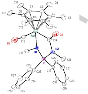
Figure 1. ORTEP diagram of 1d. Ellipsoids are shown at the 50% probability
level; hydrogen atoms are omitted for clarity. Selected bond lengths (Å) and angles (°): Ru···Arene(centroid) 1.667(1), Ru–Cl 2.415(1), Ru–N1 2.145(2), Ru–N2 2.126(3), N1–Ru–N2 68.24(9), Ru–N1–P–N2 178.43(16), Σ(N1) 357.2(6), Σ(N2) 358.2(6).
Figure 2. ORTEP diagram of the cation 2d. Ellipsoids are shown at the 50%
probability level; hydrogen atoms and the anion are omitted for clarity. Selected bond lengths (Å) and angles (°): Ru···Arene(centroid) 1.659(1), Ru–N1 2.031(2), Ru–N2 2.017(3), N1–Ru–N2 72.42(10), Ru–N1–P–N2 174.27(15), Σ(N1) 360.0(6), Σ(N2) 358.9(5).
The 18ē complex 1d exhibits a three-legged piano stool geometry with a pseudo octahedral configuration of the ligands around the ruthenium atom. The structural parameters of 1d are similar to those of its C6Me6 analogues 1a-c, except for the Ru–N
and Ru–Cl distances that are about 0.02 Å shorter (the average Ru–N and Ru–Cl bond lengths in 1a-c are 2.148–2.156(4) Å and 2.437–2.445(4) Å, respectively).[15] The p-cymene ligand is a
weaker donor and has lower steric requirements than C6Me6,
hence the lack of electron density on the ruthenium atom in 1d is compensated by shortening the bonds with the NPN and Cl ligands. The pyramidalization of the nitrogen atoms, as indicated by Σ(N), is small like for the complexes 1a,b. This is a result of the ability of the N-tolyl substituents in 1a,b,d to delocalize the unshared electron density of the nitrogen atoms, in sharp contrast with the severe pyramidalization of one of the nitrogen atoms in 1c (Σ(N1) = 344.4°), for which such delocalization is impossible. The 16ē cationic complex 2d exhibits a two-legged piano-stool geometry with the chelating NPN-ligand positioned nearly perpendicular to the p-cymene ligand. Most of the structural parameters of 2d are similar to those of its C6Me6 counterparts
2a,b, besides less significant distortion of the arene ligand. The p-cymene ligand in 2d is almost planar (Ru–C(arene) bonds in the 2.159–2.204(3) Å range) compared to the C6Me6 ring in 2a and
2b, which is bent towards a boat conformation with two considerably longer Ru–C(arene) bonds (trans to Ru–N; 2.228-2.275(2) Å) vs. the other four (2.143-2.201(2) Å).[15] This structural
peculiarity may result from weaker back-bonding from the ruthenium atom to the arene ligand in 2d.
The geometrical differences observed for the C6Me6 and
p-cymene complexes were reproduced by the density functional theory (DFT) calculation performed for 1a, 1d, 2a and 2d in the gas phase with PBE functional and def2-TZVP basis set. The optimized structures differ from the experimentally determined
ones by <0.03 Å for the distances and <2° for the bond angles (ESI, Tables S7 and S8). It is noteworthy that the DFT calculations also predict the arene orientation relative to the Cl,N,N atoms in 1a and 1d (the dihedral angle Cl–Ru–Centroid-C5 differs by <3°). Coordination of the arene ligand in the 16ē complexes 2a and 2d has a well-pronounced η2-η2 character, with the Ru–C1 and Ru–
C4 bonds being ca. 0.04 Å (2d) or 0.09 Å (2a) longer than the other four Ru–C bonds, which also indicates a stronger distortion of the planar arene ring towards a boat conformation for 2a than for 2d. This pattern can be explained by the symmetry of the ruthenium orbitals interacting with the arene ring (see below).
The 16ē complexes 2a-c exhibit a moderate intensity band in the UV-vis spectra centered at 540-550 nm,[15] which was
supposed to belong to a d-d* transition. The UV-Vis spectra of 2d exhibit a similar band, although surprisingly shifted to longer wavelengths (λmax = 590 nm). According to the calculated orbital
pattern of 2a and 2d, the highest occupied molecular orbitals (HOMO) (ESI, Fig. S10) of both cations are assigned to the antibonding combination of the ruthenium dxy orbital with the A2
-symmetric group orbital of the NPN ligand (linear combination of the nitrogen atoms py orbitals) and are located at very close
energies (-7.4 eV in the gas phase and -5.1 eV in CH2Cl2 solution,
Table 1). In both 2a and 2d, the lowest unoccupied molecular orbital (LUMO) (ESI, Fig. S11) is a combination of the ruthenium unoccupied dyz orbital and occupied B2 π-orbital of the arene
ligand. Lowering the energy of the corresponding bonding orbital (HOMO-14, see ESI, Fig. S12) is responsible for the elongation of two C-C bonds (C2–C3 and C5–C6) of the η6-arene ligand. The
greater electron-donating ability of C6Me6 leads to a higher energy
LUMO by 0.4 eV in 2a (Table 1). Hence the bands observed at 540 nm (2a) and 590 nm (2d) in the UV-vis spectra appear consistent with a ligand-to-metal charge-transfer transition, as in the 16ē arene ruthenium dithiolate complexes.[17] The
HOMO-LUMO gap for 2a and 2d are calculated in CH2Cl2 as 2.05 and
1.65 eV (Table 1), respectively, thus reflecting a significant red shift for the absorption of 2d compared to 2a.
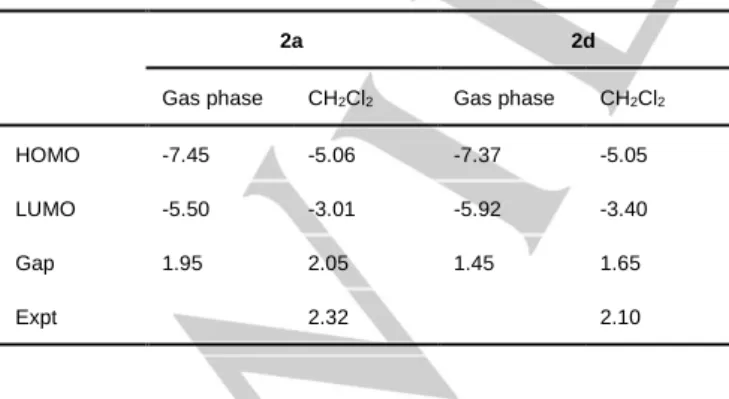
Table 1. The HOMO, LUMO energies and the HOMO-LUMO gap for 2a and 2d calculated in the gas phase and CH2Cl2 solution in comparison to the
energy of the experimentally observed band (all in eV).
2a 2d
Gas phase CH2Cl2 Gas phase CH2Cl2
HOMO -7.45 -5.06 -7.37 -5.05
LUMO -5.50 -3.01 -5.92 -3.40
Gap 1.95 2.05 1.45 1.65
Expt 2.32 2.10
It should be noted that the LUMO in complexes 2 is the orbital available to accept the electron pair donated by the external ligand (L or Cl) upon formation of the 18ē adduct. Hence, the lower energy of the LUMO in 2d is responsible for the stronger
bonding of chlorine atom in the p-cymene complex vs those containing C6Me6, as reflected by the computed and measured
Ru–Cl bond lengths (Tables S7 and S8). Coordination lability of complexes 1a-d
Despite the low symmetry (C1) of 1d in the solid state, only
two doublets at δ 5.05 and 4.87 (C6D6) were observed for the
coordinated arene in the 1H NMR spectrum, as well as two
singlets at δ 79.4, 80.7 in the 13C NMR spectrum, because of
facile rotation of the p-cymene ring around the Ru–Arene axis yielding an effective Cs symmetry in solution. The NMR spectral
feature of 2d are equally in accord with Cs-symmetry. On the other
hand, the 1H and 13C NMR spectra for the P-phenyl substituents
of 1d exhibit the expected two sets of signals in C6D6 but only one
in CDCl3, suggesting an even higher (effective C2v) point group
symmetry in the latter solvent. This can be rationalized, in addition to the rapid p-cymene ring rotation around the Ru–Arene axis, by the reversible dissociation of the Ru–Cl bond that yields an equilibrium mixture of the effectively C2v-symmetric intermediate
[2]+Cl- and undissociated 1, as has been previously proposed for
1c.[15] However, whether this process may occur in apolar
solvents and what is the dissociation enthalpy as a function of solvent, NPN substituents and type of arene ligand is still open to question. The additional studies detailed below have brought light on this point.
UV-vis spectroscopic studies of 1b and 1c in polar solvents. As we have previously reported,[15] complex 1c bearing the most
electron-releasing N-Me groups partially dissociates in the relatively polar solutions to give the corresponding cationic species [2c]+Cl- (Scheme 2), as revealed by the strong downfield
shift of its 31P NMR resonance in dichloromethane compared to
C6D6. The detailed study by means of UV-vis spectroscopy
revealed the distinct absorption bands in toluene for 1c (at 430 nm) and 2c (at 550 nm; ESI, Fig. S1A,B). The spectrum of 1c in CH2Cl2 shows a very broad absorption band located in-between
these bands (at 520 nm; Fig. 3), thus suggesting the coexistence of the dissociated and undissociated forms. The spectra are temperature dependent in the range 190-290 K, showing reversible changes with an isosbestic point at λ = 480 nm (Fig. 3). The standard enthalpy and entropy for the dissociation process (Scheme 2) were calculated from the Van‘t Hoff equation using the T-dependent molar extinction coefficient of 2c at λ = 600 nm. The dissociation process is exothermic (ΔHd = -5.0±0.2 kcal/mol),
with the cationic form dominating at low temperatures. The unexpectedly large negative entropy change (ΔSd = -27.0±0.7
cal/(mol·K)) for the dissociation process is attributed to the need to re-organize the solvent dipoles around the charged species[18]
formed upon dissociation of 1c.
Figure 3. UV-vis spectra of 1c in CH2Cl2 at 190-298 K. The insert shows the
Van’t Hoff plot of RlnKd vs 1/T.
Figure 4. UV-vis spectra of 1b in MeNO2 at 250-310 K. The insert shows the
Van’t Hoff plot of RlnKd vs 1/T.
Complex 1b, bearing an NPN-ligand with the less
electron-releasing p-tolyl substituents on the nitrogen atoms, does not show any evidence of chloride dissociation in CH2Cl2. The UV-vis
spectrum is T-independent and only the undissociated 18ē complexes is present in solution as shown by the band centered at 450 nm (ESI, Fig. S2). However, the compound predominantly exists as cationic complex [2b]+Cl- in solution of the highly polar
MeNO2 solvent, exhibiting a band with λmax = 515 nm (Fig. 4).
Analysis of the temperature dependence of this band in the range 250-310 K gave ΔHd = 3.5±0.2 kcal/mol and ΔSd = +0.3±0.6
cal/(mol·K). Compared to the dissociation of 1c in CH2Cl2, the
more positive dissociation entropy of 1b in MeNO2 is attributed to
a more efficient solvation of the charged species by MeNO2. On
the other hand, both 1a and 1d, which contain the least donating NPN-ligand, do not noticeably dissociate even in MeNO2 as
suggested by the absorption band at 450 nm in their visible spectra, even though the insufficient solubility of both compounds did not allow verification of the temperature independence of these spectra.
NMR study of 1a-d in apolar solvents. The chloride complexes 1a-c show two inequivalent P-Ph groups in their RT-NMR spectra in apolar solvents (benzene, toluene), meaning they exist as Cs
-symmetric 18ē complexes on the NMR timescale.[15] We now
report that heating the solution of 1c in C6D6 to 338 K gives one
set of signals for these groups, which is indicative of exchange. The coalescence of the two ortho-protons signals at δ 7.70 and
7.98 is observed at 323±5 K (Tc) (ESI, Fig. S3). From this
temperature and from the chemical shift difference, the rate constant (kex = 380 s-1) and activation free energy (ΔGex≠ =
15.2±0.3 kcal/mol) for the degenerative exchange process can be calculated.[19] For complex 1b with a less donor NPN-ligand,
coalescence for the P-Et methyl resonances at δ 0.40 and 1.39 could not be reached below the boiling point of toluene-d8 (ESI,
Fig. S4). However, we observed slow exchange of the ethyl groups for this compound in C6D6 and in toluene-d8 by the 2D
EXSY 1H NMR method (Fig. 5),[20] yielding the rate constants of
1.8 s-1 and 1.4 s-1 at 293 K, respectively (Table 2, lines 3,7). The
activation parameters for the exchange process were obtained from the temperature dependence of the exchange rate constants (kex) in toluene-d8 in the range 230–315 K: ΔHex≠ = 8.4±0.2
kcal/mol and ΔSex≠ = -29.2±0.7 cal/(mol·K) (ESI, Fig. S5, Table
S1).
Figure 5. 2D EXSY 1H NMR of 1b in toluene-d
8 at 291K and the mixing time of
Table 2. Exchange rate constant kex (s-1) and activation free energy ΔGex≠ (kcal/mol) for the P-bound substituent exchange in 1a, 1b and 1d from the 2D EXSY 1H
NMR investigations.
Complex Solvent Additives C(Ru), mM C(add), mM T, K kex, s-1 ΔGex≠, kcal/mol
1 1a C6D6 - 10 293 <0.1 >18.5 2 1a C6D6 H2O 10 10 294 0.79 17.4 3 1b C6D6 - 20 293 1.8 16.8 4 1b C6D6 H2O 20 5 295 >15 <15.7 5 1b Toluene - 40 295 1.6 17.0 6 1b Toluene - 8 295 1.6 17.0 7 1b Toluene - 46 293 1.4 17.0 8 1d C6D6 10 293 <0.1 >18.5 9 1d C6D6 H2O 10 0.7 293 0.72 17.3 10 1d C6D6 H2O 10 2.4 293 2.6 16.6 11 1d C6D6 Et4N+Cl- 10 0.7 292 >11 <15.7
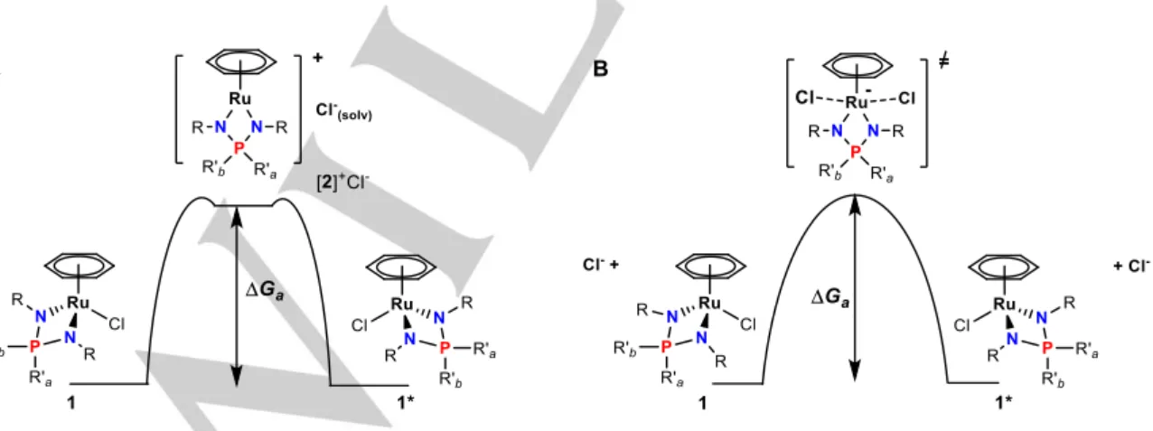
A large negative value of the activation entropy evidences a highly ordered transition state in the exchange process. The exchange between R’a and R’b substituents occurs via a
configuration inversion at the Ru atom, which can most easily be envisaged by moving the chloride ligand from one coordination side to the opposite one by ligand exchange processes. Generally, such ligand exchange may proceed either via a dissociative mechanism with a cationic 16ē intermediate (pathway A in Fig. 6A) or via a SN2-like associative mechanism through a 20e
transition state (pathway B in Fig. 6B). In the latter case, however, configuration inversion with exchange of the phosphorus R’a and
R’b substituents could take place only when the entering ligand is
Cl-. Given that the NMR and UV studies outlined above were
carried out in non-coordinating solvents and coordinating ligands L are not present, the exchange in 1b most likely takes place via the dissociated complex [2b]+Cl-, which requires an entropically
unfavorable solvation of the chloride-ion by apolar molecules (Fig. 6A), similarly to the dissociation of 1c in CH2Cl2 described above.
We have further confirmed that the exchange rates are independent on the concentration of 1b (1.6 s-1 in both 40 mM and
8 mM toluene-d8 solutions at 295 K, Table 2, lines 5,6). The
P-substituent exchange for complexes 1a and 1d, which bear the least electron-donating NPN-ligand, is not observed by 2D EXSY
1H NMR even at t
m = 1 s, hence the rates are slower than 0.1 s-1
and the activation free energies (ΔGex≠) are greater than 18.5
kcal/mol.
In principle, addition of Cl- should either slow down or
accelerate the exchange depending on whether the mechanism is A or B, respectively. Addition of 0.07 equiv. of Et4N+Cl- to 1d
strongly enhances the exchange process (kex > 11 s-1). This result
strongly suggests that the exchange occurs by the associative mechanism B under these conditions (Table 2, line 11, and ESI, Fig. S6). Therefore, it seems that both mechanisms A and B are possible, the former in the absence of added Cl- and the latter in
its presence.
Assistance by an external ligand L may also result in Cl- release.
While the subsequent attack of the same Ru-L intermediate by Cl
-would not result in any P substituent exchange, attack of another Ru-Cl complex achieves a degenerative self-exchange with configuration inversion and P substituent exchange (Fig. 7). Thus, the P-phenyl group exchange for these compounds becomes observable after adding water to the C6D6 solutions (cf. lines 1,2
for 1a and lines 8,9 for 1d) and the rate constants are found to
increase with the increase of the water content (Table 2, lines 9,10; ESI, Fig. S7). For the 1d sample with [H2O] = 0.7 mM (0.07
equiv.), the activation parameters could be derived from the temperature dependence (291-335 K) of kex (ESI, Fig. S8, Table
S2). The calculated activation enthalpy[21] ΔH
ex≠ = 11.8±0.2
kcal/mol of the associative exchange can be used as a lower limit for the ΔHex≠ in dissociative exchange of 1d in the absence of
external ligands. It is pertinent to underline that water may have a double role in the promotion of the associative chloride exchange: as a ligand to replace Cl- in the coordination sphere of the Ru
atom, and as a proton donor to stabilize the dissociated Cl- by
H-bonding. Interestingly, the effect of water for 1a is much smaller; a rate constant of 0.79 s-1 is achieved only in the presence of an
equimolar amount of H2O in C6D6. Perhaps the sterically bulky
C6Me6 ligand hampers the Cl- or external ligand coordination thus
increasing the activation free energy.
Figure 7. Associative exchange promoted by water-induced chloride dissociation.
We would like to underline that the activation enthalpy ΔHex≠
for the exchange process in dissociative mechanism A should be very close to the Ru–Cl bond dissociation enthalpy, ΔHd. The
latter depends predominantly on the Ru–Cl bond strength and on the solvent polarity. Hence, the ΔHd value for complex 1c may be
estimated from its ΔG≠ at 323 K as ΔH
d ~ ΔHex≠ = ΔGex≠ + TΔSex≠,
where ΔSex≠ can be assumed to be close to that for 1b (-29.2
cal/(mol·K)). This gives an estimate for the ΔHd of 1c as ca. 5.7
kcal/mol. Analogously, the ΔHd of 1a and 1d can be estimated as
>10 kcal/mol. Thus, we have proved that the Ru–Cl bond in 18ē arene ruthenium iminophosphonamide complexes can dissociate even in apolar solvents. The ΔHd for 1a-d expectedly lowers with
an increase of the ligand donating ability: over 10 kcal/mol (1a, 1d) > 8.4 kcal/mol (1b) > 5.7 kcal/mol (1c).
Coordination of ligands to the 16e cationic complexes 2 We have previously mentioned that the 16ē cationic complex 2a interacts with MeCN and CO, but failed to isolate these unstable adducts.[15] We have now studied the activity of the 16ē
complexes 2 towards coordination of external ligands L (MeCN, pyridine, CO) in detail by means of UV-Vis and NMR spectroscopies (Scheme 3) to determine the thermodynamics of the process and with regard to the conditions allowing isolation. Since 1a is, among the 18ē chloride complexes 1, the least prone to chloride dissociation, the corresponding 16ē complex 2a is expected to display the highest affinity toward saturation by coordination of L ligands. Hence, the coordination of relatively weak donors MeCN and Py was studied for 2a only.
Scheme 3.
The titration of 2a with MeCN in CH2Cl2 at 296 K with UV-Vis
monitoring shows that the intensity of the 535 nm band does not significantly change until >100 equiv. of MeCN are added (ESI, Fig. S9A), while a similar amount of pyridine significantly extinguishes this band to give rise a new band of the pyridine adduct at 445 nm (ESI, Fig S9B). The estimated equilibrium constants Kc are <2 M-1 for 2a·MeCN and 32±7 M-1 (from UV-vis),
37±1 M-1 (from NMR, ESI, Fig. S9C) for 2a·Py, (ESI, Tables
S3-S5). The UV-vis measurements carried out for the 1:2 mixture of 2a:Py in the temperature range 200-296 K show that 2a dominates at room temperature, while the equilibrium shifts towards formation of the 18ē adduct 2a·Py at lower temperatures. The conversion becomes essentially complete below 220 K (Fig. 8). The analysis of the Van’t Hoff’s plot yields ΔHc = -12.4±0.5
kcal/mol and ΔSc = -36±2 cal/(mol·K). Thus, pyridine coordination
required to observe coordination at room temperature because of high association entropy. This is in sharp contrast with the previously reported irreversible coordination of pyridine to the 16ē arene ruthenium amidinate complex [(C6H6)Ru(t
BuN-C(Ph)-NtBu}](BArF 4).10
Figure 8. UV-vis spectra of reversible coordination of pyridine to 2a in CH2Cl2
at 200-296K. The insert shows the Van’t Hoff dependence of RlnKc vs 1/T.
In comparison with acetonitrile and pyridine, carbon monoxide is a stronger π-acceptor and reacts readily with 2a to give the CO adduct 3a. However, the reaction is reversible and facile CO decoordination occurs upon solvent evaporation to give back 2a. Nevertheless, 3a could be isolated by precipitation from its CH2Cl2
solution upon addition of excess diethyl ether. Analogously, the reaction of 2d with CO resulted in formation of 3d, which is stable only under a CO atmosphere. This product was isolated, like 3a, by using CO-saturated CH2Cl2 solution and diethyl ether.
Although 3a and 3d slowly evolve CO in the solid state under vacuum, satisfactory elemental analyses were obtained for both. It should be noted that 3a is stable in a CO-saturated solution, whereas 3d degrades within days to unknown arene-free carbonyl complexes, perhaps similar to the previously observed ones resulting from the decomposition of [(p-Cymene)Ru(CO)(SXyl)2].[22] The molecular structures of 3a,d
were determined by single crystal X-ray diffraction (ESI, Table S6). Selected structural parameters and views of the molecules are shown in Figures 9 and 10.
Figure 9. ORTEP diagram of the cation 3a. Ellipsoids are shown at the 50%
probability level. The hydrogen atoms and the anion are omitted for clarity. Selected bond lengths (Å) and angles (°): Ru···Arene(centroid) 1.769(1), Ru– C(O) 1.871(3), Ru–N1 2.112(2), Ru–N2 2.116(2), C–O 1.145(3), N1–Ru–N2 69.20(9), Ru–N1–P–N2 164.60(15), Σ(N1) 348.3(5), Σ(N2) 359.3(5).
Figure 10. ORTEP diagram of the cation 3d. Ellipsoids are shown at the 50%
probability level. The hydrogen atoms and the anion are omitted for clarity. Selected averaged bond lengths (Å) and angles (°): Ru···Arene(centroid) 1.768(3), Ru–C(O) 1.879(9), Ru–N1 2.095(7), Ru–N2 2.120(6), C–O 1.145(10), N1–Ru–N2 68.9(3), Ru–N1–P–N2 165.8(4), Σ(N1) 353.7(13), Σ(N2) 355.3(14).
Complexes 3a and 3d are the first crystallographically characterized arene ruthenium carbonyl derivatives with any κ2
-N,N-anionic chelate ligand. The asymmetric unit of 3d contains two independent molecules, which exhibit identical metric parameters within experimental error. Both complexes exhibit a three-legged piano stool geometry with a pseudo octahedral configuration of the ligands around the ruthenium atom. The coordinated arene adopts a nearly staggered (3a) or eclipsed (3d) conformation relative to the Ru-CO axis, with C4– Arene(centroid)–Ru–CO angles of 25.7° and 5.9–6.4°, respectively. The presence of the CO ligand significantly increases the Ru–Arene(centroid) distance by ~0.1 Å in both 3a and 3d relative to 2a (1.662(2) Å) and 2d. The Ru–C(arene)
distances vary in the range 2.208–2.333(3) Å with the longest bonds being trans to the CO ligand. The C–C bonds length (1.389(12)–1.452(4) Å) in the coordinated arene slightly alternate with the shortest C–C bond (1.389–1.403 Å) located trans to the CO ligand. A similar Ru–Arene(centroid) (1.810 Å), Ru–C(O) (1.846(4) Å) and carbonyl C–O (1.133(7) Å) distances, as well as elongations for the two Ru–C(arene) bonds trans to CO (2.389– 2.396 Å) were previously reported for the arene ruthenium dithiolate carbonyl complex [(η6-C
6Me6)Ru(CO)(S2C6H4)] (4).[17]
The Ru–N bonds are shorter than those in the neutral 18ē complexes 1a-d by ca. 0.04 Å, reflecting the presence of the positive charge, but longer (by 0.05-0.08 Å) than in the 16ē complexes 2a-d, in which they are strengthened by π-donation
from the NPN-ligand. The chelate N1–Ru–N2 angles in 3a and 3d are similar to those of the other 18ē NPN-complexes 1a-d. However, unlike in the related chloride complexes 1a and 1d, the Ru–N1–P–N2 metallacycles in 3a and 3d are folded by 13–15.6° from planarity at the N1∙∙∙N2 hinge and the nitrogen atoms (N1 in 3a; N1 and N2 in 3d) are noticeably pyramidalized as shown by the Σ(N) parameter. There are several close H∙∙∙F contacts between the cation and the PF6- anion in both structures, although
none of these is shorter than the sum of the van der Waals radii of H and F (2.56 Å).[23]
Complexes 3a and 3d are sufficiently stable in concentrated solutions to allow recording their NMR spectra, although the fraction of the corresponding 16ē complexes increases with time. The 31P NMR resonance (δ 61.7 for 3a and δ 60.2 for 3d) is
downfield shifted by ca. 10 ppm relative to the corresponding 16ē cationic complexes 2a and 2d. In the 1H and 13C{1H} NMR spectra
recorded in CDCl3 there are two sets of signals for the
magnetically inequivalent phenyl groups at the phosphorus atom, as expected for the Cs-symmetric complexes, hence the
decoordination of CO is a slow process with a relatively high activation barrier. The resonance of the coordinated CO ligand in the 13C NMR spectra is observed at high field (δ 199.2, 193.8), as
is typical of terminal linear coordinated CO ligands (cf. to δ 197.8
for 4[17] and δ 203-208 reported for the cyclopentadienyl
ruthenium complexes with N,N-ligands).[9, 24]
Both carbonyl adducts 3a and 3d exhibit strong CO stretching vibration band in the IR spectrum at 1984 cm-1 (3a) and 2012 cm -1 (3d). The higher ν
CO in 3d evidences weaker Ru-CO
π-back-bonding relative to 3a that possesses the electron-richer C6Me6
arene. These frequencies are substantially lower than those of the more electron-deficient arene ruthenium amidinate carbonylic complex [(η6-C
6H6)Ru(CO){PhC(NtBu)2}](BArF4) (νCO = 2050 cm -1),[10] but closer to those of the arene ruthenium complex with
dianionic ditiolate ligands 4 (νCO = 1951 cm-1),[17] [(η6
-C6Me6)Ru(CO)(SXyl)2] (νCO = 1965 cm-1)[22] and of the
cyclopentadienyl complex with neutral κ2-N,N-ligand [(η5
-C5H5)Ru(CO)(TMEDA)](BAr4) (νCO = 1968 cm-1).[24]
Surprisingly, bubbling CO into the solution of 2c yielded a stable product, 3c, resulting from the addition of two CO molecules, one of which has inserted into one of the two Ru–N bonds. The product has been fully characterized, including by single crystal X-ray diffractometry (Figure 11, Table S6 in ESI).
Figure 11. ORTEP diagram of the cation 3c. Ellipsoids are shown at 30%
probability level. The hydrogen atoms and the anion are omitted for clarity. Selected bond lengths (Å) and angles (°): Ru···Arene(centroid) 1.828(1), Ru– N1 2.107(2), Ru–C(O)N 2.042(3), C=O 1.211(4), N–C(O) 1.414(3), Ru–C(O) 1.859(3), C–O 1.143(4), N1–Ru–C(O) 83.15(10), Σ(N1) 349.7(5), Σ(N2) 359.3(6).
The overall geometry of 3c is similar to that of 3a,d except for the expanded metallacycle with a CO group. The addition of two CO molecules leads to a significant increase of the Ru– Arene(centroid) distance relative to 2c by ca. 0.18 Å as a result of elongation of the Ru–C(arene) bonds trans to the terminal CO (2.299–2.312(3) Å) and to the carbamoyl C=O group (2.373– 2.381(3) Å). The arene C–C bonds trans to the terminal CO (1.404(6) Å) and to C=O group (1.392(5) Å) are shorter than the other four bonds (1.417–1.438(5) Å). Similarly to 3a and 3d, the terminal carbonyl group is linear. The carbamoyl C=O and C–N bonds have similar length to or are slightly longer than those in organic amides. The carbamoyl N2 atom is planar, while N1 is considerably pyramidalized as in the corresponding 18ē chloride complex 1c. There are two intermolecular close contacts in the structure of 3c that fall below the sum of the van der Waals radii,[23] implicating hydrogen atoms of the C
6Me6 ligand and either
a PF6- F atom or the carbamoyl oxygen atom, H(8A)∙∙∙F (2.269 Å)
and H(10B)∙∙∙O(2)C (2.418 Å), respectively.
The 13C{1H} NMR spectrum of 3c reveals two signals
corresponding to carbonyl C nuclei: a singlet at δ 198.2 and a
doublet at δ 192.8 (2JСР = 19.6 Hz). All the N- and P-substituents
in 3c are inequivalent and give rise to two sets of signals for both methyl and phenyl groups in the 1H and 13C{1H} spectra. In the IR
spectra 3c shows two strong carbonyl bands νCO at 1984 cm-1
(metal bound CO) and at 1644 cm-1 (carbamoyl C=O), the latter
frequency being typical of organic amides.
Apparently, the carbonylation of 2c is a two-step reaction involving: 1) coordination of a first CO molecule to give an intermediate C, which is structurally similar to 3a,d; and 2)
insertion of the terminal CO into a Ru–N bond and subsequent occupation of the resulting vacant site at the ruthenium atom by a second CO molecule (Scheme 4). This conclusion is further supported by the results of an IR spectroscopic monitoring at low temperatures (Fig. 12). Bubbling CO at -40°C for 10 s fully converts 2c to an orange complex characterized by a strong CO band at 1975 cm-1, which can be attributed to the mono-adduct C.
The latter is unstable: even at -40°C and in the absence of additional CO it slowly disappears to be replaced by 3c. The coordination of the first CO molecule is reversible, hence dissociation from the mono-adduct C provides the needed CO for the second carbonylation step. The rearrangement is complete within a few minutes at low temperatures and yields a 1:1 mixture of 2c and 3c, which were isolated upon evaporation of the solvent.
Scheme 4.
Figure 12. The reaction of 2c with CO monitored by IR. The sample was
dissolved in CH2Cl2 at -40°C and CO was bubbled for 10 s. The spectra were
recorded (a) immediately after the preparation, (b) after 40 s at room temperature, (c) after 5 min of keeping (a) at -40°C, (d) after evaporation of (c) and redissolution in CH2Cl2.
The peculiarity of the mono-carbonyl intermediate C is the high nucleophilicity of the nitrogen atoms, which is related to the presence of more electron-donating alkyl substituents. Although facile CO-insertion into M–N bonds is a known phenomenon for certain metal aminopyridinates,[25] triazenides,[26] amidinates,[27-30]
and aminophosphines[31] this reaction has not been previously
reported to the best of our knowledge for arene ruthenium complexes. The process that most resembles the formation of 3c involves a carbene insertion into a Ru–N bond in the arene ruthenium amidinate complex [(C6Me6)Ru{(iPrN)2CMe}](PF6),
followed by CO coordination to give [(C6Me6)Ru(CO){(i
PrN)-CMe(NiPr)CHSiMe
3}](PF6) (5).[32] The spectral and structural data
of 5 are similar to those of 3c: the νCO is observed at 1963 cm-1
and the carbonyl resonance appears at δ 203 in the 13C NMR
spectrum; the Ru–Arene(centroid) distance is 1.828 Å with the elongation of the Ru–C(arene) bonds trans to CO and to the inserted carbene to 2.320–2.362 Å; the CO ligand is linear (Ru–
C–O is 170.2°), and the Ru–CO and C–O bond lengths are 1.836 Å and 1.159 Å, respectively.
Conclusions
Our detailed spectroscopic study has allowed to investigate the stabilization of the coordinative unsaturation in half-sandwich ruthenium iminophosphonamide complexes depending on the N-,P-substituents, arene and other co-ligands. By 2D EXSY 1H
NMR spectroscopy we have proven that the dynamic exchange process observed for chlorides 1a-d is attributed to the dissociation of the chloride ligand via a 16ē intermediate in both polar and apolar solvents. The chloride dissociation enthalpy (ΔHd) determined for the 18ē chloride complexes 1a-d and the Cl
-coordination enthalpy (ΔHc) to the 16ē complex 2a quantitatively
estimate the stabilization effect of the NPN ligand, which is mostly provided by highly localized negative charges at the nitrogen atoms via additional π-donation. Hence the NPN ligands with
N-alkyl groups destabilize the 18ē complexes and compensate the metal electron deficiency much more effectively than the NPN ligands with N-aryl groups. Thus, the N-Me chloride complex 1c dissociates exothermically (ΔHd = -5.0 kcal/mol) even in CH2Cl2,
whereas the dissociation is always endothermic for the N-Tol complexes (1a, 1b and 2d), even in polar nitromethane. On the other hand, the less donating arene ligand (p-cymene vs C6Me6)
affects the stability of the 18ē complexes counterintuitively: it destabilizes the carbonyl adducts due to the lack of electron density provided for π-backdonation (3d vs 3a), but enhances the stability of the chloride complex (1d vs 1a) due to lower energy of the orbital corresponding to Ru-Cl bond. The coordination of ligands to 16ē complexes 2 appears to be unambiguously exothermic, while the highly negative association entropy makes the 18ē complexes stable only at low temperatures. Nevertheless, stronger π-acceptors (CO > pyridine > MeCN) give more stable adducts due to significant contribution of π-backdonation to Ru-L bonding; this fact allowed us to isolate and structurally
characterize complexes 3a and 3d, which are rare examples of cationic 18ē arene ruthenium carbonyl complexes. The unexpectedly facile insertion of CO into the Ru–N bond observed in the 18ē adduct 2c∙CO to give the carbonyl-carbamoyl complex 3c proves that arene ruthenium iminophosphonamide complexes can react in diverse manners and suggests that they have potential for application in organic synthesis and (bifunctional) catalysis. Further investigation of these complexes in catalytic transfer hydrogenation is in progress and will be reported soon.
Experimental Section
General procedures. All manipulations were carried out using standard
Schlenk techniques under an atmosphere of dry argon. Absolute solvents were used for both synthesis and spectroscopic studies; solvents were purified by standard methods and distilled prior to use. The 1H, 31P and 13C
NMR spectra were obtained on Bruker Avance 600 or Bruker Avance 400 spectrometers and referenced to the residual signals of deuterated solvent (1H and 13C), and to 85% H
3PO4 (31P, external standard). The UV spectra
were recorded on a Varian Cary 50 WinUV spectrometer in quartz cells (l = 2.2 mm or 10 mm). The IR spectra were obtained on a Fourier spectrometer Nicolet 6700 in KBr cells (l = 0.514 mm). The elemental analyses were carried out on a Carlo Erba 1106 CHN analyzer. The following compounds were prepared according to described procedures: [(η6-p-Cymene)RuCl
2]2,[33] Ph2P(N-p-Tol)(NH-p-Tol) (A),[15] 1a-c, 2a-c.[15] Synthesis of [(η6-p-Cymene)RuCl{Ph
2P(N-p-Tol)2}] (1d). To a solution
of A (0.79 g, 2.00 mmol) in benzene (60 mL) a 2.0 M solution of NaHMDS in THF (1.10 mL, 2.20 mmol) was added and the resulting solution was stirred for 2 h. Then solid [(η6-p-Cymene)RuCl2]2 (0.61 g, 1.00 mmol) was
added and the reaction mixture was stirred overnight. The reaction mixture was filtered and the solvent from the filtrate was removed under reduced pressure. The residue was washed with hexane (2x10 mL), Et2O (2x5 mL),
and then recrystallized from hot benzene (20 ml). The dark-red crystalline was filtered off, washed with Et2O (5 mL) and dried in vacuo. Yield 1.00 g
(88%). Anal. calcd for C36H38ClN2PRu: C, 64.90; H, 5.75%. Found: C,
64.85; H, 5.84%. 31P NMR (CDCl3): δ 43.8 (s, PPh2). 1H NMR (CDCl3): δ
7.87 (dd, 3J
HP = 10.8, 3JHH = 8.0, 4H, o-HPh), 7.50 (m, 2H, p-HPh), 7.39 (m,
4H, m-HPh), 6.90 (d, 3JHH = 8.0, 4H, C6H4(Tol)), 6.82 (d, 3JHH = 8.4, 4H,
C6H4(Tol)), 6.31 (d, 3JHH = 5.6, 2H, C6H4(Cym)), 5.41 (d, 3JHH = 5.6, 2H,
C6H4(Cym)), 2.89 (sept, 3JHH = 6.8, 1H, CHMe2), 2.19 (s, 6H, MeTol), 2.11
(s, 3H, MeCym), 1.21 (d, 3JHH = 6.8, 6H, CHMe2). 31P NMR (C6D6): δ 42.9. 1H NMR (C6D6): δ 8.03 (m, 4H, o-HPh), 7.27 (d, 3JHH = 7.8, 4H, C6H4(Tol)),
7.22 (m, 3H, (m+p)-HPh), 6.93 (d, 3JHH = 7.8, 4H, C6H4(Tol)), 6.75 (m, 3H,
(m+p)-HPh’), 5.05 (d, 3J
HH = 6.0, 2H, C6H4(Cym)), 4.87 (d, 3JHH = 6.0, 2H,
C6H4(Cym)), 2.88 (sept, 3JHH = 6.6, 1H, CHMe2), 2.15 (s, 6H, MeTol), 1.86
(s, 3H, MeCym), 1.05 (d, 3JHH = 6.8, 6H, CHMe2). 13C NMR (CDCl3): δ 145.8
(d, 2JСР = 4.4, i-C
Tol(N)), 133.4 (br.s, о-CPh), 131.9 (d, 4JСР = 2.7, p-CPh),
129.0 (s, β-CHTol), 128.2 (d, 3JСР = 11.2, m-CPh), 127.5 (s, i-CTol(Me)),
122.9 (d, 3JСР = 9.7, α-CH
Tol), 102.0 (s, i-CCym), 94.8 (s, i-CCym), 80.5 (s,
CHCym), 79.6 (s, CHCym), 30.9 (s, CHMe2), 22.5 (s, СHMe2), 20.5 (s, MeTol),
18.9 (s, MeCym). 13C NMR (C6D6): δ 146.9 (s, 2JСР = 4.2, i-CTol(N)), 136.5
(d, 1J CP = 95.1, i-CPh), 135.0 (d, 2JCP = 11.0, o-CPh), 132.6 (d, 2JСР = 9.8, o-CPh’), 132.0 (d, 4JСР = 2.7, p-C Ph), 131.8 (d, 4JСР = 2.9, p-CPh’), 130.2 (d, 1J CP = 84.0, i-CPh’), 129.4 (s, β-CHTol), 128.6 (d, 3JСР = 12.5, m-CPh), 128.5
(s, i-CTol(Me)), ~127.7 (overlapped, m-CPh’), 123.9 (d, 3JСР = 9.7, α-CH Tol),
102.4 (s, i-CCym), 94.8 (s, i-CCym), 80.7 (s, CHCym), 79.4 (s, CHCym), 31.2 (s,
CHMe2), 22.5 (s, СHMe2), 20.7 (s, MeTol), 18.8 (s, MeCym). UV-vis (CH2Cl2;
λmax, nm; ε, M-1 cm-1): 450 (450, shoulder).
Synthesis of [(η6-p-Cymene)Ru{Ph
2P(N-p-Tol)2}](PF6) (2d). To a
solution of 1d (0.39 g, 0.59 mmol) in CH2Cl2 (15 ml), solid AgPF6 (0.16 g,
0.62 mmol) was added, causing the color to immediately change from red to deep violet. The reaction mixture was stirred for 2 h and then filtered through a bed of Celite. The solvent was removed under reduced pressure, and the residue was dried in vacuo to give violet-black 2d. Yield 0.41 g (91%). Anal. calcd for C36H38F6N2P2Ru: C, 55.74; H, 4.94%. Found: C,
55.47; H, 4.99%. 31P NMR (CDCl 3): δ 71.3 (s, PPh2), -144.1 (sept, JPF = 712, PF6-). 1H NMR (CDCl3): δ 7.65 (m, 2H, p-HPh), 7.52 (m, 4H, m-HPh), 7.47 (m, 4H, o-HPh), 7.02 (d, 3JHH = 8.0, 4H, C6H4), 6.84 (dd, 3JHH = 8.0, 4J HP = 1.2, 4H, C6H4), 5.88 (d, 3JHH = 6.8, 2H, C6H4(Cym)), 5.84 (d, 3JHH =
6.8, 2H, C6H4(Cym)), 2.68 (sept, 3JHH = 6.8, 1H, CHMe2), 2.26 (s, 3H,
MeCym), 2.25 (s, 6H, MeTol), 1.31 (d, 3JHH = 6.8, 6H, CHMe2). 13C NMR
(CDCl3): δ 144.1 (d, 2JСР = 3.6, i-CTol(N)), 134.4 (d, 4JCP = 2.2, p-CPh), 134.1
(s, i-CTol(Me)), 132.4 (d, 2JCP = 10.3, о-CPh), 130.2 (s, β-CHTol), 129.5 (d, 3JСР = 12.5, m-C
Ph), 124.7 (d, 1JCP = 89.4, i-CPh), 123.7 (d, 3JСР = 8.0, α-CHTol), 99.6 (s, i-CCym), 89.6 (s, i-CCym), 81.0 (s, CHCym), 78.7 (s, CHCym),
31.8 (s, CHMe2), 22.8 (s, СHMe2), 20.9 (s, MeTol), 19.7 (s, MeCym). UV-vis
(CH2Cl2; λmax, nm; ε, M-1 cm-1): 590 (1880).
Reaction of 2a,2c,2d with CO. General procedure. A stream of CO was
slowly bubbled through a stirred solution of 2a (0.12 g, 0.15 mmol) in CH2Cl2 (5 mL) for 10 min. The color quickly changed from deep violet to
orange. The product was precipitated with Et2O (20 ml) as a yellow-orange
crystalline solid, which was filtered off and dried in vacuo for 1 hr. Yield 0.12 g (96%). Anal. calcd for C39H42F6N2OP2Ru: C, 56.32; H, 5.09%.
Found: C, 56.34; H, 4.95%. 31P NMR (СD 2Cl2): δ 61.7 (s, PPh2), -144.5 (sept, 1J PF = 713, PF6-). 1H NMR (СD2Cl2): δ 7.84 (t, 3JHH = 8.6, 1H, p-HPh), 7.81 (dd, 3J HP = 12.8, 3JHH = 8.0, 2H, o-HPh), 7.69 (dt, 3JHH = 7.2, 4JHP = 3.2, 2H, m-HPh), 7.48 (dt, 3JHH = 7.2, 5JHP = 1.6, 1H, p-HPh’), 7.24 (dt, 3J HH = 8.0, 4J HP = 3.2, 2H, m-HPh’), 7.19 (dd, 3J HP = 10.8, 3JHH = 8.0, 2H, o-HPh’), 6.90 (d, 3J HH = 8.0, 4H, C6H4(Tol)), 6.46 (dd, 3JHH = 8.0, 4JHP = 2.0, 4H, C6H4(Tol)), 2.21 (s, 6H, MeTol), 2.11 (s, 18H, C6Me6). 13C NMR (СD2Cl2): δ 199.2 (s, Ru-CO), 140.8 (d, 2J СР = 2.2, i-CTol(N)), 135.8 (d, 1JСР = 97.2, i-CPh), 134.9 (d, 4JСР = 2.6, p-CPh), 133.6 (d, 4JСР = 2.7, p-CPh’), 132.9 (d, 2JСР = 9.3, o-C
Ph), 132.7 (d, 2JСР = 11.0, o-CPh’), 132.8 (s, i-CTol(Me)), 130.4
(d, 4J
СР = 1.3, β-CHTol), 130.0 (d, 3JСР = 12.1, m-CPh), 128.9 (d, 3JСР = 11.4, m-CPh’), 128.3 (d, 1JСР = 83.3, i-CPh’), 126.0 (d, 3JСР = 7.0, α-CH
Tol), 110.3
(s, C6Me6), 20.8 (s, MeTol), 17.1 (s, C6Me6). UV-vis (CH2Cl2; λmax, nm; ε, M -1 cm-1): 450 (100, shoulder). IR (CH
2Cl2, ν, cm-1): 1984 (RuСO).
Analogously, from 2c (0.10 g, 0.15 mmol) in CH2Cl2 (3 ml), complex 3c
was obtained as a stable yellow crystalline solid. Yield 0.10 g (95%). Anal. calcd for C28H34F6N2O2P2Ru•H2O: C, 46.35; H, 5.00%. Found: C, 46.49; H,
5.00%. 31P NMR (CD 2Cl2): δ 55.2 (s, PPh2), -144.5 (sept, 1JPF = 713, PF6-). 1H NMR (CD 2Cl2): δ 7.83 (ttd, 3JHH = 7.8, 4JHH = 1.8, 5JHP = 1.2, 1H, p-HPh), 7.75 (ttd, 3J HH = 7.8, 4JHH = 1.2, 5JHP = 1.2, 1H, p-HPh’), 7.71 (dt, 3J HH = 7.8, 4J HP = 3.6, 2H, m-HPh), 7.63 (ddd, 3JHP = 12.6, 3JHH = 7.8, 4JHH = 1.2, 2H, o-HPh), 7.60 (dt, 3JHH = 7.8, 4JHP = 3.6, 2H, m-HPh’), 7.42 (ddd, 3J HP = 12.6, 3J HH = 7.8, 4JHH = 1.2, 2H, o-HPh’), 2.73 (d, 3J HP = 8.4, 3H, NMe(CO)), 2.64 (d, 3J HP = 17.4, 3H, NMe), 2.24 (s, 18H, C6Me6). 13C NMR (CD2Cl2): δ 198.2 (s, Ru-CO), 192.8 (d, 2JСР = 19.6, NC=O), 135.1 (d, 4JСР = 2.4, p-C Ph), 135.0 (d, 4J СР = 2.4, p-CPh’), 133.6 (d, 2J СР = 10.6, o-CPh’), 132.6 (d, 2J СР = 10.6, o-CPh), 130.3 (d, 3JCP = 12.8, m-CPh), 130.2 (d, 3JСР = 12.6, m-CPh’), 124.2 (d, 1J CP = 112.2, i-CPh), 122.2 (d, 1JCP = 98.6, i-CPh’), 113.4 (s, i-C6Me6), 39.3 (d, 3JСР = 4.2, NMe), 29.3 (d, 3J СР = 8.4, NMe(CO)), 17.0 (s, C6Me6),. IR (CH2Cl2, ν, cm-1): 1983 (RuCO), 1644 (RuC(O)N).
Analogously, carbonylation of 2d (0.11 g, 0.14 mmol) gave a red solution of 3d. The product was precipitated by Et2O (20 ml) saturated with CO,
filtered and dried in vacuo for 30 min. Yield 0.09 g (82%). Anal. calcd for C37H38F6N2OP2Ru: C, 55.29; H, 4.77%. Found: C, 55.05; H, 4.86%. 31P
NMR (CDCl3): δ 60.2 (s, PPh2), -144.2 (sept, 1JPF = 713, PF6-). 1H NMR
1H, p-HPh), 7.73 (dt, JHH = 7.6, JHP = 2.8, 2H, m-HPh), 7.50 (t, JHH = 7.2, 1H, p-HPh’), 7.35 (dd, 3J HP = 10.2, 3JHH = 7.2,2H, o-HPh’), 7.30 (dt, 3J HH = 7.6, 3J HH = 3.2, 2H, m-HPh’), 6.87 (d, 3J HH = 8.0, 4H, C6H4(Tol)), 6.51 (d, 3J
HH = 8.0, 4H, C6H4(Tol)), 6.29 (br. s, 4H, C6H4(Cym)), 2.65 (sept, 3JHH =
6.8, 1H, CHMe2), 2.18 (s, 6H, MeTol), 2.11 (s, 3H, MeCym), 1.22 (d, 3JHH =
6.8, 6H, CHMe2). 13C NMR (CDCl3): δ 193.8 (s, Ru-CO), 143.1 (d, 2JСР = 3.4, i-CTol(N)), 134.5 (d, 4JСР = 2.6, p-CPh), 133.7 (d, 4JСР = 2.4, p-CPh’), 132.7 (d, 2J
CP = 10.3, o-CPh), 132.2 (d, 2JСР = 11.3, o-CPh’), 131.1 (d, 5JСР
= 0.8, i-CTol-Me), 130.0 (s, CHTol), 129.9 (d, 3JСР = 12.3, m-CPh), 129.0 (d, 3JСР = 11.7, m-CPh’), 128.6 (d, 1J
CP = 105.2, i-CPh), 126.8 (d, 1JCP = 80.6,
i-CPh’), 123.4 (d, 3JСР = 8.6, α-CH
Tol), 121.2 (s, i-CCym), 119.5 (s, i-CCym), 97.8
(s, CHCym), 95.2 (s, CHCym), 32.1 (s, СHMe2), 22.7 (s, СHMe2), 20.6 (s,
MeTol), 19.5 (s, MeCym). IR (CH2Cl2, ν, cm-1): 2012 (RuCO). UV-vis (CH2Cl2;
λmax, nm; ε, M-1 cm-1): 480 (140).
EXSY 1H NMR. 2D 1H-1H EXSY spectra were collected on a Bruker
Avance 600 spectrometer at 25°С using the standard Bruker library noesygpph pulse program. At least three experiments with different values of the mixing time tm were performed to find an optimum mixing time,
resulting in sufficiently large exchange cross-peaks without significant relaxation contribution. The rate constants k = kAB + kBA for the A ↔ B
exchange reactions were calculated using simple two-site model[19] using
Equations (1), (2), where kAB and kBA are the rate constants of the direct
and inverse reactions, IAB, IBA and IAA, IBB are the cross-peak and the
diagonal peak integral intensities, respectively. In case of exchange between equally populated states (kAB = kBA), the exchange rate constant
is kex = k/2. 𝒌 =𝟏 𝒕𝒎𝐥𝐧( 𝒓+𝟏 𝒓−𝟏) (1) 𝒓 =𝑰𝑨𝑨+𝑰𝑩𝑩 𝑰𝑨𝑩+𝑰𝑩𝑨 (2)
The activation free energy ΔG≠ was calculated from the Eyring equation
(3) and the activation enthalpy ΔH≠ and entropy ΔS≠ were derived by linear
fitting of Rln(kex/T) plotted vs. 1/T according to the equation (4).
∆𝑮≠= −𝑹𝑻𝒍𝒏𝒌𝒆𝒙𝒉 𝒌𝒃𝑻 (3) 𝑹𝒍𝒏𝒌𝒆𝒙 𝑻 = − ∆𝑯≠ 𝑻 + ∆𝑺 ≠+ 𝑹𝒍𝒏𝒌𝒃 𝒉 (4)
UV-Vis spectroscopic study. The UV-vis monitored titrations were
performed in a 10 mm quartz cell, while the spectra at low temperatures were recorded in a 2.2 mm quartz cell. In most experiments the initial concentrations of ruthenium complex (c0
Ru) were 2.5×10⁻³ M, while in other
cases c0
Ru was in the range 5–8×10⁻⁴ M. Neat MeCN and pyridine were
used for the titrations. To calculate the dissociation constants Kd for 1b,c
(Eq. 5) and the constant Kc for the ligand coordination to 2a (Eq. 6), the
equilibrium concentrations of the 16ē (𝒄𝟏𝟔ē) and 18ē (𝒄𝟏𝟖ē) complexes were
obtained from the UV-vis spectra according to the Beer-Lambert law. The absorption of the equilibrium mixture D(λi) was measured at a wavelength
λi (Eq. 7), at which the absorption coefficient 𝜺𝟏𝟖ē for 18ē complexes (1 or 2a(L)) is close to zero, while the 𝜺𝟏𝟔ē for 16ē species (2a-c) is still high
enough. Particularly, at the chosen λi = 600 nm, 𝜺𝟏𝟔ē is in the range 550 –
650 M-1 cm-1, whereas 𝜺
𝟏𝟖ē is < 30 M-1 cm-1. Therefore, the equilibrium
concentration 𝒄𝟏𝟔ē can be approximated as in Equation (8).
𝑲𝒅= 𝒄 𝑹𝒖𝑪𝒍 (5) 𝑲𝒄= 𝒄𝑹𝒖𝑳+ 𝒄𝑹𝒖+𝒄𝑳 (6) 𝑫(𝝀𝒊) = 𝜺𝟏𝟔ē(𝝀𝒊)𝒄𝟏𝟔ē𝒍 + 𝜺𝟏𝟖ē(𝝀𝒊)𝒄𝟏𝟖ē𝒍 (7) 𝒄𝟏𝟔ē~ 𝑫(𝝀𝟔𝟎𝟎) 𝜺𝟏𝟔ē(𝝀𝟔𝟎𝟎)𝒍 (8)
In the titration of 2a with L, the initial concentrations c016ē of 2a for every i-titration point was corrected for the dilution factor (𝒄𝟏𝟔ē𝟎 (𝒊)) according to
Equation (9).
𝒄𝟏𝟔ē𝟎 (𝒊) = 𝒄𝟏𝟔ē𝟎 𝑽𝟏𝟔ē𝟎 𝑽𝟏𝟔ē𝟎 +𝑽𝑳
(9)
where 𝑽𝟏𝟔ē𝟎 and 𝑽𝑳 are the initial volume of 2a solution and the added
volume of L, correspondingly.
Computational details. The geometry optimizations were carried out with
the PBE functional and def2-TZVP[34] basis set for all atoms without any
symmetry restrictions in the gas phase using the Gaussian09 package.[35]
The obtained stationary points were confirmed to have no imaginary frequencies. The orbital energies were computed with the SMD[36] solvent
model using the gas phase optimized geometry.
X-ray crystal structure determination. Single crystals 1d, 2d, 3a, 3c, 3d
were obtained by slow diffusion of Et2O into CH2Cl2 solutions; in the case
of 3d the solution was saturated with CO. The data collection for samples
1d and 2d were performed on a Bruker APEX DUO diffractometer, and
those for 3a, 3c and 3d on a Bruker SMART APEX II diffractometer, both equipped with an Apex II CCD detector and operating with MoKα radiation (λ = 0.71073 Å). Frames were integrated using the Bruker SAINT software package[37] by a narrow-frame algorithm. A semiempirical absorption
correction was applied with the SADABS[38] program using the intensity
data of equivalent reflections. The structures were solved with direct methods and refined by the full-matrix least-squares technique against F2
hkl in anisotropic approximation with the SHELX[39] software package.
The positions of the hydrogen atoms were calculated, and all hydrogen atoms were refined using the riding model with 1.5Ueq(Cm) and 1.2Ueq(Ci), where Ueq(Cm) and 1.2Ueq(Ci) are respectively the equivalent thermal parameters of methyl and all other carbon atoms to which corresponding H atoms are bonded. The structure of 1d contains an interstitial CH2Cl2 molecule, which is disordered about an inversion center.
In the structure of 3d two crystallographically independent cations and anions are related by an approximate non-crystallographic inversion center. Detailed crystallographic information is given in Table 4. Crystallographic data have been deposited to the Cambridge Crystallographic Data Centre, CCDC numbers 1536180-1536184. Copies of the data can be obtained free of charge via http://www.ccdc.cam.ac.uk/data_request/cif, or by e-mailing [email protected], or by contacting The Cambridge Crystallographic Data Centre, 12 Union Road, Cambridge CB2 1EZ, UK; fax: +44(0)1223-336033.
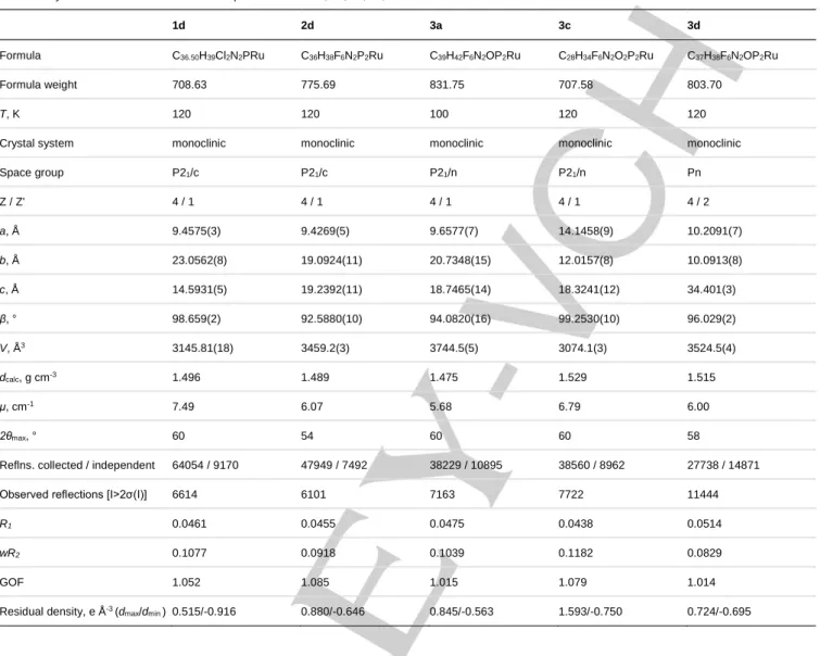
Table 3. Crystal data and structure refinement parameters for 1d, 2d, 3a, 3c, 3d.
1d 2d 3a 3c 3d
Formula C36.50H39Cl2N2PRu C36H38F6N2P2Ru C39H42F6N2OP2Ru C28H34F6N2O2P2Ru C37H38F6N2OP2Ru
Formula weight 708.63 775.69 831.75 707.58 803.70
T, K 120 120 100 120 120
Crystal system monoclinic monoclinic monoclinic monoclinic monoclinic
Space group P21/c P21/c P21/n P21/n Pn Z / Z' 4 / 1 4 / 1 4 / 1 4 / 1 4 / 2 a, Å 9.4575(3) 9.4269(5) 9.6577(7) 14.1458(9) 10.2091(7) b, Å 23.0562(8) 19.0924(11) 20.7348(15) 12.0157(8) 10.0913(8) c, Å 14.5931(5) 19.2392(11) 18.7465(14) 18.3241(12) 34.401(3) β, ° 98.659(2) 92.5880(10) 94.0820(16) 99.2530(10) 96.029(2) V, Å3 3145.81(18) 3459.2(3) 3744.5(5) 3074.1(3) 3524.5(4) dcalc, g cm-3 1.496 1.489 1.475 1.529 1.515 μ, cm-1 7.49 6.07 5.68 6.79 6.00 2θmax, ° 60 54 60 60 58
Reflns. collected / independent 64054 / 9170 47949 / 7492 38229 / 10895 38560 / 8962 27738 / 14871
Observed reflections [I>2σ(I)] 6614 6101 7163 7722 11444
R1 0.0461 0.0455 0.0475 0.0438 0.0514 wR2 0.1077 0.0918 0.1039 0.1182 0.0829 GOF 1.052 1.085 1.015 1.079 1.014 Residual density, e Å-3 (d max/dmin ) 0.515/-0.916 0.880/-0.646 0.845/-0.563 1.593/-0.750 0.724/-0.695
Acknowledgements
The authors thank the Russian Science Foundation (grant no. 14-13-00801) for financial support. I.S. thanks the French Ministry of Foreign Affairs for a Ph.D. scholarship through the French Embassy in Moscow.
Keywords: ruthenium iminophosphonamides • electron-deficient complexes • carbonyl complexes • CO insertion • 2D EXSY NMR [1] T. Naota, H. Takaya, S.-I. Murahashi, Chem. Rev. 1998, 98, 2599-2660.
[2] R. Poli, Chem. Rev. 1996, 96, 2135-2204.
[3] T.J. Johnson, K. Folting, W.E. Streib, J.D. Martin, J.C. Huffman, S.A. Jackson, O. Eisenstein, K.G. Caulton, Inorg. Chem. 1995, 34, 488-499. [4] A.D. Phillips, G. Laurenczy, R. Scopelliti, P.J. Dyson, Organometallics
2007, 26, 1120-1122.
[5] A.D. Phillips, O. Zava, R. Scopelitti, A.A. Nazarov, P.J. Dyson,
Organometallics 2010, 29, 417-427.
[6] A.D. Phillips, K. Thommes, R. Scopelliti, C. Gandolfi, M. Albrecht, K. Severin, D.F. Schreiber, P.J. Dyson, Organometallics 2011, 30, 6119-6132.
[7] P. Dejan, G. Thomas, B. Thomas, G.H. Cristian, R. Sören, G.J. Peter, T. Matthias, Eur. J. Inorg. Chem. 2007, 3472-3475.
[8] T. Glöge, D. Petrovic, C. Hrib, P.G. Jones, M. Tamm, Eur. J. Inorg.
Chem. 2009, 4538-4546.
[9] Y. Yamaguchi, H. Nagashima, Organometallics 2000, 19, 725-727. [10] T. Hayashida, Y. Yamaguchi, K. Kirchner, H. Nagashima, Chem. Lett.
2001, 954-955.
[11] H. Kondo, Y. Yamaguchi, H. Nagashima, J. Am. Chem. Soc. 2001, 123, 500-501.
[12] H. Nagashima, H. Kondo, T. Hayashida, Y. Yamaguchi, M. Gondo, S. Masuda, K. Miyazaki, K. Matsubara, K. Kirchner, Coord. Chem. Rev. 2003,
245, 177-190.
[13] J.I. Terasawa, H. Kondo, T. Matsumoto, K. Kirchner, Y. Motoyama, H. Nagashima, Organometallics 2005, 24, 2713-2721.
[14] T.A. Peganova, A.V. Valyaeva, A.M. Kalsin, P.V. Petrovskii, A.O. Borissova, K.A. Lyssenko, N.A. Ustynyuk, Organometallics 2009, 28, 3021-3028.
[15] T.A. Peganova, I.S. Sinopalnikova, A.S. Peregudov, I.V. Fedyanin, A. Demonceau, N.A. Ustynyuk, A.M. Kalsin, Dalton Trans. 2016, 45, 17030-17041.
[16] P.J. Bailey, K.J. Grant, S. Parsons, Organometallics 1998, 17, 551-555. [17] S. Tsukada, T. Sagawa, T. Gunji, Chem. Asian J. 2015, 10, 1881-1883. [18] N.V. Belkova, L.M. Epstein, E.S. Shubina, Eur. J. Inorg. Chem. 2010, 3555-3565.
[19] A.D. Bain, Prog. Nucl. Magn. Reson. Spectrosc. 2003, 43, 63-103. [20] C.L. Perrin, T.J. Dwyer, Chem. Rev. 1990, 90, 935-967.
[21] Although the Van’t Hoff plot for 1d allowed us to find the entropy term ΔS = -18.7±0.4 cal/(mol•K), it requires correction for chloride concentration. In
fact it consists of ΔSex and the constant Rln[Cl] as the rate of associtive
process depends on the concentration of the Cl-. Since the stationary [Cl-] is
unknown the true ΔSex≠ cannot be calculated. Nevertheless, the calculated
activation enthalpy ΔHex≠ does not require any correction due to the change of
the mechanism.
[22] K. Mashima, H. Kaneyoshi, S.-I. Kaneko, A. Mikami, K. Tani, A. Nakamura, Organometallics 1997, 16, 1016-1025.
[23] R.S. Rowland, R. Taylor, J. Phys. Chem. 1996, 100, 7384-7391. [24] C. Gemel, J.C. Huffman, K.G. Caulton, K. Mauthner, K. Kirchner, J.
Organomet. Chem. 2000, 593–594, 342-353.
[25] A. Zamorano, N. Rendón, J.E.V. Valpuesta, E. Álvarez, E. Carmona,
Inorg. Chem. 2015, 54, 6573-6581.
[26] C.J. Adams, R.A. Baber, N.G. Connelly, P. Harding, O.D. Hayward, M. Kandiah, A.G. Orpen, Dalton Trans. 2007, 1325-1333.
[27] C. Jones, C. Schulten, R.P. Rose, A. Stasch, S. Aldridge, W.D. Woodul, K.S. Murray, B. Moubaraki, M. Brynda, G. La Macchia, L. Gagliardi,
Angew. Chem. Int. Ed. 2009, 48, 7406-7410.
[28] T.J.J. Sciarone, C.A. Nijhuis, A. Meetsma, B. Hessen, Organometallics
2008, 27, 2058-2065.
[29] H. Brunner, J. Wachter, J. Chem. Res. (S) 1978, 136-137. [30] E. Jellema, T.J.J. Sciarone, N.M. Navarrete, M.J. Hettinga, A. Meetsma, B. Hessen, Eur. J. Inorg. Chem., 2011, 91-100.
[31] Ö. Öztopcu, B. Stöger, K. Mereiter, K. Kirchner, J. Organomet. Chem.
2013, 735, 80-87.
[32] T. Hayashida, H. Nagashima, Organometallics 2001, 20, 4996-4998. [33] S.B. Jensen, S.J. Rodger, M.D. Spicer, J. Organomet. Chem. 1998,
556, 151-158.
[34] F. Weigend, R. Ahlrichs, Phys. Chem. Chem. Phys. 2005, 7, 3297-3305.
[35] M.J. Frisch, G.W. Trucks, H.B. Schlegel, G.E. Scuseria, M.A. Rob, J.R. Cheeseman, J.A.M. Jr, T. Vreven, K.N. Kudin, J.C. Burant, J.M. Millam, S.S. Iyengar, J. Tomasi, V. Barone, B. Mennucci, M. Cossi, G. Scalmani, N. Rega, G.A. Petersson, H. Nakatsuji, M. Hada, M. Ehara, K. Toyota, R. Fukuda, J. Hasegawa, M. Ishida, T. Nakajima, Y. Honda, O. Kitao, H. Nakai, M. Klene, X. Li, J.E. Knox, H.P. Hratchian, J.B. Cross, V. Bakken, C. Adamo, J. Jaramillo, R. Gomperts, R.E.Stratmann, O. Yazyev, A.J. Austin, R. Cammi, C. Pomelli, J.W. Ochterski, P.Y. Ayala, K. Morokuma, G.A. Voth, P. Salvador, J.J. Dannenberg, V.G. Zakrzewski, S. Dapprich, A.D. Daniels, M.C. Strain, O. Farkas, D.K. Malick, A.D. Rabuck, K. Raghavachari, J.B. Foresman, J.V. Ortiz, Q. Cui, A.G. Baboul, S. Clifford, J. Cioslowski, B.B. Stefanov, G. Liu, A. Liashenko, P. Piskorz, I. Komaromi, R.L. Martin, D.J. Fox, T. Keith, M.A. Al-Laham, C.Y. Peng, A. Nanayakkara, M. Challacombe, P.M.W. Gill, B. Johnson, W. Chen, M.W. Wong, C. Gonzalez, J.A. Pople. Gaussian 09,
Revision D.01. Gaussian, Inc., Wallingford, CT, 2009, 2009.
[36] A.V. Marenich, C.J. Cramer, D.G. Truhlar, J. Phys. Chem. B 2009, 113, 6378-6396.
[37] SAINT v8.34A., Bruker AXS, Madison, Wisconsin, USA, 2013.
[38] G.M. Sheldrick. SADABS, v. 2.03, Bruker/Siemens Area Detector Absorption Correction Program, Bruker AXS, Madison, Wisconsin, 2003. [39] G.M. Sheldrick, Acta Crystallogr. C 2015, C71, 3-8.
Entry for the Table of Contents
FULL PAPER
The lone pair of the highly basic iminophosphonamide N-atoms in arene ruthenium complexes can both stabilize 16ē species and attack an external ligand in 18ē species.
Iana S. Sinopalnikova, Tat'yana A. Peganova, Valentin V. Novikov, Ivan V. Fedyanin, Oleg A. Filippov, Natalia V. Belkova, Elena S. Shubina, Rinaldo Poli, Alexander M. Kalsin*
Page No. – Page No.
Coordinatively labile 18-electron arene ruthenium
iminophosphonamide complexes


![Thiolato-Bridged Arene–Ruthenium Complexes: Synthesis, Molecular Structure, Reactivity, and Anticancer Activity of the Dinuclear Complexes [(arene)2Ru2 (SR)2Cl2]](data:image/gif;base64,R0lGODlhAQABAIAAAP///wAAACH5BAEAAAAALAAAAAABAAEAAAICRAEAOw==)