HAL Id: hal-03045492
https://hal.sorbonne-universite.fr/hal-03045492
Submitted on 8 Dec 2020
HAL is a multi-disciplinary open access archive for the deposit and dissemination of sci-entific research documents, whether they are pub-lished or not. The documents may come from teaching and research institutions in France or abroad, or from public or private research centers.
L’archive ouverte pluridisciplinaire HAL, est destinée au dépôt et à la diffusion de documents scientifiques de niveau recherche, publiés ou non, émanant des établissements d’enseignement et de recherche français ou étrangers, des laboratoires publics ou privés.
RECIST and iRECIST criteria for the evaluation of
nivolumab plus ipilimumab in patients with
microsatellite instability-high/mismatch repair-deficient
metastatic colorectal cancer: the GERCOR NIPICOL
phase II study
Romain Cohen, Jaafar Bennouna, Aurélia Meurisse, Christophe Tournigand,
Christelle de la Fouchardière, David Tougeron, Christophe Borg, Thibault
Mazard, Benoist Chibaudel, Marie-Line Garcia-Larnicol, et al.
To cite this version:
Romain Cohen, Jaafar Bennouna, Aurélia Meurisse, Christophe Tournigand, Christelle de la Fouchardière, et al.. RECIST and iRECIST criteria for the evaluation of nivolumab plus ipilimumab in patients with microsatellite instability-high/mismatch repair-deficient metastatic colorectal cancer: the GERCOR NIPICOL phase II study. Journal for Immunotherapy of Cancer, BMJ Publishing Group 2020, 8 (2), pp.e001499. �10.1136/jitc-2020-001499�. �hal-03045492�
RECIST and iRECIST criteria for the
evaluation of nivolumab plus
ipilimumab in patients with
microsatellite instability- high/mismatch
repair-deficientmetastaticcolorectal
cancer: the GERCOR NIPICOL phase
II study
Romain Cohen ,1 Jaafar Bennouna,2 Aurélia Meurisse,3 Christophe Tournigand,4 Christelle De La Fouchardière,5 David Tougeron,6 Christophe Borg,7
Thibault Mazard,8 Benoist Chibaudel,9 Marie- Line Garcia- Larnicol,10 Magali Svrcek,11 Dewi Vernerey,3 Yves Menu,12 Thierry André 1
To cite: Cohen R, Bennouna J,
Meurisse A, et al. RECIST and iRECIST criteria for the evaluation of nivolumab plus ipilimumab in patients with microsatellite instability- high/ mismatch repair- deficient metastatic colorectal cancer: the GERCOR NIPICOL phase II study. Journal for ImmunoTherapy of Cancer 2020;8:e001499. doi:10.1136/jitc-2020-001499 ►Additional material is published online only. To view please visit the journal online (http:// dx. doi. org/ 10. 1136/ jitc- 2020- 001499).
Accepted 18 September 2020
For numbered affiliations see end of article.
Correspondence to Dr Thierry André; thierry. andre@ aphp. fr © Author(s) (or their employer(s)) 2020. Re- use permitted under CC BY. Published by BMJ.
ABSTRACT
Background Immune checkpoint inhibitors (ICIs) are
highly effective in patients with microsatellite instability/ mismatch repair- deficient (MSI/dMMR) metastatic colorectal cancer (mCRC). Response Evaluation Criteria in Solid Tumors (RECIST) 1.1 criteria may underestimate response to ICIs due to the pseudoprogression
phenomenon. The GERCOR NIPICOL phase II study aimed to evaluate the frequency of pseudoprogressions in patients with MSI/dMMR mCRC treated with nivolumab and ipilimumab.
Methods Patients with MSI/dMMR mCRC previously
treated with fluoropyrimidines, oxaliplatin, and irinotecan with/without targeted therapies received nivolumab 3 mg/ kg plus ipilimumab 1 mg/kg every 3 weeks for four cycles then nivolumab 3 mg/kg every 2 weeks until progression or a maximum of 20 cycles. Computed tomography scan tumor assessments were done every 6 weeks for 24 weeks and then every 12 weeks. The primary endpoint was disease control rate at 12 weeks according to RECIST 1.1 and iRECIST by central review.
Results Of 57 patients included between December
2017 and November 2018, 48.0% received ≥3 prior lines of chemotherapy, 18.0% had BRAFV600E mutation,
and 56.0% had Lynch syndrome- related cancer. Seven patients (12.0%) discontinued treatment due to adverse events; one died due to a treatment- related adverse event. The disease control rate (DCR) at 12 weeks was 86.0% with RECIST 1.1% and 87.7% with iRECIST. Two pseudoprogressions (3.5%) were observed, at week 6 and at week 36, representing 18% of patients with disease progression per RECIST 1.1 criteria. With a median follow- up of 18.4 months, median progression- free survival (PFS) and overall survival (OS) were not reached. The 12- month PFS rate was 72.9% with RECIST 1.1% and 76.5% with iRECIST. The 12- month OS rate was 84%. Overall
response rate was 59.7% with both criteria. RAS/BRAF status, sidedness, Lynch syndrome, and other baseline parameters were not associated with PFS.
Conclusion Pseudoprogression is rare in patients with
MSI/dMMR mCRC treated with nivolumab and ipilimumab. This combined ICI therapy confirms impressive DCR and survival outcomes in these patients.
Trial registration number NCT03350126.
INTRODUCTION
Microsatellite instability (MSI) is caused by the deficiency of the DNA mismatch repair (MMR) system, resulting from a germline mutation in MMR genes (Lynch syndrome) or from an epigenetic extinction of MLH1 gene (sporadic cases).1 Sporadic MSI/MMR- deficient (dMMR) colorectal cancers (CRCs) are frequently associated with the BRAFV600E
mutation, a well- known negative prognostic factor. Approximately 5% of metastatic CRC (mCRC) are MSI/dMMR and one- third of them are BRAFV600E- mutated.2
Tumors with MSI/dMMR are character-ized by a high tumor mutational burden, with highly immunogenic neoantigens arising from frameshift mutation and by a high infiltration of cytotoxic T lymphocytes. The upregulation of immune checkpoints by tumor cells protects them from this hostile microenvironment.3 4 Immune checkpoint inhibitors (ICIs) such as anti- programmed death 1 (PD1) or anti- programmed death ligand 1 (PD- L1) monoclonal antibodies, alone or in combination with anti- CTLA-4
on December 7, 2020 by guest. Protected by copyright.
Open access
agents, have demonstrated high clinical activity with durable responses in patients with MSI/dMMR mCRC.5–9 The KEYNOTE-177 phase III study showed a significant improvement of progression- free survival (PFS) with first- line pembrolizumab compared with standard- of- care chemotherapy plus targeted therapy.10
The initial effect of ICIs on tumor can include an increase of tumor diameter due to the immune cell infiltra-tion. Conventional criteria for the evaluation of treatment responses, namely Response Evaluation Criteria in Solid Tumors (RECIST) 1.1, fail to identify a phenomenon of pseudoprogression and may falsely conclude that there is disease progression (PD). These criteria have been shown to underestimate the benefits of pembrolizumab in terms of overall response rate and PFS in approximately 15% of patients with melanoma.11 12 Consequently, modified RECIST 1.1 for immune- based therapeutics (iRECIST) have been developed, requiring the confirmation of PD to rule out or confirm pseudoprogression.13 14 Although pseudoprogression is well recognized and reported in up to 10% of patients with melanoma or lung cancer,15–18 its frequency in patients with MSI/dMMR treated with ICIs has never been evaluated. This knowledge is of great value, though, given the negative consequences of either discon-tinuation of an effective treatment or maintenance of an ineffective drug beyond PD. Therefore, we designed the GERCOR NIPICOL phase II study to evaluate RECIST1.1 and iRECIST criteria in patients with MSI/dMMR mCRC treated with the nivolumab and ipilimumab combination. PATIENTS AND METHODS
Study design and population
This a single- arm, open- label, multicenter phase II study (NIPICOL) was designed (GERCOR) to evaluate disease control rate (DCR) by RECIST and iRECIST at 12 weeks in patients with MSI/dMMR mCRC. Patients were recruited from eight French hospitals. Eligible patients were ≥18 years old, had histologically confirmed mCRC locally assessed as MSI/dMMR, measurable disease per RECIST 1.1 criteria, and an Eastern Cooperative Oncology Group performance status (ECOG PS) of 0 or 1. Patients developed PD or were intolerant to approved standard therapies (fluoropyrimidine, oxaliplatin, irino-tecan, antiangiogenics, and antiepidermal growth factor receptor agents if their tumors harbored wild- type RAS/RAF). Eligible patients had absolute neutrophil count ≥1500 cells per mm³, platelet count ≥100 x 109/L,
hemoglobin ≥9 g/dL, serum creatinine level <150 µM, aspartate aminotransferase and alanine aminotransferase ≤3 × upper limit of normal (ULN; or ≤5 × ULN in the case of known liver metastases), alkaline phosphatase <5 × ULN, and total bilirubin ≤1.5 × ULN. Main exclu-sion criteria included: prior treatment with an anti- PD1, anti- PD- L1, anti- CTLA-4, or other agent targeting T- cell co- stimulation or immune checkpoint pathways, condi-tions requiring corticosteroids (prednisone equivalents >10 mg/day) or other immunosuppressive medication
within 2 weeks before starting treatment, other serious or uncontrolled medical disorders, active brain or leptome-ningeal metastases, or prior malignancy within the previous 5 years except for cured select localized cancers. Procedures
Patients received nivolumab 3 mg/kg intravenously over 60 min and ipilimumab 1 mg/kg intravenously over 90 min every 3 weeks for four cycles (induction phase) and then nivolumab 3 mg/kg intravenously every 2 weeks until PD, discontinuation because of toxicity, death, with-drawal of consent, or for a maximum of 20 infusions, equivalent to 1 year of therapy. Dose modifications were not permitted.
Outcomes
The primary endpoint was DCR at 12 weeks according to RECIST 1.1 and iRECIST criteria by central review. DCR was defined as the number of patients with stable disease (SD), partial response (PR), or complete response divided by the total number of patients. Secondary endpoints were safety, PFS, overall survival (OS; time from the first dose to death, whatever the cause), and association between baseline clinicopathological parameters and PFS. PFS by RECIST 1.1 was calculated from the first ICI infusion to the first documented PD or death resulting from any cause, whichever occurred first. PFS by iRECIST was calculated from the first dose to the first documented PD with subsequent confirmation or death resulting from any cause, whichever occurred first.
Assessments
Baseline radiologic tumors assessments were assessed ≤28 days before the first dose (baseline) of treatment followed by every 6 weeks for 24 weeks and every 12 weeks thereafter by CT. RECIST and iRECIST evaluations were centrally reviewed by one radiologist (YM). Pseudopro-gression was defined as an unconfirmed PD according to iRECIST.14 If pseudoprogression was suspected, the same images were independently blindly reviewed by a second radiologist. In case of discrepancy, a final decision was reached by consensus. Safety was assessed per National Cancer Institute Common Terminology Criteria for Adverse Events (NCI CTCAE) V.4.0.
The MSI/dMMR status was evaluated before screening by local laboratories and defined by high level of MSI by PCR testing (two instable markers or more; with recom-mended panel including: BAT25, BAT26, NR21, NR24, and NR27) and/or loss of expression of minimum one MMR protein by immunohistochemistry (testing of four MMR proteins: MLH1, PMS2, MSH2, and MSH6). Patho-logical and molecular biology reports were reviewed by the study sponsor prior to patients’ inclusion. Tumor samples (archival or fresh biopsy specimen from primary or meta-static lesions) were collected for central laboratory confir-mation of the MSI/dMMR status (Saint- Antoine Hospital, Paris, France) and additional translational studies.
on December 7, 2020 by guest. Protected by copyright.
http://jitc.bmj.com/
Tumors were considered related to Lynch syndrome in case of known MMR gene germline mutation, loss of expression of MSH2 and/or MSH6, or loss of expression of MLH1, and/or PMS2 without BRAFV600E mutation nor
hypermethylation of MLH1 promoter. Tumors with loss of MLH1 and/or PMS2 harboring BRAFV600E mutation and/
or a hypermethylation of MLH1 promoter were consid-ered as sporadic. In all other cases, Lynch syndrome status was defined as unknown.19
Statistical analysis
The study was designed considering an estimated 12- week DCR of 70% with RECIST 1.1. Using iRECIST, a 12 week DCR of 85% was expected (H1: alternative hypothesis), whereas a 12- week DCR of 70% was considered as unin-teresting (H0: null hypothesis). According to a A'Hern single- stage phase II design20 21 with a one- sided 5% type I error and a power of 80%, 49 evaluable patients were needed for the analysis of the primary endpoint. Considering a minimal 15% rate of patients not evalu-able for the primary endpoint, 57 patients were planned to be included. Statistical analyses were performed on an intention- to- treat basis.
Continuous and categorical variables were described by medians (IQR) and frequencies (percentage), respec-tively. The median PFS and OS, and the proportion of patients meeting these endpoints at specific time points were estimated by the Kaplan- Meier method. The 95% CI were calculated using log- log transformation. Median follow- up was calculated by the reverse Kaplan- Meier method.
Cox proportional- hazard models were used to estimate HRs and their 95% CIs for baseline factors associated with PFS. The association was evaluated in a prespecified exploratory analysis using the univariate Cox model and then parameters with p values of <0.1 were entered into a final multivariate Cox regression model.
A waterfall plot illustration was used to present the best percentage change in target lesion size from baseline during the first year of treatment. Duration of treatment and response in patients with at least one evaluation with SD or better were summarized in a swimmer plot. All analyses were performed using SAS V.9.4 (SAS Institute).
RESULTS Population
Between December 2017 and November 2018, 57 patients with MSI/dMMR mCRC were enrolled. Table 1 shows baseline clinical and pathological characteristics. Most patients were male (52.6%), with ECOG PS of 1 (64.9%), right- sided tumors (54.4%), two metastatic sites or more (71.9%). Twenty- eight (49.1%) and 10 (17.5%) patients’ tumors were RAS- mutated and BRAFV600E- mutated,
respectively. Overall, 47.4% of patients were treated with ≥3 prior lines of treatment, including fluoropyrimidines (100.0%), oxaliplatin (100.0%), irinotecan (96.5%),
antiangiogenics (57.9%), and anti- epidermal growth factor receptors (45.6%; table 1).
The database was locked on December 11, 2019, with a median follow- up of 18.1 months (95% CI 14.1 to 19.2). A total of 36 patients (63.2%) completed the predefined 1- year duration of treatment, five patients died from disease- related event during the induction phase (12
Table 1 Patient baseline characteristics
Characteristics N=57
Age (years), median (Q1–Q3) 56.5 (45.8–63.8) Sex, n (%) Male 30 (52.6) Female 27 (47.4) ECOG PS, n (%) 0 20 (35.1) 1 37 (64.9)
Primary tumor location, n (%)
Right- sided 31 (54.4) Left- sided 25 (43.9) Right and left location 1 (1.8) Mutation status, n (%)
RAS/RAF wild type 19 (33.3)
RAS mutation 28 (49.1)
BRAF mutation 10 (17.5)
Origin of MMR deficiency, n (%)
Lynch- related 32 (56.1) Known germline mutation 19 (59.4) Sporadic 16 (28.1) Unknown 9 (15.8) Number of metastatic sites, n (%)
1 16 (28.1)
2 25 (43.9)
>2 16 (28.1) Number of prior lines, n (%)
1 5 (8.8) 2 24 (42.1) >2 27 (47.4) Missing 1 (1.8) Prior treatments, n (%) 5- FU/capecitabine 57 (100.0) Oxaliplatin 57 (100.0) Irinotecan 55 (96.5) Trifluridine/tipiracil 4 (7.0) Regorafenib 5 (8.8) Bevacizumab/aflibercept 33 (57.9) Cetuximab/panitumumab 26 (45.6)
ECOG PS, The Eastern Cooperative Oncology Group Performance Status; 5- FU, 5- fluorouracil; MMR, DNA mismatch repair.
on December 7, 2020 by guest. Protected by copyright.
Open access
weeks). Other reasons of treatment discontinuation were progressive disease (n=8), adverse event (n=7) including one toxic death, and a patient’s wish (n=1).
Safety
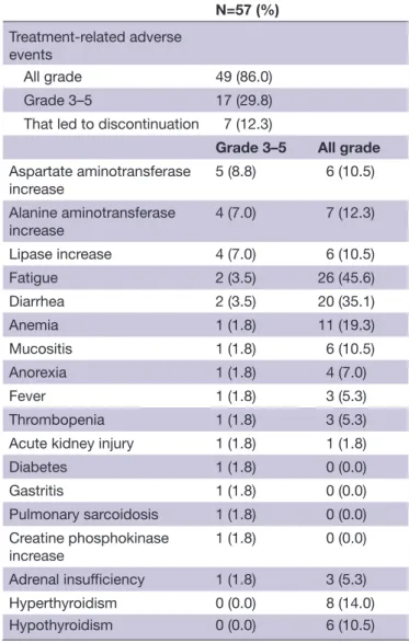
Thirty- two patients (56.1%) experienced grade ≥3 adverse events. Grade 3–5 treatment- related adverse events (TRAEs) were reported for 17 patients (29.8%), with one patient who died from septic shock while being treated with corticosteroid for a potentially immune- related hepatitis (table 2). Most frequent grade 3–5 TRAEs were increased transaminases (8.8%), increased serum lipase level (7.0%), diarrhea (3.5%), and fatigue (3.5%). Endocrine- related TRAEs included grade 3 adrenal insufficiency (n=1; 1.8%), grade 3 diabetes (n=1; 1.8%), grade 2 hypothyroidisms (n=3; 5.3%), and grade 2 hyperthyroidism (n=1; 1.8%). One patient was diagnosed with grade 3 sarcoidosis and one with grade 2 uveitis. Efficacy
For eligible patients (n=57), the 12- week DCR was 86.0% by RECIST 1.1.% and 87.7% by iRECIST (central review; kappa coefficient=0.92, 95% CI 0.77 to 1.0 with no
significant difference). Three patients died from disease- related cause prior to any tumor assessment and were considered as progressive. Five patients experienced PD at the first CT scan evaluation (week 6). Of these, two died from disease- related cause before the second CT scan and were considered as confirmed PD, two (who continued treatment) had confirmed PD on the subse-quent CT scan at 12 weeks, and one had pseudoprogres-sion. At 12 weeks, 2 (3.5%) CR and 18 (31.6%) PR were observed (table 3; online supplemental file 1).
Overall, two pseudoprogressions were observed during follow- up (out of 57, 3.5%). The first pseudoprogression occurred at week 6 in a 68- year- old man treated for peri-toneal metastases from a MLH1/PMS2- negative, MSI, RAS/RAF wild- type right- sided mucinous colon carci-noma. Study treatment was maintained, and the patient experienced PR after 11 months of treatment. The second pseudoprogression occurred at week 36 in a 47- year- old woman with a KRAS- mutated, MSI, MSH2/MSH6- negative mucinous, rectal carcinoma harboring mesenteric lymph node metastases. Best observed response was SD. Study treatment was interrupted in this patient 2 months after pseudoprogression due to grade 3 diarrhea. Both patients were alive and free of progression 22 and 27 months after treatment initiation, respectively.
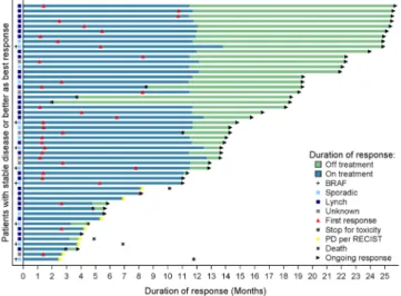
Best observed responses by RECIST 1.1 were 11 CR (19.3%), 23 PR (40.3%), 17 SD (29.8%), 3 PD (5.3%), and 3 non- evaluable cases (5.3%; table 3). Best percentage changes in target lesion size from baseline are displayed in figure 1. Median time to response was 5.7 months (95% CI 2.7 to 8.2). Figure 2 shows the duration of treat-ment and response of patients who experienced disease control. Median duration of response was not reached. Kaplan- Meier curves of PFS per RECIST 1.1 and OS are displayed in figure 3. Median survivals were not reached.
Table 2 Treatment- related adverse events
N=57 (%)
Treatment- related adverse events
All grade 49 (86.0) Grade 3–5 17 (29.8) That led to discontinuation 7 (12.3)
Grade 3–5 All grade
Aspartate aminotransferase increase 5 (8.8) 6 (10.5) Alanine aminotransferase increase 4 (7.0) 7 (12.3) Lipase increase 4 (7.0) 6 (10.5) Fatigue 2 (3.5) 26 (45.6) Diarrhea 2 (3.5) 20 (35.1) Anemia 1 (1.8) 11 (19.3) Mucositis 1 (1.8) 6 (10.5) Anorexia 1 (1.8) 4 (7.0) Fever 1 (1.8) 3 (5.3) Thrombopenia 1 (1.8) 3 (5.3) Acute kidney injury 1 (1.8) 1 (1.8) Diabetes 1 (1.8) 0 (0.0) Gastritis 1 (1.8) 0 (0.0) Pulmonary sarcoidosis 1 (1.8) 0 (0.0) Creatine phosphokinase increase 1 (1.8) 0 (0.0) Adrenal insufficiency 1 (1.8) 3 (5.3) Hyperthyroidism 0 (0.0) 8 (14.0) Hypothyroidism 0 (0.0) 6 (10.5)
Table 3 Tumor response at 12 weeks and overall best observed response per RECIST 1.1 criteria
At 12 weeks N=57 Overall best response N=57 Complete response, n (%) 2 (3.5) 11 (19.3) Partial response, n (%) 18 (31.6) 23 (40.4) Objective response rate, % 35.1 59.6 Stable disease, n (%) 29 (50.9) 17 (29.8) Progressive disease, n (%) 5 (8.8) 3 (5.3) Confirmed progressive disease
per iRECIST criteria 4 (7.0) 2 (3.5) Pseudoprogression per
iRECIST criteria 1 (1.8) 1 (1.8) Non- evaluable, n (%) 3 (5.3) 3 (5.3) Disease control rate per
RECIST1.1/iRECIST, % 86.0/87.7 89.5/91.2
RECIST, Response Evaluation Criteria in Solid Tumors.
on December 7, 2020 by guest. Protected by copyright.
http://jitc.bmj.com/
The 12- month PFS and OS per RECIST 1.1 were 72.9% (95% CI 59.0% to 82.7%) and 84.0% (95% CI 71.4% to 91.3%), respectively.
Parameters associated with efficacy
The MSI/dMMR status was centrally confirmed in 11 of 15 patients with a PFS event. Four tumor samples were not evaluable. Overall, eight patients had dMMR and MSI tumors and three pMMR and microsatellite stable (MSS) as assessed by central review. Of the latter three patients, one achieved PR of liver metastases, but had confirmed PD of the primary lesion 24 weeks after treatment initia-tion, and died due to disease- related event thereafter. The patients with MSS/pMMR mCRC were excluded from the Cox models analysis.
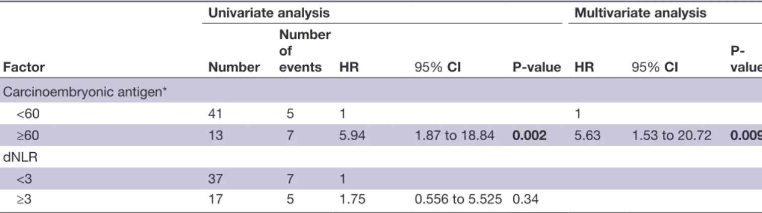
In univariate Cox model (table 4), a carcinoembry-onic antigen (CEA) cutoff of 60 ng/mL (identified as the optimal cutoff by the restricted cubic splines method) was strongly associated with PFS (p=0.0025). The KRAS/BRAF mutational status tended to be associ-ated with PFS (p=0.0973). Compared with patients who were wild type for KRAS/BRAFV600E, HR was 1.7 (95% CI
0.46 to 6.34) and 0.33 (95% CI 0.08 to 1.39) for those harboring BRAFV600E and RAS mutations, respectively.
In multivariate model, the CEA level of ≥60 ng/mL was the only baseline parameter associated with poorer PFS (HR=5.63, 95% CI 1.53 to 20.7, p=0.0094).
DISCUSSION
In this open- label, multicenter, phase II study, the combi-nation of nivolumab and ipilimumab was associated with a low frequency of pseudoprogression and high clinical activity in patients with MSI/dMMR mCRC.
This is the first report of the pseudoprogression inci-dence in a large group of patients with mCRC with MSI/ dMMR tumors. Only two patients (3.5%) experienced pseudoprogression in our study, with the frequency lower than expected value. The incidence of pseudoprogres-sion in previous studies of patients with melanoma, lung, or renal cancers did not exceed 10% with anti- PD1 mono-therapy.12 15 22 23 Of note, this estimation is impaired by the fact that the definition of pseudoprogression varied across different studies.24 Here, we defined pseudopro-gression an unconfirmed PD, per central review following the iRECIST guidelines, with CT scans performed every 6 weeks for 24 weeks.14 Even if one pseudoprogression was detected after 36 weeks of treatment in our study, it
Figure 1 Best percentage change in target lesion size from baseline by central review. + sign: BRAF- mutated tumors, yellow triangle: pseudo- progression.
Figure 2 Duration of treatment and response in patients with stable disease or better disease control per RECIST v1.1 criteria. PD, disease progression; RECIST, Response Evaluation Criteria in Solid Tumors.
Figure 3 Kaplan- Meier survival curves of progression- free survival per RECIST v1.1 (A), per iRECIST (B) and overall survival (C) NE, not estimable
on December 7, 2020 by guest. Protected by copyright.
Open access
Table 4 Univariate and multivariate Cox regression analysis for progression- free survival
Factor
Univariate analysis Multivariate analysis
Number Number of events HR 95% CI P- value HR 95% CI P- value Age (years) <65 43 8 1 ≥65 11 4 2.26 0.68 to 7.53 0.18 Sex Male 29 5 1 Female 25 7 1.82 0.58 to 5.74 0.31 Body mass index
≤20 14 4 1.58 0.44 to 5.59
20–25 28 6 1 0.77
25–30 8 1 0.527 0.06 to 4.38
≥30 4 1 1.16 0.14 to 9.68
Primary tumor location
Right 29 8 1 Left / rectum 24 4 0.556 0.17 to 1.85 0.34 Grade Well differentiated 8 1 1 Moderately differentiated 21 4 1.66 0.18 to 14.83 Poorly differentiated 17 4 2.09 0.23 to 18.82 0.93 Undifferentiated 1 0 – Stage II 4 1 1 III 17 3 0.79 0.08 to 7.63 IV 25 6 1.08 0.13 to 9.01 0.91 Number of metastatic sites
1–2 40 10 1
>2 14 2 0.61 0.13 to 2.79 0.52 Number of anterior therapeutic sequences
1–2 28 5 1
>2 25 6 1.54 0.47 to 5.04 0.48 Antibiotics before inclusion
No 45 9 1
Yes 6 3 2.86 0.77 to 10.65 0.12 Mutational status
Wild type 18 5 1 1
BRAFV600E- mutated 9 4 1.70 0.46 to 6.34 0.68 0.16 to 2.94
RAS- mutated 27 3 0.33 0.08 to 1.39 0.10 0.32 0.07 to 1.34 0.30 Origin of MMR deficiency Sporadic 15 4 1 Lynch- related 31 5 0.55 0.15 to 2.06 0.38 Carcinoembryonic antigen (median) <12 28 4 1 ≥12 26 8 2.46 0.74 to 8.19 0.14
on December 7, 2020 by guest. Protected by copyright.
http://jitc.bmj.com/
is thought to be an early phenomenon that might not be detected with a conventional interval of 8 weeks between tumor assessments.25 One might hypothesize that the frequency of pseudoprogression might vary depending on the type of ICI (anti- PD- L1 alone or in combination with anti- CTLA-4) used. However, data are lacking to make comparison of the pseudoprogression frequency in patients with MSI/dMMR mCRC treated with anti- PD- L1 alone, of which the associated pseudoprogression inci-dence is currently unknown, with those treated by anti- PD- L1 and anti- CTLA-4 agents. Still, our results suggest that RECIST 1.1 can be safely used as the primary criteria for response- based endpoints in randomized trials, espe-cially those evaluating combinations of anti- PD1 and anti- CTLA-4 monoclonal antibodies versus conventional chemotherapy.
Of the 11 patients diagnosed with radiologic PD according to RECIST 1.1 criteria in our study, two had pseudoprogression (18.0%) and were still alive and free of progression at the last follow- up visit, whereas the remaining (82.0%) had confirmed PD of whom five died during follow- up. In other words, patients experiencing PD per RECIST 1.1 criteria while treated with nivolumab and ipilimumab are more likely to have confirmed PD and poor outcomes than pseudoprogression. Given the observed activity of ICI extended beyond toxicity- related treatment interruption, it might be more beneficial to promptly change antitumor treatment rather than main-tain ICI until the next CT scan assessment. This should be discussed between the patient and their providers for a shared decision making when considering treatment continuation after PD.17 26
In this study with heavily pretreated patients, the combination of nivolumab and ipilimumab was associ-ated with impressive efficacy and manageable toxicity. All 36 patients who completed the predefined 1 year of therapy were alive and free of progression at the last follow- up visit. The objective response rate was 59.6%, and the 12- month PFS and OS estimates were 72.9% and 84.0%, respectively. These results, with 1 year of
immunotherapy, are consistent with previously published data on the combination cohort from the CheckMate-142 study, in which patients with controlled disease were mainly treated for longer than 2 years.7 Longer follow- up in this study is necessary to evaluate if the control of the disease continues after ICI treatment discontinuation (all patients received up to 1 year of therapy) and to determine median duration of response. Interestingly, the number of patients harboring disease resistance to immune checkpoint blockade with nivolumab and ipilim-umab seems lower than with anti- PD- L1 monotherapy.5 8–10 In the KEYNOTE-177 phase III trial that demonstrated the superiority of pembrolizumab over first- line chemo-therapy, 29.0% of patients in the experimental arm had PD by RECIST 1.1 as best observed response, while only 10.0% of patients in the NIPICOL study had PD or death as the best response.10 The CheckMate- 8HW phase III study comparing nivolumab and nivolumab plus ipilim-umab to chemotherapy should provide valuable data into this topic.
Given the durable activity of ICIs among responders, the main challenge from a clinical and scientific perspec-tive will be to develop biomarkers that may predict the resistance of MSI/dMMR tumors to immunotherapy. We have reported, similarly to others, a significant amount of resistant cases related to a misdiagnosis of the MSI/ dMMR status, with MSS/pMMR tumors incorrectly considered as MSI/dMMR.27 28 A review of pathological and molecular reports was therefore mandatory prior to the study enrollment. Despite that, a central reassessment detected three misdiagnosed patients (5.3%), who expe-rienced PD during the study treatment. This observation highlights the importance of the accurate diagnosis of MSI/dMMR status in routine practice.
In addition to MSI/dMMR misdiagnosis, a CEA level of ≥60 ng/mL was the only factor significantly associated with decreased PFS, though this cutoff is more likely to be a negative prognostic factor than a predictor of resis-tance to immunotherapy. We acknowledge that, with only 15 PFS events, our analysis lacks of statistical power.
Factor
Univariate analysis Multivariate analysis
Number
Number of
events HR 95% CI P- value HR 95% CI P- value
Carcinoembryonic antigen* <60 41 5 1 1 ≥60 13 7 5.94 1.87 to 18.84 0.002 5.63 1.53 to 20.72 0.009 dNLR <3 37 7 1 ≥3 17 5 1.75 0.556 to 5.525 0.34
N stands for number of (changed) ; P value stands for P- value (changed). *Modeled with the restricted cubic splines method.
dNLR, a derived neutrophils/leukocytes minus neutrophils ratio; MMR, DNA mismatch repair. Table 4 Continued
on December 7, 2020 by guest. Protected by copyright.
Open access
Centrally assessed responses were observed irrespective of the mechanism underlying the MMR deficiency (sporadic vs Lynch- related), the RAS/BRAF status, and the primary tumor sidedness. Whereas results from the KEYNOTE-177 study suggested that the efficacy of pembrolizumab might vary depending on the RAS mutational status, our results are in line with previous reports demonstrating the clin-ical activity of ICIs whatever the disease subset.5 7 10 Other potential determinants of treatment efficacy including the use of antibiotics at baseline,29 30 the derived neutro-phils/(leukocytes minus neutrophils) ratio,31 or the body mass index32 did not predict patients’ outcomes in our study. Other small cohort studies have shown that tumor mutational load and immune infiltrate are interesting parameters deserving further research.27 33 34 Translational studies with tumor samples from this trial are currently evaluating potential biological predictive factors.
In summary, this phase II study confirms high activity of nivolumab plus ipilimumab for four cycles followed by nivolumab alone for maximum 1 year of therapy in patients with MSI/dMMR mCRC. The ongoing Check-Mate- 8HW phase III study should provide useful informa-tion on the clinical benefit of nivolumab plus ipilimumab versus nivolumab alone in this setting. In patients with MSI/dMMR who exhibit PD per RECIST 1.1 criteria, the likelihood of pseudoprogression is low compared with the risk of real progression. Close clinical monitoring and shared information with patients are required if treat-ment continues beyond progression.
Author affiliations
1Sorbonne University, Department of Medical Oncology, Saint- Antoine Hospital,
APHP, Paris, France
2Department of Medical Oncology, University Hospital of Nantes, Nantes, France 3Department of Oncology, Besançon University Hospital, Methodology and Quality of
Life Unit, Besançon, France
4Department of Gastroenterology and Digestive Oncology, Henri Mondor University
Hospital, APHP, Creteil, France
5Department of Medical Oncology, Centre Léon Bérard, Lyon, France 6Department of Gastroenterology, Poitiers University Hospital and University of
Poitiers, Poitiers, France
7Department of Medical Oncology, University Hospital of Besançon, Besançon,
France
8Department of Medical Oncology, Institut de Recherche en Cancérologie de
Montpellier (IRCM), INSERM, Montpellier University, INSERM U1194, Montpellier, France
9Department of Medical Oncology, Franco- British Hospital, Fondation Cognacq- Jay,
Levallois- Perret, France
10Multidisciplinary Group in Oncology (GERCOR), Paris, France
11Sorbonne University, Department of Pathology, Saint- Antoine Hospital, AP- HP,
Paris, France
12Sorbonne University, Department of Radiology, Saint- Antoine Hospital, AP- HP,
Paris, France
Acknowledgements Authors acknowledge Magdalena Benetkiewicz (ScD) for the editorial assistance.
Contributors RC, M- LG- L, MS, DV, YM and TA contributed to study concept and design. RC, AM, M- LG- L, MS, DV, YM, and TA analyzed the data. All authors interpreted the data. RC, AM, DV, and TA contributed to manuscript writing. All authors reviewed the manuscript. All authors approved the final manuscript. Funding This work was supported by Bristol- Myers Squibb (BMS) and GERCOR. BMS had no role in study design, data collection, analysis, and interpretation, or writing of the report.
Competing interests TA reports consulting/advisory role and/or received honoraria from Amgen, Bristol- Myers Squibb, Chugai, Clovis, Gritstone Oncology, HalioDx, MSD Oncology, Pierre Fabre, Roche/Ventana, Sanofi, Servier, and Tesaro and has received travel, accommodations, and expenses from Roche/Genentech, MSD Oncology, and Bristol- Myers Squibb. JB reports consulting/advisory role and or received honoraria from Amgen, Bristol- Myers Squibb, MSD Oncology, Roche, Bayer, Servier, and AstraZeneca and has received travel and accommodations from Roche, MSD Oncology, and AstraZeneca. CB reports consulting/advisory role and/ or received honoraria from Bayer, Sanofi, and Roche, and research grant from Roche. RC reports honoraria from Amgen, MSD Oncology, Sanofi, and Servier, and research grant from Servier Institute. TM reports honoraria from Amgen, Sanofi, Bristol- Myers Squibb, and Sandoz, travel, accommodations, or expenses by Amgen and research funding from Roche and Amgen. YM received honoraria from Bristol- Myers Squibb. MS reports consulting/advisory role and/or received honoraria from Bristol- Myers Squibb, Astellas, MSD Oncology, and Sanofi and has received travel, accommodations, and expenses from Bristol- Myers Squibb and Ventana/Roche. DT reports consulting/advisory role and/or received honoraria from Bristol- Myers Squibb, MSD Oncology, and Merck Serono and has received travel, accommodations, and expenses from MSD Oncology and Bristol- Myers Squibb. Patient consent for publication Not required.
Provenance and peer review Not commissioned; externally peer reviewed. Data availability statement Data are available upon reasonable request. Supplemental material This content has been supplied by the author(s). It has not been vetted by BMJ Publishing Group Limited (BMJ) and may not have been peer- reviewed. Any opinions or recommendations discussed are solely those of the author(s) and are not endorsed by BMJ. BMJ disclaims all liability and responsibility arising from any reliance placed on the content. Where the content includes any translated material, BMJ does not warrant the accuracy and reliability of the translations (including but not limited to local regulations, clinical guidelines, terminology, drug names and drug dosages), and is not responsible for any error and/or omissions arising from translation and adaptation or otherwise. Open access This is an open access article distributed in accordance with the Creative Commons Attribution 4.0 Unported (CC BY 4.0) license, which permits others to copy, redistribute, remix, transform and build upon this work for any purpose, provided the original work is properly cited, a link to the licence is given, and indication of whether changes were made. See https:// creativecommons. org/ licenses/ by/ 4. 0/.
ORCID iDs
Romain Cohen http:// orcid. org/ 0000- 0001- 9602- 5162
Thierry André http:// orcid. org/ 0000- 0002- 5103- 7095
REFERENCES
1 Cohen R, Rousseau B, Vidal J, et al. Immune checkpoint inhibition in colorectal cancer: microsatellite instability and beyond. Target Oncol
2020;15:11–24.
2 Venderbosch S, Nagtegaal ID, Maughan TS, et al. Mismatch repair status and BRAF mutation status in metastatic colorectal cancer patients: a pooled analysis of the Cairo, CAIRO2, coin, and focus studies. Clin Cancer Res 2014;20:5322–30.
3 Llosa NJ, Cruise M, Tam A, et al. The vigorous immune microenvironment of microsatellite instable colon cancer is balanced by multiple counter- inhibitory checkpoints. Cancer Discov
2015;5:43–51.
4 Marisa L, Svrcek M, Collura A, et al. The balance between cytotoxic T- cell lymphocytes and immune checkpoint expression in the prognosis of colon tumors. J Natl Cancer Inst 2018;110:68–77. 5 Le DT, Kim TW, Van Cutsem E, et al. Phase II open- label study of
pembrolizumab in treatment- refractory, microsatellite Instability- High/Mismatch repair- deficient metastatic colorectal cancer: KEYNOTE-164. J Clin Oncol 2020;38:11–19.
6 Le DT, Uram JN, Wang H, et al. Pd-1 blockade in tumors with mismatch- repair deficiency. N Engl J Med 2015;372:2509–20. 7 Overman MJ, Lonardi S, Wong KYM, et al. Durable clinical benefit
with nivolumab plus ipilimumab in DNA mismatch Repair- Deficient/ Microsatellite Instability- High metastatic colorectal cancer. J Clin Oncol 2018;36:773–9.
8 Overman MJ, McDermott R, Leach JL, et al. Nivolumab in patients with metastatic DNA mismatch repair- deficient or microsatellite instability- high colorectal cancer (CheckMate 142): an open- label, multicentre, phase 2 study. Lancet Oncol 2017;18:1182–91.
on December 7, 2020 by guest. Protected by copyright.
http://jitc.bmj.com/
9 Kim JH, Kim SY, Baek JY, et al. A Phase II Study of Avelumab Monotherapy in Patients with Mismatch Repair- Deficient/ Microsatellite Instability- High or <i>POLE</i>-Mutated Metastatic or Unresectable Colorectal Cancer. Cancer Res Treat 2020. doi:10.4143/crt.2020.218. [Epub ahead of print: 24 Apr 2020]. 10 Andre T, Shiu K- K, Kim TW, et al. Pembrolizumab versus
chemotherapy for microsatellite instability- high/mismatch repair deficient metastatic colorectal cancer: the phase 3 KEYNOTE-177 study. JCO 2020;38:LBA4.
11 Eisenhauer EA, Therasse P, Bogaerts J, et al. New response evaluation criteria in solid tumours: revised RECIST guideline (version 1.1). Eur J Cancer 2009;45:228–47.
12 Hodi FS, Hwu W- J, Kefford R, et al. Evaluation of immune- related response criteria and RECIST v1.1 in patients with advanced melanoma treated with pembrolizumab. J Clin Oncol
2016;34:1510–7.
13 Gerwing M, Herrmann K, Helfen A, et al. The beginning of the end for conventional RECIST - novel therapies require novel imaging approaches. Nat Rev Clin Oncol 2019;16:442–58.
14 Seymour L, Bogaerts J, Perrone A, et al. iRECIST: guidelines for response criteria for use in trials testing immunotherapeutics. Lancet Oncol 2017;18:e143–52.
15 Long GV, Weber JS, Larkin J, et al. Nivolumab for patients with advanced melanoma treated beyond progression: analysis of 2 phase 3 clinical trials. JAMA Oncol 2017;3:1511–9.
16 Blumenthal GM, Theoret MR, Pazdur R. Treatment beyond progression with immune checkpoint Inhibitors- Known unknowns.
JAMA Oncol 2017;3:1473–4.
17 Martin- Romano P, Castanon E, Ammari S, et al. Evidence of pseudoprogression in patients treated with PD1/PDL1 antibodies across tumor types. Cancer Med 2020;9:2643–52.
18 Mulkey F, Theoret MR, Keegan P, et al. Comparison of iRECIST versus RECIST V.1.1 in patients treated with an anti- PD-1 or PD- L1 antibody: pooled FDA analysis. J Immunother Cancer
2020;8:e000146.
19 Sinicrope FA. Lynch syndrome- associated colorectal cancer. N Engl J Med 2018;379:764–73.
20 Fleming TR. One- sample multiple testing procedure for phase II clinical trials. Biometrics 1982;38:143–51.
21 A'Hern RP. Sample size tables for exact single- stage phase II designs. Stat Med 2001;20:859–66.
22 Brahmer J, Reckamp KL, Baas P, et al. Nivolumab versus docetaxel in advanced squamous- cell non- small- cell lung cancer. N Engl J Med
2015;373:123–35.
23 George S, Motzer RJ, Hammers HJ, et al. Safety and efficacy of nivolumab in patients with metastatic renal cell carcinoma treated beyond progression: a subgroup analysis of a randomized clinical trial. JAMA Oncol 2016;2:1179–86.
24 Borcoman E, Kanjanapan Y, Champiat S, et al. Novel patterns of response under immunotherapy. Ann Oncol 2019;30:385–96. 25 Ferrara R, Caramella C, Besse B, et al. Pseudoprogression in non-
small cell lung cancer upon immunotherapy: few drops in the ocean?
J Thorac Oncol 2019;14:328–31.
26 Fujimoto D, Yoshioka H, Kataoka Y, et al. Pseudoprogression in previously treated patients with non- small cell lung cancer who received nivolumab monotherapy. J Thorac Oncol 2019;14:468–74. 27 Cohen R, Hain E, Buhard O, et al. Association of primary resistance
to immune checkpoint inhibitors in metastatic colorectal cancer with misdiagnosis of microsatellite instability or mismatch repair deficiency status. JAMA Oncol 2019;5:551–5.
28 Loupakis F, Depetris I, Biason P, et al. Prediction of benefit from checkpoint inhibitors in mismatch repair deficient metastatic colorectal cancer: role of tumor infiltrating lymphocytes. Oncologist
2020;25:481–7.
29 Pinato DJ, Howlett S, Ottaviani D, et al. Association of prior antibiotic treatment with survival and response to immune checkpoint inhibitor therapy in patients with cancer. JAMA Oncol
2019;5:1774–8.
30 Derosa L, Hellmann MD, Spaziano M, et al. Negative association of antibiotics on clinical activity of immune checkpoint inhibitors in patients with advanced renal cell and non- small- cell lung cancer. Ann Oncol 2018;29:1437–44.
31 Mezquita L, Auclin E, Ferrara R, et al. Association of the lung immune prognostic index with immune checkpoint inhibitor outcomes in patients with advanced non- small cell lung cancer. JAMA Oncol
2018;4:351–7.
32 Kichenadasse G, Miners JO, Mangoni AA, et al. Association between body mass index and overall survival with immune checkpoint inhibitor therapy for advanced non- small cell lung cancer. JAMA Oncol 2020;6:512–8.
33 Mandal R, Samstein RM, Lee K- W, et al. Genetic diversity of tumors with mismatch repair deficiency influences anti- PD-1 immunotherapy response. Science 2019;364:485–91.
34 Schrock AB, Ouyang C, Sandhu J, et al. Tumor mutational burden is predictive of response to immune checkpoint inhibitors in MSI- high metastatic colorectal cancer. Ann Oncol 2019;30:1096–103.
on December 7, 2020 by guest. Protected by copyright.