HAL Id: hal-03048620
https://hal.archives-ouvertes.fr/hal-03048620
Submitted on 9 Dec 2020
HAL is a multi-disciplinary open access
archive for the deposit and dissemination of
sci-entific research documents, whether they are
pub-lished or not. The documents may come from
teaching and research institutions in France or
abroad, or from public or private research centers.
L’archive ouverte pluridisciplinaire HAL, est
destinée au dépôt et à la diffusion de documents
scientifiques de niveau recherche, publiés ou non,
émanant des établissements d’enseignement et de
recherche français ou étrangers, des laboratoires
publics ou privés.
mitochondrial import pathway
Camille Reinhardt, Giuseppe Arena, Kenza Nedara, Ruairidh Edwards,
Catherine Brenner, Kostas Tokatlidis, Nazanine Modjtahedi
To cite this version:
Camille Reinhardt, Giuseppe Arena, Kenza Nedara, Ruairidh Edwards, Catherine Brenner, et al..
AIF meets the CHCHD4/Mia40-dependent mitochondrial import pathway. Biochimica et Biophysica
Acta - Molecular Basis of Disease, Elsevier, 2020, 1866 (6), pp.165746. �10.1016/j.bbadis.2020.165746�.
�hal-03048620�
Contents lists available atScienceDirect
BBA - Molecular Basis of Disease
journal homepage:www.elsevier.com/locate/bbadis
AIF meets the CHCHD4/Mia40-dependent mitochondrial import pathway
Camille Reinhardt
a, Giuseppe Arena
a, Kenza Nedara
a, Ruairidh Edwards
b, Catherine Brenner
c,
Kostas Tokatlidis
b,⁎, Nazanine Modjtahedi
a,⁎,1aUniversité Paris-Saclay, Institut Gustave Roussy, Inserm, Radiothérapie Moléculaire, 94805 Villejuif, France
bInstitute of Molecular Cell and Systems Biology, College of Medical, Veterinary and Life Sciences, University of Glasgow, University Avenue, Glasgow G12 8QQ, Scotland,
UK
cUniversité Paris-Saclay, Inserm, Signalisation et Physiopathologie Cardiovasculaire, 92296 Châtenay-Malabry, France
A R T I C L E I N F O
Keywords: Mitochondria Metabolism
Respiratory chain machinery Mitochondrial protein import Disulfide relay system
A B S T R A C T
In the mitochondria of healthy cells, Apoptosis-Inducing factor (AIF) is required for the optimal functioning of the respiratory chain machinery, mitochondrial integrity, cell survival, and proliferation. In all analysed species, it was revealed that the downregulation or depletion of AIF provokes mainly the post-transcriptional loss of respiratory chain Complex I protein subunits. Recent progress in thefield has revealed that AIF fulfils its mi-tochondrial pro-survival function by interacting physically and functionally with CHCHD4, the evolutionarily-conserved human homolog of yeast Mia40. The redox-regulated CHCHD4/Mia40-dependent import machinery operates in the intermembrane space of the mitochondrion and controls the import of a set of nuclear-encoded cysteine-motif carrying protein substrates. In addition to their participation in the biogenesis of specific re-spiratory chain protein subunits, CHCHD4/Mia40 substrates are also implicated in the control of redox reg-ulation, antioxidant response, translation, lipid homeostasis and mitochondrial ultrastructure and dynamics. Here, we discuss recent insights on the AIF/CHCHD4-dependent protein import pathway and review current data concerning the CHCHD4/Mia40 protein substrates in metazoan. Recentfindings and the identification of disease-associated mutations in AIF or in specific CHCHD4/Mia40 substrates have highlighted these proteins as potential therapeutic targets in a variety of human disorders.
1. Introduction
It was about 20 years ago that Apoptosis-inducing factor (AIF), which is confined to mitochondria of normal healthy cells, was iden-tified by Kroemer and colleagues [1] as thefirst caspase-independent cell death effector. Upon mitochondrial outer membrane permeabili-zation (MOMP) - a characteristic of most apoptotic pathways [2] - AIF is released from mitochondria and translocates to the nucleus, where it contributes to chromatin condensation and DNA degradation, two features that are classically associated with apoptosis [1,3,4]. Today, accumulated published data indicate that the contribution of AIF to cellular demise depends not only upon the cell type but also on the nature of the apoptotic insult (see the following reviews that discuss the role of AIF in cell death [5–8]). Interestingly, protein structure analysis, biochemical, metabolic and genetic-based experimental approaches revealed that in addition to its apoptotic activity in the nucleus of dying cell, the phylogenetically conserved flavoprotein AIF plays an indis-pensable physiological role in cell survival, proliferation and
differentiation by regulating the optimal functioning of the respiratory chain Complex I in the mitochondrion [6]. The recent isolation of the first mitochondrial interactor of mammalian AIF, a protein called coiled-coil-helix-coiled-coil-helix domain containing 4 (CHCHD4) [9,10], has shed new light on the mitochondrial pro-survival function of AIF, by linking it to the evolutionary conserved redox-regulated CHCHD4-dependent import machinery that operates in the inter-membrane space (IMS) of the organelle [11–13]. CHCHD4 is the human homolog of yeast Mia40 that controls the import of a set of nuclear-encoded cysteine-motif-carrying protein substrates [11–13]. Given the variety of mitochondrial processes that are covered by potential CHCHD4/Mia40 substrates [11,14–16], AIF cannot be solely viewed as a regulatory factor for respiratory chain Complex I (CI). Rather, the emerging idea is that AIF in mammals is a pivotal component of the CHCHD4-dependent import machinery that, in addition to its role in the biogenesis of respiratory chain subunits, it has also the capacity to regulate additional activities ranging from protein import to intra-mi-tochondrial lipid homeostasis, anti-oxidant response, calcium storage,
https://doi.org/10.1016/j.bbadis.2020.165746
Received 27 October 2019; Received in revised form 19 February 2020; Accepted 20 February 2020
⁎Corresponding authors.
E-mail addresses:Kostas.Tokatlidis@glasgow.ac.uk(K. Tokatlidis),nazanine.modjtahedi@gustaveroussy.fr(N. Modjtahedi).
1Present address: Université Paris-Saclay, Institut Gustave Roussy, CNRS, Metabolic and systemic aspects of oncogenesis (METSY), 94805, Villejuif, France. BBA - Molecular Basis of Disease 1866 (2020) 165746
Available online 24 February 2020
0925-4439/ © 2020 The Authors. Published by Elsevier B.V. This is an open access article under the CC BY license (http://creativecommons.org/licenses/BY/4.0/).
mitochondrial translation or mitochondrial membrane ultrastructure and dynamics [11–13,17].
Here, we will only focus our attention on the mitochondrial activity of AIF, which is related to its physical and functional interaction with the CHCHD4-dependent mitochondrial import pathway, review recent findings and discuss the relevance of the AIF/CHCHD4 pathway to human health.
2. Expression, processing and regulation of AIF
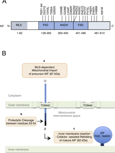
Through phylogenetic studies, it was established that the mi-tochondrialflavoprotein AIF belongs to a family of proteins that are found across eukaryotes and have in common structural and functional characteristics [1,3,5,18–20]. In man, the AIFM1 gene is located on chromosome X (Xq25-Xq26) [1] and the tissue-specific alternative splicing of its precursor transcript allows the biosynthesis of multiple isoforms [6]. Two major splice variants (AIF1 and AIF2) are produced through the alternative usage of exon 2 (2a or 2b) [21]. The ubiqui-tously expressed isoform AIF1 (which uses exon 2a and is commonly called AIF) carries an N-terminal positively-charged bipartite mi-tochondrial localization signal (MLS) that allows its import into the mitochondrion [22] (Fig. 1). Upon mitochondrial import, the precursor
AIF protein (67 kDa) is cleaved by a mitochondrial peptidase that eliminates thefirst N-terminal 53 residues of the MLS [22]. Then, with the help of the remaining C-terminal part of the MLS, which contains an inner membrane sorting signal and a transmembrane segment, the mature AIF protein (62 kDa) is targeted and inserted in the inner membrane (IM) facing the intermembrane space (IMS) [22]. In the mitochondrion, the fully processed protein goes through a conforma-tional maturation that requires the incorporation of its co-factorflavin adenine dinucleotide (FAD) [1,23]. Protein structure analyses have revealed that AIF contains two FAD binding sequences (residues 128 to 262 and 401 to 480) that frame one nicotinamide adenine dinucleotide (NAD+/NADH) binding segment (residues 263 to 400) [3,18] (Fig. 1).
A homology found between the central cofactor-binding segments of AIF and the bacterial NADH-dependent ferredoxin reductase suggested that AIF could be a NADH-dependent enzyme acting on yet to be identified substrates [1,3,18]. Ubiquinone could be one of these sub-strates, as under specific in vitro experimental conditions, AIF, which shares homology with the yeast NADH:ubiquinone oxidoreductase Ndi1, was found to oxidize NADH by acting on ubiquinone [20].
AIF exists both in monomer and dimer forms in the mitochondrion and in vitro experiments coupled to non-reducing SDS/PAGE analyses have revealed that the monomer/dimer equilibrium shifts towards the
A
B
128-262 263-400 401-480 481-613
MLS FAD NADH FAD
N- -C
R201 del D237G V243L T260A G262S
hAIF
G338E L344F G360R
G308E R422W R422Q R430C R451Q A472V P475L Y492H E493V V498M I591M
1-82
- Inner membrane insertion - Cofactor -assisted Refolding
of mature AIF (62 kDa) MLS-dependent
Mitochondrial Import of precursor AIF (67 kDa)
Proteolytic Cleavage between residues 53-54 a b N--C c Cytoplasm Mitochondrial matrix Mitochondrial intermembrane space TOM40 TOM40 Outer membrane Inner membrane AIF FAD / NADH
Fig. 1. A) Schematical presentation of the characteristic domains of human AIF. The domains are: Mitochondrial Localisation Signal (MLS, residues 1 to 82), FAD-binding domain (residues 128 to 262 and 401 to 480), NADH binding domain (residues 263 to 400), C-terminal domain (residues 481 to 613). The position and the corresponding amino acid substitutions for disease-aggregating mutations are shown (seeTable 1for references). B) Mitochondrial import of the precursor AIF protein: a) the precursor AIF protein is im-ported in the mitochondrion using an N-terminal MLS; b) upon import thefirst N-terminal 53 residues are cleaved off by a mitochondrial peptidase; c) the mature AIF is inserted into the inner mitochondrial membrane, via its N-terminal transmembrane region. Concomitant with its membrane in-sertion AIF adopts its physiological conformation by inter-acting with its cofactors (FAD, NAD/NADH).
dimer upon the interaction of AIF with NADH or NADPH [24–29]. At the molecular level, the interaction of AIF with NAD(P)H triggers the formation of tight, dimeric, and stable FADH-NAD(P) charge-transfer complexes (CTC) that are inefficient in electron transfer [24–29]. Based on the formation of stable CTCs and the subsequent conformational reorganization of AIF, it was proposed that the redox-driven process could control AIF functions by regulating its interaction with protein partners, nucleic acids (DNA or RNA) and/or its still unknown enzy-matic activity [9,24–30].
While AIF1 is ubiquitously expressed, the expression of AIF2 (which uses exon 2b) was revealed to be tissue-specific and found only in the brain and retina [21,31]. Protein sequence alignments revealed that the alternative usage of exon 2a and 2b introduced a difference between AIF1 and AIF2 that only targeted the C-terminal part of their MLS, which carries the transmembrane segment [21]. Like AIF1, AIF2 is imported to mitochondria and inserted in the IM facing the IMS where it can support the biogenesis of respiratory chain CI protein subunits [9,21]. In addition to their role in the regulation of the respiratory chain activity, AIF1 and AIF2 could participate to other functions that implicate the structural organization and/or stability of the IM. The potential IM-organizing activity of AIF1 and AIF2 may reside in their respective transmembrane segments that exhibit differences in their membrane anchorage capacities and affect differentially the mi-tochondrial morphology [21]. Even though the tissue-specific
require-ment for the expression of AIF2 is not yet understood, in vivo ob-servations point towards coordination in the expression of the two isoforms. Indeed, it was observed that the loss of Aif2 mRNA in Aif2 knockout (KO) (exon 2b-specific KO) mice was accompanied by an increase in the expression level of Aif1 mRNA, which became even more pronounced in aged Aif2 KO mice compared to their wild-type (WT) littermates [32]. While no phenotypic difference was detected between
Aif2 KO mice and their WT littermates under physiological conditions, it was revealed that in a model of hypoxia/Ischemia (HI)-induced neuronal death, Aif2 mRNA depletion enhanced oxidative stress and aggravated HI-induced neonatal brain injury [32]. In addition to AIF1 and AIF2, several short AIF isoforms (AIFsh1, AIFsh2 and AIFsh3) have also been described [6,33,34]. Proteomics and functional studies will be required to assess the precise cell-specific distribution and function of each one of the isoforms.
Several proteins have been involved in the regulation of AIF ex-pression [6]. For instance, the regulation of AIF mRNA expression by basal levels of p53 is suggested to contribute to the metabolic action of p53 that is known to preserve the mitochondrial respiratory chain ac-tivity [35,36]. A CCAAT-enhancer binding protein alpha
(C/EBPα)-mediated transcriptional upregulation of AIF was implicated in the differentiation of adipocytes [37]. In many colon cancer cell lines, AIF expression was also regulated by hypoxia-inducible factor-1 (HIF-1) [38]. In particular, using luciferase reporter and chromatin im-munoprecipitation (CHIP) assays, Xiong et al. [38] showed that under hypoxic conditions, HIF-1 repressed the transcription of AIF via binding to a hypoxia-responsive element (HRE) in the AIF promoter. The ob-served downregulation of AIF correlated with the hypoxia-induced epithelial-mesenchymal transition (EMT) program [38].
3. AIF and its prosurvival function in the mitochondrion
3.1. The pro-survival mitochondrial function of AIF
Initially, the mitochondrial pro-survival function of AIF was sus-pected by the revelation of a significant homology between the internal, non-apoptotic, segment of AIF and bacterial NADH-oxidoreductases [1,3,18]. It was only later that the molecular and phenotypic char-acterization of the mutant Harlequin (Hq) mouse, a model of late-onset neurodegeneration, revealed the importance of AIF in mitochondria and highlighted its impact on cell survival, proliferation and di ffer-entiation [6,39]. Hq mice were first noted for growth retardation,
alopecia and the development of a progressive ataxia and blindness due to an oxidative stress-associated loss of terminally differentiated cere-bellar and retinal neurons [39,40]. Monitoring the neuroanatomical features of Hq mice revealed that the downregulation of AIF in the Hq brain provoked an early alteration of the mitochondrial ultrastructure that preceded the progressive neurodegeneration observed in various brain regions [41]. All observed phenotypes of Hq mice are due to a retroviral insertion in thefirst intron of Aifm1 gene that provokes an 80% reduction in AIF expression [6,39]. Even though AIF down-regulation is observed in almost all the organs of Hq mice, the impact of AIF hypomorphic Hq mutation on cell metabolism and survival appears to be cell-specific [39,42–45]. In addition to the loss of various neurons and skeletal muscle cells [39,41,44], Hq mice exhibit a T-cell lineage (but not B lineage) development deficiency that can be overcome either by antioxidant treatment or by the overexpression of a wild type AIF protein [45].
Our understanding of the mitochondrial pro-survival function of AIF has also largely benefited from the characterization of additional in vitro and in vivo AIF deficiency models [6]. As AIF is indispensable for embryonic development [46], all attempts to generate AIF null mice by homologous recombination failed and consequently only tissue-specific genetic deletion of Aifm1 gene was used for the characterization of the in vivo role of AIF protein in cell survival, differentiation and organo-genesis [6]. For instance, the specific deletion of Aifm1 in the
pro-spective midbrain and cerebellum revealed that AIF plays a pivotal role in the cell-type specific neurogenesis during brain development [47]. A tissue-specific deletion of Aifm1 in the telencephalon highlighted a major impact of AIF on the neuronal survival and cortical development [48]. The conditional deletion of Aifm1 in organs such as muscle and liver brought to the light an important role of AIF on the whole-body metabolism as, compared to control littermates, these mutant mice exhibited resistance to diet-induced obesity and diabetes [49]. With age, muscle-specific loss of AIF leads to a progressive skeletal muscle atrophy and a dilated cardiomyopathy [50]. Recently, the relevance of the mitochondrial function of AIF in the hematopoietic development was addressed in conditional Aifm1 KO mice models [51,52]. While Milasta et al. [51] showed that the loss of AIF provoked metabolic defects only in mature T (but not B) lymphocytes, a more recent in-dependent study, realized by Susin and coworkers, indicated that AIF deficiency could also affect B cells and erythroid lineages [52]. Dis-crepancies between the studies are probably explainable by differences in the knockout strategy that could impact on the developmental timing of AIF depletion.
3.2. AIF regulates the optimal functioning of the respiratory chain machinery
The detection of oxidative stress in degenerating Hq mutant neurons combined to the observation of enhanced sensitivity of various Hq cells to oxidant-triggered cell death, brought out the idea that AIF could have a free-radical scavenging role in the mitochondrion [39,40,53–56]. Nonetheless, the implication of AIF in this particular activity appeared controversial as conflicting results generated in var-ious experimental AIF-deficiency models failed to reveal AIF as a direct regulator of reactive oxygen species (ROS) [42,43,49,57–62].
As an alternative interpretation for the increase of oxidative stress in AIF-deficient mice, it was proposed that the loss or downregulation of the unknown enzymatic activity of AIF could provoke a dysfunction in the oxidative phosphorylation and/or perturb specific redox-regulated mitochondrial pathways [63]. Biochemical and metabolic analyses of organs from Hq mice, or murine and human cells lacking AIF as a result of homologous recombination or knockdown by RNA interference al-lowed Kroemer and colleagues [42] to validate this hypothesis and show that AIF is indeed necessary for the optimal functioning of the mitochondrial respiratory chain machinery and is involved in redox metabolism. In the same study, it was also revealed that among
mitochondrial respiratory chain multi-protein complexes, CI (nicoti-namide adenine dinucleotide (NADH): ubiquinone oxidoreductase; EC 1.6.5.3) was the most disturbed upon AIF deficiency and that the ob-served enzymatic dysfunction of CI was caused by a post-transcriptional loss of CI protein subunits [42]. The impact of AIF depletion on CI was also confirmed in vitro in ES cells, in vivo in murine embryos, skeletal muscle, heart, liver, brain and hematopoietic cells in which Aifm1 was conditionally knocked out [9,42,49–52,64]. Occasionally, cell-specific
or tissue-specific dysfunctions of complexes CIII and CIV have also been associated with the loss of AIF but, so far, the contribution of these supplementary defects to the phenotype of AIF-deficient mice remains elusive [9,42,49–52]. Even though the main AIF isoform (AIF1) is ex-pressed in almost all organs, one intriguing observation is that the impact of its dysfunction is not equal in all cell types [39,42,43,45,49,51]. For instance, in Hq mice CI dysfunction is mainly confined to specific brain areas that undergo degeneration and there is a clear correlation between the downregulation of AIF in degenerating neurons, the progressive aggravation of CI dysfunction and the phe-notypic evolution of the disease [43]. Similarly, the phenotypic char-acterization of additional AIF-deficiency animal models supports the notion that the impact of the mitochondrial dysfunction, which is caused by the loss of AIF, is rather cell-type specific [48,49,51,52,65]. The molecular and metabolic bases for the vulnerability of specific cells to AIF deficiency are not yet clearly understood. However, studies conducted by Germain et al. [65] indicate that the resistance of certain types of neurons to AIF deficiency correlates with the activation of a metabolic compensatory mechanism implicating the LKB1 kinase.
The requirement for AIF in the regulation of oxidative phosphor-ylation is conserved during evolution. The knockout of zygotic Drosophila melanogaster AIF (DmAIF) mRNA resulted in a larval growth arrest that was accompanied by the enzymatic dysfunction of the complexes CI and CIV [66]. Recently, Troulinaki et al. [67] showed that the knockdown of WAH-1/AIF in C. elegans shortens nematode's life-span by perturbing the mitochondrial respiratory activity. Constitutive antisense-mediated downregulation of AIF in Dictyostelium discoideum was reported to perturb growth and the development of the model organism and the observed effects were accompanied by a reduction in the levels of mitochondrial ATP level and an enhancement in glucose-dependency [68]. In Saccharomyces cerevisiae (which lacks complex CI), the knockout of AIF ortholog (AIF1/YNR074C protein) negatively im-pacted on the respiration and growth on non-fermentable carbon sources [42].
3.3. AIF interacts with the mitochondrial CHCHD4/Mia40-dependent import pathway
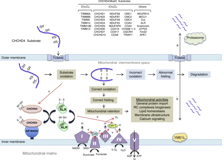
Neither downregulation nor depletion of AIF had major effects on the mRNA levels of respiratory chain subunits [42], indicating that AIF was either necessary for the post-transcriptional biogenesis/assembly of CI protein subunits or for the maintenance of the structure of the multi-protein complex [5]. The isolation of thefirst mitochondrial interactor of AIF, namely CHCHD4, revealed the implication of AIF in the redox-regulated CHCHD4-dependent protein import pathway, thus providing a molecular explanation for the involvement of AIF in the biogenesis/ assembly of the respiratory chain protein subunits (Fig. 2) [9,10]. CHCHD4 is the human homolog of yeast mitochondrial intermembrane space import and assembly protein 40 (Mia40), which is the core component of a disulfide relay-dependent import machinery that con-trols the import of small nuclear-encoded cysteine motif-carrying pro-teins (called substrates) into the IMS [11,17,69–73]. Published data indicated that AIF regulates the CHCHD4-dependent import pathway by interacting with and controlling the mitochondrial import of CHCHD4 [9]. In the proposed molecular model, the mitochondrial bioenergetics dysfunctions that are provoked in the absence of AIF are secondary to the deficiency of CHCHD4 and the subsequent loss of CHCHD4-specific protein substrates [9–11]. In agreement with this
possibility, the progressive CHCHD4 deficiency, which was detectable in the Hq brain postpartum, precedes the loss of CI subunits that manifests during early Hq mice adulthood [9]. The overexpression of CHCHD4 in Hq mutant mouse embryonic fibroblasts (MEF), or in murine and human cells lacking AIF as a result of homologous re-combination or knock-down by RNA interference, restored levels of CHCHD4 substrates and corrected CI deficiency, confirming that CHCHD4 operates downstream of AIF [9,10].
Homozygous knockout of Chchd4 gene leads to embryonic lethality [9]. This is reminiscent of the fact that genes encoding critical protein import components result in embryonic lethality when knocked out, in line with their essentiality for viability in simple eukaryotic cells like S. cerevisiae. However, mice bearing a heterozygous deletion of Chchd4 are viable and do not exhibit obvious phenotypic changes [9]. Interestingly, similar to mice carrying the Hq mutation in the whole-body or tissue-specific knockout of Aifm1 in liver or muscle, animals with a hetero-zygous knockout of Chchd4 exhibit resistance to weight gain when ex-posed to a high-fat diet [49,74]. These data [49,74] combined with the observation that CHCHD4 is downregulated in many organs of Hq mice [9,10] indicate that the resistance of AIF-deficient mice to high fat
diet-induced obesity could be secondary to the dysfunction of the CHCHD4-dependent import pathway.
4. The CHCHD4/Mia40 mitochondrial import pathway
4.1. The CHCHD4/Mia40-dependent import machinery from yeast to man
It was about 15 years ago that the oxidative folding system (also known as the disulfide relay system) that operates in the mitochondrial IMS and implicates the CHCHD4/Mia40-dependent import machinery was discovered in the yeast Saccharomyces cerevisiae [69–72,75]. Initial results showed that small, cysteine-rich proteins of the IMS must be folded through oxidation of their internal cysteines to form in-tramolecular disulphide bonds [75,76]. Subsequently, it was discovered that Mia40 is the key component that catalyses this oxidative folding mechanism in mitochondria [69–72]. To perform its key role as the core component of this redox-regulated machinery, Mia40 recognizes its substrates (a set of small nuclear-encoded cysteine-rich proteins) by combining two separate functions. Thefirst one is cysteine-independent and underpins the initial recognition of the preprotein, as the latter emerges from the TOM (Translocase of the Outer Membrane) entry gate in the outer membrane. This step has been described as the‘sliding step’ in which the peptide orients itself to the hydrophobic cleft of Mia40 directed by hydrophobic residues within the internal IMS-specific Tar-geting Signal (called ITS or MISS) of the incoming preprotein. This hydrophobic non-covalent packing of the substrate to the cleft of Mia40 positions the preprotein in such a manner so that its docking cysteine can now bind covalently to the redox-active cysteine-proline-cysteine (CPC) motif of Mia40 in what is called the‘docking step’. Now the substrate protein is bound covalently to Mia40 via an intermolecular disulfide bond [77–80]. The dual activity of Mia40 therefore consists of a chaperone function that induces the folding of the substrate in the first ‘sliding’ step, followed by a disulfide donor function in the second ‘docking’ step. The combination of these two activities leads to the catalysis of the oxidative folding of the preprotein in the IMS, which thereby traps the protein and retains it in the IMS [16,81–83]. During this process, Mia40 donates its disulfide to the substrate whilst its CPC motif becomes reduced. It is therefore critical that the CPC motif can be reoxidised back to its functional state to ensure another cycle of sub-strate oxidation. This critical reoxidation step of the CPC motif is en-sured by the second key component of the oxidative folding machinery, the FAD-linked sulfhydryl oxidase Erv1 (human equivalent: ALR) [84–86]. Specifically, the N-terminal segment of Erv1 that contains a CX2C motif and is natively disordered mediates the reoxidation of the
CPC motif [87]. The pair of electrons released after thefirst interaction of the substrate with Mia40 is subsequently transferred in a cascade of
transfer reactions (i) from Mia40 to the N-terminal cysteines of Erv1 (ii) from the N-terminal shuttle of Erv1 to the cysteine pair that is proximal to the FAD domain in the core of Erv1, (iii) from the FAD-proximal cysteine pair to FAD, (iv) from the FAD to cytochrome c and (v) from cytochrome c to the cytochrome oxidase (COX) complex and finally molecular oxygen [88]. In addition to molecular oxygen, there are several otherfinal electron acceptors such as the cytochrome c perox-idase (Ccp1) or the protein Osm1 that operate under anaerobic condi-tions [89]. In human cells, the reoxidation of the CPC motif of CHCHD4 is ensured by its recycling enzyme ALR (homolog of the S. cerevisiae enzyme Erv1) [72,78,90,91] (Fig. 2). By developing a protein oxidation assay in intact human cells, Fischer et al. [92] have shown that the coupled import and oxidative folding of CHCHD4 substrates require the CHCHD4 oxidase as the main catalytic component in the process and its recycling partner ALR.
Structural and functional comparisons of the yeast and higher
eukaryote Mia40 (human equivalent: CHCHD4) show that the principal catalytic and structural features of Mia40, in particular within the segment that contains the redox active cysteine-proline-cysteine (CPC) and the (CX9C)2motifs, have been conserved throughout eukaryotic
evolution [9,72,92–96]. However, some important differences have also emerged. Probably the most interesting feature is that during evolution, the yeast Mia40 (~40 kDa) has lost its N-terminal pre-sequence and its membrane anchoring segment (which comprise almost two thirds of the entire molecule and are dispensable for its function) and has become the mammalian CHCHD4 (16 kDa), a soluble protein that is no longer imported via the TIM23 translocase (which in yeast cells interacts with the yeast Mia40 N-terminal segment) but rather relies on a self-catalytic import process [9,72,93–95]. It is plausible that, during evolution, the import of CHCHD4 would become more tightly coupled to the IMS-localized disulfide relay system, presenting an advantage to sustain adaptive responses in mitochondria of
Mitochondrial matrix
CHCHD4 Substrate Substrate oxidation S I S S I S Outer membrane Inner membraneMitochondrial intermembrane space TOM40 H+ H+ H+ CoQ Cyto C NADH NAD+ Fumarate Succinate O2 H2O ADP ATP ALR e -e -e -e -CHCHD4 CPC SH SH TOM40 H+ ALR AIF/NADH CHCHD4 CP C S - S SH SH S - S e- Correct oxidation Mitochondrial retention Abnormal folding Degradation YME1L a b Proteasome (Cx3C)2 (Cx9C)2 CHCHD1 CHCHD6 CHCHD3 NDUFA8 NDUFB7 NDUFS5 NDUFS8 UQCRH COX6B 1/ 2 CHCHD10 CHCHD4 TRIAP1 MICU1 CHCHD2 CMC1 CMC2 CMC4 COA4 COA6 COX17 COX19 CHCHD7 CHCHD4/Mia40 Substrates TIMM8A TIMM8B TIMM9 TIMM10 TIMM10B TIMM13 NDUFB10 NDUFAF8 CHCHD5 COA5 Others Ccs ALR Anamorsin ATP23 Tim22 APE1 Mitochondrial activities General protein import RC complexes biogenesis Lipid homeostasis Membrane ultrastructure Calcium signaling O2 Incorrect oxidation Correct folding FAD FAD
Fig. 2. CHCHD4/Mia40-dependent import pathway in metazoan. Nuclear-encoded substrates carrying (CX3C)2or (CX9C)2motifs or non-conventional cysteine motifs
are translated by cytoplasmic ribosomes and imported, in their reduced form, into the mitochondrial inter membrane space (IMS), by passing through the outer membrane-localized TOM translocase. In the IMS, the oxidase CHCHD4/Mia40 regulates the import and/or the post-import folding of the substrates by catalysing the formation of disulfide bonds. The mitochondrial import and optimal functioning of CHCHD4 requires its interaction with the AIF/NADH complex. Electrons (red arrow) generated by the oxidation of the substrate are transferred to the CPC motif of CHCHD4/Mia40, to ALR, to cytochrome c (Cyto c), to complex CIV and oxygen (O2). A direct transfer of electron from ALR to O2is an alternative possibility. Electronflow generated by the activity of the respiratory chain complexes is also
depicted (red arrow). Upon vectorial import, the correctly oxidized and folded CHCHD4/MIA40 substrate is retained in the mitochondrion and engaged in a wide panel of activities such as: general protein import (via (CX3C)2motif-carrying small TIM proteins). The mitochondrion-imported (CX9C)2motif-carrying and the
non-conventional substrates of CHCHD4 participate to a vast spectrum of activities that include protein import, lipid homeostasis, mitochondrial ultrastructure and dynamics, Ca2+storage, respiratory chain complexes biogenesis and assembly and mitochondrial protein translation. The dysfunction of the major constituents of the
machinery (CHCHD4, ALR and AIF) or mutations in the cysteine motifs of the substrates can inhibit or alter the oxidation-dependent folding of the substrate. In this case, a quality control check will identify and eliminate the misfolded protein either via its degradation a) in the IMS via the proteolytic activity of YME1L or b) in the cytoplasm, via the activity of the proteasome. This latest possibility requires the retrotranslocation of the substrate from the mitochondrion to the cytoplasm.
specialized cells. Such responses could involve for example fast and/or signal-dependent metabolic changes. The acquisition of the capacity to interact with a new partner, the IM-bound flavoprotein AIF (Fig. 2), may represent an additional evolutionary advantage for CHCHD4 that is still not clearly understood [9,10,97]. The N-terminal 27 residues of CHCHD4 are necessary and sufficient to establish a direct interaction with AIF [9]. Nuclear magnetic resonance (NMR) spectroscopy in-dicates that the AIF-binding domain of CHCHD4 resides within the N-terminal unstructured lid segment that includes the redox-active center CPC [72]. However, far-UV circular dichroism (CD) spectroscopy ana-lysis suggests that upon its interaction with AIF, the AIF-binding seg-ment of CHCHD4 undergoes important conformational rearrangeseg-ments that may well affect the interactions of the CPC domain of CHCHD4 with its incoming substrates or its reoxidising partner ALR [9]. Of note, recent quantitative proteomic characterization of CHCHD4 binding partners in human cells, confirmed previous in vitro observations in-dicating that mutation of either cysteine of the CPC motif (C53S or C55S) has no impact on the interaction of CHCHD4 with its partner AIF [9,91]. On the other hand, the conformational state of AIF, which is determined by the binding of its cofactor NADH [25,29], appears cri-tical for its interaction with CHCHD4 [9]. Addition of NADH or NADPH enhances the interaction between CHCHD4 and AIF, yet fails to do so when AIF is mutated (G308E) in its NADH-binding domain [9] (Fig. 1;
Table 1). Even though it is not yet clear how the interaction with AIF affects the redox-state and availability of CHCHD4, AIF has been shown to be an important actor that is required for the cotranslational import of CHCHD4 into the IMS [9] (Fig. 2). Downregulation or abnormal functioning of CHCHD4 leads to the loss of its substrates [9,10,91–93,98]. Based on all available data so far it is thought that CHCHD4 is the main catalytic component in the oxidative folding pathway, whereas the FAD-binding proteins AIF and ALR are regulators of the CHCHD4 import function.
4.2. CHCHD4/Mia40 substrates and their implication in mitochondrial activities
The majority of the substrates of the CHCHD4/Mia40 import pathway are proteins of a relatively small size (usually less than 25 kDa) and share a conserved coiled coil-helix1-coiled coil-helix 2 (CHCH) domain (CHCHD). In each of the helices there are two cysteines separated by 3 (CX3C) or 9 (CX9C) residues, with the two cysteine
motifs juxtaposed to each other and connected by intramolecular dis-ulfide bonds [14,82,114–116] (Fig. 2).
Thefirst (CX3C)2-containing substrates to be studied in detail were
yeast small Tim proteins [114], which are IMS-localized chaperones that facilitate the mitochondrial import of hydrophobic membrane proteins by ushering them from the TOM entry gate in the outer membrane to theirfinal destination in the inner or outer membranes [114]. Among the small Tim proteins Tim9 and Tim10 are encoded by essential genes in S. cerevisiae, whereas the Tim8 and Tim13 genes are dispensable. In human cells however, deletion or loss-of-function mu-tations in the gene encoding the human homolog of yeast Tim8 called DDP1/TIMM8A [117] is responsible for a rare X-linked recessive neu-rological disorder called the deafness-dystonia or Mohr-Tranebjaerg syndrome [118,119]. In mammalian cells, the loss of AIF disturbs the mitochondrial import of CHCHD4 and consequently impacts on the biogenesis of DDP1/TIMM8A indicating that the mitochondrial activity of AIF is not limited to the regulation of the biogenesis of CI [9].
The second family of Mia40 substrates contains a twin (CX9C) motif
and controls oxidative phosphorylation (OXPHOS) either directly through the regulation of the biogenesis/assembly of the subunits of the respiratory chain complexes, or indirectly through the control of the mitochondrial ultrastructure, or lipid homeostasis of the mitochondrial IM [11,14,16,115,120].
Studies conducted in yeast revealed that among the (CX9C)2motif
carrying substrates, the evolutionarily conserved proteins Cmc1, Cmc2, Cox6B, Coa4, Coa5, Coa6, Cox17, Cox 19 and Cox23 are implicated directly in the biogenesis or in the assembly of the respiratory chain cytochrome c oxidase complex (COX/CIV) [11,14–16,115,120,121]. In Table 1
Human disease-associated AIFm1 genetic alterations.
Nucleotide alteration aa change X chromosome-Linked disease Reference
c.422C > T T141I Progressive ataxia, brain atrophy and auditory neuropathy [99]
c.513G > A M171I Early childhood-onset axonal polyneuropathy [100]
c.601delAGA R201 deletion Mitochondrial Encephalomyopathy [101]
c.629T > C F210S Early childhood-onset axonal polyneuropathy [102]
c.630C > G F210L Late-onset axonal polyneuropathy [103]
c.705G > C Q235H Hypomyelinating leukodystrophy and
spondylometaphyseal dysplasia
[104]
c.710A > T D237V Hypomyelinating leukodystrophy and spondylometaphyseal dysplasia [104]
c.710A > G A237G Hypomyelinating leukodystrophy and spondylometaphyseal dysplasia [104]
c.710A > G D237G Spondyloepimetaphyseal dysplasia with neurodegeneration [105]
c.727G > T V243L Progressive muscular atrophy, ataxia and hearing loss [106]
c.778A > G T260A Familial auditory neuropathy spectrum disorder [107]
c.784 G- > A G262S Slowly progressive encephalopathy [108]
c.923 G- > A G308E Early prenatal ventriculomegaly [109]
c.1013G > A G338 E early-onset encephalopathy, muscular atrophy, and motor axonal neuropathy [110]
c.1019T > C M340T Progressive ataxia, brain atrophy and auditory neuropathy [99]
c.1030C > T L344F Familial and sporadic auditory neuropathy spectrum disorder [107]
c.1078G > C G360R Sporadic auditory neuropathy spectrum disorder [107]
c.1264C > T R422W Familial and sporadic auditory neuropathy spectrum disorder [107]
c.1265G > A R422Q Familial auditory neuropathy spectrum disorder [107]
c.1288C > T R430C Sporadic auditory neuropathy spectrum disorder [107]
c.1352G > A R451Q Familial auditory neuropathy spectrum disorder [107]
c.1415C > T A472V Sporadic auditory neuropathy spectrum disorder [107]
c.1424C > T P475L Sporadic auditory neuropathy spectrum disorder [107]
c.1436A > G Q479R Neonatal mitochondriopathy [111]
c.1474T > C T492H Neonatal mitochondriopathy [112]
c.1478A > T E493V Cowchock syndrome (CMTX4). Hearing and sensorial loss, mental retardation, muscle weakness and axonal neuropathy [113]
c.1492G > A V498M Sporadic auditory neuropathy spectrum disorder [107]
human cells, the abundance or the normal functioning of CHCHD4 was correlated with the respiratory chain activity and the abundance of several of these proteins (e.g. CMC1, COA4, COX6B1, COX17 and COX19) [9,91–93]. The negative impact of AIF deficiency on CIV ac-tivity, which is observed in a tissue-specific manner, could be explained at least partially, by the loss of these specific substrates of CHCHD4. This possibility is supported by the observation that the depletion of AIF in human cells is accompanied by downregulation of the copper cha-perone COX17 [9]. The overexpression of CHCHD4 restores the ex-pression level of COX17 that is secondary to the depletion of AIF [9].
Additional twin CX9C motif-carrying respiratory chain complex
subunits, which have appeared during the evolution and were sug-gested to be substrates of CHCHD4/Mia40, are the subunits NDUFB7, NDUFS5, NDUFA8, and NDUFS8 of CI (Fig. 2) [15,92]. Among this group of substrates, NDUFB7 and NDUFA8 are localized in the IMS and likely stabilize the assembled CI by binding to its surface [122]. In mammalian cells, the correlation between the abundance of CHCHD4 (and its binding partner AIF) and the expression of many of these proteins is a clear indicator of the direct implication of the CHCHD4-dependent import pathway in the biogenesis of CI [9,10,91,92]. How-ever, it is noteworthy to mention that, in mammalian cells, AIF deple-tion negatively impacts on the expression of addideple-tional protein subunits of CI (NDUFA9, NDUFS7, NDUFB6, NDUFB8, and NDUFA13), CIII (UQCR1, UQCR2, and UQCRFS1) and CIV (COX2) that are not direct substrates of the CHCHD4 [9,42]. This secondary consequence of AIF depletion can be corrected by the overexpression of CHCHD4, estab-lishing the key role of the CHCHD4-dependent import pathway in the indirect control of these specific set of respiratory chain protein sub-units [9]. One could speculate that the expression of the above-men-tioned proteins could be indirectly controlled by the activity of a set of CHCHD4 substrates that regulate the mitochondrial ultrastructure (e.g. CHCHD2, CHCHD3, CHCHD6, CHCHD10 or TRIAP1) [11]. It is now established that the organization of the mitochondrial inner membrane and the formation of cristae are finely tuned processes that have a major impact not only on the formation of the respiratory chain su-percomplexes but also on the activity of many protein complexes en-gaged in various mitochondrial processes, or in the translation of mi-tochondrion-encoded proteins [123–125]. There is no doubt that a more in depth characterization of these specific CHCHD4 substrates will help to better understand the molecular basis for the mitochondrial fragmentation and perturbed mitochondria-lysosome cross-talk that accompanies OXPHOS dysfunction in various AIF deficiency models [21,42,48–50,65,126]. It is worth mentioning that many of the above-mentioned (CX9C)2motif-carrying proteins were also found mutated or
abnormally expressed in various human disorders (Supplemental Table 1). Obviously, molecular and functional characterization of in-dividual proteins will be instrumental in the evaluation of the relevance of their dysfunction in the development of specific disease condition.
Although most studies on the substrates of CHCHD4/Mia40 have focused so far on proteins that contain either (CX3C)2or (CX9C)2motifs,
there is an increasing number of substrate proteins not carrying (CXnC)2
motifs. The CHCHD4/Mia40 binding repertoire needs to be expanded to include larger proteins with cysteine motifs that are not organized structurally in a CHCH domain [11,127]. A typical example of this type of protein is Erv1 itself, which contains a CX16C motif [128]. NDUFB10,
a new addition to the list of CHCHD4 substrates, is an accessory subunit of complex I, which carries an atypical cysteine motif (CX6C/CX11C). A
mutation in the conserved cysteine C107 of NDUFB10 caused a severe CI-related mitochondriopathy and was reported to inhibit the CHCHD4-dependent oxidation of the protein and its mitochondrial accumulation [129].
Furthermore, another type of CHCHD4/Mia40 substrate is re-presented by MICU1, a mitochondrial regulator of the Ca2+uniporter (MCU) [97]. In human cells, the import of MICU1 in the IMS requires an N-terminal targeting sequence while its interaction with CHCHD4 and cysteine oxidation are not as critical as its membrane
potential-dependent import requirement [97]. This latter example underscores the versatile implication of the CHCHD4/Mia40 system in multiple distinct import pathways.
4.3. The CHCHD4/Mia40 pathway and quality control/YME1L/Yme1 protease/cytoplasmic proteasome
Mia40-orchestrated oxidative protein folding is an intricate and highly organized process that requires particular orientation of the substrate protein in the hydrophobic binding cleft of Mia40 through the substrates' ITS/MISS (IMS-specific Targeting Signal) followed by precise oxidation of the substrates cysteine motif. But what happens when a Mia40 substrate is cysteine-trapped on Mia40 or misfolded? In recent years, quality control processes that govern the fate of an aberrant Mia40 substrate have been uncovered. The yeast mitochondrial inner membrane-anchored AAA protease, Yme1 (Yeast mitochondrial escape 1), is involved in the disaggregation and proteolytic degradation of IMS proteins, particularly Mia40 substrates [130–139]. Two studies fo-cusing on the dispensability of any single cysteine residue within the Mia40 substrates Tim9 and Tim10 showed that unstable forms of Tim9 or Tim10 resulted in degradation by Yme1, particularly if the inner disulfide bond was missing [140,141]. These were in agreement with a previous study that showed the inner disulfide bond to be more im-portant than the outer disulfide for maintaining the structure of the small Tims [142]. Yme1 also possesses chaperone-like activity and has been shown to aid and facilitate the disaggregation of the Mia40 sub-strates Tim10 and Erv1 [143,144]. The human homolog of Yme1 (YME1L) was also reported to be responsible for the degradation and elimination of CHCHD4 substrates (e.g. COX17 and NDUFA8) that are probably not correctly processed in cells in which AIF was down-regulated as a result of knockdown by RNA interference [9]. This pathway could fulfil the function of a quality control system operating on poorly folded or damaged Mia40 substrates where initially Yme1 attempts to salvage these proteins via its chaperone activity before eventually degrading them.
Although misfolded Mia40 substrates pose a challenge to mi-tochondrial function, an unfolded and trapped Mia40 substrate prob-ably presents an even greater challenge. If a Mia40 substrate forms a mixed disulfide with Mia40 and is not released, the abnormal protein complex not only blocks other Mia40 substrates from import but also negatively impacts on the import of the whole cohort of nuclear-en-coded proteins that pass through the TOM complex. To overcome this hurdle reducing enzymes found in the IMS are responsible for the re-lease of the disulfide-bonded Mia40 substrate. In mammalian cells, the reducing enzyme glutaredoxin 1 (GRX1) is present in the IMS and has been shown to modulate the oxidation of the CHCHD4 substrate COX17 [91]. A similar role may be ensured by the yeast thioredoxin 1 (Trx1), which also localises to the IMS [145]. This would be an interesting and plausible mechanism because the glutaredoxin and thioredoxin redu-cing pathways have overlapping functions and usually operate in the same compartments. Another mechanism that may operate to clear a subset of substrates of Mia40 that stay unfolded in the IMS involves a retrotranslocation step back to the cytosol. In yeast, it has been shown that certain Mia40 substrates can escape from the IMS in an unfolded state [146]. However, not all Mia40 substrates have the capacity to retrotranslocate as the overall length of the protein as well as its 3D structure limit such an escape route back to the cytosol. This retro-translocation is thought to take place through the TOM channel al-though the exact mechanism for this retrograde movement is still un-known. Once back into the cytosol these Mia40 substrates get degraded by the cytosolic ubiquitin-proteasome system (UPS). Several studies have highlighted the role of the UPS in the degradation of CHCHD4/ Mia40 substrates [91,146,147]. Interestingly, the UPS not only de-grades Mia40 substrates that have failed to be retained in the IMS after import, but may also compete for cytosolic Mia40 substrates not yet imported by degrading them [147]. This competition is likely due to
slower kinetics of import of certain Mia40 substrates and could serve as a regulatory mechanism to control the levels of Mia40 substrates inside mitochondria in response to different metabolic requirements of the cell. One could speculate that deubiquitinating enzymes may also play a role in this regulation.
The studies mentioned above would suggest that the optimal func-tion of the CHCHD4/Mia40-dependent import pathway relies on a mitochondrial protein quality control checkpoint, where misfolded proteins are either directly degraded in the organelle, or delivered to the default proteasome degradation machinery in the cytosol. The end result is to ensure the elimination of toxic proteins, whilst maintaining some level of adjustment to specific metabolic needs (Fig. 2). 4.4. The AIF/CHCHD4-dependent import pathway in health and disease
4.4.1. AIF in health and disease
The characterization of the mitochondrial function of AIF in mul-tiple model organisms including yeast,flies, worms and the observation that mice affected by a hypomorphic AIF mutation (Harlequin mice) or bearing tissue-specific knockout of AIF develop fatal neuromuscular mitochondriopathies [6] had let the community to speculate that AIF dysfunction could be associated with cases of X chromosome-linked human mitochondriopathies. These disorders (affecting 1 in every 5000 individuals) include many heterogeneous, systemic or tissue-specific metabolic diseases that primarily affect infants and are caused by in-born or progressive defects in the respiratory chain activity [148]. Since 2010, several pathogenic AIF mutations have been discovered asso-ciated with a large panel of human mitochondrial diseases that differ in their clinical phenotype and severity (Fig. 1;Table 1).
Among mitochondrial disease-segregating AIF mutations (R201del, V243L, G262S, G308E, G338E and Q479R), which have been found associated with a pronounced reduction in the expression levels of protein subunits and enzymatic activity of the respiratory chain com-plexes, AIF R201del and G308E are the most characterized and are until now the only ones reported for their interference with the AIF/CHCHD4 import pathway [9,10,29,111,149]. AIF R201del mutation, which consists of the deletion of three base pairs coding for the arginine re-sidue 201 of the precursor AIF protein, was the first AIF mutation identified as the genetic basis for the X-linked mitochondriopathy of two male infants born from monozygotic twin sisters and unrelated fathers [101]. The biochemical examination of fibroblasts from both patients revealed a major defect in CIII and CIV that was partially corrected by the overexpression of recombinant wild type AIF or by continuous culture of mutantfibroblasts in the presence of riboflavin [101]. Moreover, muscle biopsies from both patients revealed a severe loss of mitochondrial DNA that could be responsible for the multi-complex (CI, CIII and CIV) dysfunction [101]. Molecular modelling of the mutant AIF, as well as in vitro experiments indicated that the pa-thogenic AIF was unstable and exhibited altered folding and redox properties [29,101,149,150]. The instability of AIF R201del was also reflected by its low expression in patient-derived fibroblasts and in various organs (liver, heart, cerebellum and muscle) of the equivalent knockin mouse model for the human AIF R201del mutation (AIF R200del) [10,151]. Interestingly, the reduction in the expression levels of AIF R201del in patient-derivedfibroblasts correlated with CHCHD4 protein deficiency and loss CI protein subunits similarly to glial cells and organs (e.g. brain) of Hq mice [10]. This observation suggested that disease-segregating mutations of AIF reducing the protein stability could become pathogenic by triggering a CHCHD4 deficiency-related dysfunction of respiratory chain complexes. Studying the phenotypic and metabolic behaviour of the Aifm1-R200del knockin mouse model, Bano and colleagues [151] showed that mice expressing the pathogenic variant of AIF had a normal fur, weighed slightly less than WT litter-mates, and developed an early onset myopathy that was mainly asso-ciated with the loss of CI and CIV protein subunits. Contrary to AIF hypomorphic Hq mutant mice, which exhibit an early onset progressive
CHCHD4 deficiency followed by a CI dysfunction-related neurodegen-eration within the cerebellum, neither CHCHD4 downregulation nor signs of neurodegeneration could be detected in the cerebellum of Aifm1-R200del knockin murine model [151]. More in-depth investiga-tions should help to better understand the lack of correlation between the expression levels of AIF R200del and that of CHCHD4 in the brain of the knockin mouse. Reduced expression levels of V243L, G262S, G338E AIF variants were detected in patient-derived cells, but so far there is no indication on the impact of the above-mentioned mutations on the stability of AIF variants and on the expression levels of CHCHD4.
Linkage analysis followed by exome sequencing led to the descrip-tion of the missense pathogenic mutadescrip-tion of AIF (G308E) that is asso-ciated with a severe prenatal encephalo-myopathy and complexes CI and CIV defects [109]. Structural and biochemical analyses of this particular AIF variant revealed that the G308E substitution did not affect the general conformation or stability of the protein but rather perturbed its capacity to bind its cofactor NAD(P)H and to catalyse redox reactions [28,29,150,152]. So far, the G308E mutation is the only one to be reported to inhibit the capacity of AIF to bind to its mi-tochondrial interactor CHCHD4, shedding new light on the molecular basis for the pathogenicity of this particular mutation [9].
Other AIF mutations, which are exemplified by F210L and E493V substitutions, have been associated with late onset and slowly pro-gressing mitochondriopathies that exhibit relatively mild clinical phe-notypes [29,103,113]. The AIF F210L variant segregated with a late onset axonal polyneuropathy, and biochemical analyses of patient-de-rivedfibroblasts revealed that the expression of AIF F210L was asso-ciated with a loss of respiratory chain protein subunits that was limited to CI and CIII (levels of CIV protein subunits were not affected) [103]. Moreover, it was observed that the mutation did not affect the capacity of AIF to interact with CHCHD4, suggesting that the observed pertur-bation in CI and CIII biogenesis was independent of AIF/CHCHD4 complex formation [103]. The pathogenic AIF mutation E493V, which was reported to enhance the apoptotic activity of the protein (without affecting its general conformation or expression levels), is another mutation of AIF that did neither affect the in vitro interaction of AIF with CHCHD4 nor its capacity to control respiratory chain activity [9,29,113,150].
While AIF mutations have been mainly associated with human neurodegenerative mitochondriopathies, in the context of diseases such as cancer published data report mainly changes in its expression levels [153–160]. For instance immunohistochemical analysis of AIF in 103 colorectal carcinoma revealed that in the majority of analysed cases, AIF expression was much higher in tumour cells compared to sur-rounding normalfibroblasts or mucosal epithelial cells [153]. In the same study, mutation analysis of AIF (exon 10 to 15) by single-strand conformation polymorphism (SSCP) in 48 colorectal, 48 gastric, 48 breast, 48 hepatocellular carcinomas and 48 leukemias indicated that AIF mutation was an extremely rare event in cancer [153]. High ex-pression levels of AIF protein were observed in the majority of Diffuse large B Cell lymphoma (DLBCL) [155,156] and associated to a better overall survival of the patients [155]. Wilkinson and colleagues [158] reported that AIF levels were increased in prostate tumours and showed that the mitochondrial enzymatic activity of AIF that regulates the optimal function of the respiratory chain complexes was required for the tumour growth and invasiveness of advanced prostate cancer. Even though the elevation of the expression levels of AIF in pancreatic tu-mours was considered modest compared to normal tissue, knockdown of AIF in a set of pancreatic cancer cells that differed in their metabolic status inhibited cell growth and correlated with the loss of respiratory chain complexes and glucose consumption [161]. In patients with non-small cell lung cancer, high AIF expression was associated with poor prognosis [160]. Deletion of Aifm1 in a KrasG12D-driven mouse model of lung cancer negatively impacted on tumour initiation and malignant progression and ameliorated the overall survival of the mice [160]. At the biochemical level, the growth disadvantage, which was caused by
the deletion of Aifm1, was accompanied by the respiratory chain dys-function and a metabolic switch towards glycolysis [160]. The re-ex-pression of both WT and mutant AIF (which was proficient for its mi-tochondrial activity but disabled for its nuclear translocation and apoptotic activity) in Aifm1-knockout KrasG12D mice restored mi-tochondrial respiratory chain activity, rescued tumour growth and re-duced mice survival [160]. In vitro knockdown of AIF or CHCHD4 in human lung adenocarcinoma cell line A549 similarly provoked re-spiratory chain dysfunction and inhibited proliferation [160]. Taken together, these data highlight the crucial role of the mitochondrial bioenergetics in the growth of lung tumour and confirms the critical role of AIF and CHCHD4 in the control of OXPHOS and mitochondrial metabolism.
4.4.2. CHCHD4 in health and disease
Until now, only changes in the expression levels of CHCHD4 (and not its mutations) have been reported in disease conditions. For in-stance CHCHD4 protein downregulation was observed in multiple or-gans of mutant Hq mice [9,10]. The recent report of reduced levels of CHCHD4 protein in Friedreich's ataxia patient-derived B lymphocytes suggests that CHCHD4 import pathway dysfunction could also be im-plicated in some aspects of the autosomal recessive ataxia that is caused by deficiency of the mitochondrial protein frataxin [162].
CHCHD4 overexpression was reported in various cancer settings and associated with tumour progression and reduced patient survival. The protein is seen as a negative prognostic marker in glioma and breast cancers [11,163]. A number of signalling pathways as well as inter-actors have reinforced the link CHCHD4 has with cancer. In mamma-lian cells, CHCHD4 has been shown to mediate the mitochondrial translocation of the tumour suppressor and transcription factor p53 under respiratory conditions and oxidative stress [164,165]. P53 con-tains two redox active cysteine pairs, a CX5C and CX1C motif. These
cysteine pairs are unconventional when compared to CHCHD4 sub-strates although they are involved in the interaction and translocation into the IMS [164]. Within mitochondria p53 is thought to preserve the mitochondrial genome although how exactly p53 executes this function is not yet known. The question remains as to how p53 either translo-cates to the matrix to protect the mtDNA or signals to proteins in the matrix to facilitate the same function.
Hypoxia is defined as the reduction of oxygen levels within a certain area of tissue. It is a common characteristic found in many cancers and is correlated with cancer progression and resistance to anticancer treatments. In hypoxic conditions, CHCHD4 was shown to influence hypoxia inducible factor (HIF1-α)-dependent signalling through its capacity to control HIF1-α stability [163,166]. Elevated CHCHD4 ex-pression in patient tumours (breast cancer, glioma) was correlated with hypoxia gene signature, decreased patient survival and increased tu-mour grade [163]. Intriguingly, under normoxic conditions, the nega-tive regulator of HIF1-α, the Von-Hippel Lindau (VHL) protein, which is also known for its capacity to regulate mitochondrial functions, was revealed to regulate CHCHD4 expression [167]. In renal cell carci-nomas lacking VHL, subsequent re-expression of the protein resulted in increased expression of CHCHD4, increased oxygen consumption rate and alterations in metabolism of glucose and glutamine [167]. Further work is required to understand the finely tuned mechanism whereby VHL elevates CHCHD4 expression, as data published by Briston et al. [167] suggest that the VHL-induced increase in the levels of CHCHD4 and other mitochondrial proteins is separable from VHL-induced reg-ulation of HIF1-α.
In recent years, the ability for CHCHD4 to regulate tumour cell metabolism and proliferation has become ever increasing. Converging in vitro and in vivo data indicate that CHCHD4 participates to the metabolic plasticity of cancer cells and provides growth advantage to these cells by mainly controlling the optimal functioning of the re-spiratory chain complexes [9,10,98,163,168]. In vitro experiments in-dicate that despite the growth advantage that the overexpression of
CHCHD4 provides to cancer cells, it sensitized them to CI inhibitors, through CI-related overproduction of ROS [98,168]. Yet another ex-ample of the tight control CHCHD4 has on tumour cell metabolism is its ability to signal through mTORC1, a nutrient sensing complex critical for the production of amino acids and nucleotides [169]. Thomas et al. [168] propose that through a complex CI-mediated oxidation of NADH, CHCHD4 controls the TCA cycle and thus the synthesis of nucleotides and amino acids, which will then signal through the mTORC1 pathway. Finally, increased CHCHD4 expression was associated with down-reg-ulation of genes associated with the epithelial to mesenchymal transi-tion, a hallmark of metastasing tumours [168]. This appears counter-intuitive as CHCHD4 expression results in increased tumour grade and decreased patient survival suggesting more invasive tumours. However, the detrimental effects of CHCHD4 overexpression may arise through other signalling with HIF1-α as suggested by Thomas et al. [168]. 4.4.3. ALR in health and disease
The sulfhydryl oxidase, ALR, the third component of the redox-regulated CHCHD4-dependent import pathway in mammals, has also been implicated in various disease settings [170–183]. Several reports have highlighted congenital conditions related to mutations in the ALR gene [172,180,182]. Three cases of siblings from the same family showed mutations in ALR that resulted in a progressive mitochondrial myopathy, respiratory chain deficiency, hearing loss, congenital cat-aracts and lactic acidosis. The identified mutant variant of ALR (ALR R194H) was reported to mislocalize mainly to the cytosol and its ex-pression resulted in reduced respiratory chain activity [172]. In yeast, the equivalent mutation in Erv1 leads to decreasedfitness, decreased cytochrome oxidase activity and a rho−phenotype (partial deletion of the mitochondrial DNA) [172]. The mutation ALR R194H was also associated with two additional families [182], suggesting that this mutation is strongly linked to mitochondrial disease characterized by congenital cataracts, progressive encephalomyopathy and hypotrophy. Interestingly, two of the patients carried also heterozygous mutations in acylglycerol kinase (AGK), a protein involved in the mitochondrial IM carrier import pathway [182]. Mutations in the AGK gene have been associated with Sengers syndrome. ALR has also been linked to hepa-tocellular carcinoma and steatohepatitis [177,179]. One study high-lighted that liver-specific deletion of ALR resulted in accelerated de-velopment of hepatocellular carcinoma (HCC) [177]. Authors suggested that the increased ROS production and mitochondrial damage in ALR depleted liver cells could be implicated in the progression of hepato-cellular carcinoma. One striking fact from this study was that 60% of mice that had liver specific knockout of ALR developed HCC by 1 year [177]. In other studies, the upregulation of ALR in various cancers was associated with increased tumour cell survival [170,171,174,177,181]. Dayoub et al. [174] highlighted the negative correlation between ALR expression and tumour grade. Furthermore, they found the short form of ALR to be expressed in HCC. When the short form was re-expressed, the HCC cells had decreased migration and invasion. In two different human brain-derived cancer cells, it was also shown that ALR could have a role in the protection against oxidative stress related cell death [173,175]. ALR was also implicated in blood cancers. In leukemic T cells, the upregulation of ALR was shown to reduce sensitivity to the anticancer agent vincristine [176]. A recent study has highlighted a role for ALR in ischemia-reperfusion-induced acute kidney injury (I/R-in-duced AKI) whereby ALR regulates the cell death pathway, ferroptosis, via the lipid peroxidase Gpx4 [183]. Knockdown of ALR in an in vitro model of I/R-induced AKI resulted in an increased susceptibility to ferroptosis, a form of cell death that is caused by the accumulation of lipid peroxides [183]. Altogether, these studies highlight the im-portance of ALR in a disease setting. Further research will be required to elucidate the molecular mechanisms that underpin the role of ALR in disease, particularly cancer.
5. Concluding remarks
In mammals, the mitochondrial disulfide relay-dependent protein import machinery is now established as a key player in the coupling of the mitochondrial IMS to the nucleus. The central component of this machinery is the disulfide donor/chaperone CHCHD4/Mia40 while AIF and ALR are important regulators for CHCHD4/Mia40. This machinery critically controls the import and/or folding of specific sets of cysteine motif-carrying proteins [6,11,12]. The discovery that AIF regulates the biogenesis of the respiratory chain protein subunits by interacting with the CHCHD4 import pathway opens a newfield of investigation that will definitely help the elucidation of the tissue-specific mode of action of AIF. The hypomorphic or deletion mutant animal models of AIF are considered up to now the most reliable models of CI deficiency, which is accountable for about 30% of human mitochondrial disorders [6,148]. CI-related mitochondriopathies are incurable degenerative disorders with multi organ manifestations and symptoms that vary in their degrees of severity [148]. In this context, AIF hypomorphic Hq mouse model, which was recently revealed to be also a model for CHCHD4 deficiency could be instrumental for the development of di-agnostic and/or therapeutic strategies aimed at correcting tissue-spe-cific CI deficiency or reducing its metabolic consequences [6,9,10]. In this respect, understanding the molecular mechanisms through which AIF controls the mitochondrial import and activity of CHCHD4 is a major future goal. Moreover, in order to better understand the tissue-specific effects of AIF/CHCHD4-dependent import pathway dysfunc-tion, special attention must be paid to the functional characterization and tissue-specificity of CHCHD4/Mia40 substrates that are so far mainly characterized in the yeast model organism [11]. The common metabolic phenotype of AIF and CHCHD4 deficiency mice models suggests that dysfunction of the CHCHD4/Mia40-dependent import pathway may be at the origin a broader whole-body metabolic repro-gramming at least in murine models. It would be exciting to tease out in future studies the specific features of such a metabolic reprogramming, given the important links of the pathway to several conditions of human disease.
A better characterization of the redox-regulated AIF/CHCHD4-de-pendent import pathway may also help to better understand the neu-roprotective effect of AIF downregulation (for reviews see [5–8,184]). Neuroprotection provided by the downregulation of AIF is mainly ex-plainable by the downregulation of the nuclear translocation of AIF, which is a prerequisite for the activation of its proapoptotic function [1,5–8,184]. Nonetheless, published data suggest that the observed neuroprotective effects could also implicate the metabolic con-sequences of the lowering of mitochondrial activity of AIF [185]. In this hypothetical scenario, the reduction in the mitochondrial activity of AIF could trigger a metabolic “preconditioning state” that in turn could protect cells against injury. It was recently shown that the down-regulation of CHCHD4 in Chchd4+/−heterozygous mice reduced brain injury in response to hypoxia-ischemia and the observed neuroprotec-tion was accompanied by still elusive mitochondrial changes that re-duced the apoptotic release of mitochondrial proteins [186]. More in depth biochemical and metabolic investigations will be required to evaluate the impact of the downregulation of the AIF/CHCHD4-de-pendent import pathway on the mitochondrial reprogramming of the cells that resist to the injury.
ALR, the key regulatory component of the oxidative folding pathway which functions as the de novo oxidase in this pathway, oxi-dizes CHCHD4/Mia40 to render it active. On the other hand, ALR has been shown to have an antioxidant role, possibly via modulation of ferroptosis [183], although the exact mechanism remains elusive. It is still unclear though whether these antioxidant and anti-ferroptosis functions of ALR are in any way related to its role in the oxidative folding pathway as a partner of CHCHD4 and AIF.
While full knockout of Aifm1 or Chchd4 genes causes embryonic lethality in mice [9,46], specific mutations of AIF, ALR or substrates of
CHCHD4 have been associated with a panel of human neurodegen-erative diseases (Table 1and Supplemental Table 1). The fact that pa-thogenic AIF mutations (G308E and R210del) reduce AIF binding to CHCHD4 and/or impact on the expression levels of CHCHD4 underlines the importance of the interaction between AIF and CHCHD4 in normal cells [9,10]. In the future, monitoring the interaction between AIF and CHCHD4 should help to classify AIF mutations and better evaluate the molecular basis for their pathogenicity.
Given its importance for primary mitochondrial diseases, common neurodegenerative disorders and cancer and its likely underpinning effect in whole-body metabolic reprogramming, the AIF/CHCHD4/ALR import and folding pathway emerges as an important therapeutic target. Understanding the precise links of this machinery to dysfunction in specific tissues, possibly involving different sets of substrates, holds promise for a much-improved understanding at the fundamental me-chanistic level and for more targeted therapeutic approaches.
Supplementary data to this article can be found online athttps:// doi.org/10.1016/j.bbadis.2020.165746.
CRediT authorship contribution statement
Camille Reinhardt: Writing - review & editing. Giuseppe Arena: Writing - review & editing.Kenza Nedara: Writing - review & editing. Ruairidh Edwards: Writing - review & editing. Catherine Brenner: Writing - review & editing, Funding acquisition. Kostas Tokatlidis: Conceptualization, Writing - review & editing, Supervision, Funding acquisition. Nazanine Modjtahedi: Conceptualization, Writing - re-view & editing, Supervision, Funding acquisition.
Declaration of competing interest
The authors declare that they have no known competingfinancial interests or personal relationships that could have appeared to influ-ence the work reported in this paper.
Acknowledgments
CR was supported by PhD fellowships from Gustave Roussy course of Excellence in Oncology-Fondation Philanthropia and Fondation pour la recherche contre le cancer (ARC). G.A. was supported by the French National Cancer Institute (INCa 2017-1-PL BIO-08). K.N. received a PhD fellowship from the French Ministry of higher education, research and innovation. N.M. was supported by French National Cancer Institute (INCa 2017-1-PL BIO-08) and Fondation EDF. C.B. was sup-ported by French National Cancer Institute (INCa 2017-1-PL BIO-08). RE was supported by a Lord Kelvin-Adam Smith PhD studentship from the University of Glasgow. K.T. was supported by the UKRI-BBSRC (grant BB/R009031/1), EU COST Action CA15133 (‘FeSBioNet’) and a joint BBSRC-EPSRC Impact Accelerator grant (University of Glasgow).
References
[1] S.A. Susin, H.K. Lorenzo, N. Zamzami, I. Marzo, B.E. Snow, G.M. Brothers,
J. Mangion, E. Jacotot, P. Costantini, M. Loeffler, N. Larochette, D.R. Goodlett, R. Aebersold, D.P. Siderovski, J.M. Penninger, G. Kroemer, Molecular
character-ization of mitochondrial apoptosis-inducing factor, Nature 397 (1999) 441–446.
[2] D.R. Green, G. Kroemer, The pathophysiology of mitochondrial cell death, Science
305 (2004) 626–629.
[3] H. Ye, C. Cande, N.C. Stephanou, S. Jiang, S. Gurbuxani, N. Larochette, E. Daugas,
C. Garrido, G. Kroemer, H. Wu, DNA binding is required for the apoptogenic action
of apoptosis inducing factor, Nat. Struct. Biol. 9 (2002) 680–684.
[4] J.Z. Parrish, D. Xue, Functional genomic analysis of apoptotic DNA degradation in
C. elegans, Mol. Cell 11 (2003) 987–996.
[5] N. Modjtahedi, F. Giordanetto, F. Madeo, G. Kroemer, Apoptosis-inducing factor:
vital and lethal, Trends Cell Biol. 16 (2006) 264–272.
[6] E. Hangen, K. Blomgren, P. Benit, G. Kroemer, N. Modjtahedi, Life with or without
AIF, Trends Biochem. Sci. 35 (2010) 278–287.
[7] L. Delavallee, L. Cabon, P. Galan-Malo, H.K. Lorenzo, S.A. Susin, AIF-mediated
caspase-independent necroptosis: a new chance for targeted therapeutics, IUBMB
![Table 1). Even though it is not yet clear how the interaction with AIF a ff ects the redox-state and availability of CHCHD4, AIF has been shown to be an important actor that is required for the cotranslational import of CHCHD4 into the IMS [9] (Fig](https://thumb-eu.123doks.com/thumbv2/123doknet/14700875.564742/7.892.59.837.703.1116/table-interaction-availability-chchd-important-required-cotranslational-import.webp)
