New basal synapsid supports Laurasian origin
for therapsids
JUN LIU, BRUCE RUBIDGE, and JINLING LI
Liu, J., Rubidge, B., and Li, J. 2009. New basal synapsid supports Laurasian origin for therapsids. Acta Palaeontologica Polonica 54 (3): 393–400. DOI: 10.4202/app.2008.0071.
The distant evolutionary ancestry of mammals is documented by a rich therapsid fossil record. While sphenacodontid synapsids are considered the sister−group of therapsids, the place of origin of therapsids is an enigma, largely because of a long standing morphological and temporal gap (Olson’s Gap) in their fossil record. We describe a new large predatory synapsid, Raranimus dashankouensis gen. et sp. nov., from the Middle Permian of Dashankou in China which has a unique combination of therapsid and sphenacodontid features. This specimen is of great significance as it is a basal therapsid which is the sister taxon to all other therapsids. The fact that it was found in association with Early Permian tetrapods (Anakamacops and Belebey) suggests that it is the oldest therapsid and provides the first evidence of therapsid−bearing rocks which cover Olson’s Gap. It further supports that therapsids may have had a Laurasian rather than Gondwanan origin. Key words: Therapsida, Dashankou, Permian, Laurasia, China.
Jun Liu [[email protected]] and Jinling Li [[email protected]], Key Laboratory of Evolutionary Systematics of Ver− tebrates, Institute of Vertebrate Paleontology and Paleoanthropology, Chinese Academy of Sciences, Beijing 100044, China;
Bruce Rubidge [[email protected]], Bernard Price Institute for Palaeontological Research, School for Geos− ciences, University of the Witwatersrand, Private Bag 3, WITS, Johannesburg, 2050, South Africa.
Received 18 November 2008, accepted 5 May 2009, available online 16 July 2009.
Introduction
A rich, nearly continuous 315 million year fossil record docu− ments the evolutionary history of a diverse clade of synapsid amniotes that includes extant mammals and their stem−group, often called “mammal−like reptiles” (Rubidge and Sidor 2001; Kemp 2005). One of the major remaining problems in synap− sid research is the presence of a morphological and temporal gap (so−called Olson’s Gap) between the earliest therapsids and their supposed sphenacodontian−grade ancestors (Hopson 1991; Sidor and Hopson 1998; Kemp 2005). Even at their first appearance in the fossil record therapsids had already diversi− fied into several distinct groups including small and large her− bivores and predators (Chudinov 1983; Rubidge 1995).
The Middle Permian Dashankou fauna from Gansu Prov− ince, China is known to have produced a wide variety of basal tetrapod fossils (Battail 2000; Li 2001). Recently a remarkable new specimen, comprising the partial snout of a tetrapod, was discovered at the Dashankou locality and contributes to the di− versity of this fauna. Although fragmentary, the fossil reveals a unique combination of therapsid and sphenacodontid fea− tures. This find helps us understand the morphological transi− tion from sphenacodonts to therapsids and provides new in− sight into the long−standing debate on whether basal thera− psids had a Laurasian or Gondwanan origin.
Institutional abbreviations.—BPI, Bernard Price Institute for Palaeontological Research, Johannesburg, South Africa;
IGCAGS, Institute of Geology, Chinese Academy of Geo− logical Sciences; IVPP, Institute of Vertebrate Paleontology and Paleoanthropology, Beijing, China.
Geological setting
The Dashankou locality is located about 20 km southwest of Old Yumen City, 50 km west of Jiayuguan City, 2500 m above the sea level. It lies on the north side of the Qilian Mountains. This single locality produced all known tetrapods of the Dashankou Fauna. The Dashankou Fauna, including the taxon described in this paper, is from the Xidagou Formation. The Xidagou Formation is fluvial deposit which is character− ized by a reddish medium to coarse sandstone containing peb− bles, but the vertebrate fossils occur in a red mudstone in the upper part of the unit.
Material and methods
The skull, which is preserved in a red mudstone, was exca− vated in 1998. The specimen was prepared mechanically us− ing an air−driven engraver fitted with a tungsten carbide stylus. A phylogenetic analysis was performed with PAUP 4.0b10 (Swofford 2001). See Appendices 1, 2 for the list of characters, sources and data matrix.
DOI: 10.4202/app.2008.0071
Systematic palaeontology
Therapsida Broom, 1905
Genus Raranimus nov.
Etymology: From Latin raro− (rare) and animus (soul, spirit). Type species: Raranimus dashankouensis sp. nov.
Diagnosis.—As for the type species by monotypy.
Raranimus dashankouensis gen. et sp. nov.
Figs. 1, 2.
Etymology: Specific name from Dashankou, the name of the fossil locality. Holotype: IVPP V15424.
Type locality: Dashankou Locality, Yumen, Gansu Province, China. Type horizon: Xidagou Formation, Middle Permian (Li et al. 2004). Diagnosis.—A plesiomorphic therapsid characterised by: choana short, with the posterior margin lying at the level of the first pair of canines; long facial process of septomaxilla; presence of one precanine and two functional linguo−labially compressed canines on maxilla; six incisors.
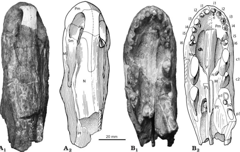
Description.—The specimen consists of a well preserved though slightly laterally crushed, slender snout (length of 100 mm, height of 65 mm, width between canines 33 mm)
with a marginal tooth series comprising incisors, precanines, canines and the roots of three postcanines (Figs. 1B, 2). Re− curved and slender incisor teeth and the presence of serra− tions on the posterior edge of the second canine suggest that it belonged to a large predator with the complete skull proba− bly exceeding 16 cm in length.
A large oval external naris (Figs. 1A, 2) is positioned close to the anterior margin of the snout. The dorsal process of the premaxilla makes up most of the internarial bar, and termi− nates posteriorly beyond the posterior margins of the external nares where it is overlapped by the nasals. Paired nasals ex− tend backwards from the posterodorsal margin of the external naris to meet the prefrontal posteriorly, and ventrally form a long sutural contact with the maxilla and septomaxilla. The latter bone comprises the floor of the external naris with its posterodorsal process wedged between the maxilla and the na− sal and extending further posteriorly on the snout than the dor− sal process of the premaxilla. While the posteriormost extent of the maxilla is not preserved, it contacts the nasal dorsally and the prefrontal posterodorsally. Anteriorly the vertical su− ture between the maxilla and the premaxilla descends from the front of the external naris to a point between the third and fourth incisor, and continues posteriorly along the ventral edge labial to the incisors before turning medially to reach the 20 mm
Fig. 1. Partial skull of the basal therapsid Raranimus dashankouensis gen. et sp. nov., IVPP V15424 (holotype) from Middle Permian Xidagou Formation, Dashankou, Xumen, Gansu, China, in dorsal (A) and ventral (B) views. Photographs (A1, B1) and explanatory drawings (A2, B2). Abbreviations: c1–2, ca− nine 1–2; ch, choana; en, external naris; i1–6, incisor 1–6; M, maxilla; N, nasal; p1–3, postcanine 1–3; Pf, prefrontal; Pl, palatine; Pm, premaxilla; Pt, pterygoid; rc, replacement canine; ri, replacement incisor; Sm, septomaxilla; sp, small precanine maxillary tooth; V, vomer.
choana in front of the precanine. In lateral view the ventral margin of the maxilla turns sharply downwards forming a notch between the last incisor and canine. Bone sculpturing is present on the snout with small pits on the anterior surface of the premaxilla and radial striations converging on the concave area above the root of the canine on the maxilla, while longitu− dinal striations occur on the rest of the snout.
In palatal view the premaxilla forms the anterior and most of the lateral margin of the choana up to the level of the precanine, while being anteroventrally overlain by the ante− rior process of the vomer as in dinocephalians. Long, thin and edentulous paired vomers form the medial border of the choana. Their ventral surface is flat with the anterior section being slightly ventrally convex and the lateral edges of the posterior interchoanal portion forming weak ridges. The choanae are short, extending from the level of the fourth inci− sor to that of the first canine, a character unknown in other
therapsids. Only the anterior part of the left palatine is pre− served. It underlies the maxilla, possibly contacts the vomer medially, and extends anteriorly to the level of the first post− canine. No palatine teeth are evident and only the anterior portions of the pterygoids are present.
Six incisors were present on each premaxilla. Those with preserved crowns show them to be similar in size, recurved and unserrated, and therefore resembling the morphology of those of most theriodont therapsids (Fig. 1B). A diastema is present between the last incisor and the first canine on the left side and the last incisor and precanine on the right. Two re− curved canines, ovoid in cross section, are present in each maxilla. The complete left second canine (c2 in Fig. 2B) is considered to be newly erupted as it only partially occupies its alveolus. No serrations are preserved on the first canine, but they do exist on the posterior ridge of the right second canine. A small replacement tooth, lingual to the left first canine indi− DOI: 10.4202/app.2008.0071 20 mm
Pf
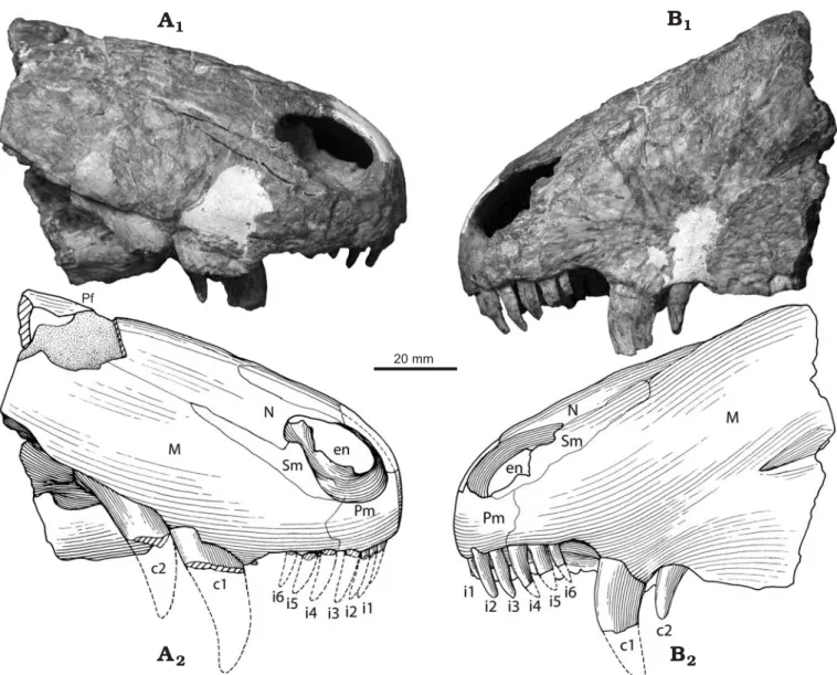
Fig. 2. Partial skull of the basal therapsid Raranimus dashankouensis gen. et sp. nov., IVPP V15424 (holotype) from Middle Permian Xidagou Formation, Dashankou, Xumen, Gansu, China in right lateral (A) and left lateral (B) views.. Photographs (A1, B1) and explanatory drawings (A2, B2). Abbreviations: c1–2, canine 1–2; ch, choana; en, external naris; i1–6, incisor 1–6; M, maxilla; N, nasal; p1–3, postcanine 1–3; Pf, prefrontal; Pl, palatine; Pm, premaxilla; Pt, pterygoid; rc, replacement canine; ri, replacement incisor; Sm, septomaxilla; sp, small precanine maxillary tooth; V, vomer.
cates that the two canines are not simply replacements of one another, but functioned simultaneously. This makes Rara− nimus the only therapsid with two functional canines, a condi− tion reminiscent of the caniniform teeth seen in the large pred− atory sphenacodontids (Romer and Price 1940; Reisz 1986). These canines, despite being doubled as in basal synapsids, have a more derived therapsid morphology in being quite slen− der and compressed linguo−labially, rather than having the massiveness seen in similarly sized sphenacodontids.
A small precanine with fine serrations on its anterior ridge is present in the maxilla anterior to the right first canine (Fig. 1B) and is reminiscent of the small precanine teeth known in Dimetrodon (Romer and Price 1940) and Tetraceratops (Laurin and Reisz 1996). Roots of three postcanines are pre− served in the left maxilla but the rest of this bone is missing. Judging by root diameter, the postcanines vary in size but are all much smaller than the canines.
Discussion
To explore the phylogenetic position of Raranimus and to ex− amine the effects of the new data upon current hypotheses of relationships amongst basal synapsids and therapsids, we built upon the data matrices from Sidor and Hopson (1998), Sidor and Rubidge (2006), and Rubidge et al. (2006). Therocepha− lians and cynodonts are excluded from this analysis because their position as advanced therapsids is confirmed in the pri− mary analyses. Haptodus is used as the outgroup, and the alleged basalmost therapsid Tetraceratops is also included. Laurin and Reisz (1996) stated that the interpterygoid vacuity is closed posteriorly by an additional posteromedian flange of pterygoid. As we are unable to verify this we have coded char− acter 41 as unknown. From all the characters used in analysis, Tetraceratops has only two derived states (Appendix 2) and our analysis supports that Tetraceratops is better considered as a sphenacodontid as suggested by Conrad and Sidor (2001). Our phylogenetic analysis shows Raranimus to be the most basal therapsid as it is closely allied to other well known therapsids (Fig. 3). Raranimus retains a number of plesio− morphic sphenacodontid characters (vomerine process of pre− maxilla absent, more than one functional canine, concave diastema with postero−ventrally sloping alveolar margin of the premaxilla, and nearly parallel−sided internarial portion of vomer) (Romer and Price 1940; Reisz 1986) which are un− known in any other therapsid. However, the presence of a greatly elongated dorsal process of the premaxilla, septo− maxilla with a long facial process, maxilla which is increased in height so as to contact the prefrontal, and ventral surface of the vomer with lateral ridges and median trough distinguish Raranimus as a therapsid (Hopson and Barghusen 1986; Hop− son 1991; Sidor and Hopson 1998). The very short choana which extends posteriorly only as far as the anterior margin of the canine, and six incisors are considered as autapomorphies.
While Broom (1910) pointed out the similarities between “pelycosaurs” and therapsids, there has always been a mor−
phological gap between the two groups (Kemp 2005, 2006). Pelycosaurian−grade synapsids are known predominantly from Carboniferous–Middle Permian rocks of North America, Europe and Asia, while therapsids are known predominantly from Middle Permian or younger rocks of South Africa and Russia, with little temporal overlap between them apart from some varanopid relicts in Russia and South Africa (Dilkes and Reisz 1996; Modesto et al. 2001; Botha−Brink and Modesto 2007), and caseids in Russia (Reisz 1986). Because the earliest Russian therapsid faunas are more primitive than those from the Tapinocephalus Assemblage Zone of South Africa it was considered that therapsids had their origin in Russia (Laurasia) and arrived in southern Africa (Gondwana) by overland dis− persal (Boonstra 1969). More recent discovery of a basal therapsid fauna from the underlying Eodicynodon Assem− blage Zone of South Africa resulted in the opposite proposal of a Gondwanan origin for several therapsid clades (Rubidge 1995; Modesto and Rubidge 2000; Modesto and Rybczynski 2000; Abdala et al. 2008). Unfortunately, although the oldest
Burnetia Haptodus Dimetrodon Raranimus Biarmosuchus Hipposaurus Herpetoskylax Lycaenodon Lemurosaurus Proburnetia Gorgonops Lycaenops Cyonosaurus Biseridens Patranomodon Suminia Stenocybus Syodon Titanophoneus Estemmenosuchus Styracocephalus Jonkeria to mammals 2 3 2 3 4 299 270.6 268 265.8 260.4 PERMIAN Ch Ro W Ca Wu BIARMOSUCHIA DINOCEPHALIA ANOMODONTIA GORGONOPSIA PEN
Cisuralian Guadalupian Lopingian
Tetraceratops 2 1 4 1 4 2 2 2 1 2 2 1 THERAPSIDA 253.8 251 Ma
Fig. 3. Phylogeny of Raranimus among basal therapsids. Tree (tree length = 169, consistency index = 0.54, retention index=0.75) is the strict consensus tree of four shortest trees resulting from our PAUP analysis (version 4.0b10, branch and bound search, with unordered multistate characters) of 71 cranial and dental characters. Numbers on tree indicate decay index of the respective clade. Shaded area indicates “Olson’s Gap”. Abbreviations: Ca, Capitanian; Ch, Changhsingian; Ma, Million years; PEN, Pennsylva− nian; Ro, Roadian; W, Wordian; Wu, Wuchiapingian.
and most basal therapsid faunas are known from Russia, South Africa and China (Battail 2000; Modesto and Rybczynski 2000; Li 2001; Kemp 2005), the current lack of reliable radio− metric dates limits accurate age correlation of these geographi− cally spaced faunas.
Roadian tetrapod faunas from North America are very dif− ferent from the oldest faunas from South Africa, Russia, and China with the major difference being the lack of therapsids in the North American faunas (Reisz and Laurin 2001; Lucas 2002, 2004, 2006). Tetraceratops from the Early Permian of Texas, has been considered the oldest therapsid (Laurin and Reisz 1996) but its therapsid identity has since been ques− tioned (Sidor and Hopson 1998; Conrad and Sidor 2001) and our analysis shows it to be more basal than Raranimus (Fig. 3). Lack of a therapsid record in the early Roadian and their first appearance as an already diverse group at the Roadian–Wor− dian transition, suggests a gap (dubbed Olson’s Gap) in the early therapsid fossil record (Lucas 2004; Ivakhnenko 2005), a crucial interval in which the initial evolution of this group must have occurred (Abdala et al. 2008).
One of the great remaining unsolved problems in synapsid history is the sphenacodontid−therapsid transition and the early diversification of therapsids. It has been suggested that the origin and early diversification of the main therapsid lin− eages occurred either by a rapid process of apomorphy accu− mulation, or by gradual acquisition of apomorphies during an extended temporal interval of up to 35 Ma (Kemp 2006; Abdala et al. 2008). Choosing between these two scenarios is possible only if therapsid−bearing rocks from Olson’s Gap are found. The presence of Raranimus at Dashankou, the basal− most Middle Permian therapsid known, in association with the dissorophoid Anakamacops, the bolosaurid Belebey (both families occur together only in the Early Permian) and the very primitive therapsids Biseridens, Stenocybus, and Sinophoneus (known only from China, Li et al. 1996; Cheng and Li 1997; Li and Cheng 1997; Li 2001), support the hypothesis of an early Roadian age for this locality, and helps to fill in Olson’s Gap. In addition, the discovery of a new basal Laurasian therapsid which cannot be assigned to any major therapsid clade, suggests that the initial evolutionary radiation of therapsids occurred in Laurasia.
Acknowledgements
We acknowledge the assistance of Zhang Hong, Wang Zhao (IVPP) for preparation, Yang Minwang (IVPP) for illustration, Carl Mehling (American Museum of Natural History, New York, USA) for accessing Tetraceratops. Fernando Abdala (BPI), Tom Kemp (Oxford Univer− sity, Oxford, UK), Michel Laurin (Université Paris 7, France), Sean Modesto (Cape Breton University, Sydney, Canada), Robert Reisz (University of Toronto, Canada), Adam Huttenlocker and Christian Sidor (University of Washington, Seattle, USA) are acknowledged for comments on early drafts. This study is supported by Knowledge Inno− vation Project of CAS (KZCX2−YW−BR−07), Ministry of Science and Technology of China (“973” project 2006CB806403) and the DST, NRF and PAST of South Africa.
References
Abdala, F., Rubidge, B.S., and Heever, J.A. van den 2008. The oldest therocephalians (Therapsida,Eutheriodontia) and the early diversifica− tion of Therapsida. Palaeontology 51: 1011–1024.
Battail, B. 2000. A comparison of Late Permian Gondwanan and Laurasian amniote faunas. Journal of African Earth Sciences 31: 165–174. Boonstra, L.D. 1936. The cranial morphology of some titanosuchid deinoce−
phalians. Bulletin of the American Museum of Natural History 72: 99–116. Boonstra, L.D. 1969. The fauna of the the Tapinocephalus Zone (Beafort
Beds of the Karoo). Annals of the South African Museum 56: 1–73. Botha−Brink, J. and Modesto, S.P. 2007. A mixed−age classed “pelycosaur”
aggregation from South Africa: earliest evidence of parental care in amniotes? Proceedings of the Royal Society B: Biological Sciences 274: 2829–2834.
Broom, R. 1910. A comparison of the Permian reptiles of North America with those of South Africa. Bulletin of the American Museum of Natural
History 28: 197–234.
Cheng, Z. and Li, J. 1997. A new genus of primitive dinocephalian—the third report on Late Permian Dashankou lower tetrapod fauna [in Chi− nese with English summary]. Vertebrata Palasiatica 35: 35–43. Chudinov, P.K. 1960. Upper Permian therapsids from the Ezhovo locality
[in Russian]. Paleontologičeskij žurnal 1960: 81–94.
Chudinov, P.K. 1983. Early therapsids [in Russian]. Trudy Paleontolo−
gičeskogo Instituta AN SSSR 202: 1–230.
Conrad, J. and Sidor, C.A. 2001. Re−evaluation of Tetraceratops insignis (Synapsida. Sphenacodontia). Journal of Vertebrate Paleontology 21: 42A.
Currie, P.J. 1977. A new haptodontine sphenacodont (Reptilia: Pelyco− sauria) from the Upper Pennsylvanian of North America. Journal of Pa−
leontology 51: 927–942.
Dilkes, D.W. and Reisz, R.R. 1996. First record of a basal synapsid (“mam− mal−like reptile”) in Gondwana. Proceedings of the Royal Society of
London Series B Biological Sciences 263: 1165–1170.
Hopson, J.A. 1991. Systematics of the nonmammalian Synapsida and impli− cations for patterns of evolution in Synapsida. In: H.−P. Schultze and L. Trueb (eds.), Origins of the Higher Groups of Tetrapods: Controversy
and Consensus, 635–693. Cornell University Press, Ithaca.
Hopson, J.A. and Barghusen, H.R. 1986. An analysis of therapsid relation− ships. In: N. Hotton III, P.D. MacLean, J.J. Roth, and E.C. Roth (eds.),
The Ecology and Biology of Mammal−like Reptiles, 83–106. Smithso−
nian Institution Press, Washington.
Ivakhnenko, M.F. 1999. Biarmosuches from the Ocher Faunal Assemblage of Eastern Europe. Paleontological Journal 33: 289–296.
Ivakhnenko, M.F. 2000. Estemmenosuchus and primitive theriodonts from the Late Permian. Paleontological Journal 34: 189–197.
Ivakhnenko, M.F. 2005. Comparative survey of Lower Permian tetrapod faunas of eastern Europe and South Africa. Paleontological Journal 39 (1): 66–71.
Kemp, T.S. 2005. The Origin and Evolution of Mammals. 331 pp. Oxford University Press, Oxford.
Kemp, T.S. 2006. The origin and early radiation of the therapsid mam− mal−like reptiles: a palaeobiological hypothesis. Journal of Evolution−
ary Biology 19 (4): 1231–1247.
Laurin, M. 1993. Anatomy and relationships of Haptodus garnettensis, a Pennsylvanian synapsid. Journal of Vertebrate Paleontology 13: 200–229.
Laurin, M. and Reisz, R.R. 1996. The osteology and relationships of
Tetraceratops insignis, the oldest known therapsid. Journal of Verte− brate Paleontology 16: 95–102.
Li, J. 2001. The most primitive lower tetrapod fauna in China. Science in
China (Series D) 44: 47–51.
Li, J. and Cheng, Z. 1997. First discovery of eotitanosuchian (Therapsida, Synapsida) of China [in Chinese with English summary]. Vertebrata
Palasiatica 35: 268–282.
Li, J., Rubidge, B.S., and Cheng, Z. 1996. A primitive anterosaurid dino−
cephalian from China—implications for the distribution of the earliest therapsid faunas. South African Journal of Science 92: 252–253. Li, Y.a., Li, P., Sun, D., and Cheng, Z. 2004. Paleomagnetic Study of the
Permian–Triassic in the Yumen Area, Gansu [in Chinese with English abstract]. Geological Review 50: 407–412.
Lucas, S.G. 2002. The reptile Macroleter: First vertebrate evidence for correla− tion of Upper Permian continental strata of North America and Russia: Discussion. Geological Society of America Bulletin 114: 1174–1175. Lucas, S.G. 2004. A global hiatus in the Middle Permian tetrapod fossil re−
cord. Stratigraphy 1: 47–64.
Lucas, S.G. 2006. Global Permian tetrapod biostratigraphy and biochrono− logy. In: S.G. Lucas, G. Cassinis, and J.W. Schneider (eds.), Non−Ma−
rine Permian Biostratigraphy and Biochronology, 65–93. Geological
Society, London.
Modesto, S. and Rubidge, B. 2000. A basal anomodont therapsid from the lower Beaufort Group, Upper Permian of South Africa. Journal of Ver−
tebrate Paleontology 20: 515–521.
Modesto, S.P. and Rybczynski, N. 2000. The amniote faunas of the Russian Permian: implications for Late Permian terrestrial vertebrate biogeo− graphy. In: M.J. Benton, M.A. Shishkin, D.M. Unwin, and E.N. Ku− rochkin (eds.), The Age of Dinosaurs in Russia and Mongolia, 17–34. Cambridge University Press, Cambridge.
Modesto, S., Sidor, C.A., Rubidge, B.S., and Welman, J. 2001. A second varanopseid skull from the Upper Permian of South Africa: Implica− tions for late Permian “pelycosaur” evolution. Lethaia 34: 249–259. Orlov, Y.A. 1958. Carnivorous dinocephalians from the fauna of Ishev
(Titanosuchia) [in Russian]. Trudy Paleontologičeskogo Instituta AN
SSSR 71: 1–114.
Reisz, R. 1986. Pelycosauria. 102 pp. Gustav Fischer Verlag, Stuttgart. Reisz, R.R. and Laurin, M. 2001. The reptile Macroleter: First vertebrate evi−
dence for correlation of Upper Permian continental strata of North Amer− ica and Russia. Geological Society of America Bulletin 113: 1229–1233. Romer, A.S. and Price, L.I. 1940. Review of the Pelycosauria. Geological
Society of America Special papers 28: 1–538.
Rubidge, B.S. 1995. Biostratigraphy of the Eodicynodon Assemblage Zone.
South African Committee fot Stratigraphy, Biostratigraphic Series1: 3–7.
Rubidge, B.S. and Hopson, J.A. 1996. A primitive anomodont therapsid from the base of the Beaufort Group (Upper Permian) of South Africa.
Zoological Journal of the Linnean Society 117: 115–139.}
Rubidge, B.S. and Sidor, C.A. 2001. Evolutionary patterns among Permo− Triassic therapsids. Annual Review of Ecology and Systematics 32: 449–480.
Rubidge, B.S. and Sidor, C.A. 2002. On the cranial morphology of the basal therapsids Burnetia and Proburnetia (Therapsida: Burnetiidae). Jour−
nal of Vertebrate Paleontology 22: 257–267.
Rubidge, B.S. and van den Heever, J.A. 1997. Morphology and systematic position of the dinocephalian Styracocephalus platyrhynchus. Lethaia 30: 157–168.
Rubidge, B.S., Sidor, C.A., and Modesto, S.P. 2006. A new burnetiamorph (Therapsida : Biarmosuchia) from the Middle Permian of South Africa.
Journal of Paleontology 80: 740–749.
Rybczynski, N. 2000. Cranial anatomy and phylogenetic position of Suminia
getmanovi, a basal anomodont (Amniota: Therapsida) from the Late
Permian of Eastern Europe. Zoological Journal of the Linnean Society 130: 329–373.
Sidor, C.A. 2003. The naris and palate of Lycaenodon longiceps (Therapsida, Biarmosuchia), with comments on their early evolution in the Therapsida.
Journal of Paleontology 77: 977–984.
Sidor, C.A. and Hopson, J.A. 1998. Ghost Lineages and “Mammalness”: As− sessing the Temporal Pattern of Character Acquisition in the Synapsida.
Paleobiology 24: 254–273.
Sidor, C.A. and Rubidge, B.S. 2006. Herpetoskylax hopsoni, a new Biarmo− suchian (Therapsida: Biarmosuchia) from the Beaufort Group of South Africa. In: M.T. Carrano, R.W. Blob, T.J. Gaudin, and J.R. Wible (eds.), Amniote Paleobiology: Perspectives on the Evolution of Mam−
mals, Birds, and Reptiles, 76–113. The University of Chicago Press,
Chicago.
Sidor, C.A. and Welman, J. 2003. A second specimen of Lemurosaurus
pricei (Therapsida: Burnetiamorpha). Journal of Vertebrate Paleontol− ogy 23: 631–642.
Sigogneau, D. 1970. Révision systématique des Gorgonopsiens sud−africains.
Cahiers de Paleontologie: 417.
Sigogneau−Russell, D. 1989. Theriodontia 1. Phthinosuchia, Biarmosuchia,
Eotitanosuchia, Gorgonopsia. 127 pp. Gustav Fischer Verlag, Stuttgart.
Swofford, D.L. 2001. PAUP*. Phylogenetic Analysis Using Parsimony (* and
Other Methods). Version 4.0b10. Sinauer Associates, Sunderland, MA.
Appendix 1
List of characters and character states used to construct the cladogram. The number preceding the character definition corresponds to that of the col− umns in the data matrix. Most of the characters are cited from SH: Sidor and Hopson (1998); SR: Sidor and Rubidge (2006); RSM: Rubidge et al. (2006). When an asterisk follows the citation, it denotes that the character definition has been modified or character state(s) has been added/deleted. Coding of characters is based on the coding of selected characters in original references, sources listed in the end of character list, and personal ob− servation.
1. Dorsal surface of snout: oblique convex (0), near straight and flat (1). (SH: 46*)
2. Snout width/height ratio: height greater than width (0), height equal to width (1), height less than width (2). (SH: 45)
3. External nares: terminal (0), retracted (1). (RSM: 4)
4. Length of dorsal process of premaxillae: short (0), long, reaching to a level posterior to that of the upper canine (1). (SH: 1*; SR: 2; RSM: 2)
5. Premaxilla alveolar margin shape: downturned (0), horizontal or slightly upturned (1), greatly upturned (2). (SH: 2*; SR: 3*; RSM: 3*)
6. Antorbital region: long (0), short (1). (SR: 4)
7. Septomaxilla: contained within external naris (0), escapes to have a short (1) or long facial exposure (2). (SH: 6; SR: 5*, 6*; RSM: 5)
8. Maxilla contacts prefrontal: absent (0), present (1). (SH: 8; SR: 8; RSM: 7)
9. Shape of dorsal surface of nasals: flat (0), with median boss (1). (SR: 9; RSM: 9*)
10. Supraorbital margin: thin (0), moderately to greatly thickened (1). (SR: 12; RSM: 12)
11. Orbit size smaller than that of the temporal fenestra: absent (0), present (1)
12. Adductor musculature originates on lateral surface of postorbital: absent (0), present (1), on both postorbital and postfrontal (2). (SR: 13*, 17*; RSM: 13*)
13. Postorbital bar: thin (A−P length less than one−third of height) (0), thickened such that A−P length is greater than 40% of its height (1). (RSM: 16)
temporal fenestra (0), descends onto posterior margin of lateral temporal fenestra (1). (RSM: 14)
15. Boss above postorbital bar: absent (0), present (1). (RSM: 15) 16. Postfrontal: without (0) or with (1) posterior extension along its
medial contact with the frontal. (SR: 16; RSM: 18)
17. Shape of dorsal surface of parietal surrounding parietal fora− men: flat (0), low and diffuse swelling (1), forms well−defined chimney (2). (SH: 21*; SR: 18; RSM: 19)
18. Temporal fenestra: small (0), expanded posterodorsally (1) so that adductor musculature origination on squamosal visible in dorsal view. (SH: 14*; SR: 19; RSM: 20)
19. Intertemporal region: wider (0) or narrower (1) than interorbital region. (SH: 18*; SR: 20; RSM: 21)
20. Ventral surface of zygomatic arch and suborbital bar: smooth (0), with bosses (1). (SR: 21, RSM: 22)
21. Zygomatic arch elevated above margin of upper tooth row so as to fully expose quadrate and quadratojugal in lateral view: ab− sent (0), present (1). (SR: 22)
22. Anterior extension of anterior ramus of squamosal: stops under temporal fenestra (0), beyond the anterior margin of the tempo− ral fenestra (1). (SR: 23*; RSM: 23*)
23. Squamosal external auditory meatus groove: absent (0), present (1) (SH: 52*)
24. Preparietal: absent (0), present (1). (SH: 48; SR: 24; RSM: 24*) 25. Supratemporal: present (0), absent (1). (SH: 22; SR: 25; RSM: 25) 26. Tabular: contacts paroccipital process of opisthotic (0), re−
stricted dorsally (1). (SH: 54*; SR: 26*)
27. The position of the posterior border of choana: close to the inci− sor (0), far behind the incisor (1)
28. Length of vomerine process of premaxilla: short (0); long, ex− tending posteriorly and forming part of the medial margin of the inner choana (1); absent in ventral view (2) so that vomer abuts body of premaxilla. (SH: 3*; SR: 1*; RSM: 1*)
29. Vomer: paired (0), unpaired (1). (SH: 25*, 26*; SR: 27; RSM: 26) 30. Vomer internarial part: nearly parallel−sided or slightly ex−
panded backward (0), widest nearly middle (1), strongly con− straining backwards (2). (SH: 23*)
31. Interchoanal portion of vomer where it meets the postchoanal por− tion: broad (0), forms median ridge (1). (SH: 23*; RSM: 27) 32. Vomer ventral surface: flat to convex (0), lateral ridges and me−
dian trough (1). (SH: 24*)
33. Choanal and postchoanal portions of vomer: meet at similar level on palate (0), choanal portion is offset ventrally from postchoanal portion (1). (SR: 28)
34. Lateral margin of the choana formed by the palatine: less than 1/3 (0), over 1/3 (1)
35. Two palatines: separated by the vomer and pterygoid (0), join in midline (1)
36. Palatine dentition: broadly distributed (0), restricted to small area (1), absent (2). (SH: 36*; SR: 29; RSM: 28*)
37. Dentition on palatal ramus of pterygoid: present (0), absent (1). (SH: 37; SR: 33)
38. Row of teeth on transverse flange of pterygoid: present (0), ab− sent (1). (SR: 30*; RSM: 29)
39. Position of transverse flange of pterygoid: under posterior half of orbit (0), under anterior half of orbit (1), preorbital (2). (SH: 73*; SR: 31; RSM: 30)
40. Pterygoid: without (0) or with (1) shelf posterior to its trans− verse flange. (SR: 32; RSM: 31)
41. Basicranial rami of pterygoids: broadly separated (0), narrowly separated with median trough formed (1), broadly contacting anterior to basicranium (2). (SR: 34; RSM: 32)
42. Medial edge of pterygoid basicranial ramus forms parasagittal ridge on ventral surface: absent (0), present (1). (RSM: 33) 43. Basipterygoid articulation located: high above primary palate (0),
just dorsal to basicranial ramus of pterygoid (1), at level basi− cranial ramus (i.e., suture visible in ventral view) (2). (SR: 35) 44. Ectopterygoid teeth: present (0), absent (1). (SH: 39; SR: 36;
RSM: 34)
45. Shape of postparietal: wider than tall (0), approximately square (1), or taller than wide (2). (SR: 37; RSM: 35)
46. Forward rotation of occiput: none (0), moderate (= vertical) (1), pronounced (2). (SH: 42; SR: 38; RSM: 36)
47. Paroccipital process orientation: strongly posteroventral and lateral (0), moderately posteroventral and lateral (1), transverse (2) (SH: 65)
48. Quadrate contact: primarily paroccipital process (0), about equal paroccipital process and squamosal (1), mostly squamosal (2) (SH: 58*)
49. Stapedial foramen: present (0), absent (1). (SH: 76; SR: 39; RSM: 37) [this foramen is present in Scylacops, and coded as 0 for all taxa of Gorgonopsia here]
50. Dentary height in canine versus anterior postcanine regions: nearly equivalent (0), shows pronounced difference (1). (SH: 79*; SR: 40; RSM: 38)
51. Dentary: coronoid eminence (0), coronoid process (1) (SH: 80) 52. Dentary−angular suture: runs diagonally across lateral surface
of mandible (0), posterior margin of dentary deeply incised (1). (SR: 41; RSM: 39)
53. Coronoid (posterior): present (0), absent or greatly reduced (1). (SH: 91*; SR: 47)
54. Lateral mandibular fenestra: absent (0), present (1). (SH: 93, 94*; SR: 46)
55. Angular reflected lamina dorsal notch: near articular (0), midway between articular and dentary (1), close to dentary (2) (SH: 97) 56. Angular with pattern of ridges and fossae on its lateral surface:
absent (0), present (1). (SH: 98*; SR: 42; RSM: 40)
57. Dorsal edge of surangular just posterior to dentary with laterally projecting ridge: absent (0), or present (1). (SR: 43; RSM: 41) 58. Foramen between prearticular and angular (sometimes bor−
dered by splenial as well) on medial surface of lower jaw: ab− sent (0), present (1). (SR: 44; RSM: 42)
59. Articular dorsal process: absent (0), present (1). (SR: 45; RSM: 43)
60. Differentiation of upper tooth row: more than one caniniform teeth (0), one canine (1), barely differentiated (1). (SR: 48*) 61. Premaxillary teeth number: 5 (0), 4 or less (1), 6 (2)
62. Upper and lower incisors intermesh: absent (0), present in ante− rior incisors (1), present in all incisors (2). (SH: 105*;SR: 49; RSM: 44)
63. Incisor heels: absent (0), present (1) (SH: 106)
64. Upper incisors: much larger (0) or roughly equivalent in size to postcanines (1). (SR: 50; RSM: 46)
65. Precanine maxillary teeth: present (0), absent (1) (SH: 110) 66. Lower canine: fits into choana (0), or into fossa roofed by
premaxilla and maxilla (1), or passes anterior and external to upper canine (2). (SR: 51; RSM: 47)
67. Upper and lower canines: without heels (0) or small heels pres− ent (1). (SR: 52; RSM: 45)
68. Postcanine diastema on upper jaw: absent (0), present (1) 69. Number of upper postcanines: twelve or greater (0), fewer than
12 (1). (SH: 112; SR: 53; RSM: 48)
70. Postcanine teeth with triangular crown bearing coarse serrations along both anterior and posterior carinae: absent (0), present (1). (SH: 113*; SR: 56; RSM: 51)
71. Upper postcanine teeth confluent with upper incisor row medial to canine: absent (0), present (1). (SR: 55; RSM: 50)
Taxa included and the coding basis: Haptodus (Currie 1977; Laurin 1993) Dimetrodon (Romer and Price 1940)
Tetraceratops AMNH4526 (Laurin and Reisz 1996; Conrad and
Sidor 2001)
Raranimus IVPP V15424
Biarmosuchus (Chudinov 1960; Ivakhnenko 1999) Hipposaurus (Sigogneau 1970; Sigogneau−Russell 1989) Herpetoskylax (Sidor and Rubidge 2006)
Lycaenodon (Sidor 2003)
Lemurosaurus (Sidor and Welman 2003)
Proburnetia and Burnetia (Rubidge and Sidor 2002) Syodon and Titanophoneus (Orlov 1958)
Stenocybus IGCASGS V361 (Cheng and Li 1997) Styracocephalus (Rubidge and van den Heever 1997) Jonkeria (Boonstra 1936)
Estemmenosuchus (Chudinov 1960; Ivakhnenko 2000)
Biseridens IGCAGS V 632, IVPP V 12009 (Li and Cheng 1997) Patranomodon (Rubidge and Hopson 1996)
Suminia (Rybczynski 2000)
Gorgonops, Lycaenops, and Cyonosaurus (Sigogneau 1970)
Appendix 2
Character matrix used to analyze the phylogenetic position of Raranimus
Taxon 1 1111111112 2222222223 3333333334 4444444445 5555555556 6666666667 7 1234567890 1234567890 1234567890 1234567890 1234567890 1234567890 1234567890 1 Haptodus 0000000000 0000000000 00000??000 0000000000 0000?0000− 0000−00000 00000?0000 0 Dimetrodon 0000000000 0000000000 0000000000 0000000000 ?000000000 0000−00000 1000010000 0 Tetraceratops 000000000? ??0000???? ???????00? ?????00000 ?0?1?????0 ?0?0??0??0 10000000?0 0 Raranimus 00010?2100 ?????????? ??????0000 01000????? ?????????? ?????????0 2?0?0?00?0 ? Biarmosuchus 0001102100 0100002000 00101000?1 ??10?00010 20?1111101 00?0011??1 0?011?0110 0 Hipposaurus 0001102100 000101200? 0?11101??1 111??00010 1021111111 0(01)0001?111 01011?0110 0 Herpetoskylax 0000102100 000?012000 (01)111101?1? ?111?10110 10211111?1 0100011011 01011?0110 0 Lycaenodon 00001?2100 000?012?0? ???1??1111 1111010110 11?1?????? ?????????1 0?011?011? 0 Lemurosaurus 000??02101 00010?2001 011??01??1 1111?10110 11?1111111 01?00110?1 ?1011?0110 0 Proburnetia 0000111?11 000?1?1001 011??111?0 11?1010110 11211112?1 ?100?1???1 ?1011?0?10 ? Burnetia 00???1??11 100?1?1001 0?1??111?1 ?1?1010?10 11?1?11??? ?????????1 ????1????? ? Syodon 0111201100 1210002110 0010101201 0101?10?10 2011211100 0100100101 0210111010 0 Titanophoneus 0111201101 1210002110 0010101201 0101011010 2011211100 00001001?1 0210111010 0 Stenocybus 0001201100 0210002110 00101????? ??????00?0 20??211??0 ?0?010???1 02101?1?10 0 Styracocephalus 021?10?101 ?11?1?1100 0?1?111?00 0?0??10021 2011021111 0100???0?1 ????????1? ? Jonkeria 0211102101 1100001110 01101?1200 0101021121 2011?211?0 010?100001 1210121001 1 Estemmenosuchus 0101101?11 1?0?102100 0?101?1211 0101?00021 20???21100 01?01001?1 0210020001 1 Biseridens ?????1?1?0 1101002100 101?1????? ??????0??? ?????012?0 0???2?0??? ?????????? ? Patranomodon 0110?1?100 1100000100 1111111001 1111021100 1011002210 0011211002 ????−?− − 10 0 Suminia 0111111100 11000?2100 111011101? ??11021100 1011102210 0111210102 1?00−?− − 11 0 Gorgonops 1100102100 1100002100 0011101112 1111110010 2111002201 1000110?10 0000100110 ? Lycaenops 1000102100 1100002100 0011101112 1111121120 2111002201 1000110?10 0000100110 0 Cyonosaurus 1100102100 1100002100 0011101112 1111110110 2111002201 1000110?10 00001?0110 0