THE WILHEMINE E. KEY 2004 INVITATIONAL LECTURE
New Perspectives on Eye Development
and the Evolution of Eyes and
Photoreceptors
W. J. G
EHRINGFrom the Department of Cell Biology, Biozentrum, University of Basel, Klingelbergstrasse 70, 4056 Basel, Switzerland Address correspondence to Walter Gehring at the address above, or e-mail: [email protected]
Walter J. Gehring is Professor at the Biozentrum of the University of Basel, Switzerland. He obtained his Ph.D. at the University of Zurich in 1965 and after two years as a research assistant of Professor Ernst Hadorn he joined Professor Alan Garen’s group at Yale University in New Haven as a postdoctoral fellow. In 1969 he was appointed as an associate professor at the Yale Medical School and 1972 he returned to Switzerland to become a professor of developmental biology and genetics at the Biozentrum of the University of Basel. He has served as Secretary General of the European Molecular Biology Organization and President of the International Society for Developmental Biologists. He was elected as a Foreign Associate of the US National Academy of Sciences, the Royal Swedish Academy of Science, the Leopoldina, a Foreign Member of the Royal Society of London for Improving Natural Knowledge and the French Acade´mie des Sciences.
Walter Gehring has been involved in studies ofDrosophila genetics and development, particularly in the analysis of cell determination in the embryo and transdetermination of imaginal discs. He has made significant contributions to the study of the heat shock genes, various transposons and the homeotic genes which are involved in the genetic control of
development. He and his group have discovered the homeobox, a DNA segment characteristic for homeotic genes which is not only present in arthropods and their ancestors, but also in vertebrates up to man. He has been involved in the development and application of enhancer trapping methods. He and his collaborators have identifiedPax 6 as a master control gene for eye development, which led to a new theory about the monophyletic origin of the eyes in evolution.
Abstract
Recent experiments on the genetic control of eye development have opened up a completely new perspective on eye evolution. The demonstration that targeted expression of one and the same master control gene, that is, Pax6 can induce the formation of ectopic eyes in both insects and vertebrates, necessitates a reconsideration of the dogma of a polyphyletic origin of the various eye types in all the animal phyla. The involvement of Pax6 and six1 and six3 genes, which encode highly conserved transcription factors, in the genetic control of eye development in organisms ranging from planarians to humans argues strongly for a monophyletic origin of the eye. Because transcription factors can control the expression of any target gene provided it contains the appropriate gene regulatory elements, the conservation of the genetic control of eye development by Pax6 among all bilaterian animals is not due to functional constraints but a consequence of its evolutionary history. The prototypic eyes postulated by Darwin to consist of two cells only, a photoreceptor and a pigment cell, were
(Photograph by Ch. Scholz.)
Journal of Heredity 2005:96(3):171–184 ª 2005 The American Genetic Association
doi:10.1093/jhered/esi027
as secondary chloroplasts and in some species evolved into the most sophisticated eye organelles, as found, for example, in some dinoflagellates like Erythropsis and Warnovia, which lack chloroplasts. Because dinoflagellates are commonly found as symbionts in cnidarians, the dinoflagellates may have transferred their photoreceptor genes to cnidarians. In cnidarians such as Tripedalia the step from photoreceptor organelles to multicellular eyes has occurred. These two hypotheses, the cellular differentiation and the symbiont hypothesis, are not mutually exclusive and are the subject of further investigations.
Introduction
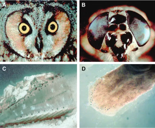
In the course of evolution several basically different eye types have been generated, like the camera-type eye, the compound eye, and the mirror eye (Figure 1). These eye types are different not only with respect to their morphology and physiology but also with respect to their mode of development. This has led to the dogma that eyes have evolved in all animal phyla 40 to 60 times independently (Salvini-Plawen and Mayr 1961). However, recent genetic experiments cast serious doubts on this notion and argue strongly in favor of a monophyletic origin of the various eye types followed by divergent, parallel, and convergent evolution. In his latest book, Ernst Mayr (2001) admits that his earlier notion may no longer be correct. In the following article I discuss the evidence arguing for a mono-phyletic origin of the eyes, and I shall follow the eye back to its origins from single-celled photoreceptors.
The Genetic Control of Eye Development
Mutations affecting eye development are easily detectable and the eyeless (ey) mutation in Drosophila was discovered as early as 1915 by Hoge. A similar mutation was found in mice and designated as Small eye because the heterozygous animals have reduced eyes, whereas the homozygous fetuses that die in uterolack not only the eyes but also the nose and a large part of the forebrain, including the pineal organ (Hill et al. 1991). A hereditary syndrome called aniridia causes a very similar phenotype in humans; reduced iris in heterozygous individuals, and two homozygous mutant aborted fetuses have been described that lacked eyes and nose completely and suffered brain damage. The Small eye and Aniridia genes were cloned by Walther and Gruss (1991) and Ton et al. (1991), respectively, and correspond to the highly conserved
Pax6gene. The Pax6 homolog of Drosophila was cloned by Quiring et al. (1994) and surprisingly turned out to correspond to the eyeless (ey) gene of Hoge. The fact that small eye, aniridia, and eyeless are mutations in homologous genes suggested to me that Pax6 might be a master control gene specifying eye development in both vertebrates and insects.
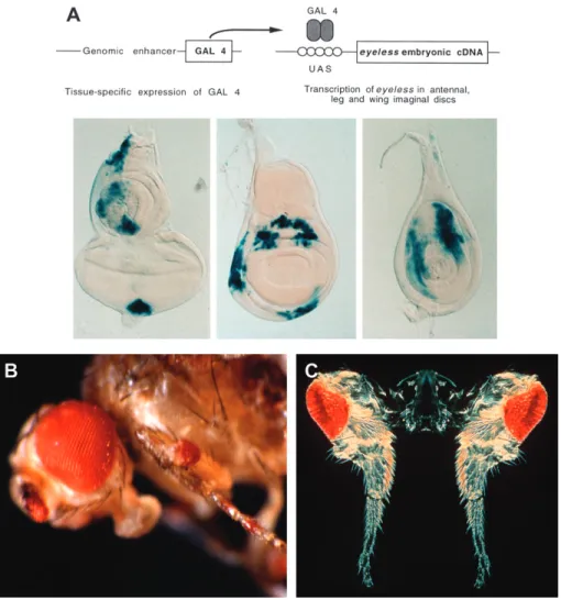
To test this hypothesis I decided to construct a Pax6 gain-of-function mutation to express Pax6 ectopically in an attempt to induce ectopic eye structures. It is relatively easy to abolish eye formation but more difficult to induce an eye ectopically. Two of my collaborators, Georg Halder and Patrick Callaerts, used the yeast transcription factor gal4 to drive eyeless cDNA into imaginal discs other than the eye disc (Figure 2). After several months of failure, they succeeded in inducing ectopic eye structures on the antennae, legs, and wings, which made the front page of the New York Times with an article titled ‘‘Scientists Out Do Hollywood.’’ Callaerts later showed by recording electroretinogrammes that some of the ectopic eyes on the antennae are fully functional.
Subsequently, we also tested the mouse Pax6 gene in Drosophilaand showed that it is capable of inducing ectopic eyes in Drosophila (Figure 3). These eyes are, of course, Drosophila eyes and not mouse eyes, because we have only exchanged the main switch to trigger eye development and all of the other genes required for forming an eye (which we estimate to be ; 2,000) are provided by the Drosophila host. These experiments lead to the conclusion that Pax6 is a master control gene on the top of the genetic cascade leading to eye morphogenesis and that this master switch can initiate eye development both in insects and mammals.
Pax6encodes a gene regulatory protein with two DNA binding domains—a paired domain as well as a homeodo-main. The various Pax genes found in mice and flies clearly illustrate the principle of evolutionary tinkering (Jacob 1977).
The nine members of the gene family did not arise inde-pendently de novo but rather by recombination of bits and pieces of preexisting genes like paired domains, octapeptides, homeodomains, and parts thereof. Pax6 has a paired domain and a homeodomain but lacks the octapeptide motif. Its nearest relatives are Pax2, 5, and 8, which have a paired domain plus an octapeptide, but only a partial homeodomain consisting of the N-terminal arm and the first a-helix. It will be interesting to identify their last common precursor, which may still exist in some cnidarians (see later discussion).
Decyphering the Eye Genetic Program
Because the first spontaneous ey mutants, ey2 and eyR are caused by transposon insertions (Quiring et al. 1994) they are not null mutations. They still produce a normal transcript in parts of the nervous system, but they fail to be expressed in the eye disc because of the fact that the two transposons in ey2 and eyRhave inserted into an eye-specific enhancer located in the first intron of the gene (Hauck et al. 1999). Therefore, we induced a true null mutation in ey by mutagenesis with ethylmethanesulfonate (Flister et al. unpublished data; Punzo et al. 2004). However, this null mutation only removed the compound eyes in most flies and not the ocelli, suggesting the presence of a second gene with partially redundant function. Indeed, in collaboration with M. Busslinger we discovered a second Pax6 homolog in Drosophila (Czerny et al. 1999) which we designated twin of eyeless (toy). This second gene is
only found in holometabolous insects, Drosophila, and silkworms but not in the more primitive hemimetabolous insects, like grasshoppers or springtails. Null mutations in toy have a much more severe phenotype (Flister et al. un-published data; Kronhamn et al. 2002), they are essentially headless, forming only the proboscis and the thorax but no antennae and head structures, except two spheres of eye facets found back in the thorax, which presumably reflect the residual activity of the eyþgene, which is still present in these flies (Punzo et al. 2004). Ectopic expression of toy is also capable of inducing ectopic eyes (Figure 2B). My analysis indicates that ey and toy arose from a duplication and diverged in function, so that toy acts upstream of ey and activates it in normal development leading to the intercalation of an additional gene into the eye morphogenetic pathway (see later discussion).
We also identified direct target genes of ey in particular sine oculis(so), a homeobox gene that is also conserved in both vertebrates and invertebrates (Niimi et al. 1999). The so enhancer contains five binding sites for toy and ey (Punzo et al. 2002) all of which are recognized by TOY protein, whereas EY binds only to two of them. All five binding sites have to be be mutated to abolish enhancer activity completely. So protein forms a heterodimer with eyes absent (eya) a protein phospha-tase. The so gene alone, when ectopically expressed, does not induce ectopic eyes; however, in combination with eya eye structures are induced, but less efficiently than by ey, and there is a positive feedback loop from so/eya to ey (Figure 4). Figure 1. Different eye types. (A) Camera-type eye of an owl. (B) Compound eye of a horsefly. (C) Mirror-type eye of a scallop. (D) Prototypic eyes consisting of a single photoreceptor cell and a pigment cell in the planarian Polycelis auricularia.
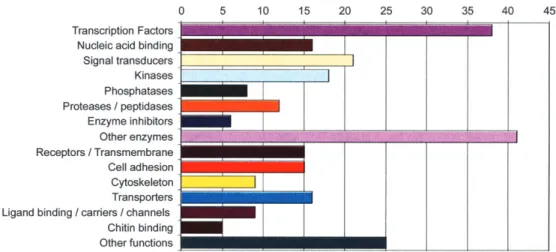
Following these studies on single genes, I undertook a more global approach to decypher the entire eye morpho-genetic pathway of Drosophila and eventually compare it to that of the mouse. My colleagues and I have begun to use DNA microarrays to determine the patterns of gene expres-sion at consecutive stages of eye development. By comparing the genes expressed in a leg disc in which an eye field has been induced with those in a normal leg disc, we have deter-mined the earliest genes expressed on the top of the genetic hierarchy. The large fraction of these early genes are tran-scription factors (Figure 5), whereas very few of the structural genes are expressed at this early stage (Michaut et al. 2003).
Darwin and the Problem of Eye Evolution
Charles Darwin in The Origin of Species (1882) had great difficulties with eye evolution and devoted an entire chapter to it, ‘‘Difficulties of the Theory,’’ in which he discusses ‘‘organs of extreme perfection and complication:
To suppose that the eye with all its inimitable contrivances for adjusting the focus to different distances, for admitting different amounts of light and for the correction of spherical and chromatic aberration, could have been formed by natural selection, seems, I freely confess, absurd in the highest degree.
But then he continues:
Reason tells me, that if numerous gradations from a simple and imperfect eye to one complex and perfect can be shown to exist, each grade being useful to its possessor, as is certainly the case; if further, the eye ever varies and the variations be inherited, as is likewise certainly the case; and if such variations should be useful to any animal under changing conditions of life, then the difficulty of believing that a perfect and complex eye could be formed by natural selection, though insuperable by our imagination, should not be considered as subversive of the theory.
Figure 2. Targeted expression of eyeless and twin of eyeless and induction of ectopic eyes in Drosophila (Halder et al. 1995). (A) Targeted expression of ey cDNA using a genomic enhancer to induce the gal4 transcription factor in various imaginal discs. Gal 4 binds to the upstream activating sequences (UAS) and drives the expression of ey into the respective areas of eye-antennal, wing, and leg discs. (B) Ectopic eyes induced by eyeless (ey). (C) Ectopic eyes induced by twin of eyeless (toy). Courtesy of Urs Kloter and Georg Halder.
This pushes the question of eye evolution back to the problem of how the first primitive eye, the prototype, evolved. The evolution of an eye prototype would seem to be a highly improbable stochastic event, because selection can only work after the various components are assembled into a prototype that is at least partially functional as a photore-ceptor organ. ‘‘The simplest organ which can be called an eye consists of an optic nerve, surrounded by pigment-cells and covered by translucent skin, but without any lens or other refractive body.’’
Such primitive eyes are found, for example, in certain flatworms. Hesse (1897) described the eyes of Planaria torva, which consists of three photoreceptor cells and a single pigment cell only. There is a planarian species Polycelis auricularia, which has multiple eyes with one photoreceptor cell and one pigment cell only, which corresponds exactly to the Darwinian prototype (Figure 1D).
For the present discussion we adopt Darwin’s definition of an eye as an organ consisting of at least two different cell types, photoreceptor cells and pigment cells. The ‘‘eyes’’ of protists are organelles (not organs) formed within a single cell and arise by the assembly of molecules within a cell rather than by the assembly of different cells, which is a fundamental difference with respect to the genetic control of morphogenesis.
Because the evolution of a prototypic eye is a highly improbable stochastic event that is not driven by selection, the hypothesis of a polyphyletic origin of the eyes, arising 40 to 65 times independently, is extremely unlikely and incom-patible with Darwin’s ideas. Furthermore, all three major eye types (the camera-type, the compound eye, and the mirror eye) are found within the same class of molluscs, in the bival-via, making an independent evolutionary origin even more unlikely.
Our finding of the same master control gene for eye development in mammals and insects suggested that Pax6 might be the universal master control gene in both
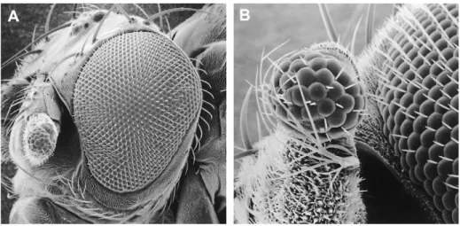
vertebrates and invertebrates. To test this hypothesis we tested the mouse Pax6 gene in Drosophila and reciprocally the Drosophila eyand toy genes in Xenopus (Onuma et al. 2002). As shown in Figures 3 and 6 in both cases ectopic eye structures can be induced, suggesting that the genetic cascade leading to eye formation is similar in vertebrates and invertebrates. Furthermore, not only Pax6 is highly conserved in eye development, but also so, the direct target gene of Pax6 is involved in morphogenesis of the vertebrate and invertebrate eyes, suggesting that the camera-type eye and the compound eye share similar eye morphogenetic programs. However, we have also found clear differences, for example, the Retina Figure 3. Ectopic Drosophila compound eye induced on the antenna by targeted expression of the mouse Small eye
(Sey) (¼ Pax6) gene. (A) Overview. (B) Higher magnification. Scanning electron micrograph (REM Laboratory, University of Basel). ey eya dac ? so toy eye development Pax 6 initiation feedback regulation
Figure 4. Gene regulatory network controlling eye determination in Drosophila.
homeoboxgene (Rx) (Mathers et al. 1997) is expressed in the retina of the mouse, but not in Drosophila, where it is expressed in the brain only (Eggert et al. 1998). Nevertheless, the eye morphogenetic programs of mice and flies are so similar that they suggest a common evolutionary ancestry.
Monophyletic Origin of the Different Eye
Types in Bilateria
True Pax6 homologs (orthologs) have been found in all bilateria analyzed so far, ranging from planarians to humans, Figure 5. Repartition of ey-induced genes in third instar larval leg discs in which an eye morphogenetic field is induced (Michaut et al. 2003).
Figure 6. Induction of ectopic eye structures in Xenopus embryos by injection of Drosophila ey and toy mRNA at the two-cell stage (Onuma et al. 2002). (A) Control embryo noninjected. (B) Embryo injected with 0.5 ng ey mRNA showing expansion of the retinal pigment epithelium. (C) Embryo injected with 2 ng toy mRNA showing a duplication of the retina. (D) Cross-section of an embryo injected with 0.25 ng ey mRNA showing a duplication of the lens and retina.
including C. elegans. This nematode has lost its eyes because it lives underground, but it has retained its Pax6 gene. The reason for retaining the Pax6 gene lies in the pleiotropic function of Pax6, which specifies not only the eye but also the nose and parts of the brain. Therefore, there is selective pressure to maintain Pax6 despite of the reduction of the eyes. This interpretation is supported by the fact that C. elegans has lost its rhodopsin gene(s); because the only function of rhodopsin is light reception, there is no selective pressure to maintain the rhodopsin gene. For C. elegans, the squid (Loligo), the seasquirt (Phallusia), and the lancelet (Amphioxus) we have shown that their Pax6 genes are capable of inducing ectopic eyes in Drosophila. The only exception to this rule are the two Pax6 genes of the planarian (Dugesia tigrinina), which apparently have diverged too much. However, we also have obtained functional genetic evidence that Pax6 and six (so) genes are also involved in eye morphogenesis in Dugesia.
By injection of double-stranded RNA (RNA interfer-ence) against sine oculis we have been able to prevent eye regeneration in Dugesia (Pineda et al. 2000). We have also developed methods for electroporation of planarians in toto that allowed us to apply a different strategy for testing the genetic control of eye development by Pax6. If Pax6 is highly conserved, it is to be expected that its target sites are also conserved to specify the same genetic circuitry. Therefore, we analyzed the Pax6-responsive P3-enhancer, which is
found in front of the rhodopsin genes ranging from fruitflies to humans (Berghammer et al. 1999). By fusing three copies of P3 to a minimal promoter driving green fluorescent protein (GFP) Horn and Wimmer (2000) have constructed transposon-derived vectors that function in a wide variety of organisms. Transgenic animals generally express the P3-GFP construct in their eyes (Figure 7A).
By electroporation we transformed the totipotent neo-blasts with the P3-GFP construct. These stem cells are capable of replacing somatic cells as well as germ cells, which are turning over. When the transformed neoblasts enter the eye region, they begin to express the GFP marker and gradually become photoreceptor cells until the eyes of the host are green fluorescent (Figure 7C and D). Neoblasts can also colonize the gonad and become germ cells, which allowed us to obtain F1 progeny with completely green eyes (Figure 7B) and to establish stable transgenic lines from them. These results indicate that not only Pax6 is conserved but also its target sites and the genetic circuits leading to eye morphogenesis.
The functional conservation of Pax6 and so from planarians to humans strongly suggests a monophyletic origin of the bilaterian eye. Because transcription factors can control the expression of any target gene (provided it contains the appropriate gene regulatory elements), there are no functional constraints linking Pax6 to eye development. Therefore, the link between Pax6 and so to eye development Figure 7. Planarians transformed by electroporation with GFP constructs with the Pax6-responsive P3-enhancer in the hermes transposon vector of Horn et al. (2000). (A) Transgenic Drosophila control. (B) Transgenic planarian line with homogenously green eyes 12 months after electroporation (after Gonza´lez-Este´vez et al. 2003). (C) Transformed neoblasts turn green as they enter the eye field. (D) Mosaic eyes in which a large fraction of the original photoreceptor cells are replaced by transformed neoblasts.
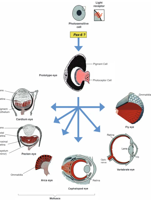
must simply be a consequence of a common evolutionary history. Therefore, I have proposed that all bilaterian eye types go back to a single root, a Darwinian prototype as, for example, found in planarians (Figure 1; Hesse 1897).
Starting from this prototype, selection has generated in-creasingly more performant eyes and that the various eye types arose by divergent, parallel, and convergent evolution (Figure 8).
Figure 8. Hypothetical evolution of photosensitive cells containing rhodopsin as a light receptor and monophyletic
evolution of the various eye types. The eye prototype consisting of a photoreceptor cell and a pigment cell is assembled under the control of Pax6 (after Gehring and Ikeo 1999).
From Unicellular Photoreceptors to
Multicellular Eyes
In cnidarians eyes are found only sporadically in some Hydrozoans, like Cladonema (Figure 9) and in box-jellyfish (cubozoans) like Tripedalia, and we do not know whether other jellyfish have lost their eyes in the course of evolution or whether they never acquired them. Neither in corals (Miller et al. 2000; Plaza et al. 2003) nor jellyfish (Gro¨ger et al. 2000; Kozmik et al. 2003; Sun et al. 1997, 2001) a bonafide Pax6gene has yet been found, but in Tripedalia Kozmik et al. (2003) have described a PaxB gene that might be an ancestral gene of Pax6. The paired domain of PaxB of Tripedalia shows 82% sequence identity to mammalian Pax2 and 75% to Pax6, and it also contains an octapeptide like Pax2, but it has a complete homeobox like Pax6, whereas Pax2 retains only the N-terminal part of the homeobox. This indicates that PaxB is structurally a mosaic between Pax2 and Pax6, which is also supported by functional studies. In Drosophila PaxB can partially complement Pax2 mutants (sparkling) and also induce ectopic eyes like Pax6 (Kozmik et al. 2003). Therefore, we consider PaxB to be a candidate ancestral gene of both Pax6 and Pax2. In the hydrozoan Cladonema, Stierwald et al. (2004) have obtained evidence that the six 1 and six 3 homologs are both involved in eye regeneration
(Figure 9), the same genes that are also controlling eye development in vertebrates, which lends strong support for the hypothesis of a monophyletic origin of the eye.
In Tripedalia the transition from unicellular eye organelles to multicellular eye organs can be observed. The planula larva of Tripedalia forms unicellular photoreceptors scattered over the epidermis (Figure 10), whereas the adult jellyfish forms elaborate multicellular eyes (Nordstro¨m et al. 2003). These unicellular photoreceptors contain both the putative photo-sensory microvilli and the shielding pigment granules within the same cell, which also carries a motor cilium that enables the larva to show phototactic behavior. These unicellular photoreceptors closely ressemble some unicellular photo-sensitive protists. We propose that in the course of evolution these unicellular photoreceptors has duplicated and differ-entiated into at least two different cell types, photoreceptor cells and pigment cells, as they are found in adult Tripedalia jellyfish and in the Darwinian prototype eyes of planarians (Polycelis auricularia).
The Evolution of Eyes and Brain
Because the eye in vertebrates develops as an evagination of the brain and is part of the brain it has generally been assumed that brain evolved before the eye. Furthermore, the Figure 9. Life cycle and eye structure and regeneration of the eyes in the hydrozoan jellyfish Cladonema (after Stierwald 2004). (A) Life cycle. (B) Location of the eyes at the base of the tentacle (arrowheads). (C) Eye structure: photoreceptor cells (red), pigment cells (yellow), lens cells (blue). (D) Removal of an eye from the tentacle bulb and eye regeneration. (E) Gene expression analysis by quantitative PCR; both six 1 and 3 homologs are induced.
9) and transmit their response directly to the muscles without processing by the brain. Of course, we cannot rule out the possibility that Cladonema has lost its brain, but it seems unlikely for a free-living pelagic animal to lose its brain in the course of evolution. Therefore, I consider it likely that the eyes evolved first, before the brain. This point of view is supported by the observation that the unicellular photo-receptor organelles in Tripedalia larvae have evolved in the apparent absence of an elaborate nervous system (Nordstro¨m et al. 2003).
Retrograde and Intercalary Evolution
Horowitz (1945) considered the evolution of biosynthetic pathways, which are in general linear, leading from the original substrate(s) over many successive enzymatic steps to the final biosynthetic product. A rather simple Darwinian solution to the problem of linearity was found by Horowitz by assuming a retrograde mode of evolution (Figure 11A). Originally the respective organism could not synthesize the respective product and had to take it up from the environment. When the supply in the environment was exhausted, those organisms whose had the last enzyme in the pathway could make use of the immediate precursor and convert it to the final product, until the supply of the immediate precursor was also exhausted. Then only those organisms could survive that possessed the second last enzyme and so on until the biosynthetic pathway was completely established.
For the evolution of morphogenetic pathways, like eye morphogenesis, I propose a mechanism of intercalary evolution (Figure 11B). The eye prototype, which is due to a purely stochastic event that assembles a photoreceptor and a pigment cell into a visual organ, requires the function of at least two classes of genes, a master control gene, Pax6, and the structural genes encoding on rhodopsin, for instance, the top and the bottom of the genetic cascade. Starting from such a prototype increasingly more sophisticated eye types arose by recruiting additional genes into the morphogenetic pathway. At least two mechanisms of recruitment are known that lead to the intercalation of additional genes into the genetic cascade. These mechanisms are gene duplication and
proteins. By fusing it to a lens-specific enhancer, the Drosocrystallin gene has been recruited into the eye de-velopmental pathway. In some cases the recruitment need not be associated with gene duplication, if the gene can still fulfill its original function; in other cases the recruitment may be accompanied by a gene duplication leaving an active gene copy behind to ensure the original function of the gene.
The Origin of Photoreceptor Cells in
Metazoa
For the origin of metazoan photoreceptor cells I have put forward two hypotheses: one based on cell differentiation and a more speculative model based on symbiosis.
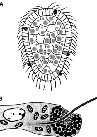
The more conventional idea is to assume that metazoa arose from a colony of flagellate-like cells like Volvox and Eudorina (Figure 12) in which all of the cells originally possessed a photoreceptor organelle, an ‘‘eyespot’’ that transmits its signals to the flagella and allows for phototactic behavior. Subsequent cellular differentiation would lead to an arrangement as found in the Tripedalia larva (Figure 10), which possesses unicellular photoreceptors scattered among the other ciliated cells in the ectoderm of the larva. The photoreceptor cells not only possess microvilli, presumably containing the visual pigment, but also melanin pigment granules shielding the basal side of the cell, as well as a cilium, which is thought to be used for steering of the larva toward or away from the light source. The next step would involve the differentiation of the unicellular photoreceptors into two cell types, a photoreceptor cell and a pigment cell and their assembly into a prototypic eye.
The alternative symbiont hypothesis is based on the observation that the eye organelle in flagellates like Volvox or Chlamydomonasis located in the chloroplast, suggesting that light perception goes all the way back to cyanobacteria that became integrated into eukaryotic cells as chloroplasts. Indeed, the sequencing of the complete genome of the cyanobacterium Nostoc has revealed the presence of a proteo-rhodopsin gene (Kaneko et al. 2001). A proteoproteo-rhodopsin gene has also been found in the dinoflagellate Pyrocystis and shown to be controlled by the internal clock (Okamoto and Hastings 2003). Another observation supporting the
symbiont hypothesis comes mainly from the work of Greuet (1965, 1969), who described the photoreceptor organelles of some dinoflagellates like Erythropsis and Warnovia, which are as elaborate as the human eye but assembled in a single cell (Figure 13). They consist of a cornea-like surface layer, a
lens-like structure, a retina-like structure with stacked mem-branes (or microvilli), and a pigment cup, all in a single cell. Because dinoflagellates are common symbionts in corals, sea anemones, and other cnidarians, dinoflagellates might have transferred the genes required for photoreception to the Figure 10. Unicellular photoreceptors in the planula larva of
the box jellyfish Tripedalia (after Nordstro¨m et al. 2003). (A) Planula larva. (B) Unicellular photoreceptor with pigment granules, microvilli, and a flagellum.
Figure 11. Models for the evolution of biosynthetic and morphogenetic pathways. (A) Retrograde evolution of biosynthetic pathways (after Horowitz 1945). (B) Intercalary evolution of the eye morphogenetic pathway (after Gehring and Ikeo 1999).
Figure 12. The colonial organization of Volvox and the structure of its photoreceptor organelle located in the chloroplast of Eudorina. (A) Colonial organization. (B) Structure of a single flagellate (somatic cell). (C) Ultrastructure of the photoreceptor organelle in the chlorplast (thylakoid membranes) of Eudorina californica (after Grell 1973).
cnidarians, which would also explain their sporadic occur-rence in different groups of cnidarians.
This symbiont hypothesis, which I call the Russian doll model, assumes that light sensitivity first arose in teria, the earliest known fossils on Earth. These cyanobac-teria were subsequently taken up by eukaryotic red algae as primary chloroplasts surrounded by an outer and inner
bacterial membrane separated by a proteoglycan layer. Subsequently, the red algae were taken up by dinoflagellates as secondary chloroplasts surrounded by an additional third membrane coming from the primary red algal host. In some species of dinoflagellates like Erythropsis and Warnovia, which do not have any chloroplasts, these secondary chloroplasts may have been transformed into elaborate photoreceptor Figure 13. Eye organelle of the unicelllular dinoflagellates Erythropsis and Warnowia. (A) Erythropsis. (B) Eye organelle of Erythropsis. (C) Warnowia. (D) Eye organelle of Warnowia. (E) Nucleus and eye organelle of Warnowia. (F) Birefringence, the retina-like structure detected in polarized light in Warnowia. (G) Ultrastructure of the eye organelle of Warnowia. (H) Ultrastructure of the retina-like structure with stacked membranes and large pigment granules. A–F courtesy of Makiko Seimiya and Jean and Colette Febvre; G–H from Greuet 1969.
organelles as suggested by Greuet. Because dinoflagellates (also called zooxanthellae) are commonly found as sym-bionts in cnidarians, dinoflagellates may have transferred their photoreceptor genes to cnidarians. This is the most speculative step in the Russian doll model, but it can be tested by looking for dinoflagellate genes in the cnidarian genomes. These two hypotheses, the cellular differentiation and the Russian doll model, are not mutually exclusive and are subject to further investigations.
Acknowledgments
This article is based on the Wilhemine E. Key plenary lecture given at the 2004 Annual Meeting of the American Genetic Association, ‘‘Genomes and Evolution 2004,’’ Pennsylvania State University, State College, PA, June 17– 20, 2004.
References
Berghammer AJ, Klingler M, and Wimmer EA, 1999. A universal marker for transgenic insects. Nature 402:370–371.
Czerny T, Halder G, Kloter U, Souabni A, Gehring WJ, and Busslinger M, 1999. Twin of eyeless, a second Pax-6 gene of Drosophila, acts upstream of eyeless in the control of eye development. Mol Cell 3:297–307.
Darwin C, 1882. The origin of species by means of natural selection, 6th ed. London: John Murray; 143–146.
Eggert T, Hauck B, Hildebrandt N, Gehring WJ, and Walldorf U, 1998. Isolation of a Drosophila homolog of the vertebrate homeobox gene Rx and its possible role in brain and eye development. Proc Natl Acad Sci USA 95:2343–2348.
Gehring WJ and Ikeo K, 1999. Pax 6: mastering eye morphogenesis and eye evolution. Trends Genet 15:371–377.
Gonza´lez-Este´vez C, Momose T, Gehring WJ, and Salo´ E, 2003. Trans-genic planarian lines obtained by electroporation using transposon-derived vectors and an eye-specific GFP marker. Proc Natl Acad Sci USA 100: 14046–14051.
Grell KG, 1973. Protozoology. New York: Springer Verlag; 314. Greuet C, 1965. Structure fine de lo´celle d’Erythropsis pavillardi Hertwig, pteridinien Warnowiidae Lindemann. CR Acad Sci (Paris) 261:1904–1907. Greuet C, 1969. Anatomie ultrastructurale des Pte´ridiniens Warnowiidae en rapport avec la differenciation des organites cellulaires. PhD diss., Universite´ de Nice.
Gro¨ger H, Callaerts P, Gehring WJ, and Schmid V, 2000. Characterization and expression analysis of an ancestor-type Pax gene in the hydrozoan jellyfish Podocoryne carnea. Mech Dev 94:157–169.
Halder G, Callaerts P, and Gehring WJ, 1995. Induction of ectopic eyes by targeted expression of the eyeless gene in Drosophila. Science 267:1788–1792. Hauck B, Gehring WJ, and Walldorf U, 1999. Functional analysis of an eye specific enhancer of the eyeless gene in Drosophila. Proc Natl Acad Sci USA 96:564–569.
Hesse R, 1897. Untersuchungen u¨ber die Organe der Lichtempfindung bei niederen Thieren. II. Die Augen der Plathelminthen. Z Wiss Zool Band 62:527–582.
Hill RE, Favor J, Hogan BL, Ton CC, Saunders GF, Hanson IM, Prosser J, Jordan T, Hastie ND, and van Heyningen V, 1991. Mouse Small eye results from mutations in a paired-like homeobox-containing gene. Nature 354: 522–525.
Hoge MA, 1915. Another gene in the fourth chromosome of Drosophila. Am Nat 49:47–49.
Horn C and Wimmer EA, 2000. A versatile vector set for animal transgenesis. Dev Genes Evol 210:630–637.
Horowitz NH, 1945. On the evolution of biochemical syntheses. Proc Natl Acad Sci USA 31:153–157.
Jacob F, 1977. Evolution and tinkering. Science 196:1161–1166. Janssens H and Gehring WJ, 1999. Isolation and characterization of drosocrystallin, a lens crystallin gene of Drosophila melanogaster. Dev Biol 207:204–214.
Kaneko T, Nakamura Y, Wolk CP, Kuritz T, Sasamoto S, Watanabe A, Iriguchi M, Ishikawa A, Kawashima K, and Kimura T, 2001. Complete genomic sequence of the filamentous nitrogen-fixing cyanobacterium Anabaenasp. strain PCC 7120. DNA Res 8:205–213.
Kozmik Z, Daube M, Frei E, Norman B, Kos L, Dishaw LJ, Noll M, and Piatigorsky J, 2003. Role of Pax genes in eye evolution: a cnidarian PaxB gene uniting Pax2 and Pax6 function. Dev Cell 5:773–785.
Kronhamn J, Frei E, Daube M, Jiao R, Shi Y, Noll M, and Rasmuson-Lestander A, 2002. Headless flies produced by mutations in the paralogous Pax6 genes eyeless and twin of eyeless. Development 129:1015– 1026.
Mathers PH, Grinberg A, Mahon KA, and Jamrich M, 1997. The Rx homeobox gene is essential for vertebrate eye development. Nature 387:603–607.
Mayr E, 2001. What evolution is. New York: Basic Books.
Michaut L, Flister S, Neeb M, White KP, Certa U, and Gehring WJ, 2003. Analysis of the eye developmental pathway in Drosophila using DNA microarrays. Proc Natl Acad Sci USA 100:4024–4029.
Miller DJ, Hayward DC, Reece-Hoyes JS, Scholten I, Catmull J, Gehring WJ, Callaerts P, Larsen JE, and Ball EE, 2000. Pax gene diversity in the basal cnidarian Acropora millepora (Cnidaria, Anthozoa): implications for the evolution of the Pax gene family. Proc Natl Acad Sci USA 97:4475– 4480.
Niimi T, Seimiya M, Kloter U, Flister S, and Gehring WJ, 1999. Direct regulatory interaction of the eyeless protein with an eye-specific enhancer in the sine oculis gene during eye induction in Drosophila. Development 126:2253–2260.
Nordstro¨m K, Wallen R, Seymour J, and Nilsson D, 2003. A simple visual system without neurons in jellyfish larvae. Proc R Soc Lond B Biol Sci 270:2349–2354.
Okamoto OK and Hastings JW, 2003. Novel dinoflagellate clock-related genes identified through microarray analysis. J Phycol 39:519–526. Onuma Y, Takahashi S, Asashima M, Kurata S, and Gehring WJ, 2002. Conservation of Pax 6 function and upstream activation by Notch signaling in eye development of frogs and flies. Proc Natl Acad Sci USA 99:2020– 2025.
Piatigorsky J and Wistow GJ, 1989. Enzyme/crystallins: gene sharing as an evolutionary strategy. Cell 57:197–199.
Pineda D, Gonzalez J, Callaerts P, Ikeo K, Gehring WJ, and Salo E, 2000. Searching for the prototypic eye genetic network: sine oculis is essential for eye regeneration in planarians. Proc Natl Acad Sci USA 97:4525–4529. Plaza S, De Jong DM, Gehring WJ, and Miller DJ, 2003. DNA-binding characteristics of cnidarian Pax-C and Pax-B Proteins in vivo: no simple relationship with the Pax-6 and Pax-2/5/8 classes. J Exp Zoolog Part B Mol Dev Evol 299:26–35.
Punzo C, Seimiya M, Flister S, Gehring WJ, and Plaza S, 2002. Differential interactions of eyeless and twin of eyeless with the sine oculis enhancer. Development 129:625–634.
Punzo C, Plaza S, Seimiya M, Schnupf P, Kurata S, Jaeger J, and Gehring WJ, 2004. Functional divergence between eyeless and twin of eyeless in Drosophila melanogaster. Development 131:3943–3953.