Control of HslUV Protease Function by Nucleotide
Binding and Hydrolysis
by
Joseph Andrew Yakamavich
B.S. Biochemistry
North Carolina State University, 2002
Submitted to the Department of Biology in partial fulfillment of the requirements for the degree of
Doctor of Philosophy in Biochemistry
at the
Massachusetts Institute of Technology
February 2008
@ 2007 Joseph A Yakamavich. All rights reserved.
The author hereby grants to MITpermission to reproduce and to distribute publicly paper and electronic copies of this thesis document in whole or in part.
Signature of Author:
Snu tr Department of Biology
January 14, 2008
Certified by:
-Robert T. Sauer Salvador E. Luria Professor of Biology Thesis Supervisor
Accepted by: Stephen
P. Bell Professor of Biology
LIBRARIES
Co-Chair, Biology Graduate Committee
ARCHIVES
OF •OHCLOO
FEB
2 2008
_Control of HslUV Protease Function by Nucleotide Binding and Hydrolysis by
Joseph Andrew Yakamavich
Submitted to the Department ofBiology on January 14, 2008 in Partial Fulfillment of the Requirements for the Degree of Doctor ofPhilosophy in Biochemistry
ABSTRACT
Many proteins act as molecular machines, using the power of nucleotide binding and hydrolysis to drive conformational changes in themselves and their target substrates. Like other AAA+ proteases, HslUV recognizes, unfolds, translocates, and degrades substrate proteins in an ATP-dependent manner. Understanding how nucleotides interact with HslU and control the activities of both HslU and HslV provides insights into the general mechanism of energy-dependent proteolysis.
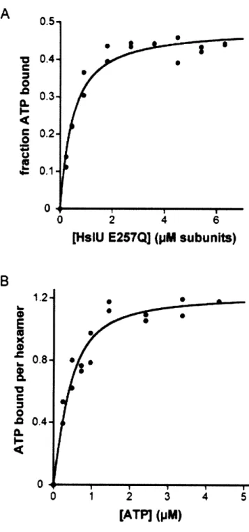
In order to better understand HslU-nucleotide interactions, I created a variant of HslU unable to hydrolyze ATP. HslU is composed of six identical subunits with a total of six nucleotide-binding sites. Moreover, many crystal structures show HslU with six bound nucleotides. Nevertheless, I found that HslU in solution is only able to bind 3-4 ATPs at saturation. This result rules out a model of ATP hydrolysis in which six nucleotides bind and are hydrolyzed together in a single power stroke and also suggests that many HslU crystal structures represent states that are not populated in the normal ATPase cycle. I also characterized the nucleotide requirement for various HslU activities. I found that at least two ATPs must be bound to HslU to support substrate binding and ATP hydrolysis, but showed that a single nucleotide is sufficient to support HslU-HslV binding and to stimulate HslV peptidase activity. I also found that the nucleotide state of HslU affects its affinity to HslV, weakening it when some subunits have ADP or no nucleotide bound. This effect is offset by an increase in HslU-HslV affinity during substrate degradation. This work suggests a simple model in which binding of a single ATP to HslU drives HslV binding, with further ATP binding acting to stabilize an HslU conformation that can bind protein substrate, hydrolyze ATP, and support substrate unfolding, translocation, and degradation.
Thesis Supervisor: Robert T. Sauer
TABLE OF CONTENTS
Paue
A bstract... ... 2 Chapter One: Chapter Two: Appendix A: Chapter Three: Chapter Four: The AAA+ proteases HsiUV, ClpXP, and the proteasome ... 6...Introduction to AAA+ proteases ... 7...
HslUV Characterization... 13
HslU and HslUV Crystal Structures ... 18
H slV ... ... ... 21
The ClpX P System ... 23
The 26S Proteasome ... ... 26
Research approach ... 29
Asymmetric nucleotide transactions of the AAA+ protease H slUV ... ... ... 39
Supplementary material for chapter two ... 77
HslU nucleotide binding site and pore-2 loop mutants ...88
HslUV cysteine mutants and work towards an HslUV FRET assay ... ... ... 101
LIST OF FIGURES & TABLES
Page Chapter One: The AAA+ proteases HslUV, CIpXP, and the proteasome
Figure 1 - Architecture of the HslUV protease ... ... 10
Figure 2 - Protein degradation by ATP-dependent proteases... 11
Figure 3 - Conformational states of the HslU C-terminal tail ... 22
Table 1 - Comparison of HslU crystal structures ... 19
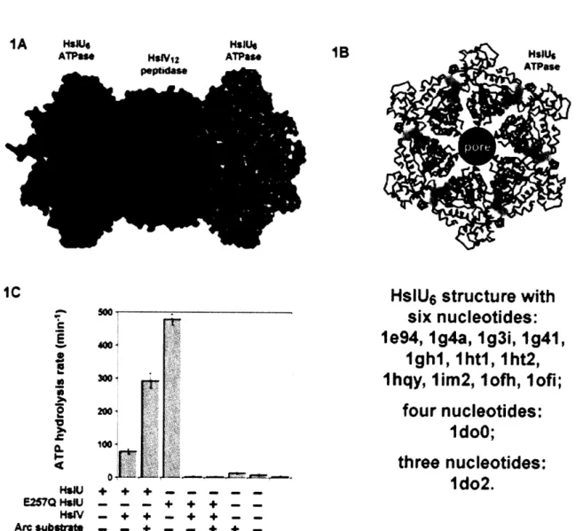
Chapter Two: Asymmetric nucleotide transactions of the AAA+ protease HslUV Figure 1 - HslU architecture and ATP interactions...68
Figure 2 - HslU binding stoichiometry... ... 69
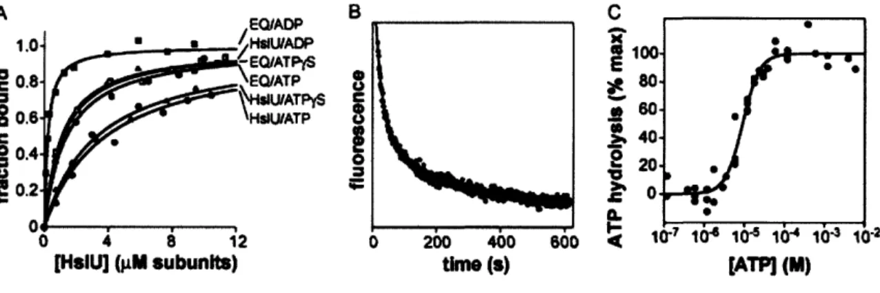
Figure 3 - Nucleotide affinity as determined by nitrocellulose filter binding assays.71 Figure 4 - Nucleotide binding, dissociation, and hydrolysis ... .. 72
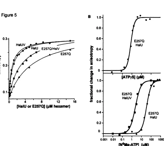
Figure 5 - HslU binding to FL-gtl, a fluorescent substrate mimic peptide...73
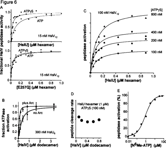
Figure 6 - H slU V binding...74
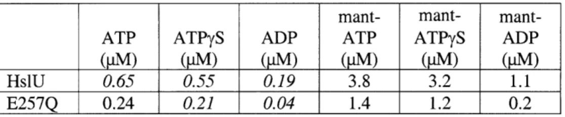
Table 1 - Nucleotide affinities ... ... ... 76
Appendix A: Supplementary material for chapter two Figure 7 - ATP-independent HslU*HslV binding ... 78
Supplemental Figure 1 - Absorbance spectra of HslU wild-type, EQ, and charcoal-treated EQ variants... 79
Supplemental Figure 2 - Charcoal-treated HslU remains able to degrade Arc repressor ... ... 79
Supplemental Figure 3 - Untreated and charcoal treated HslU activate HslV peptidase activity ... .. ... 80
Supplemental Figure 4 - Untreated and charcoal treated HslU both hydrolyze ATP sim ilarly ... ... 80
LIST OF FIGURES & TABLES (Continued)
Supplemental Figure 5 - HslU E257Q is unable to degrade Arc repressor ... 81 Supplemental Figure 6 - Peaks from chromatographic elution stimulate HslV
peptidase activity equivalently... ... 81 Supplemental Figure 7 - Nucleotide competition assay ... 82
Supplemental Figure 8 - Dissociation kinetics of mant-ATP in the presence of HslV or HslV and Arc ... 82
Chapter Three: HslU nucleotide binding site and pore-2 loop mutants
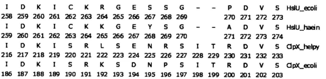
Figure 1 - Partial sequence alignment between HslU and ClpX...90
Figure 2 - HslV stimulation of ATP hydrolysis by nucleotide binding site mutant variants of HslU ... 93 Figure 3 - gtl binding by wild-type and mutant variants of HslU ... 94 Figure 4 - HslV stimulation of ATP hydrolysis by wild-type and mutant variants of
H slU ... 9 6 Figure 5 - gtl binding affinity of pore-2 mutant variants...97 Figure 6 - Arc degradation by wild-type and pore-2 mutant variants ... 98 Table 1 - Biochemical activities of wild-type and variant HslU enzymes ... 91
Chapter Four: HslUV cysteine mutants and work towards an HslUV FRET assay Figure 1 - Substitutions for C261 or C287 in E. coli HslU result in mutant enzymes
with ATP-hydrolysis rates similar to the wild-type enzyme ... 104 Figure 2 - HslV lacking cysteine cleaves peptide substrates at comparable rates to
wild-type HslV ... 105 Figure 3 - Peptidase hydrolysis by mixed complexes of HslU C261A/C287S/S338C
CHAPTER ONE
Introduction
All organisms expend energy to grow, reproduce, and react to their environment. At the cellular level, the energy from nucleotide triphosphate (NTP) binding and/or hydrolysis is transduced into physical or chemical work. Specifically, hydrolysis of NTPs (typically ATP or GTP) is a highly favorable downhill reaction from a thermodynamic perspective. Macromolecular enzymatic machines are able to harness the energy of repetitive NTP binding and hydrolysis events to drive cyclical changes in three-dimensional structure and to transmit these conformational changes to bound macromolecules. During DNA replication, for example, ATP binding and hydrolysis by the clamp loader complex is coupled to conformational changes in the 0 or PCNA clamp proteins that close the clamp ring around DNA (Naktinis et al., 1995; Baker and Bell, 1998).
In the most common class of NTPases, two conserved sequence motifs play critical roles in NTP binding and hydrolysis. The Walker-A or P-loop motif (GX4GKS/T) stabilizes
binding through contacts with the nucleotide phosphates, whereas the Walker-B motif (04DE; cD = hydrophobic) is important for nucleotide hydrolysis (Hanson and
Whiteheart, 2005). The AAA subfamily (ATPases associated with various cellular activities) is defined by a 200-250 residue ATPase domain, as well as a second region of homology (SRH) located C-terminal from the Walker-B motif. Analysis of multiple sequence alignments revealed additional related ATPases lacking the SRH, which comprise a AAA+ subfamily (Neuwald et al., 1999). AAA+ family members are involved in a wide range of cellular activities, including membrane fusion, transport,
protein disaggregation, transcription, DNA replication, DNA recombination, and proteolysis.
ATP-dependent proteolysis is a critical activity necessary for a variety of physiological functions and is carefully regulated so that only a specific set of proteins in the cell are subject to degradation at given time. There are many benefits of targeted degradation, including the removal of unwanted or deleterious proteins from the cell. Cellular levels of specific proteins vary depending on the environment. For example, under periods of stress (e.g., heat shock, starvation, acid or oxidative stress, etc.), existing cellular proteins are often damaged as a consequence of unfolding, aggregation, chemical modification, or incomplete biosynthesis. If they cannot be repaired, these non-functional and potentially dangerous proteins are generally degraded. Moreover, during stress, transcriptional responses result in the synthesis of new and different types of proteins that allow the cell to deal with the specific stress and to minimize disruption. Once the environmental stress ceases, however, the proteome must be rebalanced through changes in transcription and proteolysis in order to return cellular processes and physiology to a normal state. During normal growth, proteolysis may also be used to maintain the concentration of certain proteins within a specific range or to release dormant activities through partial proteolysis. Thus, proteolysis provides a post-translational opportunity to regulate protein
levels and activities in a variety of ways.
In Escherichia coli and related bacteria there are five distinct types of ATP-dependent AAA+ proteases: HslU/HslV (HslUV), ClpX/ClpP (ClpXP), ClpA/ClpP (ClpAP), Lon,
and FtsH (Neuwald et al., 1999; Gottesman, 1996). HslUV, ClpXP, and ClpAP are composed of separate AAA+ ATPase and peptidase components (Rohrwild et al., 1996; Katayama-Fujimura et al., 1987; Wojtkowiak et al., 1993). Lon and FtsH have both activities encoded on a single polypeptide chain (Charette et al., 1981; Tomoyasu et al., 1993). All of these enzymes are located in the cytoplasm, although FtsH is anchored to the inner side of the cytoplasmic membrane bound (Tomoyasu et al., 1993). Eukaryotes rely chiefly on the function of the 26S proteasome for degradation of cytoplasmic and nuclear proteins (Hershko and Ciechanover, 1998), although homologs of the bacterial AAA+ proteases carry out important degradation tasks within mitochondria and chloroplasts (Koppen and Langer, 2007). A closely related member of the AAA+ family powers each of these ATP-dependent proteases, and the distinct specificities of these enzymes allow cells to react to differential needs and to remodel the proteome in a dynamic fashion. In some cases, AAA+ ATPases function as disassembly chaperones to take apart macromolecular complexes and/or solubilize aggregates. For example, in the absence of protease partners, ClpX disassembles DNA-bound complexes of the MuA transposase and ClpB and Hspl04 resolubilize protein aggregates (Levchenko et al.,
1995; Goloubinoff et al., 1999; Motohashi et al., 1999; Zolkiewski, 1999).
All AAA+ proteases share generally similar architectures. Stacking of hexameric or heptameric rings creates a hollow barrel-like structure in which the proteolytic active sites are hidden within an interior chamber to prevent accidental protein degradation. Hexameric AAA+ ATPases flank one or both sides of the protease barrel and function to recognize substrates, unfold native substrate structure, and translocate the unfolded
polypeptide into the protease chamber for degradation. Conserved AAA+ domains are responsible for these shared activities, whereas family specific domains are frequently implicated in substrate or adaptor recognition (Dougan et al., 2002; Mogk et al., 2004). The AAA+ module of each ATPase subunit is composed of a large ap domain and a smaller a-helical domain, with nucleotide binding occurring at subunit-subunit interfaces (Ogura and Wilkinson, 2001). Several highly conserved residues play key roles in nucleotide hydrolysis. Mutation of a highly conserved glutamic acid in the Walker-B motif is sufficient to disrupt ATP hydrolysis in many AAA+ proteins (Hersch et al., 2005; Hanson et al., 2005). In addition, highly conserved arginines play a critical role in sensing and hydrolyzing bound ATP and in mediating conformational changes that accompany binding and hydrolysis (Ogura et al., 2003).
Figure 1
top view
side view
HslU V cutaway
-- HslU 6 -AAA+ ATPase HslVI2--peptida.se
I
/(
peptidase active sites1T
The HslUV protease is composed of HslU and HslV (Figure 1). HslU is a homohexameric AAA+ ATPase, which adopts a ring-shaped structure with a central axial pore or channel. HslV, the peptidase component, is a threonine active-site protease related to the 3 subunits of the eukaryotic 20S proteasome. The functional HslV enzyme is assembled from 12 subunits, which form two stacked hexameric rings and enclose a degradation chamber in which the active-site threonines are located. This compartment or cavity is accessible through narrow axial pores. HslU hexamers can bind to one or both faces of HslV, resulting in alignment of their respective axial pores and the creation of a narrow channel into the lumen of the protease. HslU is responsible for recognizing appropriate protein substrates, unfolding them if necessary, and then translocating the denatured polypeptide into HslV for degradation (Figure 2). The unfolding and translocation steps require ATP hydrolysis, whereas peptide-bond cleavage by HslV does not.
Figure 2
)•,& degradation native tag protein B BINDIlsil & DENATURE
IlslV __ eise ADP
peptidase
ADP
TRANSLOCATE ET PEPTIDE HYDROLYSISADP
From a structural perspective, HslUV is the best-characterized AAA+ protease. For example, there are more than 10 crystal structures of the HslU hexamer. Some structures contain isolated HslU hexamers; others contain complexes of one or two HslU hexamers
bound to HslV (Trame and McKay, 2001; Sousa et al., 2000; Wang et al., 2001a; Song et al., 2000; 2003; Bochtler et al., 2000). In some HslUV complexes, the active-site residues of HslV are chemically modified (Sousa et al., 2002; Kwon et al., 2003). The number of nucleotides bound to HslU varies from structure to structure. In some structures, for example, six ATP or ADP molecules are bound (Sousa et al., 2000; Kwon et al., 2003; Wang et al., 2001a; Song et al., 2000; Trame and McKay, 2001). In other crystal structures, only three or four nucleotides bind to the HslU hexamer (Bochtler et al., 1997; 2000). Although the abundance of structural information has allowed modeling of different conformations that may comprise the ATPase cycle (Wang et al., 2001a), it is still unclear which, if any, of the available crystal structures represent functional states of the HslUV protease. My aim in this thesis is to characterize HslUV from a biochemical perspective, thereby providing a complement to the structural information and improving our understanding of HslUV mechanism.
In the sections below, I provide an introduction to HslUV, related proteases, and their interaction with substrates and nucleotides. Although much is known about ATP-dependent proteolysis for enzymes like ClpXP and ClpAP, there have been far fewer studies of HslUV. HslV is the bacterial homolog of the eukaryotic 20S proteasome, which is an attractive pharmaceutical target given its roles in a variety of pathways, including inflammation and differentiation. With only two types of subunits, HslUV is far simpler and more accessible to dissection than the 26S proteasome, which contains more than 20 types of subunits, and yet both enzymes perform a core set of common functions. Studies of the detailed mechanisms used by HslUV to degrade substrates also facilitates
comparisons with better characterized AAA+ proteases, such as ClpXP, and should provide insights into common functions and family-specific features. When I began my thesis work, it was known that ATP was needed for many functions of HslUV. ATP or ATPyS was required for activation of HslV and for interactions between HslU and HslV (Huang and Goldberg, 1997; Yoo et al., 1997a; Burton et al., 1995). However, specific roles were unclear. How many nucleotides HslU was capable of binding, whether there were single or multiple classes of nucleotide binding sites, and how nucleotide binding was linked to specific HslUV activities were all undefined. The importance of nucleotides in AAA+ protease complex formation and substrate recognition has been documented in other systems (Bolon et al., 2004; Smith et al., 2005). Because ATP fuels the HslUV machine, determining how nucleotide binding and hydrolysis in HslU control interactions with substrate and HslV is important for understanding function.
HslUV Characterization
The HslUV protease is present in roughly 60% of eubacteria and many eukaryotic lineages (Gille et al., 2003; Ruiz-Gonzales and Marin, 2006). The tandem genes encoding HslU (also called ClpY) and HslV (also called ClpQ) were originally discovered by DNA sequencing and were found to be under the control of a heat-shock promoter (Chuang and Blattner, 1993). The names stand for heat shock locus proteins U and V. These genes and/or the encoded enzymes were also identified in other contexts (Missiakas et al., 1996; Khattar, 1997). Early studies on the physiological role of HslUV found that E. coli knockout strains were viable and showed no obvious phenotypic effects below 45 "C; above this temperature, deletion strains grew slowly or died (Katayama et al., 1996
Kanemori et al., 1997). Other heat-shock proteins were found to be overproduced in the knockout strains, and overexpression of HslUV reduced the heat-shock response (Missiakas et al., 1996). The purified HslUV protease from E. coli shows maximal degradation activity between 50 and 60 'C, a temperature range in which some AAA+ proteases, like ClpXP, denature and lose all activity (Burton et al., 2005). As a consequence, it seems likely that HslUV plays a special role in degrading cellular proteins that unfold or become damaged at high temperatures. Indeed, HslUV was shown to participate in degradation of incomplete and thus presumably unfolded proteins produced by premature puromycin-induced termination of translation (Missiakas et al., 1996). The presence of unfolded proteins in E. coli induces the heat-shock response (Parsell and Sauer, 1989). Thus, the increased levels of heat-shock proteins in strains lacking HslUV can be explained by over accumulation of denatured proteins, whereas HslUV overexpression would be expected to reduce the concentration of unfolded proteins and therefore diminish the heat-shock response. HslU has also been shown to play a role in the disaggregation of proteins, an activity shared by related AAA+ ATPases (Seong et al., 2000).
In addition to proteins damaged by thermal denaturation, several other physiological substrates of HslUV have been identified. The heat-shock transcription factor a32, which
is responsible for the expression of HslUV (Rohrwild et al., 1996), is degraded primarily by FtsH, but also by HslUV and to lesser extents by Lon and ClpXP (Kanemori et al., 1997). RcsA (the capsule-synthesis activator) and SulA (a cell-division inhibitor) are also substrates for HslUV and for Lon (Wu et al., 1999). For example, overexpression of
HslUV suppresses phenotypes caused by stabilization of both proteins in Lon-knockout strains (Kuo et al., 2004). Pulse-chase analysis showed that HslUV degradation of SulA occurs in the absence of heat shock, and thus HslUV presumably also plays roles in regulation and protein-quality control under normal physiological conditions (Seong et al., 1999). HslUV and Lon are both required for cell viability under conditions in which the signal recognition particle (SRP) is expressed at low levels. SRP is responsible for proper cotranslational insertion of proteins into the inner membrane of E. coli. When SRP activity is limited, inner-membrane proteins accumulate in the cytoplasm, suggesting that both HslUV and Lon function to remove these otherwise toxic mislocalized proteins (Bernstein and Hyndman, 2001). Taken together, these results indicate that HslUV degrades numerous intracellular substrates, often sharing overlapping functions with other ATP-dependent proteases. Removal of many of these substrates seems to be sufficiently important that multiple redundant pathways have evolved to ensure degradation.
In general, known physiological substrates of HslUV have not been very useful as model substrates for studies in vitro. For example, SulA and a32 are both relatively difficult to
express, are poorly soluble, and exhibit short intracellular half-lives under normal conditions (Nguyen et al., 1993; Kwon et al., 2004). There is little structural information for either protein (Kwon et al., 2004). SulA has been purified as a fusion protein to maltose-binding protein (MBP) and green fluorescent protein (GFP), but HslUV is incapable of degrading either the MBP or GFP portions of these fusions (Seong et al.,
unfoldase activity. The HslUV cleavage sites in SulA have been mapped by analysis of peptide products, showing that HslV prefers to cleave after hydrophobic residues (Nishii and Takahashi, 2003). However, HslUV degrades SulA very slowly in vitro, and thus non-physiological substrates have typically been used for detailed biochemical studies.
In early studies, the proteolytic activity of HslUV was usually monitored by degradation of 3H- or 14C-labeled casein or insulin B-chain (Huang and Goldberg, 1997; Seol et al.,
1997). These generic substrates have no stable tertiary structure and are degraded by most proteases, but are only bound weakly by HslUV and are degraded at slow rates. Moreover, HslUV degradation of casein does not require ATP (Huang and Goldberg, 1997). This result suggests that casein does not have to be actively translocated through HslU and thus does not serve as a good model for normal degradation.
More recently, it has been found that HslUV efficiently degrades the Arc repressor from bacteriophage P22 (Kwon et al., 2004; Burton et al. 2005). The structure of Arc, a dimeric ribbon-helix-helix transcription factor, is known and its stability to denaturation has been well characterized (Breg et al., 1990; Sauer et al., 1996). Steady-state kinetic parameters have been determined for HslUV degradation of Arc mutants of various stabilities, as well as for variants fused to the 127 domain of human titin, a protein that is very resistant to mechanical unfolding (Carrion-Vazquez et al., 1999; Burton et al., 2005). Stable Arc substrates are recognized and degraded efficiently (Km - 5 ýpM; turnover number -7 min-'). The titin domain fused to Arc is also degraded. Thus, HslUV is able to unfolded substrates that are quite stable prior to degradation. HslUV recognizes
residues at both the N- and C-terminus terminus of Arc (Kwon et al., 2004; Burton et al. 2005; Sundar and McGinness, personal communication). Based on the N-terminal sequence of Arc, peptide-array experiments have been used to identify a peptide (called gtl) that binds HslU in a nucleotide-dependent fashion and competes with binding and degradation of Arc (Burton et al., 2005).
When HslUV was initially purified, a variety of fluorogenic peptides were screened as potential substrates (Rohrwild et al., 1996). N-a-benzyloxycarbonyl-Gly-Gly-Leu-7-amido-4-methylcoumarin (Z-GGL-AMC), a substrate previously used to assay the chymotrypsin-like activity of the proteasome (Chu-Ping et al., 1992), was found to be cleaved efficiently by HslUV (Rohrwild et al., 1996). In the absence of HslU, HslV degrades Z-GGL-AMC very slowly, and this activity is inhibited by nucleotide (Huang and Goldberg, 1997; Lee et al., 2007). The physiological relevance of the latter observation is unclear. In the presence of HslU and a suitable nucleotide triphosphate (ATP, AMPPNP, or ATPyS), however, HslV cleavage of Z-GGL-AMC is stimulated roughly 500-fold (Huang and Goldberg, 1997). Thus, by itself, HslV is a poor peptidase, degrading even small peptides far more slowly in the absence than the presence of HslU. Because the axial pore of HslV is large enough to allow Z-GGL-AMC to diffuse into the peptidase chamber, the active sites of this enzyme must be largely unreactive when HslU is not bound. Because HslU in combination with ATP or the poorly hydrolyzed ATPyS analog or the non hydrolyzable AMPPNP analog stimulates the peptidase activity of HslV to comparable extents, NTP binding but not hydrolysis seems to be required to allow HslU to interact productively with HslV. As discussed below, NTP binding to
HslU appears to place its C-terminal tail in a conformation that binds and activates HslV (Seong et al., 2002; Sousa et al., 2002). In Chapter 2, I use nucleotide-dependent activation of HslUV cleavage of Z-GGL-AMC to probe the strength and requirements of HslU binding to HslV.
HslU and HslUV Crystal Structures
Like all AAA+ ATPases, HslU subunits contain a large and a small AAA+ domain, and nucleotides bind at the interface formed by both of these domains in a single subunit and by the large AAA+ domain of an adjacent subunit (Bochtler et al., 2000; Sousa et al., 2000). This architecture allows changes in nucleotide occupancy and/or identity to alter the tertiary and quaternary structure of the HslU hexamer, giving rise to conformational transitions during the ATPase cycle that presumably explain its machine-like properties. Indeed, the orientations of the large and small AAA+ domains vary in different HslU structures and do seem to depend upon nucleotide state; these observations have been used to propose a series of structural changes that occur during the nucleotide hydrolysis cycle (Wang et al., 2001a). As I discuss below, however, it is unclear how many of the subunit conformations observed in different HslU crystal structures are functionally relevant.
E. coli HslU and its orthologs contain an additional "intermediate" or I-domain. If HslU
is viewed as having the basic shape of a hexagonal nut, then the I-domain projects away from one face of the nut and HslV binds to the other face (Bochtler et al., 2000; Sousa et al., 2000). Thus, the I-domain is on the face of HslU where substrates are thought to bind
initially. HslU variants in which the I-domain is deleted cannot support HslV degradation of SulA or Arc repressor but some casein degradation is observed (Song et al., 2000; Kwon et al., 2003). Thus, the I-domain probably plays a role in the recognition of specific protein substrates.
Table
1
Organism Protein HslU Tail Conformation Year Nucleotide State PDB Accession
H. influenzae UV extended 2000 6 ATP 1G31
H. influenzae U(AI)V extended 2003 6 ADP (Pi, Mg++) 10FH E. coli UV packed 2001 6 dATP 1G4A E. coli UV packed 2000 6 AMPPNP 1E94
E. coli U packed 2000 4 ATP 1DOO
H. influenzae U packed 2001 6 ADP 1G41
E. coli U packed 1997 3 AMPPNP, 3 empty 1D02
H. influenzae U packed 2001 6 ADP 11M2 E. coli UV packed 2000 6 ADP 1HQY
E. coli UV packed 2000 6 ADP 1HT1
E. coli UV packed 2000 6 ADP 1HT2
E. coli UV packed 2000 none 1G4B
HslUV is the only ATP-dependent protease for which the complete three-dimensional structure has been solved. Crystal structures of HslU6, HslV12, HslU6HslV12, and
HslU6HslV12HslU6 are known (Bochtler et al., 1999; Sousa et al., 2000; 2002; Sousa and
McKay, 2001; Trame and McKay, 2001; Wang et al., 2001a; Kwon et al., 2003; Song et al., 2003). Table 1 lists the types and numbers of nucleotides bound to HslU in the available structures. Many of the known HslU structures are six-fold symmetric and have ATP or ADP bound to each potential nucleotide binding site, as would be expected for a model in which HslU cycled between an all-ATP state and an all-ADP state in a concerted allosteric reaction. However, one HslU structure is a trimer of dimers with AMPPNPs bound to alternating subunits, and another is a dimer of trimers with adjacent subunits in each trimer being nucleotide-free, ATP-bound, and ATP*Mg +-bound,
respectively. These non-symmetric structures suggest that HslU may operate by some type of sequential mechanism in which only a subset of bound nucleotides can be hydrolyzed.
In some cases, identification of the nucleotides bound in HslU structures has been a matter of dispute. For example, one HslU structure (1DOO) with six putatively bound AMPPNPs was found to have a structure indistinguishable from another structure (1G4A) in which six ADPs were bound (Bochtler et al., 2000; Song et al., 2000; Wang et al., 2001a; 2001b). When the 1DOO structure was recalculated, however, no electron density was observed for the y-phosphate of AMPPNP (Wang et al., 2001b). Additionally, the adenine base in the 1DOO structure was built in a syn conformation, whereas most nucleotides bound to AAA+ ATPases adopt an anti conformation (Wang et al., 2001b). In this instance, it seems clear that the AMPPNP in the 1DOO structure was misidentified and is actually ADP, presumably present as a contaminant in the AMPPNP or enzyme used for crystallization. Indeed, in another HslU structure (1DO0), nucleotides were found bound to HslU even through nucleotide had not been included in the crystallization drop. Because of these issues, the identity of nucleotides in other HslU structures has also been questioned. For example, in both asymmetric HslU structures, the adenines of ATP (1D00) or AMPPNP (1D02) were syn and the temperature factors of the y-phosphate atoms were roughly 50 A 2 higher than those of surrounding atoms,
HslV
As noted above, the peptidase active sites, including the catalytic N-terminal threonine, are sequestered in an internal cavity in the double-ring HslV dodecamer. HslV has the distinct fold shared by members of the N-terminal nucleophile (Ntn) hydrolase family. Originating from a common ancestor, the sequences of family members, which have a variety of hydrolytic activities, are now highly diverged. Other members of this family include penicillin acylases, glutamine PRPP amidotransferases, glycosylasparaginases, and notably the 20S proteasome (Brannigan et al., 1995). In the proteasome, autocatalytic processing reveals the N-terminal catalytic threonine; in HslV, removal of the initiator formyl methionine by the combined action of peptide deformylase and methionine amino-peptidase frees threonine as the N-terminal residue. Comparison of HslV structural data suggests that a nearby lysine in the active site lowers the pKa of the a-amino group of Thr', which in an unprotonated state can accept a proton from and activate the Thr' side chain for nucleophilic attack (Groll et al., 1995). Replacing Thri in
E. coli HslV with a serine results in peptidase levels similar to wild type when stimulated
by HslU (Yoo et al., 1997b). In fact, the B. subtilis HslV homolog, CodW, is the only known N-terminal serine protease. Serine-protease inhibitors inactivate CodW, and replacing Seri with threonine abolishes all enzymatic activity (Kang et al., 2001).
Comparison of HslV structures in combination with biochemical results has provided key insights into the mechanism of HslV activation by HslU. HslV structures from E. coli, Thermatoga maritima, and Haemophilus influenzae (Bochtler et al., 1997; Song et al., 2003; Sousa and McKay, 2001) have been solved. The structure of H. influenzae HslV
has the highest resolution (2.1
A),
and this enzyme has been crystallized in complex with HslU (Sousa and McKay, 2001). The structures of free and HslU-bound HslV reveal conformational differences, as expected from the finding that the active sites of HslU-bound HslV react with vinyl sulfone inhibitors, whereas the active sites of free HslV do not (Bogyo et al., 1997). Moreover, when inhibitor was soaked into crystals containing asingle HslU6 bound to HslV12, the active sites in the cis-HslV ring but not the trans-HslV
ring were modified (Kwon et al., 2003). These results suggest that HslU binding is required to stabilize an HslV conformation in which the active-site residues assume the correct catalytic geometry and that an HslU hexamer only activates the HslV ring to which it binds. This model explains why HslUV but not HslV cleaves the Z-GGL-AMC peptide efficiently.
Figure 3
HslU
HslUV
extended state
In several H. influenzae HslUV structures, the C-terminal tails of HslU form an extended conformation and pack in grooves between HslV subunits (Trame and McKay, 2001; Figure 3; upper panel). These interactions cause changes in the conformation of the apical helices of HslV, which are propagated to the active site (Sousa et al., 2002). In structures of the E. coli HslUV complex, by contrast, the C-terminal tails of HslU pack back against structural elements in HslU (Figure 3, lower panel). Mutational and peptide experiments support the importance of the C-terminal peptides of HslU in activation of HslV. Specifically, mutations that increased the size of the HslV pore did not stimulate peptidase activity in the absence of HslU, whereas deletion and point mutations in the C-terminal residues of HslU prevented HslV activation (Ramachandran et al., 2002; Seong
et al., 2002). Moreover, high concentrations of synthetic peptides corresponding to just the 8 C-terminal residues of E. coli HslU were shown to activate HslV cleavage of Z-GGL-AMC substantially (Ramachandran et al., 2002). These results raise the question of why the C-terminal peptides of HslU are extended to interact with HslV in some structures but not in others. One likely possibility is that identity of bound nucleotides determines whether the C-terminal tails are extended. This model is consistent with the observation that HslU*ADP does not activate HslV, whereas HslU*ATP, HslU*ATPyS, and HslU*AMPPNP do activate HslV.
The ClpXP system
ClpX shares significant sequence and structural homology with HslU, and the ClpXP system has been characterized extensively as a model for ATP-dependent protein
degradation. ClpX is a homohexameric AAA+ ATPase, which binds to one or both sides of ClpP, a double-ring tetradecameric serine peptidase. Like HslUV and most other ATP-dependent proteases, the architecture of ClpXP resembles an extended barrel with the proteolytic chamber of ClpP in the center. A narrow axial channel, through ClpP and
ClpX, connects the protease chamber with the outside world.
The protein substrates of ClpXP are recognized via short peptide tag sequences, usually located at the N- or C-termini. For example, ClpX recognizes -Leu-Ala-Ala-COO at the C-terminus of ssrA-tagged proteins (Gottesman et al., 1998; Flynn et al., 2001) but also recognizes different sequence motifs at both N- and C-terminal positions of natural substrates (Flynn et al., 2003). Recognition of ssrA-tagged substrates requires an ATP-bound form of ClpX (Bolon et al., 2004; Hersch et al., 2005). ATP binding is also need for HslU recognition of some protein substrates (Burton et al., 2005). Some ClpXP substrates are delivered by adaptor proteins, a theme that is common for AAA+ enzymes (Baker and Sauer, 2006). For example, the SspB adaptor binds both to ClpX and to ssrA-tagged substrates, facilitating tethering of these substrates to ClpXP (Levchenko et. al, 2000; Wah et al., 2003). SspB-mediated tethering lowers KM for ClpXP degradation of ssrA substrates about 10-fold, but modifications of the ssrA tag that reduce ClpX binding result in much larger effects and make substrate binding effectively adaptor-dependent (McGinness et al., 2006; 2007).
Studies of protein degradation by ClpXP have lead to models in which translocation of a substrate peptide tag through the narrow central channel results in the application of a
mechanical unfolding force when a folded substrate cannot fit through this pore (Kim et al., 2000; Kenniston et al., 2003). An alternative model in which ClpX simply traps globally unfolded substrates have been ruled out because ClpXP degradation of certain substrates, like GFP, occurs more than 106-fold faster than the solution-unfolding rate (Kim et al., 2000). The resistance of proteins to ClpX-induced unfolding and degradation can vary substantially and appears to correlate with the local stability of the protein region that bears the degradation tag rather than with global protein stability (Lee et al., 2001; Kenniston et al., 2003; 2004). If a native protein resists ATP-dependent unfolding by ClpXP, it is released from the enzyme and must rebind before another unfolding attempt can be made (Kenniston et al., 2005). This feature allows ClpXP to degrade the most susceptible substrates available and prevents trapping of the enzyme in futile unfolding attempts, but it also has a high energetic cost. For example, ClpXP may need to hydrolyze an average of more than 500 ATPs to ensure degradation of a single substrate that resists unfolding (Kenniston et al., 2003).
Detailed studies of ATP binding to ClpX have shown that it only binds 3-4 nucleotides at saturation and support the existence of three classes of ClpX subunits in active hexamers (Hersch et al., 2005). The N- and C-termini of ClpX subunits can also be connected by flexible linkers, allowing biochemical studies of "single-chain" hexamers (Martin et al., 2005). In these covalent hexamers, individual subunits can be independently mutated, allowing determination of the number of active subunits needed for ClpX function. A single-chain ClpX hexamer with one wild-type subunit and five subunits incapable of hydrolyzing ATP supported low levels of ClpXP degradation, indicating that the power
stroke can arise from hydrolysis in a single subunit. In addition, covalent hexamers composed of two wild-type subunits, two subunits that mimic nucleotide-free subunits, and two subunits that could bind but not hydrolyze ATP were found to degrade substrates with the same energetic efficiency as wild-type ClpXP. These studies support the idea that individual subunits in a ClpX homohexamer assume distinct functional roles, and are inconsistent with models that require concerted ATP hydrolysis or a strict pattern of sequential hydrolysis. One model, consistent with the available information, is that that ATP hydrolysis is sequential but probabilistic (Martin et al., 2005). In such a model, following ATP hydrolysis in one subunit, any of the remaining ATP-bound subunits could be the next to fire, potentially depending of which are stimulated by contact with a translocating substrate.
The 26S Proteasome
Because HslV is related to the P-subunits of the eukaryotic 20S proteasome, it has been considered to be the bacterial homolog. The 20S proteasome is a barrel-shaped protease, also called the core particle (CP), with a mass of approximately 700 kDa. Together with the 19S regulatory complex, it is responsible for most degradation of cytosolic and nuclear proteins. The 20S particle is composed of stacked rings: two P7 rings form the
core, which is then flanked by two a7 rings. This architecture creates three chambers: a large central chamber created by the P rings, in which hydrolysis of peptide bonds occurs, and two smaller chambers created at the a/p interfaces. Both the a and P subunits have diverged into unique sequences. The 3 subunits provide the proteolytic activity, although only three of the seven subunits in a ring (P1, P2, and P5) are active. These subunits
provide a trypsin-like, a chymotrypsin-like, and a caspase-like peptidase activity (Hershko and Ciechanover, 1998). Two additional activities, a preference to cleave after branched amino acids and after small neutral amino acids, are shared among the three active 0 subunits (Groll et al., 2005).
The a subunits have a fold similar to the P subunits, but their function seems to be to restrict access to the active sites in the p subunits instead of providing peptidase activity. The N-termini of the a subunits cover the top of the axial pore in an asymmetric overlapping manner, thereby physically occluding entry into the CP (Groll et al., 1997). Restructuring of the a N-termini, which occurs upon binding of regulatory proteins, causes these tails to extend distally and allows access to the proteolytic sites. Thus, activation allows enhanced peptidase, protease, or ubiquitin-protein degradation (Bajorek and Glickman, 2004). Deletion of just 9 N-terminal residues of a3 (a3AN) results in
peptidase activation equivalent to or greater than observed for the stimulated wild-type enzyme (Groll et al., 2000; Kohler et al., 2001). Interestingly, these N-terminal sequences can also be added in trans to block access (Groll et al., 2000). SDS also stimulates CP peptidase activity (Dahlmann et al., 1985), presumably by stabilizing the open
conformation.
Gate opening caused by restructuring of the N-terminal tails of the a subunits is normally induced by binding of either the 19S (PA700) regulatory particle (RP) or the 11S (PA28) regulatory complex. PA28 functions to increase production of small peptides for display by MHC class I molecules (Groettrup et al., 1996; Dick et al., 1996). However, the
mechanism of gate opening appears to be different for the 19S and PA28 complexes, as there is little sequence or structural similarity between these complexes. The base of the RP is composed of six homologous ATPase subunits, Rptl-6 (Glickman et al., 1998; Fu et al., 2001). This base complex is responsible for gate-opening activity and also plays roles in unfolding and translocation. PAN, the archaeal homolog of the base complex, PA26, the archael homolog of PA28, and the archaeal 20S peptidase provide a simpler but related model system. The archaeal 20S peptidase is composed of single types of a and 3 subunits, and the PAN/20S enzymes are easily purified and do not require ubiquitin-conjugated proteins for degradation (Smith et al., 2007). By extending into pockets between the a subunits, the C-terminal tails of PA700/PAN or PA28/PA26 play important roles in docking with and activating the CP (Whitby et al., 2000, Smith et al., 2007).
Gate opening by the PA26 heptamer requires interactions between the C-terminal tails of PA26 and the 20S particle, and also requires a contact made by conserved lysine in an activation domain located distantly from the PA26 C-termini (Zhang et al., 1998; Forster et al., 2005). In contrast, peptides corresponding to the PAN C-terminal tails can activate 20S gate opening in isolation, supporting a sufficient role in the PAN complex, although only specific Rpt tails stimulate comparable gate opening in mammalian 20S proteasomes (Smith et al., 2007). Unlike PA26/PA28, the base complex of the 19S regulatory particle and PAN are hexameric and thus are mismatched with the 7-fold symmetric 20S particle. Geometric constraints suggest that only a subset of PAN or base complex tails actually interact with the 20S complex, and thus a full set of six interactions
may not be required for gate opening (Smith et al., 2007). However, binding of the ClpX hexamer to a 7-fold symmetric ClpP ring also results in a symmetry mismatch, but recent studies show that six loops from ClpX must all contact ClpP for tight binding (Martin et al., 2007).
There are both similarities and differences between activation of HslV and the 20S proteasome. In both cases, the C-terminal tails of a binding partner play important roles in activation. HslU, like 19S/PAN, provides ATPase activities required for unfolding and translocation of substrates. However, there is no mismatch in binding of HslU hexamers to the 6-fold symmetric rings of HslV12. Additionally, HslV activation is not caused by
gate opening, but rather by conformational changes induced by the binding of HslU or its C-terminal peptides. Nevertheless, it is intriguing that some aspects of the activation mechanism have been conserved for these two distantly related proteolytic systems.
Research Approach
In this introductory chapter, I have discussed HslUV and related ATP-dependent protein degradation machines. Many of the molecular mechanisms by which these complexes interact with and degrade substrates are probably shared. Thus, studies of individual systems have the potential to illuminate common themes of protein degradation or to identify family specific activities and regulatory strategies. Understanding how these proteases interact with and utilize ATP is central to understanding their roles as ATP-powered machines. In chapter 2, I characterize nucleotide interactions with HslUV and describe how HslUV activities are regulated by the nucleotide state of HslU. Using both
wild-type HslU and a mutant defective in ATP hydrolysis, I was able to assay nucleotide binding and its linkage to coupled activities such as substrate recognition and HslV activation. I found that HslU binds a maximum of 3-4 nucleotides, suggesting the majority of HslU crystal structures do not represent functional states of the enzyme. In chapter 3, I characterize HslU mutants with alterations in the nucleotide-binding sites or in conserved pore loops to clarify how these structures facilitate substrate binding and HslV activation. In chapter 4, I discuss unsuccessful efforts to establish a solution-based physical binding assay between HslU and HslV using fluorescence resonance energy transfer (FRET).
References
Bajorek M, Glickman MH. (2004) Keepers at the final gates: regulatory complexes and
gating of the proteasome channel. Cell Mol Life Sci. 61, 1579-88.
Baker TA, Bell SP. Polymerases and the replisome: machines within machines. (1998)
Cell. 92, 295-305.
Baker TA, Sauer RT. (2006) ATP-dependent proteases of bacteria: recognition logic and
operating principles. Trends Biochem Sci. 31, 647-53.
Bernstein HD, Hyndman JB. (2001) Physiological basis for conservation of the signal recognition particle targeting pathway in Escherichia coli. JBacteriol. 183, 2187-97. Bochtler M, Ditzel L, Groll M, Huber R. (1997) Crystal structure of heat shock locus V (HslV) from Escherichia coli. Proc Natl Acad Sci USA. 94, 6070-4.
Bochtler M, Hartmann C, Song HK, Bourenkov GP, Bartunik HD, Huber R. (2000) The structures of HsIU and the ATP-dependent protease HslU-HslV. Nature. 403, 800-5. Bogyo M, McMaster JS, Gaczynska M, Tortorella D, Goldberg AL, Ploegh H. (1997) Covalent modification of the active site threonine of proteasomal beta subunits and the Escherichia coli homolog HslV by a new class of inhibitors. Proc Natl Acad Sci US A. 94, 6629-34.
Bolon DN, Grant RA, Baker TA, Sauer RT. (2004) Nucleotide-dependent substrate handoff from the SspB adaptor to the AAA+ ClpXP protease. Mol Cell. 16, 343-50. Brannigan JA, Dodson G, Duggleby HJ, Moody PC, Smith JL, Tomchick DR, Murzin AG. (1995) A protein catalytic framework with an N-terminal nucleophile is capable of self-activation. Nature. 378, 416-9.
Breg JN, van Opheusden JH, Burgering MJ, Boelens R, Kaptein R. (1990) Structure of Arc repressor in solution: evidence for a family of beta-sheet DNA-binding proteins. Nature. 346, 586-9.
Burton RE, Baker TA, Sauer RT. (2005) Nucleotide-dependent substrate recognition by
the AAA+ HslUV protease. Nat Struct Mol Biol. 12, 245-51.
Carrion-Vazquez M, Oberhauser AF, Fowler SB, Marszalek PE, Broedel SE, Clarke J, Fernandez JM. (1999) Mechanical and chemical unfolding of a single protein: a
comparison. Proc Natl Acad Sci US A. 96, 3694-9.
Charette MF, Henderson GW, Markovitz A. (1981) ATP hydrolysis-dependent protease activity of the Ion (capR) protein of Escherichia coli K-12. Proc Natl Acad Sci USA. 78, 4728-32.
Chu-Ping M, Slaughter CA, DeMartino GN. (1992) Purification and characterization of a protein inhibitor of the 20S proteasome (macropain). Biochim Biophys Acta. 1119, 303-11.
Chuang SE, Burland V, Plunkett G 3rd, Daniels DL, Blattner FR. (1993) Sequence analysis of four new heat-shock genes constituting the hslTS/ibpAB and hslVU operons in Escherichia coli. Gene. 134, 1-6.
Dahlmann B, Rutschmann M, Kuehn L, Reinauer H. (1985) Activation of the multicatalytic proteinase from rat skeletal muscle by fatty acids or sodium dodecyl sulphate. Biochem J. 228, 171-7.
Dick TP, Ruppert T, Groettrup M, Kloetzel PM, Kuehn L, Koszinowski UH, Stevanovic S, Schild H, Rammensee HG. (1996) Coordinated dual cleavages induced by the
proteasome regulator PA28 lead to dominant MHC ligands. Cell. 86, 253-62.
Dougan DA, Mogk A, Zeth K, Turgay K, Bukau B. (2002) AAA+ proteins and substrate recognition, it all depends on their partner in crime. FEBS Lett. 529, 6-10.
Flynn JM, Levchenko I, Seidel M, Wickner SH, Sauer RT, Baker TA. (2001)
Overlapping recognition determinants within the ssrA degradation tag allow modulation
of proteolysis. Proc Natl Acad Sci US A. 98, 10584-9.
Flynn JM, Neher SB, Kim YI, Sauer RT, Baker TA.(2003) Proteomic discovery of cellular substrates of the ClpXP protease reveals five classes of ClpX-recognition signals. Mol Cell. 11, 671-83.
Forster A, Masters EI, Whitby FG, Robinson H, Hill CP. (2005) The 1.9 A structure of a proteasome-11S activator complex and implications for proteasome-PAN/PA700
interactions. Mol Cell. 18, 589-99.
Fu H, Reis N, Lee Y, Glickman MH, Vierstra RD. (2001) Subunit interaction maps for the regulatory particle of the 26S proteasome and the COP9 signalosome. EMBO J. 20,
7096-107.
Gille C, Goede A, Schloetelburg C, Preissner R, Kloetzel PM, Gobel UB, Frommel C. (2003) A comprehensive view on proteasomal sequences: implications for the evolution of the proteasome. J Mol Biol. 326, 1437-48.
Glickman MH, Rubin DM, Fried VA, Finley D. (1998) The regulatory particle of the Saccharomyces cerevisiae proteasome. Mol Cell Biol. 18, 3149-62.
Goloubinoff P, Mogk A, Zvi AP, Tomoyasu T, Bukau B. (1999) Sequential mechanism of solubilization and refolding of stable protein aggregates by a bichaperone network. Proc Natl Acad Sci USA. 96, 13732-7.
Gottesman S. (1996) Proteases and their targets in Escherichia coli. Annu Rev Genet. 30,
465-506.
Gottesman S, Roche E, Zhou Y, Sauer RT. (1998) The ClpXP and ClpAP proteases degrade proteins with carboxy-terminal peptide tails added by the SsrA-tagging system. Genes Dev. 12, 1338-47.
Groettrup M, Soza A, Eggers M, Kuehn L, Dick TP, Schild H, Rammensee HG,
Koszinowski UH, Kloetzel PM. (1996) A role for the proteasome regulator PA28alpha in antigen presentation. Nature. 381, 166-8.
Groll M, Ditzel L, Lowe J, Stock D, Bochtler M, Bartunik HD, Huber R. (1997) Structure of 20S proteasome from yeast at 2.4 A resolution. Nature. 386, 463-71.
Groll M, Bajorek M, Kohler A, Moroder L, Rubin DM, Huber R, Glickman MH, Finley D. (2000) A gated channel into the proteasome core particle. Nat Struct Biol. 7, 1062-7. Groll M, Bochtler M, Brandstetter H, Clausen T, Huber R. (2005) Molecular machines for protein degradation. Chembiochem. 6, 222-56.
Hanson P., Whiteheart S. (2005) AAA+ proteins: have engine, will work. Nat Rev Mol Cell Biol. 6, 519-29.
Hersch GL, Burton RE, Bolon DN, Baker TA, Sauer RT. (2005) Asymmetric interactions of ATP with the AAA+ ClpX6 unfoldase: allosteric control of a protein machine. Cell. 121, 1017-27.
Hershko A, Ciechanover A. (1998) The ubiquitin system. Annu Rev Biochem. 67, 425-79. Huang H, Goldberg AL. (1997) Proteolytic activity of the ATP-dependent protease HslVU can be uncoupled from ATP hydrolysis. JBiol Chem. 272, 21364-72.
Kanemori M, Nishihara K, Yanagi H, Yura T. (1997) Synergistic roles of HslVU and other ATP-dependent proteases in controlling in vivo turnover of sigma32 and abnormal proteins in Escherichia coli. JBacteriol. 179, 7219-25.
Kang MS, Lim BK, Seong IS, Seol JH, Tanahashi N, Tanaka K, Chung CH. (2001) The ATP-dependent CodWX (HslVU) protease in Bacillus subtilis is an N-terminal serine protease. EMBO J 20, 734-42.
Katayama T, Kubota T, Takata M, Akimitsu N, Sekimizu K. (1996) Disruption of the hslU gene, which encodes an ATPase subunit of the eukaryotic 26S proteasome homolog in Escherichia coli, suppresses the temperature-sensitive dnaA46 mutation. Biochem Biophys Res Commun. 229, 219-24.
Katayama-Fujimura Y, Gottesman S, Maurizi MR. (1987) A multiple-component, ATP-dependent protease from Escherichia coli. JBiol Chem. 262, 4477-85.
Kenniston JA, Baker TA, Fernandez JM, Sauer RT. (2003) Linkage between ATP consumption and mechanical unfolding during the protein processing reactions of an AAA+ degradation machine. Cell. 114, 511-20.
Kenniston JA, Burton RE, Siddiqui SM, Baker TA, Sauer RT. (2004) Effects of local protein stability and the geometric position of the substrate degradation tag on the efficiency of ClpXP denaturation and degradation. J Struct Biol. 146, 130-40.
Kenniston JA, Baker TA, Sauer RT. (2005) Partitioning between unfolding and release of native domains during ClpXP degradation determines substrate selectivity and partial
processing. Proc Natl Acad Sci USA. 102, 1390-5.
Khattar MM. (1997) Overexpression of the hslVU operon suppresses SOS-mediated inhibition of cell division in Escherichia coli. FEBS Lett. 414, 402-4.
Kim YI, Burton RE, Burton BM, Sauer RT, Baker TA. (2000) Dynamics of substrate denaturation and translocation by the ClpXP degradation machine. Mol Cell. 5, 639-48. Kohler A, Cascio P, Leggett DS, Woo KM, Goldberg AL, Finley D. (2001) The axial channel of the proteasome core particle is gated by the Rpt2 ATPase and controls both substrate entry and product release. Mol Cell. 7, 1143-52.
Koppen M, Langer T. (2007) Protein degradation within mitochondria: versatile activities of AAA proteases and other peptidases. Crit Rev Biochem Mol Biol. 42, 221-42.
Kuo MS, Chen KP, Wu WF. (2004) Regulation of RcsA by the ClpYQ (HslUV) protease
in Escherichia coli. Microbiology. 150, 437-46.
Kwon AR, Kessler BM, Overkleeft HS, McKay DB. (2003) Structure and reactivity of an asymmetric complex between HslV and I-domain deleted HslU, a prokaryotic homolog of the eukaryotic proteasome. JMol Biol. 330, 185-95.
Kwon AR, Trame CB, McKay DB. (2004) Kinetics of protein substrate degradation by HslUV. J Struct Biol. 146, 141-7.
Lee C, Schwartz MP, Prakash S, Iwakura M, Matouschek A. (2001) ATP-dependent proteases degrade their substrates by processively unraveling them from the degradation
signal. Mol Cell. 7, 627-37.
Lee JW, Park E, Bang O, Eom SH, Cheong GW, Chung CH, Seol JH. (2007) Nucleotide triphosphates inhibit the degradation of unfolded proteins by HslV peptidase. Mol Cells. 23,252-7.
Levchenko I, Luo L, Baker TA. (1995) Disassembly of the Mu transposase tetramer by the ClpX chaperone. Genes Dev. 9, 2399-408.
Levchenko I, Seidel M, Sauer RT, Baker TA. (2000) A specificity-enhancing factor for the ClpXP degradation machine. Science. 289, 2354-6.
Martin A, Baker TA, Sauer RT. (2005) Rebuilt AAA + motors reveal operating principles for ATP-fuelled machines. Nature. 437, 1115-20.
Martin A, Baker TA, Sauer RT. (2007) Distinct static and dynamic interactions control ATPase-peptidase communication in a AAA+ protease. Mol Cell. 27, 41-52.
McGinness KE, Baker TA, Sauer RT. (2006) Engineering controllable protein degradation. Mol Cell. 22, 701-7.
McGinness KE, Bolon DN, Kaganovich M, Baker TA, Sauer RT. (2007) Altered
tethering of the SspB adaptor to the ClpXP protease causes changes in substrate delivery. JBiol Chem. 282, 11465-73.
Missiakas D, Schwager F, Betton JM, Georgopoulos C, Raina S. (1996) Identification and characterization of HsIV HsIU (ClpQ ClpY) proteins involved in overall proteolysis of misfolded proteins in Escherichia coli. EMBO J. 15, 6899-909.
Mogk A, Dougan D, Weibezahn J, Schlieker C, Turgay K, Bukau B. (2004) Broad yet high substrate specificity: the challenge of AAA+ proteins. JStruct Biol. 146, 90-8. Motohashi K, Watanabe Y, Yohda M, Yoshida M. (1999) Heat-inactivated proteins are rescued by the DnaK.J-GrpE set and ClpB chaperones. Proc Natl Acad Sci U S A. 96,
7184-9.
Naktinis V, Onrust R, Fang L, O'Donnell M. (1995) Assembly of a chromosomal replication machine: two DNA polymerases, a clamp loader, and sliding clamps in one holoenzyme particle. II. Intermediate complex between the clamp loader and its clamp. J Biol Chem. 270, 13358-65.
Neuwald AF, Aravind L, Spouge JL, Koonin EV. (1999) AAA+: A class of chaperone-like ATPases associated with the assembly, operation, and disassembly of protein complexes. Genome Res. 9, 27-43.
Nishii W, Takahashi K. (2003) Determination of the cleavage sites in SulA, a cell division inhibitor, by the ATP-dependent HslVU protease from Escherichia coli. FEBS Lett. 553, 351-4.
Nguyen LH, Jensen DB, Burgess RR. (1993) Overproduction and purification of sigma 32, the Escherichia coli heat shock transcription factor. Protein Expr Purif 4, 425-33.
Ogura T, Wilkinson AJ. (2001) AAA+ superfamily ATPases: common structure--diverse
function. Genes Cells. 6, 575-97.
Ogura T, Whiteheart SW, Wilkinson AJ. (2004) Conserved arginine residues implicated in ATP hydrolysis, nucleotide-sensing, and inter-subunit interactions in AAA and AAA+ ATPases. J Struct Biol. 146, 106-12.
Parsell DA, Sauer RT. (1989) Induction of a heat shock-like response by unfolded protein in Escherichia coli: dependence on protein level not protein degradation. Genes Dev. 3, 1226-32.
Ramachandran R, Hartmann C, Song HK, Huber R, Bochtler M. (2002) Functional interactions of HslV (ClpQ) with the ATPase HslU (ClpY). Proc Natl Acad Sci U S A. 99, 7396-401.
Rohrwild M, Coux O, Huang HC, Moerschell RP, Yoo SJ, Seol JH, Chung CH, Goldberg AL. (1996) HslV-HslU: A novel ATP-dependent protease complex in Escherichia coli related to the eukaryotic proteasome. Proc Natl Acad Sci USA. 93, 5808-13.
Ruiz-Gonzalez MX, Marin I. (2006) Proteasome-related HslU and HslV genes typical of eubacteria are widespread in eukaryotes. JMol Evol. 63, 504-12.
Sauer RT, Milla ME, Waldburger CD, Brown BM, Schildbach JF. (1996) Sequence determinants of folding and stability for the P22 Arc repressor dimer. FASEB J. 10, 42-8. Seol JH, Yoo SJ, Shin DH, Shim YK, Kang MS, Goldberg AL, Chung CH. (1997) The heat-shock protein HslVU from Escherichia coli is a protein-activated ATPase as well as an ATP-dependent proteinase. Eur JBiochem. 247, 1143-50.
Seong IS, Oh JY, Yoo SJ, Seol JH, Chung CH. (1999) ATP-dependent degradation of SulA, a cell division inhibitor, by the HslVU protease in Escherichia coli. FEBS Lett.
456, 211-4.
Seong IS, Oh JY, Lee JW, Tanaka K, Chung CH. (2000) The HslU ATPase acts as a molecular chaperone in prevention of aggregation of SulA, an inhibitor of cell division in Escherichia coli. FEBS Lett. 477, 224-9.
Seong IS, Kang MS, Choi MK, Lee JW, Koh OJ, Wang J, Eom SH, Chung CH. (2002) The C-terminal tails of HslU ATPase act as a molecular switch for activation of HslV peptidase. JBiol Chem. 277, 25976-82.
Smith DM, Kafri G, Cheng Y, Ng D, Walz T, Goldberg AL. (2005) ATP binding to PAN or the 26S ATPases causes association with the 20S proteasome, gate opening, and translocation of unfolded proteins. Mol Cell. 20, 687-98.
Smith DM, Chang SC, Park S, Finley D, Cheng Y, Goldberg AL. (2007) Docking of the proteasomal ATPases' carboxyl termini in the 20S proteasome's alpha ring opens the gate for substrate entry. Mol Cell. 27, 731-44.
Song HK, Hartmann C, Ramachandran R, Bochtler M, Behrendt R, Moroder L, Huber R. (2000) Mutational studies on HslU and its docking mode with HslV. Proc Natl Acad Sci
USA. 97, 14103-8.
Song HK, Bochtler M, Azim MK, Hartmann C, Huber R, Ramachandran R. (2003) Isolation and characterization of the prokaryotic proteasome homolog HslVU (ClpQY) from Thermotoga maritima and the crystal structure of HslV. Biophys Chem. 100,
437-52.
Sousa MC, Trame CB, Tsuruta H, Wilbanks SM, Reddy VS, McKay DB. (2000) Crystal and solution structures of an HslUV protease-chaperone complex. Cell. 103, 633-43. Sousa MC, McKay DB. (2001) Structure of Haemophilus influenzae HslV protein at 1.9 A resolution, revealing a cation-binding site near the catalytic site. Acta Crystallogr D
Biol Crystallogr. 57, 1950-4.
Sousa MC, Kessler BM, Overkleeft HS, McKay DB. (2002) Crystal structure of HslUV complexed with a vinyl sulfone inhibitor: corroboration of a proposed mechanism of allosteric activation of HslV by HslU. JMol Biol. 318, 779-85.
Tomoyasu T, Yamanaka K, Murata K, Suzaki T, Bouloc P, Kato A, Niki H, Hiraga S, Ogura T. (1993) Topology and subcellular localization of FtsH protein in Escherichia coli. JBacteriol. 175, 1352-7.
Trame CB, McKay DB. (2001) Structure of Haemophilus influenzae HslU protein in crystals with one-dimensional disorder twinning. Acta Crystallogr D Biol Crystallogr. 57, 1079-90.
Wah DA, Levchenko I, Rieckhof GE, Bolon DN, Baker TA, Sauer RT. (2003) Flexible linkers leash the substrate binding domain of SspB to a peptide module that stabilizes delivery complexes with the AAA+ ClpXP protease. Mol Cell. 12, 355-63.
Wang J, Song JJ, Franklin MC, Kamtekar S, Im YJ, Rho SH, Seong IS, Lee CS, Chung CH, Eom SH. (2001 a) Crystal structures of the HslVU peptidase-ATPase complex reveal an ATP-dependent proteolysis mechanism. Structure. 9, 177-84.
Wang J, Song JJ, Seong IS, Franklin MC, Kamtekar S, Eom SH, Chung CH. (2001b) Nucleotide-dependent conformational changes in a protease-associated ATPase HsIU.
Whitby FG, Masters EI, Kramer L, Knowlton JR, Yao Y, Wang CC, Hill CP. (2000) Structural basis for the activation of 20S proteasomes by 11S regulators. Nature. 408,
115-20.
Wojtkowiak D, Georgopoulos C, Zylicz M. (1993) Isolation and characterization of ClpX, a new ATP-dependent specificity component of the Clp protease of Escherichia coli. JBiol Chem. 268, 22609-17.
Wu WF, Zhou Y, Gottesman S. (1999) Redundant in vivo proteolytic activities of Escherichia coli Lon and the ClpYQ (HslUV) protease. JBacteriol. 181, 3681-7. Yoo SJ, Seol JH, Seong IS, Kang MS, Chung CH. (1997a) ATP binding, but not its hydrolysis, is required for assembly and proteolytic activity of the HslVU protease in
Escherichia coli. Biochem Biophys Res Commun. 238, 581-5.
Yoo SJ, Shim YK, Seong IS, Seol JH, Kang MS, Chung CH. (1997b) Mutagenesis of two N-terminal Thr and five Ser residues in HslV, the proteolytic component of the ATP-dependent HslVU protease. FEBS Lett. 412, 57-60.
Zhang Z, Clawson A, Rechsteiner M. (1998) The proteasome activator 11 S regulator or PA28. Contribution By both alpha and beta subunits to proteasome activation. JBiol
Chem. 273, 30660-8.
Zolkiewski M. (1999) ClpB cooperates with DnaK, DnaJ, and GrpE in suppressing protein aggregation. A novel multi-chaperone system from Escherichia coli. JBiol Chem. 274, 28083-6.
Asymmetric nucleotide transactions of the HslUV protease
Joseph A. Yakamavichl, Tania A. Baker"' 2, and Robert T. Sauerl* 'Department of Biology
2Howard Hughes Medical Institute
Massachusetts Institute of Technology Cambridge, Massachusetts, USA 02139
Total characters 53,955
Corresponding author: [email protected]; phone 617-253-3163
Abstract
ATP binding and hydrolysis are critical for protein degradation by HslUV, a AAA+ machine containing one or two HslU6 ATPases and the HslV12 peptidase. Although each
HslU homohexamer has six potential ATP-binding sites, we show that only three or four ATP molecules bind at saturation and present evidence for three functional subunit classes. Our results support an asymmetric mechanism of ATP binding and hydrolysis and suggest that molecular contacts between HslU and HslV vary dynamically throughout the ATPase cycle. Nucleotide binding prioritizes HslUV assembly and activity. For example, binding of a single ATP stabilizes HslU binding to and activation of HslV, whereas binding of multiple ATPs is required for protein-substrate recognition, for ATP hydrolysis, and for subsequent substrate unfolding and translocation. Thus, a simple thermodynamic hierarchy ensures that substrates bind to functional HslUV complexes and that ATP hydrolysis is efficiently coupled to protein degradation. Moreover, the differential effects .of ATP binding on HslU activity provide a fail-safe mechanism that ensures highly processive protein degradation.
AAA+ protease/ energy-dependent degradation/ protein turnover/ enzyme regulation/ peptidase activation/ HslVU