HAL Id: hal-00906590
https://hal-iogs.archives-ouvertes.fr/hal-00906590
Submitted on 20 Nov 2013
HAL is a multi-disciplinary open access
archive for the deposit and dissemination of
sci-entific research documents, whether they are
pub-lished or not. The documents may come from
teaching and research institutions in France or
abroad, or from public or private research centers.
L’archive ouverte pluridisciplinaire HAL, est
destinée au dépôt et à la diffusion de documents
scientifiques de niveau recherche, publiés ou non,
émanant des établissements d’enseignement et de
recherche français ou étrangers, des laboratoires
publics ou privés.
Local order and magnetic behavior of amorphous and
nanocrystalline yttrium iron garnet produced by swift
heavy ion irradiations
Jean-Marc Constantini, Jean-Michel Desvignes, Alain Pérez, F. Studer
To cite this version:
Jean-Marc Constantini, Jean-Michel Desvignes, Alain Pérez, F. Studer. Local order and magnetic
behavior of amorphous and nanocrystalline yttrium iron garnet produced by swift heavy ion
irra-diations. Journal of Applied Physics, American Institute of Physics, 2000, 87 (4), pp.1899-1907.
�10.1063/1.372110�. �hal-00906590�
Local order and magnetic behavior of amorphous and nanocrystalline
yttrium iron garnet produced by swift heavy ion irradiations
J. M. Costantinia)
CEA, DPTA/SPMC, BP 12, F-91680, Bruye`res-le-Chaˆtel, France J. M. Desvignes
CNRS, Laboratoire Charles Fabry, IOTA F-91403, Orsay Cedex, France A. Pe´rez
De´partment de Physique des Mate´riaux, Universite´ Claude Bernard, Lyon-I F-69622, Villeurbanne Cedex, France
F. Studer
CNRS, CRISMAT/ISMRa, 6, Boulevard Mare´chal Juin, F-14050, Caen Cedex, France ~Received 20 April 1999; accepted for publication 28 October 1999!
Thin epitaxial films of gallium or scandium-doped and undoped yttrium iron garnet ~Y3Fe5O12 or YIG! on nonmagnetic Gd3Ga5O12 substrates were irradiated with swift heavy ions (50 MeV 32S, 50 MeV 63Cu, and 235 MeV84Kr! in the electronic slowing down regime. The mean electronic stopping power in the films was always larger than the threshold for amorphous track formation in YIG which is around 4.5 MeV/mm in this low ion-velocity range. The local order and magnetic properties of the damaged films were then studied at room temperature by57Fe conversion electron Mo¨ssbauer spectroscopy ~CEMS! and x-ray absorption spectroscopy ~XAS! at the iron K edge in the fluorescence mode. In the case of paramagnetic gallium or scandium-substituted films ~YIG:Ga, YIG:Sc! irradiated with 32S or63Cu ions, the CEMS data show that the tetrahedral Fe31sites are preferentially damaged, while the octahedral sites are conserved. This is confirmed by the decrease of the pre-edge peak in the XAS data of the ferrimagnetic undoped YIG films showing that the number of tetrahedral iron sites is decreased in the amorphous phase obtained with 84Kr ion irradiation, due to the formation of fivefold-coordinated pyramidal sites, as already found in a previous study on undoped YIG sinters amorphized by 3.5 GeV132Xe ion irradiation. In the case of the nanophase induced by ion-beam recrystallization of the tracks with 32S or 63Cu irradiations, a further decrease of the pre-edge peak is found. This is interpreted by ~i! an increase of the fivefold-coordinated pyramidal sites and/or ~ii! a probable decomposition of the garnet into orthoferrite (YFeO3) and haematite (a-Fe2O3! under the high-pressure and high-temperature conditions in the thermal spike generated by the ions. The CEMS data of irradiated undoped YIG also show that both the amorphous and nanocrystalline phases have a paramagnetic behavior at room temperature. The nanophase magnetic behavior is analyzed on the basis of a superparamagnetic relaxation above the blocking temperature, whereas the amorphous phase behavior is ascribed to a speromagnetic state. © 2000 American Institute of Physics. @S0021-8979~00!01404-3#
I. INTRODUCTION
Swift heavy ion irradiations of magnetic insulators have been extensively studied over the last twenty years. It has been shown that amorphous latent tracks are produced above a threshold of the electronic stopping power Se
5(2dE/dx)e, corresponding to a large density of
ioniza-tions and electronic excitaioniza-tions.1Track production was also devised in order to modify the material properties. In particu-lar, yttrium iron garnet ~Y3Fe5O12 or YIG! was given much attention due to its interesting magnetic and magneto-optical properties that can be tailored in this way.2The amorphous track production threshold is around 4.5 MeV/mm in this
case, in the low ion velocity range, and around 7 MeV/mm in the high-velocity range.3The latter velocity effect was inter-preted by using a thermal spike model taking account of the radial distribution profile of the excitation energy deposited on the electronic subsystem.3,4
The amorphous phase ~a-YIG! produced by irradiations of bulk undoped YIG sinters in the high-velocity range ~3.5 GeV132Xe, Se536 MeV/mm! was studied on the basis of the
57Fe Mo¨ssbauer and x-ray absorption spectroscopy data.5 It was shown that the tetrahedral iron sites are preferentially destroyed in the damage process and that new fivefold-coordinated sites are formed in the amorphous phase. We have also shown by high-resolution transmission electron microscopy ~HRTEM! that, in the low ion velocity range, recrystallization of the amorphous tracks occurs in undoped YIG thin epitaxial films due to track overlap at a low a!
Author to whom correspondence should be addressed; electronic mail: costantini@drnsac.cea.fr at CEA/SACLAY, DMT/SEMI, F-31131, GiF-SUR-YVETTE Cedex, France
1899
0021-8979/2000/87(4)/1899/9/$17.00 © 2000 American Institute of Physics
fluence.6,7 A nanocrystalline phase is then produced under the ion beams with grain sizes around 10 nm6 and a much larger electrical conductivity.7The nanograins are embedded within a matrix made of amorphous and single-crystalline YIG with relative fractions depending on the fluence and Se.6 At a fluence of 4.531012cm22with 50 MeV63Cu ion irra-diations ~90% of amorphous phase!, the nanograins are widely separated with an average distance around 20 nm that decreases at higher fluence when the recrystallization proceeds.6The thermal spike model was then adapted to in-terpret the nanophase formation from the amorphous phase in the same way as was done in the case of amorphous track formation.7It was also found that p- or n-type doping of the YIG films respectively with divalent (Ca21) or tetravalent (Si41) ions did not change the amorphization and recrystal-lization processes in the thin films, although the electrical conductivity of the pristine films was increased up to eight orders of magnitude.8,9
In this work, we wanted to address the problem of the local atomic order in the amorphous and nanocrystalline phases obtained by swift heavy-ion irradiations of YIG. For this purpose, room-temperature 57Fe Mo¨ssbauer and x-ray absorption measurements were performed on gallium or scandium-doped and undoped YIG epitaxial films irradiated in the low ion velocity range, in the same way as was previ-ously done with bulk undoped YIG samples irradiated in the high-velocity range.5 However, contrary to the latter study, we have used doped YIG thin films that are paramagnetic and have simpler Mo¨ssbauer spectra, in order to see clearly any modification of the iron site distribution and/or oxidation state. In order to have an homogeneous damage in this case, it was necessary to use epitaxial films a fewmm thick so that the low-velocity heavy ions were transmitted through the samples with a mean electronic stopping power larger than the amorphous track formation threshold.
II. EXPERIMENTAL SECTION
We have used thin epitaxial layers of yttrium iron garnet ~Y3Fe5O12 or YIG! grown on ~111!-gadolinium gallium gar-net ~Gd3Ga5O12 or GGG; as512.383 Å! substrates with the
liquid phase epitaxy technique in a PbO1B2O3solvent at a growth temperature around 850 °C.10 Undoped YIG films have been grown as well as films doped with gallium ~Y2.7La0.30Fe2.84Ga2.16O12or YIG:Ga!. The substitution of yt-trium by lanthanum was used to reduce the film mismatch with the GGG lattice parameter (as) by increasing the film lattice parameter.11 Some 57Fe-enriched YIG films doped with scandium ~Y2.9La0.1Fe3.5Sc1.5O12 or YIG:Sc! have also been prepared in a different way in a lithium and rare-earth molybdate solvent at a growth temperature around 1000 °C on a SGGG ~GGG:Ca, Zr; as512.496 Å! substrate.11 Both
sides of the substrates were coated with film thicknesses ranging between around 2 and 10mm, whereas the substrate thickness was always of 500mm. The 2 in. wafers were cut into 1 cm2 square samples for this study.
The57Fe conversion-electron Mo¨ssbauer ~CEM! spectra were measured at room temperature using a 57CoRh source and constant acceleration triangular drive.12 The isomer
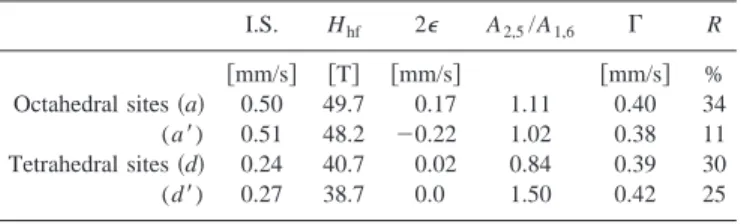
shifts ~I.S.! are corrected for the I.S. of metallic iron ~0.115 mm/s! taken as a reference. The 300 K CEM spectra of the as-grown undoped ~111!-YIG films exhibit a magnetic hy-perfine field splitting composed of four sextets due to the octahedral (16a) and tetrahedral ~24d! Fe31 lattice sites @Table I and Fig. 1~a!#.12From the areal ratios of the second or fifth lines to the first or sixth lines (A2,5/A1,6), it is con-cluded that the magnetization makes an angle around 70° relative to the sample surface normal.12 It is known indeed that YIG is a ferrimagnet with a Curie temperature (TC) of 550 K, while GGG is nonmagnetic ~antiferromagnetic order-ing of the gadolinium sublattice occurs at a Ne´el temperature of 10 K!.13 By substituting Fe31 with diamagnetic trivalent cations like Ga31and Sc31, TCdecreases so that the YIG:Ga
and YIG:Sc can become paramagnetic at 300 K for a large impurity concentration.13 This is clearly seen in the 300 K 57Fe CEM spectra of the as-grown ~111!-oriented epilayers which exhibit only two quadrupolar doublets corresponding respectively to the octahedral and tetrahedral Fe31 lattice sites, and no magnetic hyperfine field splitting @Figs. 2–4~a!#. Also, for YIG:Ga, the plot of the magnetization versus tem-perature shows that TC is around 210 K in agreement with
literature data, while for YIG:Sc, TC is given to be 100 K.13
The CEM spectra of YIG:Ga and YIG:Sc look different be-cause Ga31 sits preferentially in the tetrahedral sites while Sc31sits in the octahedral ones.13The latter effect is further enhanced by the lanthanum substitutions.11The least-squares fit of the spectra give the site distributions for YIG:Ga (octa/tetra560:40) and YIG:Sc (octa/tetra510:90) ~Tables II–IV!.
The doped films were irradiated at 300 K with 50 MeV 32S and 50 MeV 63Cu ions at the Van de Graaff Tandem accelerator in CEA Bruye`res-le-Chaˆtel, France. The undoped ones were irradiated at 300 K with 50 MeV32S and 50 MeV 63Cu ions, and with 235 MeV84Kr ions at the SARA facility in Grenoble, France. The ion beams were swept over the sample surface in order to obtain uniform irradiations. Ow-ing to the low beam current densities ~<30 nA/cm2!, only a small temperature rise around 20 K was found during the irradiations. The ion fluences were monitored by recording the backscattered ion spectra on a 100-nm-thick gold layer ring around the sample. A quantitative calibration was per-formed by measuring the beam current with a Faraday cup in the same beam conditions. Only one epitaxial film on one
TABLE I. Hyperfine parameters used for the least-squares fits of the 300 K
57Fe CEM spectrum of the ferrimagnetic pristine undoped ~111!-YIG
epi-taxial film @Fig. 1~a!#: isomer shift ~I.S.!, effective hyperfine magnetic field (Hhf), quadrupolar shift ~2e!, full width at half maximum ~G!, areal ratio of
the second or fifth lines to the first or sixth lines (A2,5/A1,6), and fraction ~R!
of the ion sites. The standard deviation is 0.01 mm/s on the I.S. and 2e values, and 0.2 T on the Hhfvalues.
I.S. Hhf 2e A2,5/A1,6 G R
@mm/s# @T# @mm/s# @mm/s# %
Octahedral sites ~a! 0.50 49.7 0.17 1.11 0.40 34
(a8) 0.51 48.2 20.22 1.02 0.38 11
Tetrahedral sites ~d! 0.24 40.7 0.02 0.84 0.39 30
(d8) 0.27 38.7 0.0 1.50 0.42 25
1900 J. Appl. Phys., Vol. 87, No. 4, 15 February 2000 Costantiniet al.
side of the substrates was irradiated to keep the film on the opposite side as a virgin reference sample. For all irradia-tions, the mean projected range of the ions ~see Table V! was higher than the film thicknesses. The major irradiation char-acteristics computed with the SRIM-96 code14 are also dis-played in Table V. It is seen that the mean electronic stop-ping powers (Se) are larger than the threshold for track
formation ~4.5 MeV/mm! in this low ion velocity range,3and at least two orders of magnitude larger than the mean nuclear stopping powers (Sn).
The 300 K 57Fe CEM spectra of the irradiated doped and undoped films were measured at the various fluences. Moreover, the x-ray absorption spectra at the iron K edge of the same as-grown and irradiated undoped films were re-corded at 300 K in the fluorescence mode between 7050 and 7200 eV by 0.5 eV steps, and between 7000 and 7700 eV by 2 eV steps. The latter experiments were performed at LURE-DCI ~Orsay, France! on the EXAFS-IV beamline with a channel-cut monochromator at an incidence angle around 20°. Prior to the measurements, an energy calibration was done with an iron metal sample at the iron K edge ~7112 eV! in the transmission mode. Then the spectra of reference
com-pounds ~a-Fe2O3 and SrFeO32x sinters! were also recorded in the transmission mode as was done in the previous study5 in order to obtain the parameters ~phase and amplitude! nec-essary for the standard x-ray absorption fine structure ~XAFS! analysis.
III. RESULTS A. Mo¨ ssbauer data
The least-squares fit of the CEM spectra of the irradiated doped films ~YIG:Ga and YIG:Sc! ~Figs. 2–4! have been performed by using two quadrupolar doublets corresponding respectively to one octahedral and one tetrahedral Fe31 site like the as-grown films as shown in Tables II–IV. An at-tempt was made to fit the spectra with three contributions corresponding to the octahedral, tetrahedral, and fivefold co-ordinated pyramidal iron sites like in a previous study.5 However, it did not improve significantly the reliability fac-tor of the least-squares fits due to the large full width at half maximum ~G! of the lines of the pristine materials. In the case of the irradiated undoped YIG films, the CEM spectra
FIG. 1. 300 K conversion electron Mo¨ssbauer spectra of undoped ~111!-YIG thin epitaxial films; as-grown ~a!, irradiated with 50 MeV32S ions:
3.731014cm22 ~b!, 7.031014cm22 ~c!, 50 MeV 63Cu ions: 1.5 31013cm22 ~d!, 2.231013
cm22 ~e!, 235 MeV 84
Kr ions: 1.031013 cm22~f!.
FIG. 2. 300 K conversion electron Mo¨ssbauer spectra of ~111!-YIG:Ga thin epitaxial films: as-grown ~a!, irradiated with 50 MeV32S ions: 631011~b!,
131012
~c!, 331012
~d!, 731012
~e!, 131013cm22~f!.
~Fig. 1! exhibit only one symmetrical doublet which was least-squares fitted with only one quadrupolar doublet corre-sponding to one Fe31iron site ~Table VI!. Tables II–IV dis-play the amorphous fractions ( fa) deduced from a Poisson’s
law: fa512exp(2Aft), where A stands for the damage or
amorphization cross-section given in Table V, andft for the
ion fluence.3 In case of undoped samples, HRTEM has
shown that the recrystallization process of the amorphous tracks is well under way after 50 MeV32S and 50 MeV63Cu ion irradiations at such high fluences ~see Table VI! and that complete amorphization has occured instead with 235 MeV 84
Kr ion irradiations at a fluence of 731013cm22 without any recrystallization.6High-resolution x-ray diffraction data show that almost full amorphization of the YIG film ( fa599.5 %) is obtained with the 84Kr irradiations at 131013cm22.15 A similar recrystallization process was found to occur in films doped with calcium or silicon.9
The analysis of the Mo¨ssbauer spectra of YIG:Ga shows that in all cases the octahedral site fraction is apparently
FIG. 3. 300 K conversion electron Mo¨ssbauer spectra of ~111!-YIG:Ga thin epitaxial films: as-grown ~a!, irradiated with 50 MeV63Cu ions: 231011~b!,
331011
~c!, 431011
~d!, 131012cm22~e!.
FIG. 4. 300 K conversion electron Mo¨ssbauer spectra of ~111!-YIG:Sc thin epitaxial films: as-grown ~a!, irradiated with 50 MeV 63Cu ions:
231011cm22~b!.
TABLE II. Hyperfine parameters used for the least-squares fits of the 300 K57Fe CEM spectra of paramagnetic ~111!-YIG:Ga epitaxial films irradiated with 50 MeV32S ions at various fluences (ft) ~Fig. 2!: amorphous
fraction ( fa), isomer shift ~I.S.!, quadrupolar splitting ~Q.S.!, full width at half maximum ~G!, and fraction ~R!
of the two sites. The standard deviation on the I.S. and Q.S. values is 0.01 mm/s.
ft @cm22#
fa
@%#
Octahedral site Tetrahedral site I.S. @mm/s# Q.S. @mm/s# G @mm/s# R @%# I.S. @mm/s# Q.S. @mm/s# G @mm/s# R @%# 0 0 0.51 0.43 0.24 61 0.21 0.84 0.22 39 631011 2 0.50 0.41 0.25 60 0.21 0.88 0.25 40 131012 3 0.52 0.46 0.29 59 0.21 0.90 0.24 41 331012 10 0.52 0.46 0.29 58 0.21 0.91 0.26 42 731012 22 0.48 0.40 0.24 32 0.30 1.04 0.24 68 131013 29 0.48 0.40 0.23 32 0.30 1.03 0.46 68
1902 J. Appl. Phys., Vol. 87, No. 4, 15 February 2000 Costantiniet al.
decreasing and the tetrahedral site fraction increasing when
ft increases ~Fig. 5!. The same behavior is found for YIG:Sc ~Table IV!. In the case of YIG:Ga, although the scaling of all the data with fais not perfect ~Fig. 6!, a similar saturation of
the site fractions occurs at fa'20 %. Moreover, it is seen
that the line of the tetrahedral iron sites broadens dramati-cally with increasing damage with a full width at half maxi-mum ~G! reaching a value around twice as much as the pris-tine sample value, while that of the octahedral ones remains almost unaltered ~Tables II–IV!. Furthermore, the isomer shift ~I.S.! and quadrupolar splitting ~Q.S.! of the tetrahedral sites also increase significantly, while the I.S. of the octahe-dral ones decrease weakly versus fa, with a saturation at
fa'20 % for both iron sites ~Fig. 7!. However, no change of
the iron oxidation state seems to occur. These observations are in agreement with the results of the previous study on undoped YIG which showed that the tetrahedral Fe31 sites are preferentially damaged and new fivefold-coordinated ~pyramidal! sites with I.S.'0.3 mm/s at 300 K are formed in the fully amorphous phase5 obtained after irradiation with 3.5 GeV132Xe ions at 531012cm22.16For the undoped YIG samples, the same quadrupolar doublet as in the a-YIG17 is found regardless of the ion and fluence: it seems then that there is no difference between the amorphous phase and the nanophase in this case ~Fig. 1!.
B. X-ray absorption data
The plot of the x-ray fluorescence yield versus photon energy is displayed in Fig. 8 in the XANES range ~80 eV! near the iron K edge for the as-grown and irradiated undoped YIG thin films. The x-ray absorption spectrum ofa-Fe2O3is also plotted. Like in the previous study,5a pre-edge peak is seen for all YIG samples, which is known to correspond to
forbidden (1s→3d) transitions that are very sensitive to the symmetry of the local order around iron. The peak increases when the number of noncentrosymmetrical sites including the tetrahedral sites increase.5 The magnification in Fig. 9 clearly shows that this pre-edge peak is quite strong in the virgin undoped YIG film with around 60% tetrahedral sites, and almost vanishes in a-Fe2O3 with no tetrahedral sites at all and only slightly distorted octahedral ones. The irradiated samples ~Table VI! spectra range in between those two: the peak height is smaller in the nanophase ~32S and63Cu irra-diations! than in the amorphous phase ~84Kr irradiation!, in which it is smaller than in the as-grown single crystal, in agreement with the previous work.5For the63Cu irradiation, the same peak height is found for the two fluences of 1.5 31013 ~not shown! and 2.231013cm22. This would mean
that the fraction of tetrahedral sites decreases from the single crystal to the amorphous then to the nanocrystalline ~nc! phase. However, no clear differences are seen in the XANES of all the irradiated samples ~Fig. 8! which give overall simi-lar features, like the CEM spectra. The EXAFS analysis was not feasible in the pristine films due to some Bragg diffrac-tion lines of the single crystals at 20° incidence angle. While the latter lines disappeared in the amorphized films, some were still present in the nanocrystallized ones, and prevented us from performing such an analysis.
IV. DISCUSSION
A. Local order and electronic structure
It is known that the grain boundaries ~GB! generally play a key role in the nanostructured materials: for a 10 nm grain size, the fraction of atoms in the GBs range between 15% for a GB thickness of 0.5 nm, and 25% for a thickness
TABLE III. Hyperfine parameters used for the least-squares fits of the 300 K57Fe CEM spectra of
paramag-netic ~111!-YIG:Ga epitaxial films irradiated with 50 MeV63Cu ions at various fluences (ft) ~Fig. 3!:
amor-phous fraction ( fa), isomer shift ~I.S.!, quadrupolar splitting ~Q.S.!, full width at half maximum ~G!, and fraction ~R! of the two sites. The standard deviation on the I.S. and Q.S. values is 0.01 mm/s.
ft
@cm22#
fa
@%#
Octahedral site Tetrahedral site
I.S. @mm/s# Q.S. @mm/s# G @mm/s# R @%# I.S. @mm/s# Q.S. @mm/s# G @mm/s# R @%# 0 0 0.51 0.43 0.24 61 0.21 0.84 0.22 39 231011 10 0.47 0.36 0.23 54 0.27 0.98 0.27 46 331011 15 0.49 0.40 0.25 38 0.29 1.06 0.43 62 431011 19 0.47 0.43 0.21 19 0.33 1.12 0.50 81 131012 41 0.48 0.42 0.20 18 0.33 1.10 0.54 82
TABLE IV. Hyperfine parameters used for the least-squares fits of the 300 K57Fe CEM spectra of
paramag-netic ~111!-YIG:Sc epitaxial films irradiated with 50 MeV63Cu ions at various fluences (ft) ~Fig. 4!:
amor-phous fraction ( fa), isomer shift ~I.S.!, quadrupolar splitting ~Q.S.!, full width at half maximum ~G!, and fraction ~R! of the two sites. The standard deviation on the I.S. and Q.S. values is 0.01 mm/s.
ft
@cm22#
fa
@%#
Octahedral site Tetrahedral site
I.S. @mm/s# Q.S. @mm/s# G @mm/s# R @%# I.S. @mm/s# Q.S. @mm/s# G @mm/s# R @%# 0 0 0.51 0.62 0.22 11 0.26 1.04 0.26 89 231011 10 0.44 0.61 0.20 5 0.30 1.12 0.40 95
of 1.0 nm.18Since the GBs are also known to contain a large amount of defects in their cores,18,19 it is not so surprising that it would resemble the amorphous phase on a large length scale owing to the large GB concentration. However, on a smaller length scale, the two phases might differ in their local order which is seen in the fact that the atomic density is much lower in GB cores than in the bulk of glasses.19 How-ever, in the case of irradiated undoped YIG, both the XANES and CEMS techniques do not see any difference between the amorphous and nc phases contrary to what was expected. HRTEM observations of the GB structure comple-mented with image simulations would certainly be of great help in this case.18 The only difference is observed in the analysis of the pre-edge peak showing that the tetrahedral iron sites are more damaged in the nc phase than in the amorphous phase. As said above, it was shown in the previ-ous study that fivefold-coordinated sites are formed in a-YIG from the decrease of the pre-edge peak height.5The present data would then hint to a further increase of the latter five-fold sites in the nc phase.
In the ion-irradiated YIG films, band tails are formed as a result of the loss of long-range order linked to the de-struction of the tetrahedral sites which is reflected in the band gap shrinkage around 1 eV seen in the optical absorp-tion spectra.20 Moreover, the same behavior of the dc con-ductivity versus temperature varying according to an exp(2T0/T)1/4law was found with large T0values indicating a multiphonon polaron hopping conduction mechanism in the amorphous and nc phases.9,21In order to account for the conductivity increase and magnetization decrease of ion-implanted or neutron bombarded YIG, it was postulated that a fraction of Fe31in the tetrahedral sites might be converted to Fe21,22 that would enhance polaron hopping between
Fe31 and Fe21. The present results show that it is not the case, in agreement with previous studies.5,23 However, the decrease of the pre-edge peak height correlated with the in-crease of fivefold-coordinated defects in the amorphous and nc phases is consistent with the conductivity increase from the single crystal to the amorphous phase,24 then to the nc phase.7
In a previous work, we also raised some doubts on the YIG phase stability, although the few discontinuous electron diffraction rings ascribed to the nc phase could be indexed in the cubic YIG crystal structure ~space group: Ia3d! as ~400!, ~420!, ~611!, ~631!, and ~732!.6,7Moreover, it is to be noted that the overall stoichiometry was not changed even at very high fluences (7.531013cm22! with 50 MeV 63Cu ion irradiation.7 Actually, it is known that Y3Fe5O12 starts to decompose under high pressure ~0.8 GPa! and high tempera-ture ~630 K! into YFeO3 ~orthoferrite! and a-Fe2O3 ~haematite!13 which both do not contain tetrahedral sites. It was found indeed that the ~200!, ~122!, ~212!, and ~240! diffraction lines of YFeO3 with an orthorhombic crystal
FIG. 5. Fractions ~R! of iron sites in ~111!-YIG:Ga thin epitaxial films vs the ion fluence (ft) after 50 MeV32S ion irradiations ~dashed lines!:
octa-hedral ~open circles! tetraocta-hedral ~full circles!, and 50 MeV63Cu ion
irradia-tions ~solid lines!: octahedral ~open squares! tetrahedral ~full squares!. The lines are guides to the eyes.
FIG. 6. Fractions ~R! of the iron sites in ~111!-YIG:Ga thin epitaxial films vs the amorphous fraction ( fa) after 50 MeV32S ion irradiations ~dashed
lines!: octahedral ~open circles! tetrahedral ~full circles!, and 50 MeV63Cu ion irradiations ~solid lines!: octahedral ~open squares! tetrahedral ~full squares!. The lines are guides to the eyes.
TABLE V. Main characteristics of the room-temperature irradiations per-formed on the YIG epitaxial films: electronic stopping power (Se), nuclear
stopping power (Sn), mean projected range (Rp), computed with theSRIM-96
code ~Ref. 14! at the entrance ion energy ~E! and damage cross-section ~A!
~Ref. 3!. Ion E @MeV# Se @MeV/mm# Sn @MeV/mm# Rp @mm# A @cm2 # 32S 50 7 9.531023 8.4 3.5310214 63Cu 50 13 5.231022 6.3 5.3310213 235Kr 235 17 2.731022 17.5 5.3310213
TABLE VI. Hyperfine parameters used for the least-squares fits of the 300 K57Fe CEM spectra of the undoped ~111!-YIG epitaxial films irradiated
with the different ions of energy E at various fluences (ft) ~Fig. 1!: isomer
shift ~I.S.!, quadrupolar splitting ~Q.S.!, full width at half maximum ~G!. The standard deviation on the I.S. and Q.S. values is 0.01 mm/s.
Ion E @MeV# ft @cm22# I.S. @mm/s# Q.S. @mm/s# G @mm/s# 32S 50 3.731014 0.40 1.15 0.54 32S 50 7.031014 0.41 1.13 0.57 63Cu 50 1.531013 0.40 1.18 0.56 63 Cu 50 2.231013 0.40 1.16 0.57 84 Kr 235 1.031013 0.40 1.17 0.56
1904 J. Appl. Phys., Vol. 87, No. 4, 15 February 2000 Costantiniet al.
structure ~space group: Pnma! were close to that of Y3Fe5O12 mentioned above, and could be used for indexing the nc phase diffraction rings. Such a phase change could also ex-plain the disappearance of the tetrahedral sites in the nanophase with respect to the amorphous phase, in the un-doped as well as in the un-doped YIG films.
We have attempted to address this ion-beam induced re-crystallization process in the frame of the thermal spike con-cept, previously used for modeling the amorphization
pro-cess in the ion tracks.7 From our simulations, it was shown that the formation of a nanophase could occur due to ion impacts on the amorphous tracks due to a smaller quench rate of the melt and melting wave-front velocity in the case of amorphous YIG with respect to the pristine crystalline phase. However, in these simplified calculations, the occur-rence of a decomposition of the melt superheated up to a maximum temperature larger in the amorphous phase than in the crystalline phase could not be taken into account. The possibility that such a phase change would have happened in the thermal spike is thus an open question.9 However, neither grain coarsening nor amorphization of the nc phase was seen even at very high fluences ~231014cm22 with
50 MeV 32S!.6 This is probably due to the stability and in-herent resistance to grain growth of the nanostructures stem-ming from their narrow grain size distribution and low en-ergy GB structures.18
B. Magnetic behavior
Another interesting issue concerns the magnetic proper-ties of the nc phase. As seen by the CEMS data, the same paramagnetic behavior as in the a-YIG phase are found, in agreement with the saturation magnetization data.25 Around the same hyperfine parameters are obtained ~Table VI! as in
FIG. 7. Isomer shift ~I.S.! of the iron sites in ~111!-YIG:Ga thin epitaxial films vs the amorphous fraction ( fa) after 50 MeV 32S ion irradiations
~dashed lines!: octahedral ~open circles! tetrahedral ~full circles!, and 50
MeV63Cu ion irradiations ~solid lines!: octahedral ~open squares!
tetrahe-dral ~full squares!. The lines are guides to the eyes.
FIG. 8. X-ray fluorescence spectra of undoped ~111!-YIG thin epitaxial films at the iron K edge: as-grown ~a!, irradiated with 235 MeV84Kr ions (1.031013cm22! ~c!, 50 MeV32
S ions (3.731014cm22) ~d!, 50 MeV 63Cu
ions (2.231013cm22) ~e!. X-ray absorption spectrum ofa-Fe 2O3~b!.
FIG. 9. Pre-edge peak of undoped ~111!-YIG thin epitaxial films at the iron
K edge: as-grown ~a!, irradiated with 235 MeV 84Kr ions (1.0
31013cm22) ~c!, 50 MeV32
S ions (3.731014cm22) ~d!, 50 MeV63Cu ions
(2.231013cm22) ~e!; pre-edge peak ofa-Fe 2O3~b!.
the a-YIG formed by 2.9 GeV 84Kr irradiation of sintered YIG, for which I.S.50.424 mm/s, Q.S.51.09 mm/s, and G 50.69 mm/s at 300 K.17
It has been shown that the critical size below which single magnetic domain formation can occur in YIG is around 34 nm.26 Since no magnetic hyperfine field splitting occurs in the nc phase at room temperature, this means that the nc phase is in the superparamagnetic state with a block-ing temperature (TB) smaller than 300 K.
27
Above TB fast
relaxation of the magnetization occurs due to the thermal fluctuations with a characteristic time constant ~t! depending of the domain size, whereas below TBthe magnetic moments
are frozen along an easy axis.27 TB is the temperature at
which t5tm, the characteristic time of the measurement
technique: for the 57Fe Mo¨ssbauer spectroscopy, t
m52.5
31028s.26,27TB increases whentmdecreases, or when the
single domain size increases for a given tm. So when t
@tmor T,TB, the magnetic sextets of the bulk should
ap-pear again, while on the contrary fort!tm or T.TB, only
a quadrupolar doublet corresponding to the average of the grain magnetization over the space and time coordinates should be seen.
If there are no interactions between particles, in the di-lute case, tis given by the relationship t5t0exp(KuV/kT),
where Kustands for the effective uniaxial anisotropy energy constant, V the particle volume, k the Boltzmann’s constant, andt0 a prefactor around 1029s.27t21is the probability of magnetization reversal that needs to overcome the energy barrier EB5KuV corresponding to the maximum anisotropy
energy. For example, in the case of magnetite ~Fe3O4 with
K1521.13105erg/cm3! particles of around 6 nm diameter,
TB'180 K, while for around 10 nm, TB.350 K, as seen by
the Mo¨ssbauer spectroscopy, while in the case of pure iron (K154.13105erg/cm3) particles of 10 nm diameter, a cal-culation gives TB around 100 K.27 However, practically
speaking, there is often a distribution of relaxation times due to the size distribution which renders difficult the determina-tion of TB. Moreover, in case of dipolar particle interactions
in dense systems,tis decreased and the Arrhenius approxi-mation does not hold, as was seen forg-Fe2O3nanoparticles dispersed in a solvent with a variable concentration.28
In the present case of YIG nanoparticles with a mean diameter of 10 nm, assuming that the interactions between grains are negligible and taking the anisotropy constant along the ^111& easy axis (K1526.73103erg/cm3) for Ku
yields a mean value of the relaxation time t at T5300 K close tot0, i.e., clearly smaller thantm. This corresponds to
TB!300 K: so it means that the nanophase should behave
like a superparamagnet at room temperature.
A similar behavior occurs in the a-YIG phase where a magnetic sextet is seen below a freezing temperature (Tf) around 70 K.5,17 The magnetic susceptibility and the zero-field cooled ~ZFC! and zero-field-cooled ~FC! magnetization mea-surements have shown that it corresponds to a speromagnetic ordering with an antiferromagnetic coupling.29In such a case the spins are locked into random orientations below Tf and
the net magnetization is zero.30However, a fraction smaller than 5% was found to be metamagnetic which is also anti-ferromagnetic at 5 K. It becomes anti-ferromagnetic under a weak
applied magnetic field that decreases when temperature is lowered.29 It was concluded that this small metamagnetic fraction results from uncomplete track overlap even at high fluences due to the Poisson’s law. The definitive proof of such a speromagnetic ordering in our case would need fur-ther in-field Mo¨ssbauer experiments at low temperature.
In the case of the nc phase examined here, it is difficult from the present results to sort out the contribution of the grain bulk supposedly superparamagnetic from the contribu-tion of the disordered GB cores which could also lead to an amorphous-like behavior. Moreover, in case of a phase de-composition as mentioned above, the orthoferrite and haema-tite are both antiferromagnets at 300 K but could also lead to a paramagnetic Mo¨ssbauer spectrum below a small particle size: for example for a-Fe2O3, a quadrupolar doublet is found below 10 nm.27 Low-temperature CEM and ZFC and FC magnetic susceptibility measurements are necessary to clear up this point.
V. CONCLUSIONS
Thin epitaxial films of yttrium iron garnet ~Y3Fe5O12or YIG! on nonmagnetic Gd3Ga5O12 substrates were irradiated with swift heavy ions ~50 MeV 32S, 50 MeV63Cu, and 235 MeV84Kr! in the electronic slowing down regime, above the amorphous track-formation threshold. The damage process leads either to an amorphous or a nanocrystalline ~nc! state, depending on the irradiation conditions.
In the case of undoped ~ferri!magnetic YIG thin films, no difference is found in the room-temperature CEMS data between the latter two phases which both have a paramag-netic behavior at room temperature. The amorphous phase is thought to be a speromagnet, whereas the nc phase appears to be a superparamagnet above the blocking temperature. The only difference is found in the x-ray absorption spectra at the iron K edge showing that the local environment in the nc phase should be more centrosymmetrical than in the amorphous phase, which is already more centrosymmetrical than the pristine single crystal, due to the preferential de-struction of the tetrahedral sites in the amorphization pro-cess, as found in the previous study on undoped ~ferri!mag-netic sintered YIG samples irradiated by 3.5 GeV132Xe ions. An explanation of this could be the formation of new sites, probably fivefold-coordinated ones, and/or a phase transfor-mation occuring in the recrystallization process from the su-perheated melt by decomposing the garnet Y3Fe5O12 at high temperature and high pressure into the orthoferrite (YFeO3) and haematite (a-Fe2O3) which both do not contain tetrahe-dral sites. In the case of paramagnetic gallium-~or scandium-! substituted films, the room-temperature57Fe con-version electron Mo¨ssbauer spectra ~CEMS! also show that no change of the iron oxidation state occurs and that the tetrahedral Fe31sites are preferentially destroyed in the dam-age process corresponding to the formation of the fivefold-coordinated ones, and/or phase decomposition.
ACKNOWLEDGMENT
The authors are indebted to Professor P. Paroli ~Univer-sita` di Roma, Italy! for providing the YIG:Sc samples.
1906 J. Appl. Phys., Vol. 87, No. 4, 15 February 2000 Costantiniet al.
1M. Toulemonde, S. Bouffard, and F. Studer, Nucl. Instrum. Methods
Phys. Res. B 91, 168 ~1994!.
2P. Hansen, H. Heitmann, and P. H. Smit, Phys. Rev. B 26, 3539 ~1982!. 3A. Meftah, F. Brisard, J. M. Costantini, M. Hage-Ali, J. P. Stoquert, F.
Studer, and M. Toulemonde, Phys. Rev. B 48, 920 ~1993!.
4
A. Meftah, J. M. Costantini, M. Djebara, N. Khalfaoui, J. P. Stoquert, F. Studer, and M. Toulemonde, Nucl. Instrum. Methods Phys. Res. B 122, 470 ~1997!.
5F. Studer, Ch. Houpert, M. Toulemonde, and E. Dartyge, J. Solid State
Chem. 91, 238 ~1991!.
6
J. M. Costantini, F. Ravel, F. Brisard, M. Caput, and C. Cluzeau, Nucl. Instrum. Methods Phys. Res. B 80Õ81, 1249 ~1993!.
7J. M. Costantini, F. Brisard, M. Toulemonde, and F. Studer, Nucl.
In-strum. Methods Phys. Res. B 122, 514 ~1997!.
8J. M. Costantini, F. Brisard, A. Meftah, M. Toulemonde, and F. Studer,
Radiat. Eff. Defects Solids 126, 233 ~1993!.
9J. M. Costantini, J. M. Desvignes, and M. Toulemonde, J. Appl. Phys.
~accepted!.
10J. M. Desvignes, J. M. Cerceau, V. B. Kratchenko, and H. Le Gall, J.
Cryst. Growth 56, 132 ~1982!.
11
G. Balestrino, P. Paroli, B. Antonini, G. Luce, and B. Maturi, J. Cryst. Growth 85, 270 ~1987!.
12G. Marest, A. Pe´rez, P. Gerard, and J. M. Mackowski, Phys. Rev. B 34,
4831 ~1986!.
13
G. Winkler, Magnetic Garnets, Vieweg Tracts in Pure and Applied Phys-ics ~Vieweg, Braunschweig, 1980!, Vol. 5.
14J. P. Biersack and L. G. Haggmark, Nucl. Instrum. Methods 174, 257
~1980!.
15J. M. Costantini, F. Brisard, L. Autissier, M. Caput, and F. Ravel, J. Phys.
D: Appl. Phys. 26, A57 ~1993!.
16F. Studer, Ch. Houpert, H. Pascard, R. Spohr, J. Vetter, J. Yun Fan, and
M. Toulemonde, Radiat. Eff. Defects Solids 116, 59 ~1991!.
17
F. Studer, N. Nguyen, G. Fuchs, and M. Toulemonde, Hyperfine Interact. 29, 1287 ~1986!.
18R. W. Siegel, J. Phys. Chem. Solids 55, 1097 ~1994!.
19H. Gleiter, Prog. Mater. Sci. 33, 223 ~1989!; Adv. Mater. 4, 474 ~1992!. 20
J. M. Costantini, F. Brisard, and A. Pe´rez, Philos. Mag. 71, 41 ~1995!.
21J. M. Costantini, F. Brisard, J. L. Flament, Philos. Mag. B 65, 873 ~1992!. 22H. Pascard, Phys. Rev. B 33, 7252 ~1986!.
23J. M. Grene`che, H. Pascard, and J. R. Regnard, Solid State Commun. 65,
713 ~1988!.
24
J. M. Costantini, F. Brisard, J. L. Flament, D. Bourgault, L. Sinopoli, and J. L. Uzureau, Nucl. Instrum. Methods Phys. Res. B 59Õ60, 600 ~1991!.
25J. M. Costantini, F. Brisard, J. L. Flament, A. Meftah, M. Toulemonde,
and M. Hage-Ali, Nucl. Instrum. Methods Phys. Res. B 65, 568 ~1992!.
26
T. V. Dimitrieva, I. S. Zhebulev, I. S. Lyubutin, O. L. Polushchenko, V. G. Tersuev, and R. I. Chalabov, Phys. Chem. Mech. Surf. 5, 1589 ~1990!.
27J. L. Dormann, Rev. Phys. Appl. 16, 275 ~1981!.
28P. Prene´, E. Tronc, J. P. Jolivet, J. Livage, R. Cherkaoui, M. Nogue`s, and
J. L. Dormann, Hyperfine Interact. 93, 1409 ~1994!.
29
Ch. Houpert, Ph.D. thesis, Universite´ de Caen ~1989!.
30C. M. Hurd, Contemp. Phys. 23, 469 ~1982!.