HAL Id: hal-02637546
https://hal.inrae.fr/hal-02637546
Submitted on 27 May 2020
HAL is a multi-disciplinary open access
archive for the deposit and dissemination of
sci-entific research documents, whether they are
pub-lished or not. The documents may come from
teaching and research institutions in France or
abroad, or from public or private research centers.
L’archive ouverte pluridisciplinaire HAL, est
destinée au dépôt et à la diffusion de documents
scientifiques de niveau recherche, publiés ou non,
émanant des établissements d’enseignement et de
recherche français ou étrangers, des laboratoires
publics ou privés.
sensitization potential of the major peanut allergen Ara
h 6
Blanche Guillon, Herve Bernard, Marie-Francoise Drumare, Stephane
Hazebrouck, Karine Adel-Patient
To cite this version:
Blanche Guillon, Herve Bernard, Marie-Francoise Drumare, Stephane Hazebrouck, Karine
Adel-Patient. Heat processing of peanut seed enhances the sensitization potential of the major peanut
allergen Ara h 6. Molecular Nutrition and Food Research, Wiley-VCH Verlag, 2016, 60 (12),
pp.2722-2735. �10.1002/mnfr.201500923�. �hal-02637546�
Received: 17-Nov-2015; Revised: 27-May-2016; Accepted: 08-Jun-2016
This article has been accepted for publication and undergone full peer review but has not been
through the copyediting, typesetting, pagination and proofreading process, which may lead to
differences between this version and the
Version of Record
. Please cite this article as
doi:
10.1002/mnfr.201500923.
Heat processing of peanut seed enhances the sensitization potential of the
major peanut allergen Ara h 6
Blanche Guillon, Hervé Bernard, Marie-Françoise Drumare, Stéphane Hazebrouck, Karine Adel-Patient#
UMR CEA-INRA Service de Pharmacologie et d’Immunoanalyse, Université Paris-Saclay, F-91991
Gif-sur-Yvette, France.
Running title: New insight in the allergenicity and immunogenicity of Ara h 6
#Corresponding author: Karine Adel-Patient, UMR INRA-CEA Service de Pharmacologie et
d’Immunoanalyse, Laboratoire d’Immuno-Allergie Alimentaire, CEA/iBiTeC-S/SPI, Bat 136, CEA de Saclay, 91191 Gif-sur-Yvette, France.
Tel.: +33 1 69084583 Fax: +33 1 69085907
E-mail: karine.adel-patient@cea.fr
ABSTRACT
Scope: Processing of food has been shown to impact IgE binding and functionality of food allergens. In the present study, we investigated the impact of heat processing on the sensitization capacity of Ara h 6, a major peanut allergen and one of the most potent elicitors of the allergic reaction.
Methods and results: Peanut extracts obtained from raw or heat-processed peanut and some fractions thereof were biochemically and immunochemically characterized. These extracts/fractions, purified Ara h 6 or recombinant Ara h 6 including Ara h 6 mutants lacking disulfide bridges were used in in vitro digestion tests and mouse models of experimental sensitization. Peanut roasting led to the formation of complexes of high molecular weight, notably between Ara h 6 and Ara h 1, which supported the induction of IgE specific to native Ara h 6. On the contrary, a fraction containing free monomeric 2S albumins or purified native Ara h 6 displayed no intrinsic allergenicity. In addition to complex formation, heat denaturation and/or partial destabilization enhanced Ara h 6 immunogenicity and increased its sensitivity to digestion.
Conclusion: These results suggest that sensitization potency and IgE binding capacity can be supported by different structures, modified and/or produced during food processing in interaction with other food constituents.
Keywords:
Different structures are responsible for sensitization and elicitation to Ara h 6 and heating is essential to sensitization potency of Ara h 6. Roasting of peanut seed induces partial denaturation and cross-linking of Ara h 6 with other peanut allergens, notably Ara h 1. The increased susceptibility to proteolytic degradation of the corresponding structures of Ara h 6 increases the immunogenicity of Ara h 6. Conversely, free monomeric Ara h 6, also found in peanut seed, is not immunogenic but is the support of IgE binding and elicitation of the allergic reaction.
Abbreviations:
DB
disulfide bridge
MW
molecular weight
r/a
reduced and alkylated
r
recombinant
PE
peanut extract
GF
gel filtration
INTRODUCTION
Peanut allergy is a major health problem, particularly in Westernized countries. This food allergy has an increasing prevalence among the pediatric and infant populations and persists until adulthood in most cases [1-6]. Clinical symptoms can be induced by traces of peanut and reactions are usually severe and potentially fatal. It is then of importance to further delineate the mechanisms, the structures and the food processing that support peanut allergenicity.
Food allergies are mainly IgE-mediated, type I hypersensitivities. This pathology relies on a biphasic adverse immune reaction: i) sensitization, which corresponds to the production of IgE specific for a food protein, ii) elicitation of the allergic reaction, that is induced after a second encounter of this food protein leading to its recognition by specific IgE antibodies bound to their high affinity IgE receptor expressed on effector cells (mastocytes and basophils) and their activation. Food allergens can be defined as food antigens that induce specific IgE responses [7]. Up to 16 allergens have been described in peanut (http://www.allergen.org). Among
them, Ara h 1, Ara h 2 and Ara h 3 have been initially identified as major peanut allergens [8-10]. Recently Ara h 6 has been given more consideration as the frequency of sensitization to Ara h 6 can be sometimes higher than that to the other 2S-albumin Ara h 2 [11-13]. Additionally, both Ara h 2 and Ara h 6 have been demonstrated to be the most potent elicitors of the allergic reaction as evaluated with skin prick test in allergic patients or with in vitro cell-based assays [14-16]. Accordingly, depletion of Ara h 2 and Ara h 6 from peanut protein extract led to a decrease of 80--90% of the allergenic activity [17-19].
Ara h 2 and Ara h 6 are storage proteins belonging to the 2S albumins protein family. 2S albumins contain major food allergens from seeds of many mono- and di-cotyledon plants and share a common compact structure that renders the proteins highly resistant to proteolysis [20]. By using site-directed mutagenesis, we recently described the importance of the disulfide bridges (DB) network in Ara h 6 resistance to trypsin digestion and in the allergenic activity of the breakdown products [21]. It is thereby usually considered that proteolytic resistance contributes to the allergenic properties of 2S albumins by allowing some intact molecules to reach the gut immune system [20;22-25]. Accordingly, IgE antibodies from allergic patients are mainly directed toward conformational epitopes since chemical or heat denaturation of purified native Ara h 6 abrogates its capacity to bind IgE and to elicit an allergic reaction [26-28].
Cooking methods have been suggested to contribute to the prevalence and severity of peanut allergy in Western countries compared to Far Eastern countries [29]. In fact, while boiling of peanut seeds, as used in Asia, leads to the loss of low molecular weight (MW) entities, including 2S albumins, in the cooking water [30], extensive heating such as roasting results in structural modifications of peanut proteins with formation of advanced glycation end products or high MW protein complexes [31]. Although heating of purified 2S albumins induced their denaturation and aggregation [27], soluble 2S albumins purified from roasted peanut still display their native monomeric forms. This shows that 2S albumins are (at least partially) protected from denaturation and aggregation within the seed [27;32].
In the present work, we aimed to further investigate the impact of heat processing of peanut seed on the sensitization to native Ara h 6, as observed in allergic patients. Our results finally provide new insights on the mechanisms and structures favoring sensitization to Ara h 6, but also suggest an inverse relationship between immunogenicity and resistance to digestion.
MATERIALS AND METHODS
1. Allergens and monoclonal antibodies 1.1 Purified allergens
Purification of Ara h 1, Ara h 2, Ara h 3 and Ara h 6 from peanut seed was performed from roasted peanut as previously described ([26;30], see supplementary data).
Reduction and S-alkylation of Ara h 6 (r/a-Ara h 6) was performed as previously described [28]. Heating of Ara h 6 was performed in solution (0,1 mg/mL, in 20 mM potassium phosphate buffer pH 7.4) at 110°C as previously described [27]. Conformation of native and chemically or heat-denaturated Ara h 6 was analyzed by circular dichroism spectroscopy (JASCO 810 spectropolarimeter, Jasco Analytic Instruments, Easton, Md) [33]. Disulfide bridges (DB) mutants of recombinant Ara h 6 (rAra h 6) correspond to Ara h 6 mutants in which DB were sequentially deleted by site-directed mutagenesis [21;28]. Disruption of DB 5 in the C-term region, a DB that is unique to Ara h 6 compared with other 2S albumins, did not affect the structure and trypsinolysis resistance of Ara h 6. On the contrary disruption of each of the four disulphide bridge (DB) in addition to DB 5 led to an increased susceptibility to hydrolysis by trypsin [21;28] although the secondary structure of Ara h 6
was preserved.
1.2 Production of mAb
Anti-Ara h 6 and anti-Ara h 1 mAb were produced by conventional techniques according to de StGroth and Scheidegger et al [34] & Grassi et al [35] using allergens purified from roasted peanut. The mAb specificity was further characterized by ELISA test and immunoblots using native and denaturated forms of Ara h 6 and Ara h 1, as previously described [33;36;37]. Additionally, immunoblots were performed using recombinant Ara h 1 and Ara h 3, kindly provided by O. Roitel and S. Jacquenet (Genclis SA, Vandœuvre-lès-Nancy), and recombinant Ara h 2 and Ara h 6 produced in our lab, confirming that the anti-Ara h 6 mAb used in the present study cross reacts neither with Ara h 1 nor Ara h 2, nor Ara h 3, and that the anti-Ara h 1 mAb used cross reacts neither with Ara h 2, nor Ara h 6, nor Ara h 3 (suppl. materials and not shown).
2. Preparation and characterization of protein extracts from raw and processed peanuts 2.1 Peanut seed processing
A typical blend consumed in Europe (50% Chinese Hsuji’s and Red Skins varieties, 50% Argentinian Runners variety) was kindly provided by Unilever (Rotterdam, Netherlands). Raw peanuts were first blanched at 130°C in an oven for 20 min to remove skin. Blanched peanuts were then either roasted, oil fried or boiled. Roasting was carried out in an oven at 180°C for 10 to 30 min. Frying was performed at 150°C for 3.5 to 10 min using a fryer and vegetable oil free of peanut oil (Frial Lesieur, Asnière sur Seine, France). Boiling was performed in water for 30 to 120 min.
2.2 Protein extraction from raw and processed peanuts
Raw and processed peanuts were ground with ultra-turrax® Tube-drive using DT-20 tubes (IKA®-Werke, Staufen, Germany) in 20 mM sodium carbonate buffer pH 9.6 in presence of protease inhibitors (Sigma, St Louis, MO, USA) until a homogeneous paste was obtained. Extraction was performed using a ratio of 50 ml of extraction buffer for 10 g of peanut. After incubation for 18 hours at 4°C on rotational shaker, protein extracts were centrifuged (3000g, 10 min, 4°C). Lipid layer was removed and supernatants, containing extracted proteins, were collected. Total protein content of each extract was estimated using BCA kit following the manufacturer’s instructions (Pierce, Thermo Scientific, Rockford, IL, USA).
2.3 Characterization of the protein extracts by electrophoresis and Western Blotting
SDS-PAGE and immunoblot analyses were performed under non-reducing or reducing conditions as described in supplementary data.
2.4 Analysis and isolation of hetero/homo oligomers of Ara h 6 in peanut extracts
Formation of heteromers between Ara h 1 and Ara h 6 was assessed by heterogeneous sandwich immunoassay. Plates (MaxiSorp Nunc, Roskilde, Denmark) were coated with a mAb directed against Ara h 6 (5 µg/ml, 50 mM phosphate buffer pH 7.4) and then saturated with EIA buffer (0.1 M phosphate pH 7,4, 0.1% BSA, 0.15 M NaCl, 0.01% sodium azide). Serial dilutions of samples in EIA buffer (or buffer for non-specific binding) were then incubated overnight at 4°C. After several washes (10 mM Phosphate buffer pH 7.4, 0.05% Tween 20), a biotinylated mAb specific for Ara h 1 (50 ng/ml; EZ Link®Sulfo-NHS-LC Biotin, Pierce) was added for 4h at 20°C. After extensive washing, neutravidin labelled with acetylcholinesterase was added for 15 min at room temperature and acethylcholinesterase-enzymatic activity was revealed after extensive washing and addition of Ellman’s reagent [38]. Absorbance at 414 nm was then measured using a reader plates (MultiskanEx, Thermo Electron Corporation).
Ara h 6 homo-oligomers were assessed following the same procedure but using a homogeneous sandwich assay: the same mAb directed against Ara h 6, i.e. recognizing the same and unique epitope (and not cross-reacting with Ara h 1), was used for both coating and detection.
2.5 Fractionation of roasted peanut extracts
Fractionation of protein extract from roasted peanut was performed by gel filtration using HR 16/50 Superdex 200 PrepGrad column (GE Healthcare) and a flow rate of 1 ml/min in 20 mM sodium carbonate pH 9.6, 0.4 M NaCl. Ten mg of roasted peanut extract was injected per run and twelve runs were carried out. Four individual fractions were harvested. After dialysis and concentration using centrifugal filter units (Millipore Merck, Darmstadt, Germany), protein contents were quantified using BCA kit. Each fraction was analyzed by electrophoresis and immunoblots as described above.
2.6 In vitro digestion model for antigen processing in antigen presenting cells
nAra h 6, r/a-Ara h 6 and rAra h 6 were subjected to cathepsin digestion. Digestion by cathepsin L (Sigma, from human liver, 2.13 U per mg of enzyme) was performed in 400 mM acetate sodium pH 5.5, 4 mM EDTA and 8 mM DTT. The enzyme:protein ratio was 91 mU:mg and samples were collected at 0.25, 0.5, 1, 2, 3, 4, 5, 6, 7 and 8 hours (2 µg protein per sampling time). Sample pH was increased to pH 8 by adding sodium bicarbonate and the enzymatic reaction was further stopped using protease inhibitors. Digestion by cathepsin S (Calbiochem-Merck, human recombinant, 97.55 U per mg of enzyme) was performed in 100 mM acetate sodium pH 5.5, 1 mM EDTA and 2 mM DTT. The enzyme:protein ratio was 20 mU:mg and samples were collected at 2, 3, 4, 5, 6, 7 and 8 hours (2 µg protein per sampling time). Sample pH was increased to pH 8 by adding sodium bicarbonate and the enzymatic reaction was further stopped using protease inhibitors. Samples were analyzed by SDS-PAGE electrophoresis.
In vitro digestion by trypsin of fractions from roasted peanut obtained by gel filtration was performed as previously described [21]. Briefly, digestion was performed with an enzyme:protein ratio of 1:20 (w:w) in 50 mM Tris-HCl pH 8. Digested fractions were collected at 1, 2 and 4 hours. Corresponding samples were analyzed by SDS-PAGE electrophoresis and anti-Ara h 6 immunoblot in non-reducing conditions.
3. Assessment of the sensitization potential of Ara h 6 in mice 3.1 Mice
Three-week-old female BALB/c mice were purchased from CERJ (Centre d’Elevage René Janvier, Le Genest-Saint-Isle, France), and were housed in filtered cages under normal specific pathogen free husbandry conditions (autoclaved bedding and sterile water). Mice were acclimated for 2 to 3 weeks before experimentation. They received a diet in which Ara h 1 was not detected using specific mAb and immunoassays developed in the laboratory (data not shown). Ara h 6 from the diet was below 5 ppm. All experiments were performed according to the European Community rules of animal care and with permission 91--368 of the French Veterinary Services.
3.2 Sensitization protocol
Allergen or protein extract adsorbed on alum (Alhydrogel 3%, Superfos, Denmark; 1 mg/injection, intraperitoneal route) was administered to mice on days 1 and 21. Eight mice per condition were tested, each receiving 10 µg of purified or recombinant allergen, 100 µg of processed peanut extracts or 50 µg of protein fractions. Naive control mice were not injected. When considering Ara h 6 mutants, mice received a second intraperitoneal boost injection at day 45 with native Ara h 6 adsorbed on alum. Individual serum samples were
collected one week after the last injection from the retro-orbital venous plexus on anesthetized mice (Isoflurane Belamont, Nicholas Piramal Limited, London, UK).
3.3 Analysis of anti- Ara h 6 IgE and IgG1 antibodies response
Sensitization of mice to Ara h 6 was assessed on individual serum by measurement of specific IgE and IgG1 using nAra h 6 or r/aAra h 6 coated plates (2.5 µg/ml, 50 mM phosphate buffer, pH 7.4) and enzyme-labelled anti-mouse IgE and IgG1, as previously described [39;40].
4. Statistical analysis
As data were not normally distributed, a non-parametric test was performed using the Kruskal-Wallis test followed by Dunn’s multiple comparisons test (DMCT). Statistical analyses were performed with GraphPad Prism 5.01 (GraphPad software, San Diego, CA, USA).
RESULTS
1. Heat-processing of peanut is necessary for the sensitization of mice to native Ara h 6
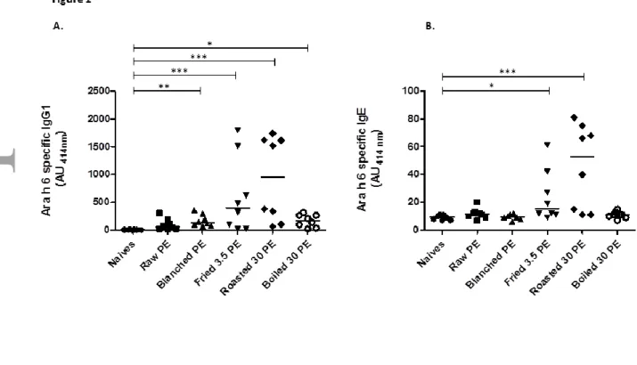
Different heat treatments such as frying, roasting or boiling were applied to blanched peanut. Proteins were extracted and administered to mice via i.p. route using alum as adjuvant in order to preserve protein structures [41]. Extracts from raw and blanched peanut were also tested. Significant production of IgG1 and IgE directed against corresponding whole peanut extract, and more particularly against Ara h 1 and Ara h 3, were obtained in all the groups of mice (data not shown). Surprisingly, no significant production of IgG1 nor IgE specific to native Ara h 6 was evidenced in mice immunized with the extract from raw peanut (Fig. 1A & 1B). Extracts from blanched or boiled peanut induced a significant production of IgG1 specific to native Ara h 6, but no specific IgE antibodies could be detected. Conversely, a significant production of IgE specific to native Ara h 6 was observed in mice treated with protein extracts from fried and roasted peanut (Fig 1B). These treated mice also demonstrated the highest production of anti-native Ara h 6 specific IgG1 antibodies (Fig. 1A).
2. Characterization of Ara h 6 from raw and heat-processed peanut
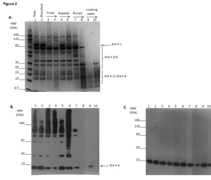
Boiling, roasting and frying processes were conducted for different durations and protein extracts were analyzed by SDS-PAGE under non-reducing conditions (Fig. 2A). In comparison to raw peanut, blanching, i.e. preliminary heating at 130°C for 20 min to remove skin but without inducing any browning of the seed, did not lead to major changes in the electrophoretic pattern (Fig. 2A, lanes 1 and 2). In contrast, after frying (lanes 3--4) or roasting (lanes 5--6), the band corresponding to monomeric Ara h 1 (at 60 kDa) progressively disappeared whereas bands above 110 kDa appeared, notably after 30 min of roasting, thus indicating protein aggregation. Conversely, after boiling (lanes 7--8), the amount of low MW entities below 30 kDa increased whereas high MW bands corresponding to both Ara h 1 and Ara h 3 tended to disappear, thus suggesting peanut protein degradation. Low MW entities were also detected in the cooking water of the corresponding boiled peanut extracts (lanes 9--10).
Extracts were then further analyzed by immunoblot using mAb specific for Ara h 6 in non-reducing (Fig. 2B) or reducing conditions (Fig. 2C). Ara h 6 immunoblot under non-reducing conditions allowed detection of Ara h 6 in its monomeric form at the expected MW in the protein extract from raw and blanched peanut (Fig. 2B lanes 1--2, MW around 15 kDa indicated by an arrow). A slight decrease of the intensity of this band was observed when comparing the protein extract from fried peanut and to a lesser extent from roasted peanut with that from raw peanut. Only very weak signal was detected after 30 min of boiling and no signal was detected after 2h of treatment (Fig. 2B lanes 7--8), whereas monomeric Ara h 6 is clearly detected in corresponding boiling water.
Interestingly, Ara h 6 immunoblot under non-reducing conditions also revealed the presence of specific bands at far higher MW than that of the monomeric form, notably after frying and roasting (Fig. 2B, lanes 3 to 6). After frying Ara h 6-specific bands were detected at MW mainly above 110 kDa, whereas they were detected at MW ranging from 30 to more than 160 kDa after roasting. Analysis under reducing conditions led to the near complete disappearance of the high MW bands in favor of monomeric Ara h 6 at its expected molecular weight (Fig. 2C, lanes 3 to 6).
These observations suggested that roasting led to complexations of Ara h 6 within high MW entities. We then further investigated the sensitization capacity of these different Ara h 6 complexes.
3. Sensitization to Ara h 6 is not induced by free native 2S albumins but required Ara h 6 high MW homo/hetero-complexes.
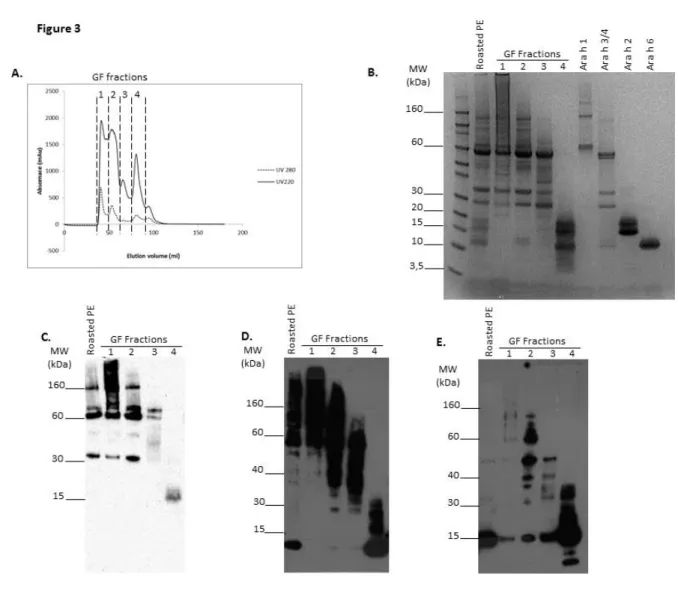
Size-fractionation by gel filtration of protein extract from roasted peanut was then performed and four fractions were obtained (Fig. 3A). Analysis by electrophoresis under non-reducing conditions (Fig. 3B) suggested the presence of Ara h 1 mainly in fractions 1 and 2, the presence of Ara h 3 in fractions 1, 2 and 3 and the presence of free 2S albumins Ara h 2 and Ara h 6 only in fraction 4. Enrichment in high MW entities was also noticed in fraction 1, and to a lesser extent in fraction 2. Immunoblot with mAb specific to Ara h 1 confirmed the presence of trimeric (180 kDa) and monomeric (60 kDa) forms of Ara h 1 in fractions 1 and 2 (Fig. 3C). A degradation product of Ara h 1 was also revealed in these fractions (30-35 KDa). It is worth noting that an intense smear was observed in fraction 1, with MW even higher than the Ara h 1 trimer, suggesting the presence of Ara h 1 in different conformations and oligomeric states. A low amount of a degradation product of Ara h 1, at MW around 15 KDa was also observed in fraction 4 (absence of cross-reactivity of the anti-Ara h 1 mAb against 2S albumins was checked). Anti-Ara h 6 immunoblot under non-reducing conditions (Fig. 3D) confirmed the presence of monomeric Ara h 6 (at 15 kDa) only in fraction 4, but also showed the presence of Ara h 6 involved in higher MW complexes, above 80 kDa in fraction 1, above 40kDa in fraction 2, and in MW entities ranging mainly between 30 and 80 kDa in fraction 3. These bands greatly decreased when analysis was performed under reducing conditions, leading to the appearance of a band corresponding to monomeric form of Ara h 6 in the corresponding fractions (Fig. 3E).
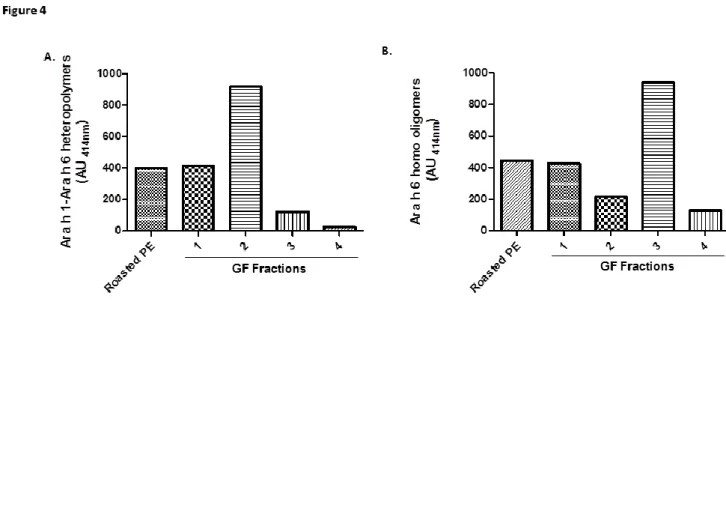
We then performed a heterogeneous immunoassay to detect Ara h 1-Ara h 6 complexes by using anti-Ara h 6 mAb as capture antibody and anti-Ara h 1 mAb as detection antibody. Ara h 6-Ara h 1 heteropolymers were detected in the extract from roasted peanut and in fractions 1 and 2 (Fig. 4A). Lower amount of heteropolymers were detected in fraction 3 whereas no significant signal could be detected in fraction 4. Ara h 1-Ara h 6 interactions in roasted peanut were also confirmed by testing Ara h 1 purified by immunoaffinity chromatography with anti-Ara h 6 immunoblot (supplementary Fig. 1). The Ara h 1-Ara h 6 heteropolymers were mainly detected after roasting and to a lesser extent after frying of peanut seed (supplementary Fig.2). Additionally, using a homogeneous sandwich assay, we observed the formation of Ara h 6 homo-oligomers mainly in fraction 3 by using the same anti-Ara h 6 mAb as capture and detection antibody (Fig. 4B).
The different fractions obtained by gel filtration of the roasted peanut extract were then administered to BALB/c mice and sensitization to native Ara h 6 was assessed. As previously observed (Fig. 1), the whole protein extract from roasted peanut led to a significant production of IgG1 (Fig. 5A) and IgE antibodies specific for native Ara h 6 (Fig. 5B). A similar intensity of response was induced by injection of fraction 2, whereas the humoral response was lower in mice receiving fraction 1. On the contrary, fraction 3 induced only the production of specific IgG1, with no significant specific production of IgE. More surprisingly, we did not detect any Arah
6-specific IgG1 or IgE production in mice receiving fraction 4, i.e. the fraction containing the monomeric form of Ara h 6.
4. Ara h 6 immunogenicity is also increased after partial denaturation, through enhanced digestibility.
Considering that free monomeric Ara h 6 contained in raw peanut or in fraction 4 displayed no sensitization capacity, we further investigated the allergenicity of purified Ara h 6. The immunogenicity of Ara h 6 either in its native form or after chemical or thermal denaturation was then evaluated using mouse models of experimental sensitization. After chemical denaturation, r/a-Ara h 6 exhibited a circular dichroism (CD) spectrum characteristic of random coiled nature. After heat denaturation at 110°C for 30 min in solution, secondary structures of Ara h 6 progressively shifted toward an unordered state with a CD spectrum indicating the presence of both native and denatured forms and/or the adoption of intermediate structures (supplementary Fig. 3). No IgE specific to native Ara h 6 could be detected, whatever the group (not shown). Significant production of native Ara h 6 specific-IgG1 was detected only in the group of mice receiving heat-denatured Ara h 6 (Fig. 6A), while three out seven mice receiving r/a-Ara h 6 were positive. Conversely, both heated Ara h 6 and r/a-Ara h 6 induced significant IgG1 responses against the denatured form of Ara h 6 (Fig. 6B).
We then tested the sensitization capacity of different recombinant Ara h 6 mutants in which some disulfide bridges (DB) were deleted by site-directed mutagenesis. These mutations did not affect the secondary structures of Ara h 6 but significantly destabilized the protein, leading to an increased susceptibility to trypsin hydrolysis [21]. Indeed, suppression of the non-canonical DB 5 at the C-terminus, in mutant Ara h 6-5, did not affect the proteolytic resistance of rAra h 6. The additional deletion of an external DB, in mutants Ara h 5.1 or Ara h 5.4, led to a lower resistance to proteolysis whereas the additional deletion of a central DB, in mutants Ara h 6-5-2 or Ara h 6-5-3, abrogated the resistance to trypsinolysis. rAra h 6-5, rAra h 6-5-4 and rAra h 6-6-5-2 were then i.p. administered to BALB/c mice. Mice injected with mutant rAra h 6-5 did not develop a significant anti-native Ara h 6 IgG1 response, as previously observed with nAra h 6 (Fig. 7). Interestingly, although no specific IgE was detected in any group (data not shown), we observed increasing anti-native Ara h 6 IgG1 responses correlating with an increasing sensitivity to trypsinolysis of the mutant proteins. The IgG1 response reached statistical significance in the group of mice receiving Ara h 6-5.2, i.e. the mutant showing the highest sensitivity to hydrolysis.
As i.p. administered allergens are not submitted to digestive enzymes such as trypsin, we also evaluated the resistance of rAra h 6 mutants to Cathepsin L (Figure 8) and Cathepsin S (data not shown), two proteases involved in antigen processing by dendritic cells. While native Ara h 6, rAra h 6-5, rAra h 1 and rAra h 6-5-4 were very resistant to both cathepsins digestion, rAra h 6-5-2 and rAra h 6-5-3 were more sensitive, with corresponding bands no more detectable after 3 hours and 6 hours of cathepsin L and cathepsin S digestion, respectively. Accordingly, r/a-Ara h 6 was promptly degraded by the two cathepsins.
Finally, we wondered whether Ara h 6 complexes in heat-processed peanut, that supported allergenicity of native Ara h 6, were also more susceptible to digestion. We then submitted the gel filtration fractions of roasted peanut to trypsin digestion and analyzed the breakdown products by electrophoresis. We observed that degradation promptly occurred in fractions 1, 2 and 3 (Fig 9A). Conversely, Ara h 6 monomers contained in fraction 4 were highly stable with no degradation clearly detected at 2 and 4 hours of digestion. Using anti-Ara h 6 immunoblot, a rapid degradation of Ara h 6 included in high MW polymers was observed in fractions 1, 2 and 3 (Fig9B). Conversely, Ara h 6 was still observed in fraction 4 after 4 hours of digestion. Complexation of Ara h
6 in heat-processed peanut actually increased its sensitivity to digestion compared to that of the monomer. Moreover, this increased digestibility also correlated with higher immunogenic/sensitizing capacity, as observed previously with partially denatured purified Ara h 6.
DISCUSSION
Ara h 2 and Ara h 6 are the most potent elicitors of the allergic reaction in peanut seeds. The IgE response in human patients is mainly directed against the native form of Ara h 6, whereas heat or chemically induced denaturation of these proteins decreases their IgE binding capacities and functionalities [27;42]. However, peanut is eaten after heating, which should lead to denaturation of Ara h 6 and then induction of IgE against the denatured form of this protein. However, despite the heating conditions of roasting, Ara h 2 and Ara h 6 extracted from roasted peanut demonstrated a native structure and an even higher IgE immunoreactivity than that of Ara h 2 and Ara h 6 purified from raw peanut [43]. It thus seems that 2S albumins are (at least partially) protected from heating degradation within the seed and could then suggest that these native proteins are responsible for their allergenicity. However, in the present study, we demonstrated that native monomeric Ara h 6 contained in raw peanut or extracted/purified from roasted peanut was unable to induce sensitization in mice, whereas extract from fried or roasted peanut do induce sensitization to native Ara h 6.
We then further analyzed the composition of the protein extracted from raw and heat-processed peanut, i.e. fried, roasted or boiled peanuts, all corresponding to forms of peanut consumed worldwide. Electrophoresis patterns clearly differed depending on the process considered. As already reported, a drastic loss of low MW proteins in the cooking water was observed during boiling [30]. We also observed a high degradation of high MW protein during boiling, as previously observed for purified Ara h 1 heated in solution [32]. Frying and roasting led to different events. As already described by Kopper et al [44], we observed that roasting or frying reduces the extractability of peanut allergens (data not shown), due to high aggregation of proteins. Using anti-Ara h 6 immunoblot in non-reducing conditions, we also identified the presence of anti-Ara h 6 in high MW entities. This phenomenon is already apparent in raw extract but is highly enhanced during frying and roasting. Our results are in accordance with those from Schmitt et al [45] demonstrating that heating process induces global aggregation of proteins and that complexes recovered in the insoluble fraction are highly IgE reactive.
By performing gel filtration of protein extracted from roasted peanut and different characterization methods (Immunoblots, homo- and hetero-immunoassays), we were able to separate complexed Ara h 6 from monomeric Ara h 6. Ara h 6 was demonstrated to be complexed either with itself (homo-complexes), or with other proteins, notably with Ara h 1 monomers, trimers and higher oligomers (MW>260kDa) (hetero-complexes). By administering the corresponding fractions to mice, we showed that immunogenicity and allergenicity of Ara h 6 is strictly dependent on this hetero-complexation allowing the induction of both IgG1 and IgE specific for native Ara h 6. Ara h 1-Ara h 6 complexes would seem good candidates for the allergenicity of Ara h 6. In fact, Ara h 1 has been demonstrated to specifically bind to dendritic cells via DC-SIGN receptor. This binding is dependent on Ara h 1 glycosylation and results in induction of Ara h 1-specific Th2 response [46;47]. Ara h 6 is not glycosylated but Ara h 6 – Ara h 1 complexation could enhance Ara h 6 uptake by dendritic cells via DC-SIGN-Ara h 1 interaction, in a Th2 environment induced by DC-SIGN-Ara h 1. Interestingly, DC-SIGN-Ara h 1 – DC-SIGN-Ara h 6 hetero-complexes were also detected in protein extracts from fried peanut, i.e. an extract that also induced sensitization to native Ara h 6, whereas they were not detected in extracts from boiled peanut that was unable to elicit anti-native Ara h 6 IgE. Ara h 3, the most abundant allergens of peanut seed, could be another candidate for an Ara h 6 carrier, as
electrophoretic analyses also suggested the formation of Ara h 6-Ara h 3 complexes. Moreover, Ara h 3 have been found in a high MW protein complexes containing also Ara h 1 and presenting IgE immunoreactivity [48]. The presence of such complexes and their role in the sensitizing potency of Ara h 6 should be further investigated.
Interestingly, we also observed that homo-polymers of Ara h 6 were more immunogenic than monomeric native Ara h 6, allowing the production of IgG1 whereas no IgE production could be evidenced. This is in agreement to the increased immunogenicity of Bet v 1d, a Bet v 1 isoform that forms a dimer, compared to that of monomeric Bet v 1. This increase was due to more efficient antigen uptake and activation of dendritic cells after dimerization [49]. The higher sensitization capacity of Ara h 6 homo-complexes in fraction 3 could then be due to a more efficient proteolytic processing in dendritic cells compared to the monomeric protein in fraction 4. The relationship between hydrolysis resistance and allergenicity of proteins is not clearly established [50;51]. Resistance to digestion of food proteins may allow reaching and stimulating the gut-associated immune system. However, proteins also have to be degraded by specific proteases within dendritic cells (DC) to be processed and then presented as allergen-derived peptides in association with major histocompatibility molecule class II (MHC II) at the cell surface of the DC. This presentation supports specific T-cell induction [52]. Studies from Delamarre et al [53;54] and Egger et al [55] suggest that resistance to lysosomal proteolysis is determinant in the immunogenicity of proteins by favoring efficient and long lasting antigen presentation in draining lymph nodes. Conversely, labile proteins failed to induce an immune response due to a too rapid degradation of the T epitopes. However, by using snake neurotoxin , a structured protein containing 4 DB, and different synthetic variants, it was demonstrated that efficacy of antigen presentation and T-cell stimulation by the antigen was inversely correlated with their conformational stability and cathepsin L sensitivity [56]. Additionally, magnitude of in vivo T-cell response, including Th2 response, and IgG production was inversely correlated to the conformational stability and endopeptidase sensitivity of hen egg lysozyme (HEL) [57;58]. Accordingly, we demonstrated the absence of immunogenicity of native Ara h 6 whereas anti-Ara h 6 immune response was induced by using more labile proteins, such as Ara h 6 denatured either by heat or chemical-modification or Ara h 6 mutants. Altogether, these results thus demonstrated an inverse relationship between stability to digestion and immunogenicity/allergenicity. These results are in accordance with previous results demonstrating that heating allows enhancing IgG and IgE Ara h 2-specific production in an oral immunization protocol of BALB/c mice [59]. These results further suggest that there is no direct and linear relationship between digestion resistance and immunogenicity/allergenicity.
It is worth noting that the presence of complexed allergens may jeopardize the interpretation of IgE-immunoblots and IgE-immunoassays when using peanut extracts and proteins purified thereof. This may thus reinforce the use of recombinant allergens in such analysis. However, we also recently demonstrated that post-translational modifications of Ara h 2 are involved in recognition of this natural allergen by IgE from most of the patients tested [28]. To really assess the respective role of the different allergens in peanut allergy, IgE reactivity of extracts and allergens should then be assessed following different experimental conditions, i.e. reducing and non-reducing conditions and using both purified natural and recombinant allergens. Such observations may be extended to other food sources, notably if consumed after heating.
In conclusion, despite being a major peanut allergen, soluble monomeric native Ara h 6 did not evidenced intrinsic sensitization capacity. Heat-processing of peanut seed, and probably peanut matrix [60], are
necessary for sensitization to native Ara h 6. We clearly displayed that heating process applied to peanut seed, such as frying and roasting, led to i) the formation of complexes of high MW between proteins, notably implicating Ara h 6 and Ara h 1, the latter probably acting as carrier for Ara h 6 uptake, DC activation and induction of specific Th2 immune response to Ara h 6, and ii) partial denaturation of Ara h 6, favouring its hydrolysis and its processing by APC and the induction of specific T cells. Nevertheless some Ara h 6 remains protected from thermal process within the seed, keeping its native conformation that supports IgE reactivity and functionality. All these results also suggest that sensitization and elicitation potency of a protein can be supported by different structures, modified and/or produced during processing in interaction with other food constituents. The sole sensitizing potential of purified proteins would then not be relevant in the global allergenicity assessment of new proteins/ingredients.
Author contributions: BG performed the whole experiments, analyzed the data and participated in the manuscript redaction. M-FD participated to production of monoclonal antibodies and characterization of Ara h 6 mutants. HB supervised purification of proteins and the heterogeneous and homogeneous assays providing characterized monoclonal antibodies. SH supervised production and characterization of Ara h 6 mutants and participated in the manuscript redaction. KA-P supervised the whole study, analyzed the data and participated in the manuscript redaction. All authors read and approved the final manuscript.
The authors have no conflicts of interest.
References
[1]Burks AW. Peanut allergy. Lancet 2008,1538-46.
[2]Gupta RS, Springston EE, Warrier MR, Smith B, Kumar R, Pongracic J, Holl JL. The prevalence, severity, and distribution of childhood food allergy in the United States. Pediatrics 2011,e9-17.
[3]Husain Z, Schwartz RA. Peanut allergy: an increasingly common life-threatening disorder. J Am Acad Dermatol 2012,136-43.
[4]Neuman-Sunshine DL, Eckman JA, Keet CA, Matsui EC, Peng RD, Lenehan PJ, Wood RA. The natural history of persistent peanut allergy. Ann Allergy Asthma Immunol 2012,326-31.
[5]Venter C, Hasan AS, Grundy J, Pereira B, Bernie CC, Voigt K, Higgins B, Dean T. Time trends in the prevalence of peanut allergy: three cohorts of children from the same geographical location in the UK. Allergy 2010,103-8.
[6]Waggoner MR. Parsing the peanut panic: the social life of a contested food allergy epidemic. Soc Sci Med 2013,49-55.
[7]Ho MH, Wong WH, Chang C. Clinical Spectrum of Food Allergies: a Comprehensive Review. Clin Rev Allergy Immunol 2012.
[8]Bernard H, Paty E, Mondoulet L, Burks AW, Bannon GA, Wal JM, Scheinmann P. Serological characteristics of peanut allergy in children. Allergy 2003,1285-92.
[9]Clarke MC, Kilburn SA, Hourihane JO, Dean KR, Warner JO, Dean TP. Serological characteristics of peanut allergy. Clin Exp Allergy 1998,1251-7.
[10]Ebisawa M, Moverare R, Sato S, Maruyama N, Borres MP, Komata T. Measurement of Ara h 1-, 2-, and 3-specific IgE antibodies is useful in diagnosis of peanut allergy in Japanese children. Pediatr Allergy
Immunol 2012,573-81.
[11]Koid AE, Chapman MD, Hamilton RG, Van RR, Versteeg SA, Dreskin SC, Koppelman SJ, Wuenschmann S. Ara h 6 Complements Ara h 2 as an Important Marker for IgE Reactivity to Peanut. J Agric Food Chem 2013. [12]Klemans RJ, van Os-Medendorp H, Blankestijn M, Bruijnzeel-Koomen CA, Knol EF, Knulst AC. Diagnostic
accuracy of specific IgE to components in diagnosing peanut allergy: a systematic review. Clin Exp Allergy 2014.
[13]Asarnoj A, Glaumann S, Elfstrom L, Lilja G, Lidholm J, Nilsson C, Wickman M. Anaphylaxis to peanut in a patient predominantly sensitized to Ara h 6. Int Arch Allergy Immunol 2012,209-12.
[14]Klemans RJ, Knol EF, Bruijnzeel-Koomen CA, Knulst AC. The diagnostic accuracy of specific IgE to Ara h 6 in adults is as good as Ara h 2. Allergy 2014, 69, 1112--4.
[15]Peeters KA, Koppelman SJ, van HE, van der Tas CW, den Hartog Jager CF, Penninks AH, Hefle SL, Bruijnzeel-Koomen CA, Knol EF, Knulst AC. Does skin prick test reactivity to purified allergens correlate with clinical severity of peanut allergy? Clin Exp Allergy 2007, 37, 108--15.
[16]Flinterman AE, van HE, den Hartog Jager CF, Koppelman S, Pasmans SG, Hoekstra MO, Bruijnzeel-Koomen CA, Knulst AC, Knol EF. Children with peanut allergy recognize predominantly Ara h2 and Ara h6, which remains stable over time. Clin Exp Allergy 2007,1221-8.
[17]Porterfield HS, Murray KS, Schlichting DG, Chen X, Hansen KC, Duncan MW, Dreskin SC. Effector activity of peanut allergens: a critical role for Ara h 2, Ara h 6, and their variants. Clin Exp Allergy 2009,1099-108. [18]Chen X, Zhuang Y, Wang Q, Moutsoglou D, Ruiz G, Yen SE, Dreskin SC. Analysis of the effector activity of
Ara h 2 and Ara h 6 by selective depletion from a crude peanut extract. J Immunol Methods 2011,65-70. [19]Chen X, Wang Q, El-Mezayen R, Zhuang Y, Dreskin SC. Ara h 2 and Ara h 6 have similar allergenic activity
and are substantially redundant. Int Arch Allergy Immunol 2013,251-8.
[20]Moreno FJ, Clemente A. 2S Albumin Storage Proteins: What Makes them Food Allergens? Open Biochem J 2008,16-28.
[21]Hazebrouck S, Guillon B, Drumare MF, Paty E, Wal JM, Bernard H. Trypsin resistance of the major peanut allergen Ara h 6 and allergenicity of the digestion products are abolished after selective disruption of disulfide bonds. Mol Nutr Food Res 2012,548-57.
[22]Astwood JD, Leach JN, Fuchs RL. Stability of food allergens to digestion in vitro. Nat Biotechnol 1996,1269-73.
[23]Lehmann K, Schweimer K, Reese G, Randow S, Suhr M, Becker WM, Vieths S, Rosch P. Structure and stability of 2S albumin-type peanut allergens: implications for the severity of peanut allergic reactions. Biochem J 2006,463-72.
[24]Sen M, Kopper R, Pons L, Abraham EC, Burks AW, Bannon GA. Protein structure plays a critical role in peanut allergen stability and may determine immunodominant IgE-binding epitopes. J Immunol 2002,882-7.
[25]Suhr M, Wicklein D, Lepp U, Becker WM. Isolation and characterization of natural Ara h 6: evidence for a further peanut allergen with putative clinical relevance based on resistance to pepsin digestion and heat. Mol Nutr Food Res 2004,390-9.
[26]Bernard H, Mondoulet L, Drumare MF, Paty E, Scheinmann P, Thai R, Wal JM. Identification of a new natural Ara h 6 isoform and of its proteolytic product as major allergens in peanut. J Agric Food Chem 2007,9663-9.
[27]Vissers YM, Blanc F, Skov PS, Johnson PE, Rigby NM, Przybylski-Nicaise L, Bernard H, Wal JM, Ballmer-Weber B, Zuidmeer-Jongejan L, Szepfalusi Z, Ruinemans-Koerts J, Jansen AP, Savelkoul HF, Wichers HJ, Mackie AR, Mills CE, Adel-Patient K. Effect of heating and glycation on the allergenicity of 2S albumins (Ara h 2/6) from peanut. PLoS One 2011, 6, e23998.
[28]Bernard H, Guillon B, Drumare MF, Paty E, Dreskin SC, Wal JM, Adel-Patient K, Hazebrouck S. Allergenicity of peanut component Ara h 2: Contribution of conformational versus linear hydroxyproline-containing epitopes. J Allergy Clin Immunol 2014.
[29]Shek LP, Lee BW. Food allergy in Asia. Curr Opin Allergy Clin Immunol 2006, 6, 197--201.
[30]Mondoulet L, Paty E, Drumare MF, Ah-Leung S, Scheinmann P, Willemot RM, Wal JM, Bernard H. Influence of thermal processing on the allergenicity of peanut proteins. J Agric Food Chem 2005,4547-53.
[31]Hebling CM, McFarland MA, Callahan JH, Ross MM. Global proteomic screening of protein allergens and advanced glycation endproducts in thermally processed peanuts. J Agric Food Chem 2013, 61, 5638--48. [32]Blanc F, Vissers YM, Adel-Patient K, Rigby NM, Mackie AR, Gunning AP, Wellner NK, Skov PS, Przybylski-Nicaise L, Ballmer-Weber B, Zuidmeer-Jongejan L, Szepfalusi Z, Ruinemans-Koerts J, Jansen AP, Bernard H, Wal JM, Savelkoul HF, Wichers HJ, Mills EN. Boiling peanut Ara h 1 results in the formation of aggregates with reduced allergenicity. Mol Nutr Food Res 2011, 55, 1887--94.
[33]Clement G, Boquet D, Mondoulet L, Lamourette P, Bernard H, Wal JM. Expression in Escherichia coli and disulfide bridge mapping of PSC33, an allergenic 2S albumin from peanut. Protein Expr Purif 2005, 44, 110--20.
[34]de StGroth SF, Scheidegger D. Production of monoclonal antibodies: strategy and tactics. J Immunol Methods 1980, 35, 1--21.
[35]Grassi J, Frobert Y, Lamourette P, Lagoutte B. Screening of monoclonal antibodies using antigens labeled with acetylcholinesterase: application to the peripheral proteins of photosystem 1. Anal Biochem 1988, 168, 436--50.
[36]Bernard H, Ah-Leung S, Drumare MF, Feraudet-Tarisse C, Verhasselt V, Wal JM, Creminon C, Adel-Patient K. Peanut allergens are rapidly transferred in human breast milk and can prevent sensitization in mice. Allergy 2014, 69, 888--97.
[37]Negroni L, Bernard H, Clement G, Chatel JM, Brune P, Frobert Y, Wal JM, Grassi J. Two-site enzyme immunometric assays for determination of native and denatured beta-lactoglobulin. J Immunol Methods 1998, 220, 25--37.
[38]Pradelles P, Grassi J, Maclouf J. Enzyme immunoassays of eicosanoids using acetylcholine esterase as label: an alternative to radioimmunoassay. Anal Chem 1985, 57, 1170--3.
[39]Adel-Patient K, Creminon C, Bernard H, Clement G, Negroni L, Frobert Y, Grassi J, Wal JM, Chatel JM. Evaluation of a high IgE-responder mouse model of allergy to bovine beta-lactoglobulin (BLG): development of sandwich immunoassays for total and allergen-specific IgE, IgG1 and IgG2a in BLG-sensitized mice. J Immunol Methods 2000, 235, 21--32.
[40]Adel-Patient K, Bernard H, Ah-Leung S, Creminon C, Wal JM. Peanut- and cow's milk-specific IgE, Th2 cells and local anaphylactic reaction are induced in Balb/c mice orally sensitized with cholera toxin. Allergy 2005, 60, 658--64.
[41]Adel-Patient K, Nahori MA, Proust B, Lapa e Silva JR, Creminon C, Wal JM, Vargaftig BB. Elicitation of the allergic reaction in beta-lactoglobulin-sensitized Balb/c mice: biochemical and clinical manifestations differ according to the structure of the allergen used for challenge. Clin Exp Allergy 2003, 33, 376--85.
[ 42] Bernard H, Drumare MF, Guillon B, Paty E, Scheinmann P, Wal JM. Immunochemical characterisation of structure and allergenicity of peanut 2S albumins using different formats of immunoassays. Anal Bioanal Chem 2009, 395, 139--46.
[43]Vissers YM, Iwan M, Adel-Patient K, Stahl SP, Rigby NM, Johnson PE, Mandrup MP, Przybylski-Nicaise L, Schaap M, Ruinemans-Koerts J, Jansen AP, Mills EN, Savelkoul HF, Wichers HJ. Effect of roasting on the allergenicity of major peanut allergens Ara h 1 and Ara h 2/6: the necessity of degranulation assays. Clin Exp Allergy 2011, 41, 1631--42.
[44]Kopper RA, Odum NJ, Sen M, Helm RM, Stanley JS, Burks AW. Peanut protein allergens: the effect of roasting on solubility and allergenicity. Int Arch Allergy Immunol 2005, 136, 16--22.
[45]Schmitt DA, Nesbit JB, Hurlburt BK, Cheng H, Maleki SJ. Processing can alter the properties of peanut extract preparations. J Agric Food Chem 2010, 58, 1138--43.
[46]Shreffler WG, Castro RR, Kucuk ZY, Charlop-Powers Z, Grishina G, Yoo S, Burks AW, Sampson HA. The major glycoprotein allergen from Arachis hypogaea, Ara h 1, is a ligand of dendritic cell-specific ICAM-grabbing nonintegrin and acts as a Th2 adjuvant in vitro. J Immunol 2006, 177, 3677--85.
[47]Buttari B, Profumo E, Capozzi A, Facchiano F, Saso L, Sorice M, Rigano R. Advanced glycation end products of human beta(2) glycoprotein I modulate the maturation and function of DCs. Blood 2011, 117, 6152--61.
[48]Boldt A, Fortunato D, Conti A, Petersen A, Ballmer-Weber B, Lepp U, Reese G, Becker WM. Analysis of the composition of an immunoglobulin E reactive high molecular weight protein complex of peanut extract containing Ara h 1 and Ara h 3/4. Proteomics 2005, 5, 675--86.
[49]Zaborsky N, Brunner M, Wallner M, Himly M, Karl T, Schwarzenbacher R, Ferreira F, Achatz G. Antigen aggregation decides the fate of the allergic immune response. J Immunol 2010, 184, 725--35.
[50]Fu TJ, Abbott UR, Hatzos C. Digestibility of food allergens and nonallergenic proteins in simulated gastric fluid and simulated intestinal fluid-a comparative study. J Agric Food Chem 2002, 50, 7154--60.
[51]Foster ES, Kimber I, Dearman RJ. Relationship between protein digestibility and allergenicity: comparisons of pepsin and cathepsin. Toxicology 2013, 309, 30--8.
[52]Li P, Gregg JL, Wang N, Zhou D, O'Donnell P, Blum JS, Crotzer VL. Compartmentalization of class II antigen presentation: contribution of cytoplasmic and endosomal processing. Immunol Rev 2005, 207, 206--17. [53]Delamarre L, Pack M, Chang H, Mellman I, Trombetta ES. Differential lysosomal proteolysis in
antigen-presenting cells determines antigen fate. Science 2005, 307, 1630--4.
[54]Delamarre L, Couture R, Mellman I, Trombetta ES. Enhancing immunogenicity by limiting susceptibility to lysosomal proteolysis. J Exp Med 2006, 203, 2049--55.
[55]Egger M, Jurets A, Wallner M, Briza P, Ruzek S, Hainzl S, Pichler U, Kitzmuller C, Bohle B, Huber CG, Ferreira F. Assessing protein immunogenicity with a dendritic cell line-derived endolysosomal degradome. PLoS One 2011, 6, e17278.
[56]Thai R, Moine G, Desmadril M, Servent D, Tarride JL, Menez A, Leonetti M. Antigen stability controls antigen presentation. J Biol Chem 2004, 279, 50257--66.
[57]So T, Ito H, Hirata M, Ueda T, Imoto T. Contribution of conformational stability of hen lysozyme to induction of type 2 T-helper immune responses. Immunology 2001, 104, 259--68.
[58]Ohkuri T, Nagatomo S, Oda K, So T, Imoto T, Ueda T. A protein's conformational stability is an immunologically dominant factor: evidence that free-energy barriers for protein unfolding limit the
immunogenicity of foreign proteins. J Immunol 2010, 185, 4199--205.
[59]Starkl P, Krishnamurthy D, Szalai K, Felix F, Lukschal A, Oberthuer D, Sampson HA, Swoboda I, Betzel C, Untersmayr E, Jensen-Jarolim E. Heating Affects Structure, Enterocyte Adsorption and Signalling, As Well as Immunogenicity of the Peanut Allergen Ara h 2. Open Allergy J 2011,24-34.
[60]Van Wijk F, Nierkens S, Hassing I, Feijen M, Koppelman SJ, de Jong GA, Pieters R, Knippels LM. The effect of the food matrix on in vivo immune responses to purified peanut allergens. Toxicol Sci 2005, 86, 333--41. LEGENDS TO FIGURES
Figure 1: Impact of peanut seed heating on sensitization to native Ara h 6. Peanut protein extracts (PE) from raw, blanched (130°C, 20 min), blanched and fried (150°C, 3.5 min), blanched and roasted (180°C, 30 min) or blanched and boiled (30 min) peanut seeds were absorbed on alum and injected to BALB/c mice (see Materials and Methods). Intra-peritoneal injections of 100 µg of protein were performed on day 1 and 21, and sera were collected on day 28. Anti-native Ara h 6 specific IgG1 (A) and IgE (B) were assayed in sera diluted 1:1000 and 1:40, respectively. Results are expressed as absorbance units at 414 nm. Each point represents an individual mouse and median is indicated. *: p<0.05, **: p<0.01, ***: p<0.001 using Kruskal-Wallis and Dunn’s multiple comparison posttest.
non-reducing conditions of peanut extract (PE) from raw (lane 1) or blanched peanut (lane 2). Blanched peanuts were then fried for 3.5 or 10 min (lanes 3 and 4), dry roasted for 10 or 30 min (lanes 5 and 6) or boiled in water for 30 or 120 min (lanes 7 and 8). Corresponding cooking waters were also analyzed (lanes 9 and 10). Immunoblot using anti-Ara h 6 mAb was performed under non-reducing conditions (B) and reducing conditions (C).
Figure 3: Fractionation of proteins from roasted peanut by gel filtration and their characterization. A. Gel filtration chromatogram. Four fractions, 1 to 4, were collected. B. SDS-PAGE under non-reducing conditions of protein extract from roasted peanut (PE), of GF fraction 1 to 4 or of purified allergens Ara h 1, Ara h 3, Ara h 2 and Ara h 6 (see Material and Methods). Immunoblot of roasted PE and GF fractions with anti-Ara h 1 mAb under non reducing conditions (C), and with anti- Ara h 6 mAb under non-reducing conditions (D) and reducing conditions (E).
Figure 4: Ara h 6 is involved in hetero and homo-polymeric structures after peanut seed roasting. A. Evidence of Ara h 1-Ara h 6 interactions using heterogeneous sandwich assay with anti-Ara h 6 mAb as capture mAb and anti-Ara h 1 as detection mAb B. Evidence of Ara h 6 homo-oligomers using homogeneous sandwich assay with the same anti-Ara h 6 mAb as capture mAb and as detection mAb. Results are expressed as absorbance units at 414 nm and are representative of three independent experiments.
Figure 5: Sensitization to Ara h 6 is not induced by the fraction containing free 2S albumins. Anti- Ara h 6 specific IgG1 (A) and IgE (B) induced in BALB/c mice after two intra-peritoneal injections of protein extract from roasted peanut and GF fractions. Sera were diluted 1:40000 and 1:40 for IgG1 and IgE assays, respectively. Results are expressed as absorbance units at 414 nm. Each point represents an individual mouse and median is indicated. *: p<0.05, **: p<0.01, ***: p<0.001 using Kruskal-Wallis and Dunn’s multiple comparison posttest.
Figure 6: Relation between the structure and the sensitization potency of Ara h 6. Mice were i.p. administered with native Ara h 6 (nAra h 6), Ara h 6 denatured by heating at 110°C for 30 minutes (nAra h 6 110) or r/a-Ara h 6. Specific IgG1 were then assayed in individual sera diluted 1:100 on plates coated with native Ara h 6 (A) or r/a Ara h 6 (B). Results are expressed as absorbance units at 414 nm. Each point represents an individual mouse and median is indicated. *: p<0.05, **: p<0.01, ***: p<0.001 using Kruskal-Wallis and Dunn’s multiple comparison posttest.
Figure 7: Relation between hydrolysis sensitivity and sensitization to native Ara h 6. Anti-native Ara h 6 specific IgG1 induced in BALB/c mice after three intraperitoneal injections of Ara h 6 DB mutants. Specific IgG1 were then assayed in individual sera diluted 1:100 on plates coated with Ara h 6. Results are expressed as absorbance units at 414 nm. Each point represents an individual mouse and median is indicated. *: p<0.05, **: p<0.01, ***: p<0.001 using Kruskal-Wallis and Dunn’s multiple comparison posttest.
Figure 8: Digestion of Ara h 6 DB mutants by Cathepsin L. Native Ara h 6, rAra h 6-5, rAra h 6-5.1, rAra h 6-5.2, rAra h 6-5.3 and rAra h 6-5.4 mutants and r/a-Ara h 6 were submitted to cathepsin L digestion with enzyme:protein ratio of 91 mU:mg. Samples were collected at various time points and were analyzed by SDS-PAGE under non-reducing conditions.
Figure 9: Complexed Ara h 6 is more sensitive to digestion by trypsin than free Ara h 6. Gel filtration fractions of roasted peanut were submitted to Trypsin digestion with enzyme:protein ratio of 1:20 (w:w). SDS-PAGE (A) and immunoblot using anti-Ara h 6 mAb (B) were performed under non-reducing conditions.