HAL Id: hal-02179723
https://hal.sorbonne-universite.fr/hal-02179723
Submitted on 11 Jul 2019
HAL is a multi-disciplinary open access
archive for the deposit and dissemination of
sci-entific research documents, whether they are
pub-lished or not. The documents may come from
teaching and research institutions in France or
abroad, or from public or private research centers.
L’archive ouverte pluridisciplinaire HAL, est
destinée au dépôt et à la diffusion de documents
scientifiques de niveau recherche, publiés ou non,
émanant des établissements d’enseignement et de
recherche français ou étrangers, des laboratoires
publics ou privés.
Quantitative neuroimaging biomarkers in a series of 20
adult patients with POLG mutations
Marion Masingue, Isaac Mawusi Adanyeguh, Maya Tchikviladze, Thierry
Maisonobe, Claude Jardel, Damien Galanaud, Fanny Mochel
To cite this version:
Marion Masingue, Isaac Mawusi Adanyeguh, Maya Tchikviladze, Thierry Maisonobe, Claude Jardel,
et al.. Quantitative neuroimaging biomarkers in a series of 20 adult patients with POLG mutations.
Mitochondrion, Elsevier, 2019, 45, pp.22-28. �10.1016/j.mito.2018.02.001�. �hal-02179723�
Quantitative neuroimaging biomarkers in a series of 20 adult patients with
POLG mutations
Marion Masingue
a,b, Isaac Adanyeguh
a, Maya Tchikviladzé
b, Thierry Maisonobe
c, Claude Jardel
d,
Damien Galanaud
a,e, Fanny Mochel
a,f,g,⁎aSorbonne Université, UPMC-Paris 6, UMR S 1127 and Inserm U 1127, and CNRS UMR 7225, and Institut du Cerveau et de la Moelle épinière, F-75013, Paris, France bAP-HP, Pitié-Salpêtrière University Hospital, Department of Neurology, Paris, France
cAPHP, Pitié-Salpêtrière University Hospital, Department of Neurophysiology, Paris, France
dAP-HP, Pitié-Salpêtrière University Hospital, Metabolic Biochemistry Department, INSERM, U1016, Institut Cochin, Paris F-75014, France eAP-HP, Pitié-Salpêtrière University Hospital, Department of Neuroradiology, Paris, France
fAP-HP, Pitié-Salpêtrière University Hospital, Department of Genetics, Paris, France gUniversity Pierre and Marie Curie, Neurometabolic Research Group, Paris, France
A B S T R A C T
Mutations in the gene encoding polymerase gamma (POLG) are a common cause of mitochondrial diseases in adults. We retrospectively analyzed volumetric and diffusion tensor imaging data from 20 adult POLG-mutated patients compared to healthy controls. We used an original clinical binary load score and electro-neuromyography to evaluate disease severity. Patients showed atrophy in the basal ganglia, amygdala, and brainstem (p < 0.05) compared to controls, as well as decreased fractional anisotropy (FA) in the cingulate gyrus, the internal capsule and the corona radiata (p < 0.05). Clinical scores correlated with decreased FA and increased radial diffusivity in several brain regions (p < 0.05).
1. Introduction
Clinical variability is major in neurometabolic disorders making their diagnosis difficult and, often, radiological findings are non-spe-cific. Treatments are nonetheless emerging so that non-invasive tech-niques are needed to evaluate their efficacy. Furthermore, numerous works have shown that mitochondrial dysfunction plays an important role in the pathophysiology of neurodegenerative disorders such as Parkinson's disease or Huntington's disease (Mochel and Haller, 2011;
Bose and Beal, 2016). Hence, identifying non-invasive biomarkers in POLG-mutated patients and other mitochondrial disorders could prove useful in the understanding of these rare neurometabolic disorders as well as more common neurological conditions.
The POLG gene encodes the catalytic subunit of the mitochondrial DNA polymerase gamma, the most important enzyme involved in mi-tochondrial DNA (mtDNA) replication and homeostasis. Patients with recessive POLG mutations have numerous clinical presentations and
constitute one of the largest groups of mitochondrial diseases in adults (Lax et al., 2012). Neurological symptoms may include sensory neuro-nopathy, cerebellar impairment, movement disorders, oculomotor dis-orders, muscle weakness, cognitive or psychiatric symptoms, neuro-sensory impairment (hearing loss, cataract) and/or stroke-like episodes (Mochel and Haller, 2011). The diagnosis is guided by elevated lactate in blood and/or CSF, mitochondrial abnormalities on muscle biopsy– ragged redfibers, i.e. myofibers with abnormal mitochondria deposits, respiratory chain deficiencies, mtDNA deletions – and is confirmed by molecular analysis of the POLG gene. To date, there is no efficient treatment for POLG-mutated patients, though several therapeutic de-velopments are in progress urging the need for biomarkers reflecting cerebral metabolism.
Findings on visual inspection of brain magnetic resonance imaging (MRI)– e.g. white matter abnormalities, sub-cortical atrophy and/or atrophy of the cerebellum, the brainstem and the corpus callosum, edema and ischemia (Van Goethem et al., 2004; Winterthun et al.,
⁎Corresponding author at: Sorbonne Université, UPMC-Paris 6, UMR S 1127 and Inserm U 1127, and CNRS UMR 7225, and Institut du Cerveau et de la Moelle épinière, F-75013, Paris,
France.
E-mail addresses:m.tchikviladze@hopital-foch.com(M. Tchikviladzé),thierry.maisonobe@aphp.fr(T. Maisonobe),claude.jardel@aphp.fr(C. Jardel),
damien.galanaud@aphp.fr(D. Galanaud),fanny.mochel@upmc.fr(F. Mochel).
Abbreviations: POLG, polymerage gamma; MRI/MRS, magnetic resonance imaging/spectroscopy; DTI, diffusion tensor imaging; FA, fractional anisotropy; RD, radial diffusivity; mtDNA, mitochondrial DNA; CSF, cerebrospinalfluid; TR, repetition time; TE, echo time; FOV, field of view; EMG, electroneuromyographic; ROI, region of interest; TBSS, tract-based spatial statistics
2005;Echaniz-Laguna et al., 2010;Kurt et al., 2010;Saneto et al., 2010;
Habek et al., 2012;Synofzik et al., 2012;Cheldi et al., 2013;Kinghorn et al., 2013;Sidiropoulos et al., 2013;Uusimaa et al., 2013;Degos et al., 2014;Bindu et al., 2015;Lam et al., 2015;Tchikviladzé et al., 2015;
Henao et al., 2016;Janssen et al., 2016;Martikainen et al., 2016)– are not specific to POLG mutations and MRI can also be normal. Only one research study has reported changes in magnetic resonance spectro-scopy (MRS) and diffusion tensor imaging (DTI) in POLG-mutated pa-tients, but it was restricted to stroke-like episodes (Tzoulis et al., 2010) that only a subset of patients present with. Yet, obtaining quantitative cerebral measures is key to understand the pathophysiology of the neurometabolic alterations associated with POLG mutations and monitor the course of the disease despite its clinical heterogeneity. MRI techniques seem particularly suited to identify biomarkers reflecting brain metabolism (Bonvento et al., 2017). Likewise, cerebral volumetry is a longstanding tool in biomarkers neuroimaging studies in order to quantify the rate at which the brain changes or atrophies due to the disease or in response to therapy. Moreover, DTI allows to further assess the integrity of nervefibers and hence gives some explanation on the underlying microstructure changes that results in the gross volume
changes. This retrospective study therefore aimed at identifying bio-markers using both volumetry and DTI on a series of 20 adult POLG-mutated patients.
2. Material and methods 2.1. Patients and controls
Initial and follow-up data were obtained retrospectively from pa-tients followed in rare disease reference centers at La Pitié-Salpêtrière hospital between 2003 and 2015. All participants provided their written informed consent for study procedures and data reporting. The POLG gene was sequenced as described (Whybra et al., 2004). The retrospective nature of our study prevented us from using clinical scales used in mitochondrial diseases, such as NMDAS (Newcastle Mitochon-drial Disease Adult Scale). Yet, disease severity was assessed by an original clinical binary load score (Fig. 1). Likewise, we used previous clinical descriptions of POLG-mutated patients (Tchikviladzé et al., 2015) and listed the main symptoms susceptible to be present in our patient series. Symptoms belonging to the same neurological category were grouped in order to ensure that each neurological dysfunction weighed equally in the score. For example, dysarthria, dysmetria, and ataxia, all attributable to cerebellar impairment, were summed and divided by 3 so that the cerebellar score ranged from 0 to 1. This scale has never been used before in mitochondrial diseases. However, such binary scoring systems have been validated before in rare neurometa-bolic diseases (Whybra et al., 2004). Here, the overall score ranged from 0 (best) to 10 (worst). Two items– cerebellar impairment and movement disorders– were used as subscores and continuous variables for correlation purposes. Controls were healthy subjects who belonged to a control cohort selected for a previous DTI study (Van der Eerden
Oculomotor disorder
(a+b)/2
/1
Ptosis
a/1
Ophtalmoplegia
b/1
1
/
y
h
t
a
p
o
r
u
e
N
Cerebellar Impairment
(c+d+e)/3
/1
Ataxia
c/1
Dysmetria
d/1
Dysarthria
e/1
Movement disorders
(f+g+h+i+j)/5
/1
Dystonia
f/1
Tremor
g/1
Myoclonus
h/1
Chorea
i/1
Hypokinesia
j/1
Muscle weakness
(k+l+m)/3
/1
UL weakness
k/1
LL weakness
l/1
Axial weakness
m/1
1
/
s
n
g
i
s
t
c
a
r
t
l
a
d
i
m
a
r
y
P
1
/
t
n
e
m
r
i
a
p
m
i
e
v
i
t
i
n
g
o
C
1
/
r
e
d
r
o
s
i
d
c
i
r
t
a
i
h
c
y
s
P
1
/
y
s
p
e
l
i
p
E
Hypoacousia
/1
0
1
/
e
r
o
c
s
l
a
t
o
T
Fig. 1. Clinical load of POLG-mutated patients. UL: upper limbs, LL: lower limbs. Table 1
Clinical and demographic characteristics of POLG-mutated patients. Volumetry (n = 20) DTI (n = 14) Age (y) 49 ± 16 (14–73) 54 ± 14 (24–73) Age at onset 30 ± 16 (0–57) 32 ± 16 (6–57) Disease evolution prior to MRI 20 ± 15 (1–51) 21 ± 15 (1–51) Sex-ratio (M/F) 8/12 5/9
Clinical load score 4.9 ± 1.1 (2.7–6.7) 4.7 ± 1.2 (2.7–6.7) Data are presented as mean ± standard deviation (range). y: year; M: male; F: female.
et al., 2014). 2.2. Brain imaging
MRI acquisitions were performed on 1.5 and 3 T MR units (General Electric, WI, USA) using the standardized protocol used in our institu-tion for the explorainstitu-tion of neuro metabolic diseases. Some POLG-mu-tated patients had a follow-up exam after 4 years. A 3D T1-weighted sequence (TR = 9.5 ms, TE = 3 ms,field of view (FOV) = 256 × 256 (n = 5), 512 × 512 (n = 15), matrix = 170 × 170 (n = 2), 180 × 180 (n = 3), 360 × 360 (n = 15)) was acquired for localization of brain regions and volumetric analyses. To evaluate the integrity of white matter microstructure, DTI was performed in a subgroup of patients (n = 14) with b value = 1000 s/mm2, 12 directions (6 directions in 2 patients), TR = 12,000 ms, TE = 80 ms, FOV = 256 × 256, slice thickness = 3 mm.
2.3. Quality control
MRI acquisitions were made as part of routine clinical follow-up. This induced, at times, loss of quality, precision and homogeneity
Table 2
Detailed mutations and clinical symptoms of the 20 POLG-mutated patients.
Patient Sex Age at onset Age atfirst MRI Mutation Clinical symptoms
Main (bold)/additional (italics) 1 F 48 73 c.1760C > T (p.Pro587Leu)a c.911 T > G (p.Leu304Arg) SANDO Hearing loss 2 F 14 55 c.1760C > T (p.Pro587Leu)a c.1760C > T (p.Pro587Leu)a SANDO
Movement disorder, Depression 3 F 44 49 c.264C > G (p.Phe88leu)
c.2243G > C (p.Trp748Ser)
MEMSA
Cerebellar ataxia, Cognitive decline 4 F 6 26 c.235C > T (p.Leu79Phe) c.895A > C (p.Met299Leu) MEMSA Pseudo-stroke 5 M 49 55 c.1943C > G (p.Pro648Arg) c.3286C > T (p.Arg1096Cys) SANDO 6 F 16 48 c.428C > T (p.Ala143Val) c.2243G > C (p.Trp748Ser) SANDO
Pyramidal syndrome, Depression 7 M 40 63 c.1760C > T (p.Pro587Leu)a c.2542G > A (p.Gly848Ser) SANDO 8 F 23 37 c.2554C > T (p.Arg852Cys) c.2665G > A (p.Ala889Thr) MEMSA Cerebellar Ataxia 9 M 15 51 c.1399G > A (p.Ala467Thr) c.1399G > A (p.Ala467Thr) SANDO 10 M 39 69 c.1760C > T (p.Pro587Leu)a c.2566G > A (p.Glu856Lys) CPEO plus Peripheral neuropathy 11 F 21 72 c.1760C > T (p.Pro587Leu)a c.2566G > A (p.Glu856Lys) CPEO plus Peripheral neuropathy 12 M 57 61 c.803G > C (p.Gly268Ala)b SANDO Glomerulopathy 13 F 0 24 c.264C > G (p.Phe88Leu) c.856-6_856-4del MIRAS
Peripheral neuropathy, Hearing loss 14 M 33 45 c.2828G > A (p.Arg943His)b SANDO
Parkinsonism, Hearing loss Cataract, Optic atrophy 15 F 16 50 c.2864A > G (p.Tyr955Cys)b SANDO
Depression 16 F 24 33 c.2243G > C (p.Trp748Ser) c.2243G > C (p.Trp748Ser) MEMSA 17 F 41 43 c.2243G > C (p.Trp748Ser) c.2243G > C (p.Trp748Ser) SANDO Palatal tremor 18 F 46 47 c.2756 T > C (p.Met919Thr) c.2956 T > G (p.Tyr986Asp) SANDO
Hearing loss, Cataract 19 M 31 40 c.2243G > C (p.Trp748Ser)
c.2243G > C (p.Trp748Ser)
SANDO
Hearing loss, Cognitive decline Pyramidal syndrome, Depression 20 M NA NA c.2243G > C (p.Trp748Ser)
c.3286C > T (p.Arg1096Cys)
MIRAS
SANDO: sensory ataxia neuropathy dysarthria and ophthalmoplegia; MEMSA: myoclonic epilepsy myopathy sensory ataxia; CPEO: chronic progressive external ophthalmoplegia; MIRAS: mitochondrial recessive ataxia syndrome; NA: non available.
aIn cis with polymorphism c.752C > T (p.Thr251Ile).
bThe pathogenicity of these mutations have been discussed inTchikviladzé et al., 2015(Table e-1).
Table 3
Brain atrophy in POLG-mutated patients compared to controls.
ROI POLG-mutated patients Controls p value Global mean 0.0057 ± 0.0011 [0.0040–0.0084] 0.0073 ± 0.0009 [0.0048–0.0088] < 0.001 Pallidum 0.016 ± 0.0004 [0.0012–0.0026] 0.0021 ± 0.0003 [0.0015–0.0025] < 0.001 Caudate nucleus 0.0040 ± 0.00007 [0.0027–0.0054] 0.0049 ± 0.0004 [0.0042–0.0056] < 0.001 Thalamus 0.0086 ± 0.012 [0.0064–0.0113] 0.0099 ± 0.0006 [0.0087–0.0111] < 0.001 Accumbens nucleus 0.0006 ± 0.0001 [0.0004–0.0009] 0.0008 ± 0.00001 [0.0006–0.0011] < 0.001 Amygdala 0.0018 ± 0.0003 [0.0014–0.0024] 0.0020 ± 0.0003 [0.0017–0.0025] 0.003 Brainstem 0.0125 ± 0.0013 [0.0093–0.0153] 0.0146 ± 0.0012 [0.0131–0.0170] < 0.001 ROI analysis found significantly decreased adjusted volumes in POLG-mutated patients in basal ganglia, amygdala and brainstem. Data are presented as mean ± standard devia-tion [range].
compared to research MRI, as shown by the difference in FOV and matrix sizes between some images. After visual and manual control, images were not included if quality was inappropriate, i.e. (i) only axial or non-3D images making segmentation not possible or unreliable (n = 7), and/or (ii) images with severe motion artifacts (n = 1 for T1 and n = 0 for DTI).
2.4. Image analyses
We used the FMRIB Software Library (FSL) tools (http://www. fmrib.ox.ac.uk) for DTI analysis. The effects of head movement and
eddy current-induced geometric distortions present on DTI images were corrected by eddy correction. Afterfitting the diffusion tensor model we generated maps of diffusion metrics – fractional anisotropy (FA) and radial diffusivity (RD). We performed tract based spatial statistics
(TBSS) on FA and RD maps and used FSL's randomize for cross-subject voxel-wise statistics with multiple comparison correction. In addition, the JHU-atlas was used to extract the mean values of FA and RD for further analysis.
3D T1 volumetric images were automatically segmented for cortical and subcortical structures using FreeSurfer 5.3 (https://surfer.nmr. mgh.harvard.edu). We minimized the bias due to skull size di ffer-ences by normalizing brain regions to the total intracranial volume of each subject to obtain the adjusted volume.
2.5. Electroneuromyography
Electroneuromyographic (EMG) studies were part of patients' follow-up. Sensory potential amplitudes and motor conduction velo-cities, motor distal latencies and composed action motor potentials
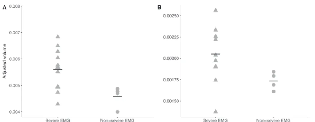
Fig. 2. Corpus callosum (A) and putamen (B) volumes in severely and non-severely affected patients on EMG. Severe EMG was defined as missing sensory potentials on lower and upper limbs. Patients with severe neuropathy had smaller corpus callosum (p = .030) and putamen (p = .030).
Fig. 3. FA values in POLG-mutated patients and controls. POLG-mutated patients had decreased FA compared to controls on the (A) left superior corona radiata (p = .004), (B) right superior corona radiata (p = .007), (C) left posterior limb of internal capsule (p = .039) and the (D) Right cingulate gyrus (p = .027).
amplitudes determined the type and the severity of neuropathy. Severe EMG profile was defined as the loss of sensory potentials on both lower and upper limbs, while maintained potentials in ulnar and/or radial nerves characterized non-severe EMG patterns.
2.6. Statistical analyses
Means were compared with the Mann and Whitney test and corre-lations were evaluated with Spearman coefficient. The Holm-Bonferroni step-down method of correction was applied for multiple tests. 3. Results
We gathered clinical and radiological data from 20 POLG-mutated patients. Their clinical and demographic characteristics are summar-ized inTable 1. The majority of our patients (n = 12) presented with SANDO (Table 2). Additionally, four patients presented with MEMSA, two patients with CPEO plus and two patients with MIRAS (Table 2). On visual inspection of brain MRI, only one patient displayed pseudo-strokes and no patient had Leigh syndrome. Two mutations were commonly found: c.2243G > C (p.Trp748Ser) in six patients, and c.1760C > T(Pro587Leu) – in cis with polymorphism c.752C > T (p.Thr251Ile) – in five patients, either at the homozygous or hetero-zygous state (Table 2). POLG-mutated patients were compared to 20 healthy controls. Four patients had a follow-up MRI after a 4-year in-terval. From the 16 POLG-mutated patients who had an EMG, 11 pa-tients displayed a sensory neuronopathy while 5 presented with an axonal length-dependent neuropathy. The EMG was realized on average
1.4 ( ± 2.4) years before the MRI. Four patients had a severe EMG profile, defined as the loss of sensory potentials on both lower and upper limbs while non-severely affected patients had maintained po-tential in ulnar and/or radial nerve.
Brain volumetry showed a global atrophy in patients, especially in the hemispheric cortex, the basal ganglia, the amygdala, and the pos-terior part of the brainstem (Table 3). The 4 patients who had succes-sive MRI showed a significant progression of atrophy in the right pal-lidum over time – adjusted volumes 0.00068 ± 0.00007 [0.00061–0.00075] versus 0.00101 ± 0.00018, [0.00079–0.00118] after 4 years, p = 0.029. The volume of the accumbens nuclei was dif-ferent between patients with and without cognitive impairment – 0.00050 ± 0.00011 [0.00036–0.00072] versus 0.00061 ± 0.00007 [0.00051–0.00072], p = 0.042. There was also a trend towards a higher clinical score associated with smaller accumbens nuclei (p = 0.130), and higher cerebellar subscore along with smaller superior cerebellar peduncle (p = 0.057, data not shown). Patients with a severe neuro-pathy on EMG had significantly smaller corpus callosum and putamen than patients with a non-severe EMG profile (Fig. 2).
DTI analyses were available for 14 POLG-mutated patients. Their clinical and demographic characteristics are summarized in Table 1. Two patients had a follow-up DTI analysis after a 4-year interval. ROI analyses showed significantly decreased FA in the corona radiata, cin-gulate gyrus and internal capsule (Fig. 3). However, the mean global FA was not different between patients and controls – 0.4649 ± 0.0339, [0.4120–0.5354] versus 0.4677 ± 0.214, [0.4231–0.4990], p = 0.607. TBSS analyses displayed significantly increased RD in the corpus cal-losum, the superior longitudinal fasciculus, internal and external cap-sules and superior cerebellar peduncles (Fig. 4). The 2 patients with follow-up DTI exhibited a tendency to decreased FA in the corona ra-diata –2.223 ± 0.0336, [2.200–2.2470] versus 2.4196 ± 0.1194, [2.4112–2.4280], p = 0.333 – and in the anterior internal capsule – 0.8854 ± 0.3248, [0.8616–0.9065] versus. 0.9454 ± 0.0107, [0.9378–0.9529], p = 0.333. There was however no significant differ-ence in RD in these two patients. The cerebellar subscore correlated with the FA in the cingulate gyrus (Spearman coefficient = −0.680, p = 0.028). Movement disorders were associated with increased RD in the internal capsule (Spearman coefficient = 0.745, p = 0.006) and the corona radiata (Spearman coefficient = 0.622, p = 0.036) while cere-bellar impairment correlated with RD in the cerebral peduncle (Spearman coefficient = 0.613, p = 0.040) (Fig. 5). Patients with hy-poacousia presented with a higher RD in the fornix than patients without hearing impairment – 0.0021 ± 0.0003 [0.0018–0.0026] versus 0.0017 ± 0.0002 [0.0014–0.0020], p = 0.004. There was no significant correlation between electrical parameters and DTI values.
Fig. 4. Increased RD in POLG-mutated patients compared to controls. Statistical map using FSL-TBSS shows voxels with significantly increased RD in corona radiata (solid arrow– axial image), in the corpus callosum (solid arrow – sagittal image), the posterior thalamic radiations (dotted arrow– axial image) and the cerebral peduncles (dotted arrow– sagittal image).
Fig. 5. Clinical correlation with DTI. (A) Cerebellar impairment correlated with low FA in the cingulate gyrus (Spearman coefficient = −0.680 p = .028) and (B) movement disorders with high RD in the internal capsule (Spearman coefficient = 0.745, p = .006).
4. Discussion
Our study shows that POLG-mutated patients displayed atrophy of the basal ganglia, amygdala and posterior cortex, consistent with the neurodegenerative process observed in adult patients. DTI also revealed abnormalities in the corona radiata, the internal capsule and the cin-gulate gyrus, possibly reflecting myelin alterations related to energy deficiency in POLG-mutated patients. Furthermore, to circumvent the wide and heterogeneous phenotypical spectrum that characterize POLG-mutated patients, we used an original binary clinical load, a method that has been validated before in rare neurometabolic diseases (Whybra et al., 2004). This score provided a simple homogenous clin-ical variable, which may prove useful in future studies but requires further validation. We also used EMG numerical data to evaluate dis-ease severity since peripheral nerve involvement is very common in POLG-mutated patients.
This is the first study associating two structural and quantitative neuroimaging modalities on a large series of adult patients with POLG mutations. Up to now, brain atrophy in POLG-mutated patients has been reported on visual inspection only (Tchikviladzé et al., 2015) while our work provides quantified measures of brain atrophy. Notably, cerebellar impairment correlated with atrophy of the cingulate gyrus, and cognitive impairment appeared to be linked to smaller basal ganglia. Using DTI, we also found correlation between cerebellar im-pairment and FA in the corona radiata of POLG-mutated patients, as well as movement disorders and RD in the corona radiata and internal capsule. This clinical study also showed altered brain diffusion in key white matter regions (cingulate gyrus, corona radiata, internal capsule) that may reflect cerebral metabolic alterations. Those results were found using both TBSS and ROI analyses, emphasizing that DTI can be a reliable tool even when performed as part of clinical routine follow-up. This is consistent with our work in other neurometabolic disorders such as Niemann Pick type C disease where we showed that DTI can identify biomarkers of disease progression and response to treatment (Masingue et al., 2017).
This study has some methodological limitations. MRI acquisitions were performed in a clinical (and not research) setting leading to suboptimal data quality. However, the coherence between the DTI findings obtained from ROI and TBSS approaches suggests that DTI may be less susceptible to artifacts generated by clinical MRI. Another lim-itation of this study is the limited size of our patient population. Nevertheless, our work reports on more patients than previous MRI studies in POLG-mutated patients. It is also the first DTI study per-formed in the macroscopically normal–appearing cerebral parenchyma, unlike previous work conducted within white matter and/or pseudo-stroke lesions from POLG-mutated patients (Tzoulis et al., 2010).
5. Conclusion
Despite its methodological limits, this original work supports the use of MRI for the identification of biomarkers in POLG-mutated pa-tients. Prospective studies are, however, needed to assess with disease progression the evolution of the volumetric and DTI parameters that we have identified.
Consent for publication
Not applicable (manuscript contains no individual person's data).
Competing interests
Authors declare nofinancial or non-financial competing interests in relation with this manuscript.
Funding sources for the study
MM was supported by the JNLF (Journées de Neurologie de langue Française) JNLF-SFN-ARSEP, 2015-2016.
Author contributions
MM and FM were involved in conception and design of the research project, analysis and interpretation of the data, and writing of thefirst draft of the manuscript. DG was involved in conception and design of the research project. MT was involved in the acquisition of clinical data, TM in the acquisition, analysis and interpretation of the EMG data and MM, IA, DG and FM in the acquisition, analysis and interpretation of the neuroimaging data. All authors read and approved the final manuscript.
Acknowledgments
We thank the following physicians for patients' referral: Sophie Demeret, Alexandra Durr, Karine Viala, Guillaume Bassez, Anthony Behin, Bruno Eymard, Pascal Laforêt, Yann Nadjar, Emmanuel Roze. References
Bindu, P.S., Arvinda, H., Taly, A.B., Govindaraju, C., et al., 2015. Magnetic resonance imaging correlates of genetically characterized patients with mitochondrial disorders: a study from south India. Mitochondrion 25, 6–16.
Bonvento, G., Valette, J., Flament, J., Mochel, F., Brouillet, E., 2017. Imaging and spec-troscopic approaches to probe brain energy metabolism dysfunction in neurodegen-erative diseases. J. Cereb. Blood Flow Metab. 1, 271678X17697989.https://doi.org/ 10.1177/0271678X17697989.
Bose, A., Beal, M.F., 2016. Mitochondrial dysfunction in Parkinson's disease. J. Neurochem. 1, 216–231.
Cheldi, A., Ronchi, D., Bordoni, A., Bordo, B., et al., 2013. POLG1 mutations and stroke like episodes: a distinct clinical entity rather than an atypical MELAS syndrome. BMC Neurol. 13 (8). https://doi.org/10.1186/1471-2377-13-8.
Degos, B., Laforêt, P., Jardel, C., Sedel, F., et al., 2014. POLG mutations associated with remitting/relapsing neurological events. J. Clin. Neurosci. 21, 186–188.
Echaniz-Laguna, A., Chassagne, M., de Sèze, J., Mohr, M., Clerc-Renaud, P., Tranchant, C., Mousson de Camaret, B., 2010. POLG1 variations presenting as multiple sclerosis. Arch. Neurol. 67, 1140–1143.
Habek, M., Barun, B., Adamec, I., Mitrović, Z., Ozretić, D., Brinar, V.V., 2012. Early-onset ataxia with progressive external ophthalmoplegia associated with POLG mutation: autosomal recessive mitochondrial ataxic syndrome or SANDO? Neurologist 18, 287–289.
Henao, A.I., Pira, S., Herrera, D.A., Vargas, S.A., Montoya, J., Castillo, M., 2016. Characteristic brain MRIfindings in ataxia-neuropathy spectrum related to POLG mutation. Neuroradiol. J. 29, 46–48.
Janssen, W., Quaegebeur, A., Van Goethem, G., Ann, L., Smets, K., Vandenberghe, R., Van Paesschen, W., 2016. The spectrum of epilepsy caused by POLG mutations. Acta Neurol. Belg. 116, 17–25.
Kinghorn, K.J., Kaliakatsos, M., Blakely, E.L., Taylor, R.W., Rich, P., Clarke, A., Omer, S., 2013. Hypertrophic olivary degeneration on magnetic resonance imaging in mi-tochondrial syndromes associated with POLG and SURF1 mutations. J. Neurol. 260, 3–9.
Kurt, B., Jaeken, J., Van Hove, J., Lagae, L., et al., 2010. A novel POLG gene mutation in 4 children with Alpers-like hepatocerebral syndromes. Arch. Neurol. 67, 239–244.
Lam, C.W., Law, C.Y., Siu, W.K., Fung, C.W., et al., 2015. Novel POLG mutation in a patient with sensory ataxia, neuropathy, ophthalmoparesis and stroke. Clin. Chim. Acta 448, 211–214.
Lax, N., Whittaker, R., Hepplewhite, P., Reeve, A., et al., 2012. Sensory neuropathy in patients harbouring recessive polymerase gamma mutations. Brain 135, 62–71.
Martikainen, M.H., Ng, Y.S., Gorman, G.S., Alston, C.L., et al., 2016. Clinical, genetic, and radiological features of extrapyramidal movement disorders in mitochondrial dis-ease. JAMA Neurol. 73, 668–674.
Masingue, M., Adanyeguh, I., Nadjar, Y., Sedel, F., Galanaud, D., Mochel, F., 2017. Evolution of structural neuroimaging biomarkers in a series of adult patients with Niemann-Pick type C under treatment. Orphanet J. Rare Dis. 12, 22–28.https://doi. org/10.1186/s13023-017-0579-3.
Mochel, F., Haller, R.G., 2011. Energy deficit in Huntington disease: why it matters. J. Clin. Invest. 121, 493–499.
Saneto, R.P., Lee, I.C., Koenig, M.K., Bao, X., Weng, S.W., Naviaux, R.K., Wong, L.J., 2010. POLG DNA testing as an emerging standard of care before instituting valproic acid therapy for pediatric seizure disorders. Seizure 19, 140–146.
Sidiropoulos, C., Moro, E., Lang, A.E., 2013. Extensive intracranial calcifications in a patient with a novel polymeraseγ-1 mutation. Neurology 81, 197–198.
Synofzik, M., Srulijes, K., Godau, J., Berg, D., Schöls, L., 2012. Characterizing POLG ataxia: clinics, electrophysiology and imaging. Cerebellum 11, 1002–1011.
Tchikviladzé, M., Gileron, M., Maisonobe, T., Galanaud, D., et al., 2015. A diagnostic flow-chart for POLG-related diseases based on signs sensitivity and specificity. J. Neurol. Neurosurg. Psychiatry 86, 646–654.
Tzoulis, C., Neckelmann, G., Mørk, S.J., Engelson, B.E., et al., 2010. Localized cerebral energy failure in DNA polymerase gamma-associated encephalopathy syndromes. Brain 133, 1428–1437.
Uusimaa, J., Gowda, V., McShane, A., Smith, C., et al., 2013. Prospective study of POLG mutations presenting in children with intractable epilepsy: prevalence and clinical features. Epilepsia 54, 1002–1011.
Van der Eerden, A.W., Khalilzadeh, O., Perlbarg, V., Dinkel, J., et al., 2014. White matter changes in comatose survivors of anoxic ischemic encephalopathy and traumatic
brain injury: comparative diffusion-tensor imaging study. Radiology 270, 506–516.
Van Goethem, G., Luoma, P., Rantamäki, M., Al Memar, A., et al., 2004. POLG mutations in neurodegenerative disorders with ataxia but no muscle involvement. Neurology 63, 1251–1257.
Whybra, C., Kampmann, C., Krummenauer, F., Ries, M., et al., 2004. The Mainz Severity Score Index: a new instrument for quantifying the Anderson-Fabry disease phenotype and the response of patients to enzyme replacement therapy. Clin. Genet. 65, 299–307.
Winterthun, S., Ferrari, G., He, L., Taylor, R.W., et al., 2005. Autosomal recessive mi-tochondrial ataxic syndrome due to mimi-tochondrial polymerase gamma mutations. Neurology 64, 1204–1208.