HAL Id: hal-01548998
https://hal.sorbonne-universite.fr/hal-01548998
Submitted on 28 Jun 2017HAL is a multi-disciplinary open access archive for the deposit and dissemination of sci-entific research documents, whether they are pub-lished or not. The documents may come from teaching and research institutions in France or abroad, or from public or private research centers.
L’archive ouverte pluridisciplinaire HAL, est destinée au dépôt et à la diffusion de documents scientifiques de niveau recherche, publiés ou non, émanant des établissements d’enseignement et de recherche français ou étrangers, des laboratoires publics ou privés.
Jean-Jacques Baudon, Francis Renault, Roberto Flores-Guevara, Marie-Paule
Vazquez
To cite this version:
Jean-Jacques Baudon, Francis Renault, Roberto Flores-Guevara, Marie-Paule Vazquez. Outcomes of Neonatal Bulbar Weakness. Pediatrics, American Academy of Pediatrics, 2016, 137 (1), pp.e20153004. �10.1542/peds.2015-3004�. �hal-01548998�
Outcomes of neonatal bulbar weakness
Jean-Jacques Baudona, MD, Francis Renaultb, MD, Roberto Flores-Guevarab,c, MD, PhD, Marie-Paule Vazquezd,e, MD.
Affiliations:
a
Faculté de Médecine Pierre et Marie Curie, Université Paris6, Paris, France
b
Clinical Neurophysiology Unit, Hôpital Armand-Trousseau, AP-HP, Paris, France
c
Facultad de Medicina, Universidad Nacional Mayor de San Marcos, Lima, Peru
d
Faculté de Médecine René Descartes, Université Paris5, Paris, France
e
Department of Maxillofacial Surgery, Hôpital Necker-Enfants Malades, AP-HP, Paris, France
Address correspondence to: Docteur Francis Renault, Unité de Neurophysiologie clinique de l’enfant, Hôpital Armand-Trousseau, 28 avenue Arnold-Netter, 75571 Paris 12, France, [docteur.frenault@wanadoo.fr], +33685208990.
Short running title: Outcomes of neonatal bulbar weakness
Funding Source: No external funding for this manuscript.
Financial Disclosure: The authors have indicated they have no financial relationships relevant to this article to disclose.
Conflict of Interest: The authors have indicated they have no potential conflicts of interest to disclose.
Abbreviations:
BRs: blink responses; BW: bulbar weakness; EMG: electromyography; EMGbf: electromyography during bottle feeding; PRS: Pierre Robin sequence
What’s known on this subject?
Neonatal bulbar weakness has various etiologiesand a broad prognostic range. Outcomes depend on both the severity of orofacial dysfunction and the nature of neuromuscular or central nervous system underlying disorders.
What this study adds?
This is the first report of long-term outcomes in a large series of infants with neonatal bulbar weakness, showing a high risk of motor or mental disabilities, and death. Orofacial
electrodiagnostic studies bring prognostic indicators regardless of underlying disorders.
Contributors’ Statements:
Dr. Baudon revised patients’ files and was in the process of writing the manuscript. Dr. Renault performed electrodiagnostic studies and was in the process of writing the manuscript.
Dr Flores-Guevara performed statistical studies and was in the process of writing the manuscript.
Dr Vazquez was in charge of most patients and critically reviewed the manuscript.
All authors approved the final manuscript as submitted and agree to be accountable for all aspects of the work.
Background/Objectives
Neonatal bulbar weakness has various etiologies and a broad prognostic range. We aimed to
report outcomes in a large series of children with neonatal bulbar weakness, and to explore
the association of orofacial electrodiagnostic data with outcome.
Method
We retrospectively reviewed the files of children who presented a facial, lingual, laryngeal or
pharyngeal weakness at birth, and who underwent electrodiagnostic studies combining
conventional needle electromyography of orofacial muscles, blink responses, and
electromyography during bottle feeding. Outcome measures included the need for prolonged
respiratory assistance and enteral feeding, as well as sensorimotor and cognitive impairments.
Results
Out of 175 patients, 73% had developmental disorders; 25% suffered from acquired brain
damage; 2% had no apparent underlying disorders. A motor or mental impairment was
observed in 71%; death occurred in 16%. Outcomes were not significantly different when
comparing developmental disorders vs acquired brain damage, and neurogenic vs normal
detection electromyography. Abnormal blink responses were associated with higher
frequencies of respiratory assistance (p=0.03), gastrostomy (p=0.025), and death (p=0.009);
moderate or severe oro-pharyngeal incoordinations were associated with higher frequencies of
respiratory assistance (p=0.006), prolonged enteral feeding (p<.0001), and gastrostomy
(p=0.0002).
Conclusion
Orofacial electrodiagnostic studies bring supplementary information to help the paediatrician
Introduction
Newborn infants with bulbar weakness (BW) present with congenital facial, lingual,
laryngeal, or pharyngeal dysfunction, alone or in combination; all of which have significant
developmental and functional consequences. Individual course may vary from rapid discharge
without any sequel to prolonged dependence on nutritional and respiratory supports,
disabilities, or death. Various etiologies include neuromuscular disorders involving the motor
neuron, neuromuscular transmission, or muscle, and cerebral palsies. The evaluation of a
child with BW requires that major emphasis be placed on an etiological definition and a
reliable prognostic assessment because outcomes depend on both the severity of orofacial
dysfunction and the nature of neuromuscular or central nervous system underlying disorder.1
Although clinical assessments have been developed,2 predicting an outcome remains a
challenging task. Available complementary investigations include videofluoroscopy,3
fiberoptic endoscopy,4,5 orofacial electrodiagnostic studies, and esophageal manometry.6 By
combining conventional detection of orofacial muscles, blink responses recording, and
functional electromyography during bottle feeding, electrodiagnostic studieshave been shown
to help diagnose cranial nerves palsies and sucking/swallowing incoordination.7,8 In this work,
we report outcomes in a large series of young infants with neonatal BW of diverse causes, and
explore the association of orofacial electrodiagnostic data with outcome.
Methods
Participants
We carried out a retrospective study. Approval by an institutional ethics’ committee was waived by our hospital’s review board. We reviewed the database of our pediatric
neurophysiology unit over a fifteen years period (07/01/1995 - 12/31/2010). We identified
692 patients who underwent orofacial electrodiagnostic studies before 6 months of age. We
presumed of obstetrical origin; and 14 patients with isolated cleft palate. As a result, we listed
258 patients with clinical signs of bulbar dysfunction who were treated in our hospital. We
excluded 59 patients with strictly isolated Pierre Robin sequence (PRS), and 24 patients lost
to follow-up before 6 months of age. Following our review, we included 175 patients and
collected clinical, radiological and genetic data that contributed to their diagnoses. The data
were de-identified prior to analysis. Diagnoses were established after clinical examination by
a neonatologist, a maxillofacial surgeon, and a clinical geneticist. Ocular, skeletal, renal, and
cardiac malformations were identified by ophthalmologic examination, X-ray, and ultrasound
scans. The brain was investigated by ultrasound scan, computed tomography, or magnetic
resonance imaging. Upper airways were examined using laryngoscopy. A karyotype was
obtained for every patient; deletion at 22q11 was tested in 166 patients. Two etiological
groups were defined: (1) developmental disorders and (2) acquired brain damage. Group 1
included patients with genetic anomalies, recognizable malformation syndromes,9 inherited
metabolic diseases, or birth defects that have not been yet classified as recognizable
syndromes. Group 2 included patients with hypoxic ischemic encephalopathy, birth asphyxia,
premature birth <32 weeks, or fetal exposure to toxic agents.
Orofacial electrodiagnostic studies
Electrodiagnostic examinations were all performed by one of us (FR) and were part of every patient’s assessment. In case of premature birth, electrodiagnostic studies were not performed before gestational age 37 weeks. Three methods, elsewhere described in detail, were
combined: conventional detection needle electromyography (EMG);10 blink responses
(BRs);11 and EMG during bottle feeding (EMGbf).10 In short, conventional detection EMG
was used to study face, tongue, and soft palate muscles, at rest and during crying stages.
Traces were analyzed manually and classified as normal; neurogenic single or reduced
decreased by at least 30 %). BRs were recorded in the orbicularis oculi muscle in response to
the electrical stimulation of the trigeminal supraorbital nerve; the presence of R1 and R2
components and R1 latency were analyzed, considering an asymmetry of up to 3 ms to be
normal. EMGbf was used to assess the pattern of suction as well as sucking/swallowing
coordination. The technique consists of a two-channel recording of the genioglossus muscle
for the oral phase, as well as the thyrohyoid muscle for the pharyngeal phase, while the infant
was drinking sugar-water from a bottle. Normal pattern features rhythmic spindle-shaped
bursts of activity separated by a quiescent period; the two muscles alternate regularly.
Abnormal patterns define oro-pharyngeal incoordination as mild, where sucking is present but
the alternation between sucking and swallowing is irregular; moderate, where sucking is
present but the pharyngeal phase is either synchronous or at random; or severe, where the
tongue does not perform rhythmic sucking activity, and the pharyngeal phase is either inactive
or tonic. Results were classified as: (1) normal oro-pharyngeal coordination or mild
anomalies; and (2) moderate or severe oro-pharyngeal incoordination.
Outcome measures
We recorded the need for respiratory assistance (oxygen or non-invasive ventilation during
more than 1 month, and tracheostomy) and enteral feeding (tube feeding during more than 6
months, and gastrostomy); motor and sensory disabilities, behavioural disorders, and
language and educational skills. Standardized neurodevelopmental evaluation was not
possible given the retrospective nature of the study and the differences in durations of the
follow-up period. Patients were considered as disabled when they suffered from cerebral
palsy, sensory hearing loss, poor or no language, and inability to follow a scholar course, even
in an adapted school. We defined the absence of disability as normal motor development and normal or slightly delayed (≤ 2 years) school course.
Numeric variables are presented as mean, median, and range; categorical variables are
presented as rates. Comparison of two independent groups was performed by the
Mann-Whitney U-test for numeric variables and the Fisher exact test for categorical variables.
Comparison of more than two groups was performed by the One-way ANOVA test for
numeric variables and the χ2 test for categorical variables. All hypotheses were constructed as 2-tailed. A P value < 0.05 was considered significant.
Results:
Participants
The series included 175 patients (male: 84, female: 91). The most frequentlypresenting
symptoms were the lack of sucking or swallowing and aspiration episodes. BW was
frequently associated with facial malformations. On general neurologic examination, main
symptoms were hypotonia, lethargy, and limb hypertonia. (Table 1) Excluding 8 patients who
died prior to age 6 months, patients were followed up to age 2 years (25/167, 15%), from 2 to
5 years (48/167, 29%), or over 5 years (94/167, 56%). Genealogical data were available for
120 patients, including 18 born to consanguineous parents. A specific diagnosis was
established for 134 patients, during the first trimester of life (110), or between 3 months and 7
years (24). The remaining patients had unidentified malformation patterns (36) or no apparent
underlying disorder (5). (Table 2) Outcomes are shownin Table 1.
Electrodiagnostic studies
Median age at electrodiagnostic examination was 40 days (range: 2-180, mean: 54). Detection
EMG distinguished 85 patients without any neurogenic EMG signs, and 90 patients with
neurogenic EMG signs in muscles innervated by the facial nerve (82/90 patients), the
pharyngeal plexus (56/90), and the hypoglossal nerve (31/90). Low amplitude traces were
recorded in soft palate muscles in 36 patients, including 32 with cleft palate. BRs were
normal coordination or mild abnormalities in 63/145 (43%) patients; and moderate or severe
oro-pharyngeal incoordination in 82/145 (57%). Regarding the 30 patients who did not
undergo EMGbf, 23 showed clinical condition precluding all attempt to oral feeding,
including 8 patients who died.
Statistical analyses
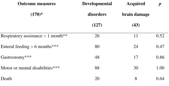
When comparing etiological subgroups - developmental disorders vs acquired brain damage -,
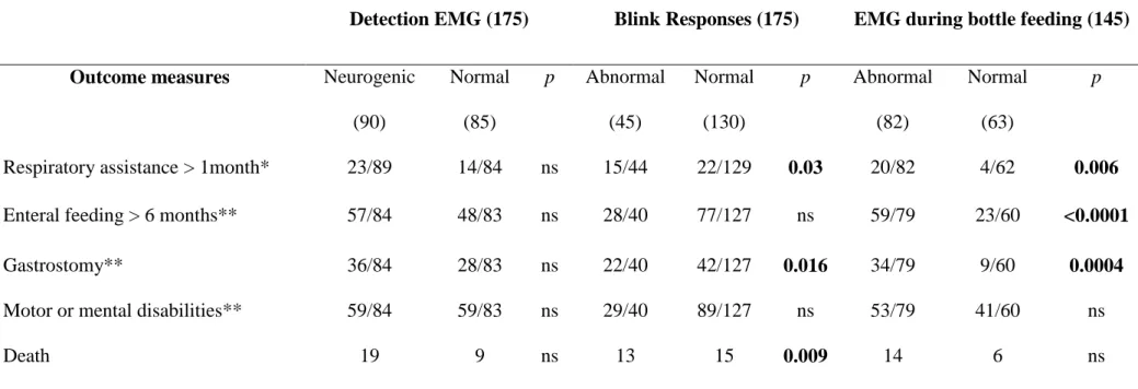
outcomes were not significantly different. (Table 3) Patients with abnormal BRs showed
higher frequencies of respiratory assistance (p=0.03), gastrostomy (p=0.016), and death
(p=0.009) than those with normal BRs. Moderate or severe oro-pharyngeal incoordination on
EMGbf was strikingly associated with the need for respiratory assistance (p=0.006),
prolonged enteral feeding (p<0.0001), and gastrostomy (p=0.0004). (Table 4)
Discussion:
By assessing bulbar pathways and oro-pharyngeal coordination, orofacial electrodiagnostic
studies gave relevant indicators for the management and prognostic evaluation of young
infants with BW. Of 175 infants, their underlying disorders notwithstanding, BW frequently
had poor outcome since 16% of the patients died and more than two thirds suffered from
motor or mental disabilities. Comparing patients with developmental disorders versus
acquired brain damage, the absence of statistically significant differences in outcomes
suggests that BW by itself underlies long-term functional and developmental consequences.
The small number of patients without apparent underlying disorders did not enable statistical
comparisons; of the five, only one required prolonged enteral feeding and not any suffered
from disability, as usually observed.12 We acknowledge that our series do not include any case
of hereditary motor neuropathy, congenital myopathy, or congenital myasthenic syndrome. In
patients with cleft palate, low-amplitude myopathic EMG signs detected in soft palate
hypoplasia. When studying patients with specific diagnoses recognizable at birth, such as
CHARGE association, Moebius syndrome, first arch syndromes, or congenital myasthenic
syndromes, different authors found widely varying or facial dysfunctions and outcomes.13-17
For patients with acquired brain damage, a possible association between early sucking and
swallowing abilities and neurodevelopment outcome is still open to debate.18 In a recent series
of preterm infants, abnormal sucking behaviour at 46 weeks of post menstrual age was
associated with neurodevelopment delay.2 Thus, a prognostic evaluation of neonatal BW
remains a challenge for the paediatrician. Video fluoroscopy and fiberoptic endoscopy are
commonly used to evaluate oro-pharyngeal dysfunction but, to our knowledge, the association
of their results with long-term outcome has not yet been established. In our practice, in the
last twenty years, the use of videofluoroscopy prior to age 6 months has progressively
declined due to the risk of aspiration and radiation exposure. Meanwhile, electrodiagnostic
studies of bulbar muscles and cranial nerves became a routine diagnostic tool to look for
bulbar involvement and investigate the mechanism and severity of dysphagia. These bulbar
electrodiagnostic studies investigate paired cranial nerves VII, IX-X, and XII, the V to VII
internuclear pathways, and the central pattern generator for swallowing involving the nucleus
of the tractus solitarius and adjacent ventromedian reticular formation.19 To that end, orofacial
electrodiagnostic studies explore small sized brain stem structures and cranial nerves that are
not routinely studied using magnetic resonance imaging (MRI).20,21 Among patients suffering
from congenital facial malformations, electrodiagnostic studies have revealed the neurological
origin of dysphagia, even in the absence of neurological signs or abnormalities in brain
images.7 EMG of the genioglossus muscle has detected an associated hypoglossal nerve
involvement in children with periventricular leukomalacia or hemorrhagic infarction.22 In
patients with PRS, the absence of neurogenic EMG signs in orofacial muscles characterized
dysphagia.8 Among patients with congenital multiple cranial neuropathy, detection EMG
could identify bulbar involvement in patients with orofacial dysfunctions attributed to a
suprabulbar vascular insult at preterm or term birth.23
In the present series, neurogenic EMG signs were detected in half of the patients.
Curiously, frequencies and durations of respiratory assistance and enteral feeding were not
statistically different in patients with or without cranial nerve involvement. These results
could be explained by the predominant involvement of cranial nerve VII, while airway
obstruction and aspiration result from disorders of nerves IX-X and XII. Interestingly, BRs
abnormalities were significantly associated with high frequencies of respiratory assistance,
gastrostomy, and death. BRs explore pathways in close proximity to the respiratory and
swallowing centers and running through the reticular formation. The R1 component of BRs
corresponds to an oligosynaptic reflex arc involving at least two and not more than three
synapses in the pons between the main sensory nucleus of cranial nerve V and the motor
nucleus of the ipsilateral cranial nerve VII. The R2 component follows polysynaptic
medullary pathways, which are more caudal and closer to the bulbar formations. Moreover,
EMGbf identified patients with moderate or severe oro-pharyngeal incoordination, who
showed more needs for respiratory assistance, long-lasting enteral feeding, and gastrostomy
than patients with normal coordination or mild abnormalities. Finally, orofacial
electrodiagnostic studies brought prognostic indicators of neonatal BW, regardless of the
underlying neuromuscular or suprabulbar disorders. This study has a limitation. Indeed,
despite the large number of patients we investigated, one can appreciate that 15 years is a long
spell of time and that diagnostic abilities and management have improved. In fact, our
investigating protocol has remained the same during that period and all patients were able to
To conclude, early orofacial electrodiagnostic studies do provide the paediatrician
with supplementary information helping anticipate the outcome of BW, given that even if
References
1 - Roig-Quilis M. Oromotor dysfunction in neuromuscular disorders: evaluation and
treatment. In: Darras BT, Jones HR, Ryan MM, and De Vivo DC (editors). Neuromuscular
disorders of infancy, childhood, and adolescence. A clinician’s approach. 2nd
edition.
Elsevier; 2015. pp 958-975
2 - Wolthuis-Stigter MI, Luinge MR, da Costa SP, Krijnen WP, van der Schans CP, Bos AF.
The association between sucking behavior in preterm infants and neurodevelopmental
outcomes at 2 years of age. J Pediatr. 2015;166:26-30
3 - Newman LA, Keckley C, Petersen MC, Hamner A. Swallowing function and medical
diagnoses in infants suspected of dysphagia. Pediatrics. 2001;108:e106-e109
4 - Hartnick CJ, Hartley BE, Miller C, Willging JP. Pediatric fiberoptic endoscopic evaluation
of swallowing. Ann Otol Rhinol Laryngol. 2000;109:996-999
5 - Da Silva AP, Lubianca Neto JF, Santoro PP. Comparison between videofluoroscopy and
endoscopic evaluation of swallowing for the diagnosis of dysphagia in children. Otolaryngol
Head Neck Surg. 2010;143:204-209
6 - Baudon JJ, Renault F, Goutet JM, Flores-Guevara R, Soupre V, Gold F, et al. Motor
dysfunction of the upper digestive tract in Pierre Robin sequence as assessed by
sucking-swallowing electromyography and esophageal manometry. J Pediatr. 2002;140:719-723
7 - Baudon JJ, Renault F, Goutet JM, Biran-Mucignat V, Morgant G, Garabedian EN,
Vazquez MP. Assessment of dysphagia in infants with facial malformations. Eur J Pediatr.
2009;168:187-193
8 - Renault F, Baudon JJ, Galliani E, Flores-Guevara R, Marlin S, Garabedian EN, Vazquez
MP. Facial, lingual, and pharyngeal electromyography in infants with Pierre Robin sequence.
Muscle Nerve 2011;43:866-871
genes and genetic disorders. Available at: htpp://omim.org
10 - Renault F. Facial and bulbar weakness. In: Brown WF, Bolton CF, Aminoff MJ (editors).
Neuromuscular function and disease. Philadelphia: WB Saunders; 2001. pp 1580-1600
11 - Vecchierini-Blineau MF, Guiheneuc P. Maturation of the blink reflex in infant. Eur
Neurol. 1984;23:449-458
12 - Heuschkel RB, Fletcher K, Hill A, Buonomo C, Bousvaros A, Nurko S. Isolated neonatal
swallowing dysfunction. A case series and review of the literature. Dig Dis Sc. 2003;48:30-35
13 - Blake KD, Hartshorne TS, Lawand C, Dailor AN, Thelin JW. Cranial nerve
manifestations in CHARGE syndrome. Am J Med Genet. Part A 2008;146A:585-592
14 - Cooper-Brown L, Copeland S, Dailey S, Downey D, Petersen MC, Stimson C, Van Dyke
DC. Feeding and swallowing dysfunction in genetic syndromes. Dev Disab Res Rev.
2008;14:147-157
15 - Khan A, Hussain N, Gosalakkal J. Bulbar dysfunction: an early presentation of
congenital myasthenic syndrome in three infants. J Pediatr Neuroscienc. 2011;6:124-126
16 - Verzijl HT, van der Zwaag B, Cruysberg JR, Padberg GW. Möbius syndrome redefined.
A syndrome of rhombencephalic maldevelopment. Neurology. 2003;61:327-333
17 - Armangue T, Macaya A, Vazquez E, Jurado MJ, Roig-Quillis M. Central hypoventilation
and brainstem dysgenesis. Pediatr Neurol. 2012;46:257-259
18 - Slattery J, Morgan A, Douglas J. Early sucking and swallowing problems as predictors of
neurodevelopmental outcome in children with neonatal brain injury: a systematic review.
Develop Med Child Neurol. 2012;54:796-806
19 - Lang IM. Brain stem control of the phases of swallowing. Dysphagia. 2009;24:333-348
20 - Quattrocchi CC, Longo D, Delfino LN, et al. Dorsal brain stem syndrome: MR imaging
location of brain stem tegmental lesions in neonates with oral motor dysfunction. Am J
21 - Sugiura H, Kouwaki M, Kato T, Ogata T, Sakamoto R, 21-Ieshima A, Yokochi K.
Magnetic resonance imaging in neonates with total asphyxia. Brain Develop. 2013;35:53-60
22 - Vijayakumar K, Rockett J, Ryan M, Harris R, Pitt M, Devile C. Experience of using
electromyography of the genioglossus in the investigation of paediatric dysphagia. Dev Med
Child Neurol. 2012;54:1127-1132
23 - Renault F, Flores-Guevara R, Baudon JJ, Vazquez MP. Congenital multiple cranial
neuropathies: Relevance of orofacial electromyography in infants. Muscle Nerve. 2015; Mar
Table 1: Clinical features and outcome in 175 patients with neonatal bulbar weakness
Number of patients (%)
Orofacial presenting symptoms
Orofacial malformations 127/175 (73)
Aspiration, choking/gaging episodes 121/175 (69)
No swallowing 115/175 (66)
No sucking 88/175 (50)
Glossoptosis 40/175 (23)
Amimia, hypomimia 25/175 (14)
Ophtalmoplegia 17/175 (10)
General neurological signs
Hypotonia 47/175 (27)
Lethargy 27/175 (15)
Limb hypertonia 14/175 (8)
Outcome
Respiratory assistance required during more than one month* 37/173 (21)
Enteral feeding during more than 6 months** 105/167 (63)
Gastrostomy** 65/167 (39)
Motor or mental disabilities** 118/167 (71)
Death 28/175 (16)
* 2 patients died prior to 1 month of age
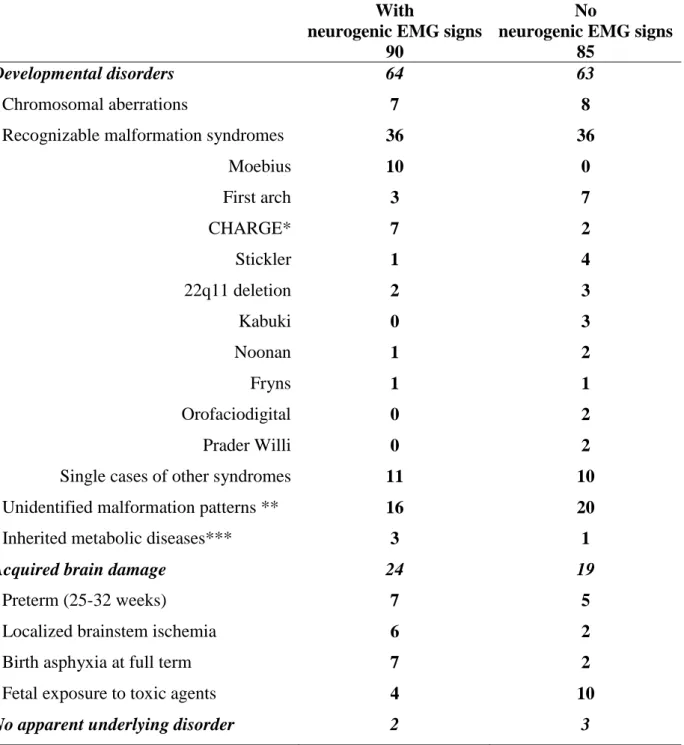
Table 2: Detection electromyography and etiology in 175 patients with neonatal bulbar dysfunction
With
neurogenic EMG signs 90
No
neurogenic EMG signs 85
Developmental disorders 64 63
- Chromosomal aberrations 7 8
- Recognizable malformation syndromes 36 36
Moebius 10 0 First arch 3 7 CHARGE* 7 2 Stickler 1 4 22q11 deletion 2 3 Kabuki 0 3 Noonan 1 2 Fryns 1 1 Orofaciodigital 0 2 Prader Willi 0 2
Single cases of other syndromes 11 10 - Unidentified malformation patterns ** 16 20 - Inherited metabolic diseases*** 3 1
Acquired brain damage 24 19
- Preterm (25-32 weeks) 7 5
- Localized brainstem ischemia 6 2 - Birth asphyxia at full term 7 2 - Fetal exposure to toxic agents 4 10
No apparent underlying disorder 2 3
* CHARGE association: colobomata, heart disease, atresia of the choanae, retarded growth and development, genital hypoplasia, and ear anomalies or deafness
** Patients showing combined malformations that are not yet classified as recognizable syndromes.
***
Including 2 patients with pyruvate dehydrogenase deficiency, 1 with abetalipoproteinemia, and 1 with dysmyelination (ADCY6 mutation)
Table 3: Outcome of neonatal bulbar weakness according to etiology* Outcome measures (170)* Developmental disorders (127) Acquired brain damage (43) p
Respiratory assistance > 1 month** 26 11 0.52
Enteral feeding > 6 months*** 80 24 0.47
Gastrostomy*** 48 17 0.86
Motor or mental disabilities*** 88 30 1.00
Death 20 8 0.64
* Excluding 5 patients without an identified underlying disorder;
** Except patients who died prior to age 1 month;
*** Except patients who died prior to age 6 months;
Table 4: Association of orofacial electrodiagnostic data and outcomes in 175 infants with neonatal bulbar weakness
*Except patients who died prior to age 1 month; **except patients died prior to age 6 months; p: Chi-squared test
Detection EMG (175) Blink Responses (175) EMG during bottle feeding (145)
Outcome measures Neurogenic
(90) Normal (85) p Abnormal (45) Normal (130) p Abnormal (82) Normal (63) p
Respiratory assistance > 1month* 23/89 14/84 ns 15/44 22/129 0.03 20/82 4/62 0.006
Enteral feeding > 6 months** 57/84 48/83 ns 28/40 77/127 ns 59/79 23/60 <0.0001
Gastrostomy** 36/84 28/83 ns 22/40 42/127 0.016 34/79 9/60 0.0004
Motor or mental disabilities** 59/84 59/83 ns 29/40 89/127 ns 53/79 41/60 ns