Modulation of placental vascular endothelial growth factor
by leptin and hCG
D.Islami, P.Bischof
1and D.Chardonnens
Department of Obstetrics and Gynaecology, Clinic of Infertility and Gynaecological Endocrinology±WHO Collaborating Centre in
Human Reproduction, University Hospital of Geneva, 32 Boulevard de la Cluse, 1211 Geneva, Switzerland
1
To whom correspondence should be addressed at: Clinique de SteÂrilite et d'Endocrinologie GyneÂcologique, HoÃpital Cantonal
Universitaire de GeneÁve, 32, Boulevard de la Cluse, 1211 GeneÁve 14, Switzerland. E-mail: [email protected]
Vascular endothelial growth factor (VEGF) has been identi®ed as an endothelium-speci®c mitogen and inducer of angiogenesis
and endothelial cell survival. Leptin and hCG have also been suggested as possible regulators of angiogenesis in various models.
In-vivo and in-vitro assays revealed that leptin has an angiogenic activity and that the vascular endothelium is a target for leptin.
Thus, we hypothesized that products of cytotrophoblastic cells may play a role in placental angiogenesis and we therefore
inves-tigated the effects of leptin and hCG on cytotrophoblast VEGF secretion. We incubated cytotrophoblastic cells (CTB) with
recombinant human leptin (rhLept) (0±4 pg/ml) or hCG (0±30 000 IU/ml) for 4 h. rhLept signi®cantly stimulated hCG
(P = 0.0045) and decreased VEGF release (P = 0.0008) by CTB in a concentration-dependent manner. On the other hand,
increasing concentrations of hCG (0±30 000 IU/ml), induced a signi®cant inhibition of leptin secretion (P = 0.0028) and a marked
dose-dependent stimulation of VEGF
165secretion (P < 0.0001). We observed an increase of >1000-fold in basal trophoblastic
VEGF secretion with physiological concentrations of hCG in vitro. An inhibitory effect of hCG on trophoblastic leptin secretion
was also observed, suggesting that hCG might exert a possible negative feedback on trophoblastic release of leptin. We
hypothe-size that trophoblastic products such as hCG and leptin are probably involved in the control of VEGF secretion at the
maternal±fetal interface.
Key words: angiogenesis/hCG/leptin/trophoblast/VEGF
Introduction
Angiogenesis is generally a quiescent process in the healthy adult
male with an extremely low turnover of endothelial cells. However, in
the female, angiogenesis is highly regulated and turned on for brief
periods of time in selected organs such as the ovary and the
endometrium. Examples of such regulated angiogenesis include
follicular growth, corpus luteum formation, endometrium
differenti-ation and repair, and embryo implantdifferenti-ation and development (Reynolds
et al., 1992; Reynolds and Redmer, 1995). Among the list of
angiogenic factors (Folkman and Klagsbrun, 1987), vascular
en-dothelial growth factor (VEGF) was identi®ed as an
endothelium-speci®c mitogen and inducer of angiogenesis and endothelial cell
survival (Plouet et al., 1989; Conn et al., 1990; Alon et al., 1995;
Gerber et al., 1998).
Analysis of the DNA sequence of a variety of human VEGF clones
indicates that VEGF may exist as one of four different isoforms as a
result of alternative splicing. VEGF
165is the major VEGF isoform
(Ferrara, 1999). In human placenta, VEGF expression has been
analysed by in-situ hybridization and immunohistochemical studies
(Ahmed et al., 2000). It is localized to the villous trophoblast, the
decidua and macrophages of both fetal and maternal origin (Sharkey
et al., 1993; Ahmed et al., 1995; Vuorela et al., 1997). Trophoblast
cells also secrete VEGF in vitro (Shore et al., 1997; Ahmed et al.,
2000). VEGF appears unique in its role as an angiogenic factor, in that
the two known human receptors for VEGF, VEGFR-1 (formerly ¯t-1)
and VEGFR-2 (formerly KDR) are expressed on endothelial cells
(de Vries et al., 1992; Terman et al., 1992). Interestingly, human
trophoblast cells, particularly the invasive extravillous cytotrophoblast
cells (CTB) also express VEGF receptors (Charnock-Jones et al.,
1994; Athanassiades et al., 1998). Both VEGF receptor forms are
present in ®rst trimester placental tissue (Ahmed et al., 1995;
Vuckovic et al., 1996).
Leptin and hCG, both produced by human trophoblasts
(Chardonnens et al., 1999), have also been suggested as possible
regulators of angiogenesis in various models (Rizk et al., 1997;
Henson and Castracane, 2000). Leptin was found to generate a
growth signal via a tyrosine kinase-dependent pathway and promote
angiogenic processes via activation of the leptin receptor (Ob-R) in
endothelial cells (Park et al., 2001). In-vivo and in-vitro assays
revealed that leptin has an angiogenic activity and that vascular
endothelium is a target for leptin (Sierra-Honigmann et al., 1998).
In contrast to the leptin-de®cient ob/ob mice, where no vascular
fenestrations are detected, capillary fenestrations are found in
leptin-producing adipose tissue in lean mice. Thus, leptin plays a critical
role in the maintenance and the regulation of vascular fenestrations
in adipose tissue. Furthermore, leptin and ®broblast growth factor
(FGF)-2 and VEGF synergistically stimulate angiogenesis (Cao
et al., 2001).
Taking these observations together, we hypothesized that products
of cytotrophoblastic cells may play a role in placental angiogenesis.
We therefore investigated the effects of leptin and hCG on
cytotrophoblast VEGF secretion.
Molecular Human Reproduction Vol.9, No.7 pp. 395±398, 2003 DOI: 10.1093/molehr/gag053
Materials and methods
CTB were isolated and puri®ed as previously described (Bischof et al., 1991). Brie¯y, trophoblast tissue was obtained from legal abortions at 8±12 weeks of normal pregnancy (from the last menstrual period). The tissue was digested with trypsin and cytotrophoblastic cells separated from blood cells and syncytia on a discontinuous Percoll gradient and immunopuri®ed by antibody-coated magnetic particles (anti-CD45; Dyna Beads, Switzerland) in order to eliminate contaminating leukocytes. After immunopuri®cation the cell suspension represents 90±97% cytotrophoblastic cells, with a 3±7% contamination by fetal stromal cells. Lymphomyeloid cells are no longer present (Bischof et al., 1991). CTB viability was assessed by Trypan Blue (Sigma) exclusion and cells diluted to 106 cells/ml with culture medium. Recombinant human leptin (rhLept) was purchased from R&D Systems (BuÈhlmann Laboratories, Switzerland). hCG (Profasi) was purchased from Ares±Serono (Switzerland).
Culture conditions
CTB (100 ml, 106cells/ml, >90% viability) were incubated in duplicates in the presence or absence of rhLept (0±4 pg/ml) or hCG (0±30 000 IU/ml). Incubation was performed in 12-well tissue culture plates (Costar, USA) under a 5% CO2and 95% air atmosphere in a humid incubator at 37°C for 4 h. Culture
medium was Dulbecco's modi®ed Eagle's medium (Gibco, Switzerland) containing 2 mmol/lL-glutamine, 4.2 mmol/l magnesium sulphate, 25 mmol/l HEPES, 1% gentamycin, 1% amphotericin B, 100 mg/ml streptomycin and 100 IU/ml penicillin, in the absence of serum. The supernatants were collected after 4 h of incubation, aliquoted and stored at ±20°C until assayed. Duplicate wells were run for each treatment condition and the experiments repeated four times with different CTB preparations.
Hormone and protein assays
VEGF165was measured in the supernatants by an enzyme-linked
immunosor-bent assay (ELISA) (R&D Systems, Germany) with a sensitivity of 5.0 pg/ml and intra- and inter-assay coef®cient of variation (CV) <6.5 and <8.5% respectively. HCG was measured in the supernatants by a microparticle enzyme immunoassay (IMX; Abbot, USA) with a sensitivity of 0.1 mIU/ml and intra-and inter-assay coef®cients of variation <4%. Leptin was measured in the supernatants by an ELISA (DRG diagnostics, Cis-Medipro, Switzerland) with a sensitivity of 0.2 ng/ml and an intra- and inter-assay CV of 4.3 and 8.6% respectively. According to the manufacturer, the leptin assay does not cross-react with human proteins such as proinsulin, C-peptide, glucagon or insulin-like growth factor-I.
Statistics
In order to normalize the distribution and minimize the inter-experimental variability, all results were expressed as a percentage of their respective controls, CTB in the absence of hCG or rhLept, and presented as mean 6 SEM. Statistical evaluations were performed with a personal computer using the Statview Program (Abascus). Tests included one-way analysis of variance with Fisher's test to compare the effects of different concentrations of rhLept or hCG. Regression analysis was performed to determine the dose dependency of rhLept or hCG effects. P < 0.05 was considered to be statistically signi®cant.
Results
Effects of leptin on hCG and VEGF secretion in human
CTB
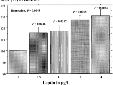
RhLept (0±4 mg/l) augmented hCG release by CTB in a
concentration-dependent manner (P = 0.0045, Figure 1). Basal hCG release was
118 6 12 IU/l. When compared to controls, hCG release was 115.7 6
6.8% (P = 0.0436), 117.3 6 6% (P = 0.0317), 127 6 5.7%
(P = 0.0058) and 130.5 6 7.5% (P = 0.0034), after incubation with
0.5, 1, 2 and 4 pg/ml of rhLept respectively.
RhLept (0±4 mg/l) decreased VEGF release by CTB in a
concen-tration-dependent manner (P = 0.0008, Figure 2). Basal VEGF release
was 7.0 6 0.5 pg/ml. When compared with control, VEGF release was
94 6 1.8% (P = 0.0013), 95.2 6 1.2% (P = 0.0059) and 92 6 1.9%
(P = 0.0001) after incubation with 1, 2 and 4 pg/ml of rhLept
respectively. However, this modest effect was still observed after
incubation for 20 h (results not shown).
Effects of hCG on leptin secretion in human CTB
Basal leptin secretion was 1.29 6 0.17 ng/ml. When incubated for 4 h
(n = 4) with increasing concentrations of hCG (0±30 000 IU/ml), a
signi®cant inhibition of leptin secretion was observed. When
compared with controls, leptin secretion was 92.3 6 2.9%
(P = 0.0095), 91.3 6 5.3% (P = 0.0042), 90.8 6 2% (P = 0.0028)
and 90.8 6 5.2% (P = 0.0028) after incubation with 5000, 10 000,
20 000 and 30 000 IU/ml of hCG respectively (Figure 3). This effect
was still observed after incubation for 20 h with removal of hCG at 4 h
(results not shown).
Figure 1. Release of hCG (% of controls) at 4 h from cytotrophoblastic cells incubated for 4 h with a range of concentrations of recombinant human leptin. Statistics were performed by analysis of variance and P values refer to differences compared with control (Leptin, 0). Error bars are SEM (n = 4 experiments in duplicate).
Figure 2. Release of vascular endothelial growth factor (VEGF) (% of controls) at 4 h from cytotrophoblastic cells incubated for 4 h with a range of concentrations of recombinant human leptin. Statistics were performed by analysis of variance and P values refer to differences compared with controls (Leptin, 0). Error bars are SEM (n = 4 experiments in duplicate).
D.Islami, P.Bischof and D.Chardonnens
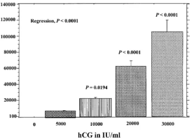
Effects of hCG on VEGF secretion in human CTB
CTB were incubated for 4 h with raising concentrations of hCG
(0±30 000 IU/ml, n = 4 experiments). Basal VEGF secretion was
111.9 6 0.1 pg/ml. There was a marked dose-dependent increase in
VEGF
165secretion (P < 0.0001) in response to hCG reaching a
maximum of 106 000 6 14 500% of controls (P < 0.0001), when
incubated with 30 000 IU/ml of hCG (Figure 4). When VEGF
secretion was measured at 8 h after incubation with hCG, VEGF
secretion was dose-dependently increased 10-fold above controls
(P < 0.0001). At 24 h after incubation, we did not ®nd any effect of
hCG on trophoblastic VEGF secretion (results not shown).
Discussion
An appropriate blood supply is essential for the development of the
feto-placental unit. Maternal blood ¯ow in the intervillous space
increases 20-fold during pregnancy due to vasomotor changes of the
distal intramyometrial portions of uteroplacental arteries and the
transformation and dilatation of decidual segments. This results
essentially from the invasive behaviour of cytotrophoblastic cells that
invade and colonize the endometrial spiral arteries allowing for a loss
of their elasticity (Fisher, 2000). The increased blood requirement is
also met by intense angiogenic processes taking place in the fetal
stroma of the placental villi and the maternal endometrium. Abnormal
angiogenesis during early pregnancy may lead to fetal growth
retardation and/or pre-eclampsia (Roberts, 1998). Although several
angiogenic factors have been identi®ed at the feto-maternal interface,
the regulation of this process remains obscure (Folkman and
Klagsbrun, 1987; Smith, 2000).
In the present study we observed an increase of >1000-fold in
trophoblastic VEGF secretion in vitro when CTB were cultured with
physiological concentrations of hCG. This observation is consistent
with other studies where VEGF expression was induced by LH in the
ovary (Geva and Jaffe 2000) and by hCG in human endometrium
(Licht et al., 2001) and in the ovary (Lee et al., 1997). This indicates
that hCG probably plays an important role in the control of
endometrial and placental vascularization by paracrine (on the
endometrium) as well as juxtacrine (on the trophoblast) mechanisms.
Since the stimulatory effect of hCG on VEGF secretion is no longer
observed at 24 h, it is tempting to speculate that hCG favours VEGF
secretion rather than synthesis.
Since leptin stimulates hCG secretion through a GnRH-dependent
pathway (Chardonnens et al., 1999) and increases the pulsatile release
of hCG in ®rst-trimester trophoblastic explants (Islami et al., 2003),
we wondered if this peptide was able to directly or indirectly (through
hCG) modify the expression of VEGF. Despite leptin stimulating
hCG, it inhibited the release of VEGF. Although this effect was
statistically signi®cant and maintained up to 24 h of culture, it was
modest compared with the effect of hCG on VEGF. The physiological
signi®cance of an inhibitory effect of leptin and a stimulatory effect of
hCG on trophoblastic VEGF secretion is far from being understood
particularly since in vivo hCG is produced by syncytiotrophoblast and
not cytotrophoblast as in in-vitro experiments. Thus our results cannot
easily be applied to an in-vivo situation.
It is also interesting to note that, in addition to the stimulatory effect
of leptin on hCG secretion, we observed an inhibitory effect of hCG on
trophoblastic leptin secretion. This effect seems to be maximal at
5000 IU/ml of hCG, possibly because of saturation of hCG receptors.
This new observation suggests that hCG might exert a possible
negative feedback on the trophoblastic release of leptin.
Our in-vitro observations are somewhat dif®cult to reconcile with
in-vivo observations in patients with pre-eclampsia. Indeed, it has
been reported that this pathology increases circulating VEGF
(Sharkey et al., 1996) as well as leptin (Mise et al., 1998). Both of
these increases can be mimicked in vitro by culturing trophoblastic
cells under hypoxic conditions (Taylor at al., 1997; Grosfeld et al.,
2001). In contrast, hCG is decreased under such hypoxic culture
conditions (Esterman et al., 1996). Thus, if these in-vitro results are
also true in vivo, then in pre-eclamptic pregnancies the stimulation of
VEGF by hCG would be reduced. One possible explanation for this
discrepancy is that hypoxia-induced VEGF release can be regarded as
a compensatory mechanism (angiogenesis) that employs a different
pathway from normoxic conditions. However, these in-vitro
experi-ments were carried out with term placentas, where the interactions
between hypoxia, hCG and VEGF could be different from ®rst
trimester trophoblast. During ®rst trimester pregnancy, we would
Figure 4. Release of vascular endothelial growth factor (VEGF) (% of controls) at 4 h from cytotrophoblastic cells incubated for 4 h with a range of concentrations of hCG (profasi). Statistics were performed by analysis of variance and P values refer to differences compared with controls (hCG, 0). Error bars are SEM (n = 4 experiments in duplicate).Figure 3. Release of leptin (% of controls) at 4 h from cytotrophoblastic cells incubated for 4 h with a range of concentrations of hCG (Profasi). Statistics were performed by analysis of variance and P values refer to differences compared with control (hCG, 0). Error bars are SEM (n = 4 experiments in duplicate).
Interactions between VEGF, leptin and hCG
speculate that hypoxia induces hCG secretion, which stimulates
trophoblastic VEGF release. Clearly the physiological interaction
between hCG, leptin and VEGF under hypoxic conditions is a
complex one and remains to be studied in an appropriate model. Our
data support the idea that trophoblastic products such as hCG and
leptin are probably involved in the control of VEGF secretion at the
maternal±fetal interface.
Acknowledgements
We thank Mrs Christine Wuillemin and Mrs Claire Gruffat for their skilful technical help. D.Islami is the recipient of a scholarship from the Novartis Research Foundation.
References
Ahmed, A., Li, X.F., Dunk, C., Whittle, M.J., Rushton, D.I. and Rollason, T. (1995) Colocalisation of vascular endothelial growth factor and its Flt-1 receptor in human placentae. Growth Factors, 12, 235±243.
Ahmed, A., Dunk, C., Ahmad, S. and Khaliq, A. (2000) Regulation of placental vascular endothelial growth factor (VEGF) and placenta growth factor (PIGF) and soluble Flt-1 by oxygen. Placenta, 21, S16±S24. Alon, T., Hemo, I., Itin, A., Peer, J., Stone, J. and Keshet, E. (1995) Vascular
endothelial growth factor acts as a survival factor for newly formed retinal vessels and has implications for retinopathy of prematurity. Nat. Med., 1, 1024±1028.
Athanassiades, A., Hamilton, G.S. and Lala, P.K. (1998) Vascular endothelial growth factor stimulates proliferation but not migration or invasiveness in human extravillous trophoblast. Biol. Reprod., 59, 643±654.
Bischof, P., Friedli, E., Martelli, M. and Campana, A. (1991) Expression of extracellular matrix-degrading metalloproteinases by cultured human cytotrophoblast cells: effects of cell adhesion and immunopuri®cation. Am. J. Obstet. Gynecol., 165, 1791±1801.
Cao, R., Brakenhielm, E., Wahlestedt, C., Thyberg, J. and Cao, Y. (2001) Leptin induces vascular permeability and synergistically stimulates angiogenesis with FGF-2 and VEGF. Proc. Natl. Acad. Sci. USA, 98, 6390±6395.
Chardonnens, D., Cameo, P., Aubert, M.L., Pralong, F.P., Islami, D., Campana, A. and Bischof, P. (1999) Modulation of human cytotrophoblastic leptin secretion by interleukin-1a and 17b-oestradiol and its effect on hCG secretion. Mol. Hum. Reprod., 5, 1077±1082.
Charnock-Jones, D.S., Sharkey, A.M., Boocock, C.A., Ahmed, A., Plevin, R., Ferrara, N. and Smith, S.K. (1994) Vascular endothelial growth factor receptor localisation and activation in human trophoblast and choriocarcinoma cells. Biol. Reprod., 51, 524±530.
Conn, G., Bayne, M.L., Soderman, D.D., Kwok, P.W., Sullivan, K.A., Palisi, T.M., Hope, D.A. and Thomas, K.A. (1990) Amino acid and cDNA sequences of a vascular endothelial cell mitogen that is homologuos to platelet-derived growth factor. Proc. Natl Acad. Sci. USA, 87, 2628±2632. deVries, C., Escobedo, J.A., Ueno, H., Houck, K., Ferrara, N. and Williams, L.T. (1992) The fms-like tyrokinase, a receptor for vascular endothelial growth factor. Science, 255, 989±991.
Esterman, A, Finlay, T.H. and Dancis, J. (1996) The effect of hypoxia on term trophoblast: hormone synthesis and release. Placenta, 17, 217±222. Ferrara, N. (1999) Molecular and biological properties of vascular endothelial
growth factor. J. Mol. Med., 77, 527±543.
Fisher, S.J. (2000) The placenta dilemma. Semin. Reprod. Med., 18, 321±326. Folkman, J. and Klagsbrun, M. (1987) Angiogenic factors. Science, 235,
442±447.
Gerber, H.P., Dixit, V. and Ferrara, N. (1998) Vascular endothelail growth factor induces expression of the antiapoptotic proteins Bcl-2 and A1 in vascular endothelail cells. J. Biol. Chem., 273, 13313±13316.
Geva, E. and Jaffe, R.B. (2000) Role of vascular endothelial growth factor in ovarian physiology and pathology. Fertil. Steril., 74, 429±438.
Grosfeld, A., Turban, S., Andre, J., Causac, M., Challier, J.C., Haugel-de-Mouzon, S. and Guerre-Millo, M. (2001) Transcriptional effect of hypoxia on placental leptin. FEBS Lett., 502, 122±126.
Henson, M.C. and Castracane, V.D. (2000) Leptin in pregnancy. Biol. Reprod., 63, 1219±1228.
Islami, D., Bischof, P. and Chardonnens, D. (2003) Possible interactions between leptin, gonadotrophin releasing hormone (GnRH I and GnRH II) and human chorionic gonadotrophin (hCG). Eur. J. Obstet. Gynecol. Reprod. Biol., in press.
Lee, A., Christenson, L.K., Patton, P.E., Burry, K.A. and Stouffer, R.L. (1997) Vascular endothelial growth factor production by human luteinised granulosa cells in vitro. Hum. Reprod., 12, 2756±2761.
Licht, P., Russu, V. and Wildt, L. (2001) On the role of human chorionic gonadotropin (hCG) in the embryo endometrial microenvironment: implications for differentiation and implantation. Semin. Reprod. Med., 19, 37±47.
Mise, H., Sagawa, N., Matsumoto, T., Yura, S., Nanno, H., Itoh, H., Mori, T., Masuzaki, H., Hosoda, K., Ogawa, Y. and Nakano, K. (1998) Augmented placental production of leptin in pre-eclampsia: possible involvement of placental hypoxia. J. Clin. Endocrinol. Metab., 83, 3225±3229.
Park, H.Y., Kwon, H.M., Lim, H.J., Hong, B.K., Lee, J.Y., Park, B.E., Jang, Y., Cho, S.Y. and Kim, H.S. (2001) Potential role of leptin in angiogenesis: leptin induces endothelial cell proliferation and expression of matrix metalloproteinases in vivo and in vitro. Exp. Mol. Med., 33, 95±102. Plouet, J., Schilling, J. and Gospodarowicz, D. (1989) Isolation and
characterisation of a newly identi®ed endothelail cell mitogen produced by AtT20 cells. EMBO J., 8, 3801±3808.
Reynolds, L.P. and Redmer, D.A. (1995) Utero-placental vascular development and placental function. J. Anim. Sci., 73, 1839±1851. Reynolds, L.P., Killilea, S.D. and Redmer, D.A. (1992) Angiogenesis in the
female reproductive system. FASEB J., 6, 886±892.
Rizk, B., Aboulghar, M., Smitz, J. and Ron-El, R. (1997) The role of vascular endothelial growth factor and interleukins in the pathogenesis of severe ovarian hyperstimulation syndrome. Hum. Reprod. Update, 3, 255±266. Roberts, J.M. (1998) Endothelial dysfunction in pre-eclampsia. Semin.
Reprod. Endocrinol., 16, 5±15.
Sharkey, A.M., Charnock-Jones, D.S., Boocock, C.A., Brown, K.D. and Smith, S.K. (1993) Expression of mRNA for vascular endothelial growth factor in human placenta. J. Reprod. Fertil., 99, 609±615.
Sharkey, A.M., Cooper, J.C., Balmforth, J.R., McLean, J., Clark, D.E. and Charnock-Jones, D.S. (1996) Maternal plasma levels of endothelial growth factor in normotensive pregnancies and in pregnancies complicated by preeclampsia. Eur. J. Clin. Invest., 26, 1182±1185.
Shore, V.H., Wang, T.H., Wng, C.L., Torry, R.J., Caudle, M.R. and Torry, D.S. (1997) Vascular endothelial growth factor, placenta growth factor adn their receptors in isolated human trophoblast. Placenta, 18, 657±665. Sierra-Honigmann, M.R., Nath, A.K., Murakami, C., Garcia-Cardena, G.,
Papapetropoulos, A., Sessa, W.C., Madge, L.A., Schechner, J.S., Savwabb, M.B., Polveriui, P.J. and Flores-Riveros, J.R. (1998) Biological action of leptin as an angiogenic factor. Science, 281, 1683±1686.
Smith, S.K. (2000) Angiogenesis and implantation. Hum. Reprod., 15 (Suppl. 6), 59±66.
Taylor, C.M., Stevens, H., Anthony, F.W. and Wheeler, T. (1997) In¯uence of hypoxia on vascular endothelial growth factor and chorionic gonadotropin production in the trophoblast derived cell lines: Jeg, Jar, and BeWo. Placenta, 18, 451±458.
Terman, B.I., Dougher-Vermazen, M., Carrion, M.E., Dimitrov, D., Armellino, D.C., Gospodorowicz, D. and Bohlen, P. (1992) Identi®cation of the KDR tyrosine kinase as a receptor for vascular endothelial cell growth factor. Biochem. Biophys. Res. Commun., 187, 1579±1586. Vuckovic, M., Ponting, J., Terman, B.I., Niketic, V., Seif, V. and Kumar, S.
(1996) Expression of the vascular endothelial growth factor receptor, KDR, in human placenta. J. Anat., 188, 361±366.
Vuorela, P., Hatva, E., Lymboussaki, A., Kaipainen, A., Joukov, V., Persico, M.G., Alitalo, K. and Halmesmaki, E. (1997) Expression of vascular endothelial growth factor and placenta growth factor in human placenta. Biol. Reprod., 56, 489±494.
Submitted on April 17, 2002; resubmitted on February 5, 2003; accepted on March 28, 2003