Modulation of human cytotrophoblastic leptin secretion by interleukin-1α and 17β-oestradiol and its effect on HCG secretion
6
0
0
Texte intégral
(2) D.Chardonnens et al. the last menstrual period, digested with trypsin, separated from blood cells and syncytia on a discontinuous Percoll gradient and immunopurified by antibody-coated magnetic particles (anti-CD45) (Dyna Beads, Milan, Geneva) in order to eliminate all contaminating leukocytes as previously reported (Bischof et al., 1995). CTB viability was assessed by Trypan Blue (Sigma, Buchs, Switzerland) exclusion and cells diluted to 106 cells/ml. IL-1β, IL-6, TNFα and M-CSF were purchased from R&D Systems (Bu¨hlmann Laboratories, Basle, Switzerland) and oestradiol from Sigma. RhLeptin was puchased from R&D Systems, gonadotrophin-releasing hormone (GnRH) from Dr J.Rivier (The Salk Institute, La Jolla, CA, USA), and Cetrorelix from Organon, Saffikon, Switzerland.. Culture conditions CTB (100 µl, 106 cells/ml, .90% viability by Trypan Blue exclusion) were incubated in duplicates in the presence or absence of IL-1α (0.01–10 ng/ml), TNFα (1–100 ng/ml), M-CSF (0.1–100 ng/ml) (Simo`n et al., 1994) or oestradiol (10–9 to 10–5 mol/l) (Petraglia et al., 1995; Licinio et al., 1998), rhLeptin (1–1000 ng/ml), GnRH (8.5310–10 to 8.5310–8 mol/l) (Bramley et al., 1992), Cetrorelix (0.1–5 µg/ml) or rhLeptin (2 µg/ml) with or without Cetrorelix (0.1 µg/ml). Concentrations of Cetrorelix and rhLeptin were chosen arbitrarily. Incubation was performed in 12-well tissue culture plate (Costar, Cambridge, MA, USA) under a 5% CO2 and 95% air atmosphere in a humid incubator at 37°C. Culture medium was Dulbecco’s modified Eagle’s medium (DMEM; Gibco, Basel, Switzerland) containing 2 mmol/l L-glutamine, 4.2 mmol/l magnesium sulphate, 25 mmol/l HEPES, 1% gentamycin, 1% amphotericin B, 100 µg/ml streptomycin and 100 IU/ml penicillin in the absence of serum. Medium was harvested on day 2 and 4 and the culture stopped on day 4. The supernatants were divided into aliquots and stored at –20°C until assayed. The cells were lysed with 200 µl Triton X-100 (2.5% in water) and stored at –20°C for total cell protein measurements. Duplicate wells were run for each treatment condition and the experiments repeated three times with different CTB preparations.. Figure 1. Effect of interleukin-1 (IL-1β) on cytotrophoblastic cell (CTB) leptin secretion (n 5 6 from three different CTB preparations). Values are expressed as means 6 SEM.. Hormone and protein assays Leptin was measured in the supernatants by an enzyme-linked immunosorbent assay (ELISA) (DRG diagnostics, Cis-Medipro, Vernier, Switzerland) with a sensitivity of 0.2 ng/ml and intra- and inter-assay coefficients of variation of 4.3 and 8.6% respectively. The leptin assay neither cross-reacts with mouse, rat leptin nor with human proteins such as proinsulin, C-peptide, glucagon or insulinlike growth factor I. HCG was measured in the supernatants by a microparticle immunoassay (MEIA) (IMX, Abbot USA) with a sensitivity of 0.1 mIU and intra- and inter-assay coefficients of variation of ,4%. Total cell proteins were measured in the cell lysate by the Bio-Rad protein assay using bovine serum albumin as the standard (Bio-Rad, Mu¨nchen, Germany).. Figure 2. Effect of 17β-oestradiol on cytotrophoblastic cell (CTB) leptin secretion (n 5 6 from three different CTB preparations). Values are expressed as means 6 SEM.. Statistical analysis Results were corrected for protein content of each individual well and expressed as percentage of their respective controls per day (CTB in the absence of cytokines or oestradiol or rhLeptin, mean 6 SEM) or as values per mg/cell protein per day (mean 6 SEM). Statistical analysis were performed by analysis of variance using Fisher’s test on the Statview 4.5 program on a Power Macintosh 7100/66 computer. The different values were compared among them and to the controls.. Effects of oestradiol Figure 2 demonstrates the effects of oestradiol on CTB leptin production. Oestradiol 10–9 to 10–6 mol/l did stimulate CTB leptin production while 10–5 mol/l did not.. Results Effects of cytokines Mean CTB leptin production under basal conditions was 25.2 6 2.6 (ng/mg prot). There was no significant change of 1078. daily basal leptin secretion over the 4-day period. Figure 1 illustrates IL-1β-induced leptin production by CTB. When compared with controls, leptin production was 320 6 16% (P , 0.0001) and 195 6 3.2% (P 5 0.0004) after incubation with 3 and 10 ng/ml of IL-1β respectively. Lower concentrations of this cytokine were without effect. In contrast to IL-1β, 1–100 ng/ml of TNFα or 0.1–100 ng/ml of M-CSF had no significant effect on CTB leptin production (results not shown).. Effects of recombinant human leptin Mean CTB HCG production under basal conditions was 5722 6 1055 mIU/mg prot. There was no significant change of daily basal HCG secretion over a 4- and even an 8-day period of CTB culture (results not shown). RhLeptin stimulated HCG production by CTB in a concentration-dependent manner (P 5 0.0039), as illustrated in Figure 3. When compared with controls, HCG production was 527 6 159% (P , 0.006) after.
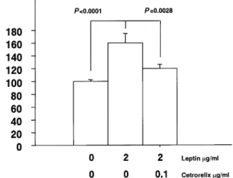
(3) Human cytotrophoblastic leptin secretion. Figure 3. Effect of human recombinant leptin on human chorionic gonadotrophin (HCG) secretion by cytotrophoblastic cells (CTB) (n 5 6 from three different CTB preparations). Values are expressed as means 6 SEM.. Figure 5. Effects of Cetrorelix on leptin-induced human chorionic gonadotrophin (HCG) secretion by cytotrophoblastic cells (CTB) (n 5 6 from three different CTB preparations). Values are expressed as means 6 SEM.. preparations may explain such a result. The addition of Cetrorelix at a concentration of 0.1 µg/ml together with rhLeptin inhibited leptin-induced HCG secretion which was then 119.5 6 6.4% (P 5 0.0028).. Discussion. Figure 4. Effect of gonadotrophin-releasing hormone (GnRH) on human chorionic gonadotrophin (HCG) secretion by cytotrophoblastic cells (CTB) (n 5 6 from three different CTB preparations). Values are expressed as means 6 SEM.. incubation with 1000 ng/ml of rhLeptin. There was no observed syncytium formation when stained with desmoplakin (results not shown).. Effects of GnRH and GnRH antagonists Figure 4 displays the effects of GnRH on HCG secretion by CTB. GnRH 8.5310–8 mol/l increased HCG secretion which was 140 6 21% of controls (P 5 0.031). Cetrorelix did not modify basal HCG secretion. Effects of GnRH antagonists on leptin-induced HCG secretion Figure 5 illustrates the effects of Cetrorelix on CTB HCG secretion induced by rhLeptin (2 µg/ml). In that set of experiments, rhLeptin raised HCG secretion to 160.2 6 13.3% of controls. Leptin-induced HCG secretion was not as high with 2 µg/ml rhLeptin when compared with a 1 µg/ml concentration. However, we know that variability from one cell preparation to the other and different gestational ages between. Circulating peripheral leptin concentrations are elevated during pregnancy (Hardie et al., 1997; Schubring et al., 1997) and the placenta has been suggested to contribute to this pregnancyrelated increase. Several recent reports have demonstrated leptin gene expression by human placental tissue or the placenta-derived cell line BeWo (Matsuzaki et al., 1997; Senaris et al., 1997). However, those only provide indirect evidence of placenta leptin production. This study represents the first direct evidence of leptin secretion by primary cultures of human cytotrophoblast. Little is known about the regulation of leptin secretion in the placenta. Placental age may be important as placental leptin gene expression is higher in the first trimester villi when compared with third trimester villi (Matsuzaki et al., 1997). This fits rather well with longitudinal measurements of peripheral concentrations of leptin during pregnancy which are highest in the second trimester (Hardie et al., 1997). Recently, insulin has been suggested as another important regulator of placental leptin secretion (Lepercq et al., 1998). Another important modulator of leptin secretion in the placenta is cell differentiation. Immunohistochemical studies have demonstrated a more intense staining for leptin in syncytiotrophoblast in comparison with cytotrophoblast (Matsuzaki et al., 1997; Senaris et al., 1997). Moreover, forskolin-induced syncytium formation in BeWo cells was shown to increase leptin secretion (Matsuzaki et al., 1997). In this study, basal leptin secretion remains constant over a 4-day period. This was observed in the absence of serum, a known promoter of syncytium formation. Staining of CTB with desmoplakin after incubation with leptin did not demonstrate syncytium formation (results not 1079.
(4) D.Chardonnens et al.. shown). Taken together, this makes syncytium formation an unlikely hypothesis to explain the results of this study. However, M-CSF has been shown to induce differentiation of cytotrophoblast (Saito et al., 1993). No such results appear to be available for IL-1. This would warrant further investigation. The results of this study indicate that IL-1β is a regulator of leptin secretion in first trimester CTB. Its effect seems to be specific, as neither TNFα nor M-CSF changed leptin secretion. IL-1β is known to stimulate in-vivo leptin secretion in humans (Janik et al., 1997) but it remains unclear whether this is a direct action of IL-1β on the adipocyte or via an endocrine cascade involving corticosteroids. In this study, we provide evidence of a direct effect of IL-1β on CTB leptin secretion. The cellular mechanism of such an effect remains unclear. However, IL-1 receptors are present on the trophoblast (Simo`n et al., 1994). A placenta-specific enhancer element (placental leptin enhancer, or PLE) has recently been identified in the promoter region of the leptin gene (Bi et al., 1997), with at least three protein binding sites. Protein binding to the PLE3 motif up-regulates the transcription of the leptin gene in the human placenta. Since IL-1 exerts similar effects and since, in other cell systems, IL-1 effects are mediated through the NF-κB transcription factor (Friedman et al., 1996; Bird et al., 1997; Reddy et al., 1997), it is tempting to speculate that IL-1β may induce leptin expression through an NF-κBdependent activation of PLE3. The specificity of IL-1β is interesting because the complete IL-1 system is present at the feto–maternal interface and is clearly involved in implantation and placentation (Simo`n et al., 1994). This raises the important question as to whether leptin is also involved in these crucial events. Oestradiol plays a role in the regulation of fetal growth (Abdul-Karim et al., 1991), onset of parturition (Chibbar et al., 1995), placental steroidogenesis (Petraglia et al., 1995, Grimes et al., 1996 ; Babishkin et al., 1997), release of neuropetides (Petraglia et al., 1990), release of glycoproteins (Wilson et al., 1984; Petraglia et al., 1995) and leptin secretion (Sivan et al., 1998). Administration of oestradiol antagonist reverses oestradiol placental actions, pointing towards a receptormediated effect. However, the exact mechanisms of oestradiol action within the placenta are still a matter of debate. Classical cytosol oestradiol receptors seem to be absent or in an extremely low number, thus escaping classical immunological and molecular detection (Rossmanith et al., 1997). This raises the possibility that oestradiol actions within the placenta are mediated by a non-classical membrane-bound receptor. This study demonstrated a concentration-dependant bimodal pattern of oestradiol on the regulation of leptin secretion. There was an increase in leptin secretion of up to 100 nmol/l of oestradiol, which is close to its physiological concentration during late pregnancy (30–50 nmol/l) (Tulchinski et al., 1972). This corroborates in-vivo studies where leptin concentrations were correlated with oestradiol concentrations during pregnancy, especially during the first trimester (Hardie et al., 1997). However, higher doses of oestradiol did not affect basal leptin values. Neither the underlying molecular mechanisms, nor the biological or pharmacological significance of this biphasic pattern of oestradiol response are known. One possible 1080. mechanism for the decrease in leptin secretion at 10 µmol/l of oestradiol may be down-regulation of the oestradiol receptor as it occurs in other reproductive tissues (Medlock et al., 1991; Simerly et al., 1991; Simo`n et al., 1994) and with glucocorticoid and thyroid hormone receptors (Raaka et al., 1981; Rosewicz et al., 1988). No clear role for placental leptin secretion has been demonstrated. So far, cord blood concentrations have been found to be correlated with birth weight and placenta weight (Harigaya et al., 1997; Hassink et al., 1997; Mantzoros et al., 1997; Matsuda et al., 1997; Schubring et al., 1997; Helland et al., 1998; Machini et al., 1998; Tamura et al., 1998), implying that the feto–placental leptin production may be involved in placental and fetal growth regulation. One putative role for leptin could be related to follicular growth, egg and embryo polarity (Antczak and van Blerkom 1997; Cioffi et al., 1997; Edwards and Beard, 1997; Antczak et al., 1998). This study provides the first direct evidence for a definite role of leptin in a fundamental placental function, i.e. HCG production. During pregnancy, trophoblast differentiates in a multistep process that converts cytotrophoblast to syncytiotrophoblast (Ringler et al., 1990). As the placenta grows and matures, there is increased formation of syncytiotrophoblast with an associated secretion of HCG. In models of trophoblast differentiation in vitro, the onset of HCG production appears to parallel the formation of syncytiotrophoblast (Kliman et al., 1986). In this study, basal HCG secretion remained constant over time. This was observed in the absence of serum, a known promoter of syncytium formation and, indeed, cultured CTB showed no syncytium formation. Trophoblast secretion of HCG is regulated by a multitude of endocrine and paracrine/autocrine factors. GnRH (Siler-Khodr et al., 1986), epidermal growth factor (EGF) (Morrish et al., 1987), IL-1 and IL-6 (Masuhiro et al., 1991), leukaemia inhibitory factor (Sawai et al., 1995), TNF (Li et al., 1992), activin (Petraglia et al., 1989) and M-CSF (Saito et al., 1991) are recognized as promoters of HCG secretion while progesterone (Szila`gyi et al., 1993), inhibin (Petraglia et al., 1989), transforming growth factor (Matsuzaki et al., 1992), GnRH antagonist (Siler-Khodr et al., 1983) and RU 486 (Das et al., 1987; Szila`gyi et al., 1993) are inhibitors of HCG secretion. However, fine tuning of HCG secretion during pregnancy is still incompletely understood and the dynamic pattern of HCG may involve an up- and down-regulation of the GnRH receptor (Lin et al., 1995). The results of this study confirm the role of GnRH in HCG secretion with a maximal stimulatory effect at 8.5310–8 mol/l. The need for such a high concentration of GnRH probably reflects the presence of low affinity GnRH receptors within the placenta (Bramley et al., 1992). Changes in intracellular phosphoinositol turnover have been proposed (Conn et al., 1986) as a possible mechanism of GnRH effect. Likewise, GnRH action within the placenta may involve Ca21 ions, similar to the situation described in the pituitary (Mathialagan et al., 1989). A high dose of GnRH may quickly desensitize the local receptors at the post-receptor level. Alternatively, high concentrations of GnRH have been demonstrated to down-regulate GnRH receptor without altering its receptor.
(5) Human cytotrophoblastic leptin secretion. mRNA content (Tstsumi et al., 1990) but with diminished polysome formation (Tsutsumi et al., 1993), raising the possibility that translational modulation may contribute to homologous down-regulation. This may explain why pulsatile GnRH is more efficient in eliciting HCG secretion in the placenta (Barnea et al., 1991). The leptin receptor mRNA has been demonstrated within the murine placenta (Hoggard et al., 1997) and leptin receptor has been demonstrated by immunohistochemistry in the human trophoblast (Castellucci, personal communication). However, it is not yet known which isoform of the leptin receptor predominates in CTB. This receptor seems functional as, in this study, leptin increases HCG secretion in CTB in a concentration-dependent way. Thus, leptin can now be added to the growing list of HCG secretion enhancers. GnRH antagonist Cetrorelix (0.1 µg/ml) inhibited leptin-induced HCG secretion. Taken together, the results suggest that the GnRH pathway may be involved in leptin-induced HCG secretion which could be put into perspective with the release of leptininduced luteinizing hormone-releasing hormone (LHRH) from the median eminence from male rats and luteinizing hormone (LH) release from their anterior pituitaries which is thought to be mediated by nitric oxide (Yu et al., 1997). However, the full-length leptin receptor has the signalling capabilities of the IL-6-type cytokine receptors (Baumann et al., 1996), and structural predictions indicate that leptin would be expected to fold into an IL-6-like structure (Madej et al., 1995) and that IL-6 would stimulate HCG secretion. These considerations can only partly explain the effects of leptin on HCG secretion since IL-6-induced HCG secretion seems different from GnRHinduced HCG secretion (Matsuzaki et al., 1992). Moreover, IL-1, a known activator of HCG secretion, acts via IL-6 and the IL-6 receptor (Masuhiro et al., 1991), and we have also observed that it stimulates trophoblast leptin secretion. Thus, the precise mechanism underlying the action of leptin on HCG secretion needs further investigation. In conclusion, these data demonstrate that human cytotrophoblastic cells secrete leptin and that IL-1α and oestradiol are important factors involved in the regulation of leptin secretion. Moreover, these results demonstrate a clear and important role of leptin in HCG secretion by CTB. GnRH appears to be involved in the pathway leading to leptin-induced HCG secretion but further investigations are required.. Acknowledgements We thank Mrs Claire Gruffat for performing the MEIA and the ELISA. D.Islami is the recipient of an Ernest Schering Foundation award.. References Abdul-Karim, R.W., Nesbitt, R.E.L., Druck, M.H. and Rizk, P.T. (1991) The regulatory effects of oestrogens on fetal growth. Am. J. Obstet. Gynecol., 109, 656–662. Antczak, M. and van Blerkom, J. (1997) Oocyte influences on early development: the regulatory proteins leptin and STAT3 are polarized in mouse and human oocytes and differentially distributed within the cells of the preimplantation stage embryo. Mol. Hum. Reprod., 3, 1067–1086. Antczak, M., Van Blerkom, J. and Clark, A. (1998) A novel mechanism of vascular endothelial growth factor, leptin and transforming growth factor-. beta2 sequestration in a subpopulation of human ovarian follicle cells. Hum. Reprod., 12, 2226–2234. Babishkin, J.S., Grimes, R.W., Pepe, G.J. and Albrecht E.D. (1997). Estrogen stimulation of P450 side-chain cleavage activity in cultures of human placental syncytiotrophoblast. Biol. Reprod., 56, 272–278. Barnea, E.R., Kaplan, M. and Naor, Z. (1991) Comparative stimulatory effect of gonadotrophin releasing hormone (GnRH) and GnRH agonist upon pulsatile human chorionic gonadotrophin secretion in superfused placental explants : reversible inhibition by a GnRH antagonist. Hum. Reprod., 6, 1063–1069. Baumann, H., White, K.K., White, D.W. et al. (1996) The full-length leptin receptor has signaling capabilities of interleukin 6-type cytokine receptors. Proc. Natl Acad. Sci. USA, 93, 8374–8378. Bi,S., Gavrilova, O., Gong, D.W. et al. (1997) Identification of a placental enhancer for the human leptin gene. J. Biol. Chem., 272,30583–30588. Bird, T.A., Schooley, K., Dower, S.K. et al. (1997) Activation of nuclear transcription factor NF-kappaB by interleukin-1 is accompanied by casein kinase II-mediated phosphorylation of the p65 subunit. J. Biol. Chem., 272, 32606–32612. Bischof, P., Haenggeli, L. and Campana, A. (1995) Gelatinase and oncofetal fibronectin secretion are dependent upon integrin expression on human cytotrophoblasts. Hum. Reprod., 10, 734–742. Bramley, T.A., McPhie, C.A. and Menzies, G.S. (1992) Human placental gonadotrophin-releasing hormones (GnRH) binding sites: I. Characterization, properties and ligand specificty. Placenta, 13, 555–581. Bray, G.A. and York, D.A. (1997) Leptin and clinical medicine: a new piece in the puzzle of obesity. J. Clin. Endocrinol. Metab., 82, 2771–2776. Chehab, F.F., Lim, M.E. and Lu, R. (1996) Correction of sterility defect in homzygous obese female mice by treatment with the recombinant leptin. Nature Genet., 12, 318–20. Chibbar, R., Wong, S., Miller, F.D. and Mitchell, B.F. (1995) Estrogen stimulates oxytocin gene expression in human chorio-decidua. J. Clin. Endocrinol. Metab., 80, 567- 572. Cioffi, J.A., van Blerkom, J., Antczak, M. et al. (1997) The expression of leptin and its receptors in pre-ovulatory human follicles. Mol. Hum. Reprod., 3, 467–472. Conn, P.M., Staley, D., Harris, C. et al. (1986) Mechanism of action of gonadotrophin releasing hormone. Ann. Rev. Physiol., 48, 495–513. Das, C. and Catt, K.J. (1987) Antifertility actions of the progesterone antagonist RU 486 include direct inhibition of placental hormone secretion. Lancet, ii, 599–601. Edwards, R.G. and Beard, H.K. (1997) Oocyte polarity and cell determination in early mammalian embryos. Mol. Hum. Reprod., 3, 863–905. Friedman, W.J., Thakur, S., Seidman, L. and Rabson, A.B. (1996) Regulation of nerve growth factor mRNA by interleukin-1 in rat hippocampal astrocytes is mediated by NFkappaB. J. Biol. Chem., 271, 31115–31120. Grimes, R.W., Pepe, G.J. and Albrecht, E.D. (1996) Regulation of human placental trophoblast low-density lipoprotein uptake in vitro by estrogen. J. Clin. Endocrinol. Metab., 81, 2675–2679. Hardie, L., Trayhurn, P., Abramovich, D. and Fowler, P. (1997) Circulating leptin in women: a longitudinal study in the menstrual cycle and during pregnancy. Clin. Endocrinol., 47, 101–106. Harigaya, A., Nagashima, K., Nako, Y. and Morikawa, A. (1997) Relationship between concentrations of serum leptin and fetal growth. J. Clin. Endocrinol. Metab., 82, 3281–3284. Hassink, S.G., de Lancey, E., Sheslow, D.V. et al. (1997) Placental leptin: an important new growth factor in intrauterine and neonatal development? Pediatrics, 100, E1. Helland, I.B., Reseland, J.E., Sugstad, O.D. and Drevon. C.A. (1998) Leptin levels in pregnant women and newborn infants: gender differences and reduction during the neonatal period. Pediatrics, 101, E12. Hoggard, N., Hunter, L., Duncan, S.J. et al. (1997) Leptin and leptin receptor mRNA and protein expression in the murine fetus and placenta. Proc. Natl Acad. Sci. USA, 94, 11073–11078. Janik, J.E., Curti, B.D., Considine, R.V. et al. (1997) Interleukin 1β increases serum leptin concentrations in the human. J. Clin. Endocrinol. Metab., 82, 3084–3086. Jokhi, P.P., King, A., Boocock, C. and Loke, Y.W. (1995) Secretion of colony stimulating factor-1 by human first trimester placental and decidual cell populations and the effect of this cytokine on trophoblast thymidine uptake in vitro. Hum. Reprod., 10, 2800–2807. Kliman, H.J., Nestler, J.E., Sermasi, E. et al. (1986) Purification characterization, in vitro differentiation of cytotrophoblasts from human term placentae. Endocrinology, 118, 1567–1582.. 1081.
(6) D.Chardonnens et al. Lepercq, J., Cauzac, M., Lahlou, N. et al. (1998) Overexpression of placental leptin in diabetic pregnancy. Diabetes, 47, 847–850. Li, Y., Matsuzaki, N., Masuhiro, K. et al. (1992) Trophoblast-derived tumor necrosis factor-alpha induces release of human chorionic gonadotrophin using interleukin-6 (IL-6) and IL-6-receptor-dependent system in the normal human trophoblasts. J. Clin. Endocrinol. Metab., 74, 184–191. Licinio, J., Negrao, A.B., Mantzoros, C. et al. (1998) Synchronicity of frequently sampled, 24-h concentrations of circulating leptin, luteinizing hormone, and estradiol in healthy women. Proc. Natl Acad. Sci. USA, 95, 2541–2546. Lin, L.S., Roberts, V.J. and Yen, S.S. (1995) Expression of gonadotropinreleasing hormone receptor gene in the placenta and its functional relationship to human chorionic gonadotropin secretion. J. Clin. Endocrinol. Metab., 80, 580–584. Machini, G., Fried, G., Ostlund, E. and Hagenas, L. (1998) Plasma leptin in infants: relations to birth weight and weight loss. Pediatrics, 101, 429–432. Madej, T., Boguski, M.S. and Bryant, S.H. (1995) Threading analysis suggests that the obese gene product may be a helical cytokine. FEBS Lett., 373, 13–18. Mantzoros, C.S., Varvarigou, A., Kaklamani, V.G. et al. (1997) Effect of birth weight and maternal smoking on cord blood leptin concentrations of fullterm and preterm newborns. J. Clin. Endocrinol. Metab., 82, 2856–2861. Mantzoros, C.S., Moschos, S., Avramopoulos, I. et al. (1997) Leptin concentrations in relation to body mass index and the tumor necrosis factorα system in the humans. J. Clin. Endocrinol. Metab., 82, 3408–3413. Masuhiro, K., Matsuzaki, N., Nishino, E. et al. (1991a) Trophoblast-derived interleukin-1 (IL-1) stimulates the release of human chorionic gonadotrophin by activating IL-6 and IL-6 receptor system in first trimester human trophoblasts. J. Clin. Endocrinol. Metab., 72, 594–601. Mathialagan, N. and Rao, A.J. (1989) A role of calcium in gonadotrophinreleasing hormone (GnRH) stimulated secretion of chorionic gonadotrophin by first trimester human placental minces in vitro. Placenta, 10, 61–70. Matsuzaki, N., Li, Y., Masuhiro, K. et al. (1992) Trophoblast-derived transforming growth factor α1 suppresses cytokine-induced, release of human chorionic gonadotropin by normal human trophoblasts. J. Clin. Endocrinol. Metab., 74, 211–216. Matsuzaki, H., Ogawa, Y., Sagawa, N. et al. (1997) Nonadipose tissue production of leptin: leptin as a novel placenta-derived hormone in the humans. Nature Med., 3, 1029–1033. Matsuda, J., Yokota, I., Iida, M. et al. (1997) Serum leptin concentration in cord blood: relationship to birth weight and gender. J. Clin. Endocrinol. Metab., 82, 1642–1644. Medlock, K.L., Lyttle, C.R., Kelepouris, N. et al. (1991) Estradiol down regulation of the rat uterine estrogen receptor. Proc. Soc. Exp. Biol. Med., 196, 293–300. Morrish, D.W., Bhardwaj, D., Dabbagh, L.K. et al. (1987) Epidermal growth factor induces differentiation and secretion of human chorionic gonadotropin and placental lactogen in normal human placenta. J. Clin. Endocrinol. Metab., 65, 1282–1290. Omigbodum, A., Coukos, G., Ziolkiewicz, P. et al. (1998) Macrophage-colony stimulating factor (M-CSF) regulates the expression of fibronectin and its alpha5 integrin receptor in human trophoblasts. Endocrinology, 139, 2190–2193. Petraglia, F., Vaughan, J. and Vale, W. (1989) Inhibin, activin modulate the release of gonadotropin-releasing hormone, human chorionic gonadotropin, progesterone from cultured human placental cells. Proc. Natl. Acad. Sci. USA, 86, 5114–5117. Petraglia, F.J., Vaughan, J. and Vale, W. (1990) Steroid hormones modulate the release of immunoreactive gonadotropin-releasing hormone from cultured human placental cells. J. Clin. Endocrinol. Metab., 70,1173–1178. Petraglia, F., de Micheroux, A.A., Florio, P. et al. (1995) Steroid-protein interaction in the human placenta. J. Steroid. Biochem. Mol. Biol., 53, 227–231. Raaka, B.M. and Samuels, H.H. (1981) Regulation of thyroid hormone nuclear receptor levels in GH1 cells by, 3,5,39-triiodo-l-thyronine. J. Biol. Chem., 256, 6883–6889. Reddy, S.A., Huang, J.H. and Liao, W.S. (1997) Phosphatidylinositol 3kinase in interleukin-1 signaling. Physical interaction with the interleukin1 receptor and requirement in NFkappaB and AP-1 activation. J. Biol. Chem., 272, 29167–29173. Ringler, G.E. and Strauss, J.F. (1990) In vitro systems for the study of human placental endocrine function. Endocr. Rev., 11, 105–123. Rosewicz, S., McDonald, A.R., Maddux, B.A. and Logsdon, C.D. (1988) Mechanism of glucocorticoid receptor down regulation by glucocorticoids. J. Biol. Chem., 263, 2581–2584.. 1082. Rossmanith, W.G., Wolfahrt, S., Ecker and Eberhardt, E. (1997) The demonstration of progesterone, but not of estrogen receptors in the developing human placenta. Horm. Metab. Res., 29, 604–610. Saito, S., Saito, M., Motoyoschi, K. and Ichijo, M. (1991) Enhancing effects of human macrophage colony-stimulating factor on the secretion of human chorionic gonadotropin by human chorionic villous cells, tP-30 cells. Biochem. Biophys Res. Commun., 178, 1099–1104. Saito, S., Saito, M., Enomot, M. et al. (1993) Human macrophage colonystimulating factor induces the differentiation of trophoblast. Growth Factors, 9, 11–19. Sawai, K., Matsuzaki, N., Kameda, T. et al. (1995) Leukemia inhibitory factor produced at the fetomaternal interface stimulates chorionic gonadotropin production: its possible implication during pregnancy, including implantation period. J. Clin. Endocrinol. Metab., 80, 1449–1456. Schubring, C., Kiess, W., Englaro, P. et al. (1997) Levels of leptin in maternal serum, amniotic fluid and arterial and venous cord blood: relation to neonatal and placental weight. J. Clin. Endocrinol. Metab., 82, 1480–1483. Senaris, R., Garcia-Caballero, T., Casabiell, X. et al. (1997) Synthesis of leptin in human placenta. Endocrinology, 138, 4501–4504. Siler-Khodr, T.M., Khodr, G.S., Vickery, B.H. and Nestor, J.J. (1983) Inhibition of hCG, ahCG, progesterone release from human placental tissue in vitro by a GnRH antagonist. Life Sci., 32, 2741–2745. Siler-Khodr, T.M., Khodr, G.S., Valenzuela, G. and Rhode, J. (1986) Gonadotropin-releasing hormone effects on placental hormone during gestation: 1. alpha-human chorionic gonadotropin, human chorionic gonadotropin, human somatomammotropin. Biol. Reprod., 34, 245–254. Simerly, R.B. and Young, B.J. (1991) Regulation of estrogen receptor mRNA in rat hypothalamus by sex steroid hormones. Mol. Endocrinol., 5, 424–432. Simo`n, C., Frances, A., Piquette, G. et al. (1994) Interleukin-1 system in the materno-trophoblast unit implantation: Immunohistochemical evidence for autocrine/paracrine function. J. Clin. Endocrinol. Metab., 78, 847–854. Sivan, E., Whittaker, P.G., Sinha, D. et al. (1998) Leptin in human pregnancy: the relationship with gestational hormones. Am. J. Obstet. Gynecol., 179, 1128–1132. Szila`gyi, A., Benz, R. and Rossmanith, W.G. (1993) Human chorionic gonadotropin secretion from the early human placenta : in vitro regulation by progesterone and its antagonist. Gynecol. Endocrinol., 7, 241–250. Tamura, T., Goldenberg, R.L., Johnston, K.E. and Cliver, S.P. (1998) Serum leptin concentrations during pregnancy and their relationship to fetal growth. Obstet. Gynecol., 91, 389–395. Tartaglia, L.A. (1997) The leptin receptor. J. Biol. Chem., 272, 6093–6096. Tulchinsky, D., Hobel, C.J., Yeager, E. and Marshall, J.R. (1972) Plasma estrone, estradiol, estriol, progesterone and 17-hydroxyprogesterone in human pregnancy. Am. J. Obstet. Gynecol., 112, 1095–1100. Tsutsumi, M., Mellon, P.L., Roberts, J.L. and Sealfon, S.C. (1990) GnRH decreases GnRH receptor mRNA activity in a rodent pituitary cell line. Soc. Neurosci. Abstr., 16, 393. Tsutsumi, M., Laws, S.C., Radic, U. and Sealfon, S.C. (1993) Translational regulation of the gonadotrophin-releasing hormone receptor in aT3–1 cells. Endocrinology, 136, 1128–1136. Wilson, E.A., Jawad, M.J. and Powel, D.E. (1984) Effect of estradiol and progesterone on human chorionic gonadotropin secretion in vitro. Am. J. Obst. Gynecol., 149, 143–148. Yanushpolski, E.H., Ozturk, M., Polgar, K. et al. (1993) The effects of cytokines on human chorionic gonadotrophin (hCG) production by a trophoblast cell line. J. Reprod. Immunol., 25, 235–247. Yu, W.H., Walczewska, A., Karanth, S. and McCann, S.M. (1998) Nitric oxide mediates leptin-induced luteinizing hormone-releasing hormone (LHRH) and LHRH and leptin-induced LH release from the pituitary gland. Endocrinology, 138, 5055–5058. Zhang, Y., Proenca, R., Maffei, M. et al. (1994) Positional cloning of the mouse obese gene and its human homologue. Nature, 372, 425–432. Zumbach, M.S., Boehme, M.W.J., Wahl, P. et al. (1997) Tumor necrosis factor increases serum leptin levels in humans. J. Clin. Endocrinol. Metab., 82, 4080–4082. Received on January 18, 1999; accepted on August 9, 1999.
(7)
Figure

Documents relatifs