Synergism between vascular endothelial growth factor and placental
growth factor contributes to angiogenesis and plasma extravasation in
pathological conditions
PETER CARMELIET1,LIEVE MOONS1,AERNOUT LUTTUN1,VALERIA VINCENTI2,VEERLE COMPERNOLLE1,MARIA
DE MOL1,YAN WU3,FRANÇOISE BONO4,LAETITIA DEW5,HEIKE BECK6,DIMITRI SCHOLZ7,TILL ACKER6,TINA
DIPALMA2,MIEKE DEWERCHIN1,AGNES NOEL5,INGEBORG STALMANS1,ADRIANO BARRA2,SYLVIA BLACHER5, THIERRY VANDENDRIESSCHE1,ANNICA PONTEN9,ULF ERIKSSON9,KARL H.PLATE6,JEAN-MICHEL FOIDART5,
WOLFGANG SCHAPER7,D.STEPHEN CHARNOCK-JONES8,DANIEL J.HICKLIN3,JEAN-MARC HERBERT4,DÉSIRÉ
COLLEN1&M.GRAZIELLA PERSICO2
1 The Center for Transgene Technology and Gene Therapy, Flanders Interuniversity Institute for Biotechnology, KU Leuven, Leuven,
Belgium
2lstituto Internazionale di Genetica e Biofisica, CNR, Naples, Italy 3ImClone Systems, New York, New York, USA
4Cardiovascular/Thrombosis Research Department, Sanofi-Synthélabo, Toulouse, France, 5Laboratory of Tumor and Developmental Biology, University Liège, Sart-Tilman, Belgium 6Department of Neuropathology, FAU Erlangen-Nürnberg, Erlangen, Germany
7Department of Experimental Cardiology, Max-Planck-Institute, Nauheim, Germany
8Reproductive Molecular Research Group, Department Obstetrics & Gynaecology, University of Cambridge, Cambridge, UK 9Ludwig Institute for Cancer Research, Stockholm, Sweden
Abstract: Vascular endothelial growth factor (VEGF) stimulates angiogenesis by activating VEGF receptor-2 (VEGFR-2). The role of its homolog, placental growth factor (PlGF), remains unknown. Both VEGF and PlGF bind to VEGF receptor-1 (VEGFR-1), but it is unknown whether VEGFR-1, which exists as a soluble or a membrane-bound type, is an inert decoy or a signaling receptor for PlGF during angiogenesis. Here, we report that embryonic angiogenesis in mice was not affected by deficiency of PlGF (Pgf-/-). VEGF-B, another ligand of
VEGFR-1, did not rescue development in Pgf-/- mice. However, loss of PlGF impaired angiogenesis, plasma
extravasation and collateral growth during ischemia, inflammation, wound healing and cancer. Transplantation of wild-type bone marrow rescued the impaired angiogenesis and collateral growth in Pgt-/- mice, indicating that
PlGF might have contributed to vessel growth in the adult by mobilizing bone-marrow-derived cells. The synergism between PlGF and VEGF was specific, as PlGF deficiency impaired the response to VEGF, but not to bFGF or histamine. VEGFR-1 was activated by PlGF, given that anti-VEGFR-1 antibodies and a Src-kinase inhibitor blocked the endothelial response to PlGF or VEGF/PlGF. By upregulating PlGF and the signaling subtype of VEGFR-1, endothelial cells amplify their responsiveness to VEGF during the 'angiogenic switch' in many pathological disorders.
Vascular endothelial growth factor (VEGF) stimulates angiogenesis by activating the VEGF tyrosine kinase receptor-2 (VEGFR-2/KDR)1,2. Little is known, however, about other VEGF homologs such as placental growth
factor (PlGF)3. Indirect evidence points to a role for PlGF in placental development4, whereas recombinant PlGF
stimulates angiogenesis in particular conditions and induces vascular permeability when co-injected with VEGF (refs. 5,6), but the role of endogenous PlGF remains unknown7,8. PlGF, VEGF and another homolog, VEGF-B,
all bind to VEGF receptor-1 (VEGFR-1/Flt-1)9,10. Loss of VEGFR-1 disrupted normal vascular development11,
but deletion of its tyrosine kinase domains allowed normal embryonic angiogenesis12, indicating that VEGFR-1
might function as an inert 'decoy' by binding VEGF and thereby regulating the availability of VEGF for activation of VEGFR-2. Such a decoy function might be particularly attributed to the soluble VEGFR-1, an alternatively processed type of VEGFR-1 which contains the extracellular ligand-binding domains, but lacks the signaling tyrosine kinase domains13,14.
PlGF has been proposed to stimulate angiogenesis by displacing VEGF from the 'VEGFR-1 sink', thereby increasing the fraction of VEGF available to activate VEGFR-2 (ref. 6). Alternatively, PlGF might stimulate angiogenesis by transmitting intracellular signals through VEGFR-1 (refs. 9,10,16). However, VEGFR-1 has weak tyrosine kinase activity and its intracellular signals are not well understood. PlGF might also affect angiogenesis by forming heterodimers with VEGF, but their role is controversial16,17. Due to lack of available
inhibitors of PlGF or VEGFR-1, their role in pathological angiogenesis could not be demonstrated thus far. To examine the role of PlGF in angiogenesis, we inactivated the gene expressing PlGF (Pgf) in mice.
Unexpectedly, the absence of PlGF—even in combination with a loss of VEGF-B—had a negligible effect on vascular development. However, PlGF deficiency reduced pathological angiogenesis, permeability and collateral
growth in ischemia, inflammation and cancer. These genetic studies unveil a synergism between PlGF and VEGF in pathological angiogenesis, which might have therapeutic implications for either stimulating or inhibiting angiogenesis.
Fig. 1 Normal vascular development but impaired tumor angiogenesis in Pgf-/-mice, a, Western blotting for VEGFR-1. Membrane-associated VEGFR-1 (Mr ~190-200 kD) represents only 2% and 30% of total VEGFR-1 in placenta and ischemic myocardium, respectively. Soluble VEGFR-1 (Mr ~100-110kD). Lanes 1-2: placental extract at E9.5 and E.15.5. Lane 3: E15.5 placental extract, mixed with VEGF and PlGF and immuno-precipitated using anti-VEGF and anti PlGF anti-bodies. Lane 4, Myocardial infarct (Ml) extract, b-d, Labeling of vitelline blood vessels upon binding of [125I]VEGF
165 to E9.5 yolk-sac sections (b). Excess cold VEGF (d) but not PlGF (c)
almost completely eliminated labeling, e and f, Toluidine blue-stained section of a corpus luteum, revealing more numerous and larger vessels in wild-type (e) than in Pgf-/- (f) mice. For clarity, vessels were traced by white lines, g-i, Macroscopic pictures of wild-type (g), Pgf
-/-(h) and Vegf-/- (i) ES-cell-derived tumors, revealing the hemorrhagic (red) appearance of the large wild-type tumor, in contrast to the small
hypovascular (white) Pgf' and Vegf-/- tumors. j, Densities (vessels/optical field) of vessels of different size in wild-type tumors in wild-type
mice, and Pgf-/- or Vegf-/- tumors in Pgf-/- mice. wild-type; Pgf-/-; Vegf-/-. *, P< 0.05 versus wild-type. k and i Endoglin
immunostaining, revealing large vessels in wild-type fibrosarcoma (k) but only small capillaries in Pgf-/- fibrosarcoma (l). Scale bars: 25 µm
(b-f), 4 mm (g-i) and 50 µm (k and l).
Normal embryogenesis and subtle vascular defects in Pgf-/- mice
We inactivated Pgf by deleting exons 3-6 and confirmed the deletion by Southern- and northern-blot analyses and ELISA (< 0.5 pg PlGF/mg in Pgf-/- mice; data not shown). Pgf-/- mice from Pgf-/- or Pgf-/- breeding pairs were born at a mendelian frequency and were healthy and fertile. Although PlGF was expressed in wild-type embryos at embryonic day (E)10.5 (33 ± 7 pg/mg protein; n = 6; mean ± s.e.m.), vascular development in Pgf-/- embryos was normal (data not shown). PlGF levels were 60% lower, however, than VEGF levels (81 ± 4 pg/mg; n = 8) and PlGF was only 10% as potent as VEGF in enlarging embryonic vessels after intracardial injection in cultured E8.5 wild-type embryos (data not shown).
VEGFR-1 is the only known signaling receptor for PlGF. However, western-blot (Fig. 1a), radioligand binding (Fig. 1b-d) and quantitative reverse transcriptase (RT)-PCR (data not shown) analyses revealed that most VEGFR-1 in the embryo from E9.5 onwards was present as a soluble form. Consistent with an inhibitory decoy role of VEGFR-1, intracardial injection of VEGFR-1 antibodies in early-stage embryos stimulated vascular development, whereas VEGFR-2 antibodies blocked it (data not shown). Although more VEGF would be bound by VEGFR-1 in the absence of PlGF, plasma VEGF levels were not reduced in Pgf-/- embryos (-65 pg/ml in
wild-type embryos versus -84 pg/mg in Pgf-/- embryos at E15.5). This might be due to a compensatory increase
in production of VEGF in Pgf-/- embryos (81 ± 4 pg/mg in wild-type embryos versus 118 ± 11 pg/mg in Pgf
-/-embryos at E10.5; n = 8; P < 0.01). Other angiogenic molecules (VEGFR-1, VEGF-B, VEGF-C and angiopoietin-1) were comparably expressed in Pgf-/- embryos (data not shown).
VEGF-B is another ligand of VEGFR-1 (ref. 18), but loss of VEGF-B (Vegfb-/-) does not affect vascular
development19. Although VEGF-B could have rescued vascular development in Pgf-/- mice, Vegfb-/- Pgf-/- mice
were born at a normal mendelian inheritance, were apparently healthy and fertile, had a normal life span and were without obvious vascular defects (data not shown). This indicates that the individual VEGFR-1 ligands may have distinct functions in angiogenesis.
We observed no structural vascular defects in Pgf-/- mice after birth, except for a subtle remodeling defect of
retinal vessels (data not shown). Fewer vessels developed in corpora lutea at 4.5 days of pregnancy in Pgf-/- mice
(vessels/mm2: 910 ± 62 in 6 wild-type mice versus 430 ± 80 in 6 Pgf-/- mice; P< 0.005; Fig. 1e and f), but did not
impair reproduction. Thus, loss of PlGF impaired VEGF-dependent retinal and luteal angiogenesis.
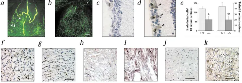
Fig. 2 Impaired retinal and myocardial angiogenesis in Pgf mice, a and b, Impaired ischemic retinal angiogenesis in Pgf mice (fluoro-angiogra-phy). Numerous foci of neovascularization (arrowheads) and large dialated and tortuous retinal vessels in wild-type (a) but not in Pgf mice (b). c and d, Immunostaining of VEGFR-1 in a wild-type mouse, revealing minimal expression during hyperoxia (P12; c) and significant upregulation in retinal vessels (arrowheads) after 1 d of ischemia (P13; d). e, Loss of PlGF reduces the number of endothelial cells and neovascular tufts in the vitreous cavity. *, P < 0.05 versus wild-type in the vitreous cavity, f and g, Thrombomodulin (TM) staining of endothelium, revealing more numerous vessels in wild-type (f) than in Pgf (g) myocardial infarcts, h and i, In situ hybridization revealing undetectable PlGF expression in normal myocardium (h) and upregulation of PlGF in ischemic cardiomyocytes and in vessels infiltrating the infarct (h and i; wild-type mice).j and k, Immunostaining revealing that VEGFR-1 expression is undetectable in quiescent myocardial vessels (j), but induced in angiogenic vessels infiltrating the infarct (j and k; wild-type mice). Scale bars: 200 µm (a and b), 25 µm (c and d), 50 µm (f, g and i-k) and 100µm (h).
Impaired tumor angiogenesis in Pgf-/- mice
PlGF is variably upregulated in tumors8.20-22 and its role in tumor angiogenesis remains ill defined. Therefore we
grew embryonic stem (ES)-cell-derived tumors of either genotype in athymic nu/nu wild-type or Pgf-/- mice for
four weeks. When both tumor and host expressed PlGF, tumors were large and highly vascularized (4 ± 1 g; n = 8; Fig. 1g and j). In contrast, when they both lacked PlGF, tumors remained small and poorly vascularized (1.0 ± 0.3 g; n = 8; Fig. 1h and j), comparable to the reduced growth and angiogenesis of VEGF-deficient tumors (0.3 ± 0.1 g; n = 10; Fig. 1 i and j). When either tumor or host produced PlGF, tumor growth and angiogenesis were intermediate, indicating that production of PlGF by tumor- and host-derived tissue contributed to tumor angiogenesis. Coverage of tumor vessels by smooth muscle cells was not affected by Pgf genotype. We confirmed the role of PlGF in tumor angiogenesis using ras-transformed fibroblasts. Pgf-/- fibrosarcomas were
smaller than wild-type fibrosarcomas when implanted in nu/nu wild-type mice (2.8 ± 0.2 g versus 5.5 ± 0.6 g; ∏
= 7; P < 0.05), and, although vascular densities were comparable in both tumors (~550 vessels/mm2), Pgf
-/-fibrosarcomas contained vessels with a smaller lumen than wild-type -/-fibrosarcomas (110 ± 18 µm2 versus 210 ±
12 µm2; n = 7; P < 0.05; Fig. 1k and 1). As revealed by in situ hybridization analysis of wild-type
fibrosarcomas, PlGF was expressed in endothelial and tumor cells, whereas VEGFR-1 was highly expressed in tumor vessels (data not shown). Wild-type tumors expressed large amounts of PlGF (85 ± 12 pg/mg protein; n = 5) and VEGF (430 ± 40 pg/mg protein; n = 5). However, reduced tumor angiogenesis in Pgf-/- tumors did not
result from reduced expression of VEGF, VEGF-B or VEGF-C (data not shown). Thus, loss of PlGF impaired growth of VEGF-dependent tumors.
Fig. 3 Impaired pathological arteriogenesis in Pgf-/-mice, a, Time course of skin wound healing. wild-type; Pgt'~ mice; n = 15 mice;
P < 0.05 versus wild-type, b and c, Macroscopic view of impaired skin wound healing in Pgf-/- (c) as compared with wild-type (b) mice, d
and e, Smooth muscle α-actin (SMA) staining, revealing impaired coverage of newly formed vessels by smooth muscle cells (arrowheads) in Pgf-/- (e) as compared with wild-type (d) mice, f and g, PlGF immunostaining of human skin, revealing minimal expression in normal skin (ƒ)
and upregulation during chronic eczema (g). Scale bars: 4 mm (b and c), 50 µm (d-g).
Reduced angiogenesis in ischemia in Pgf-/- mice
During ischemic retinopathy, increased VEGF levels stimulate vitreous neovascularization. Previous studies reported minimal changes in PlGF transcripts23 or increased PlGF protein levels24, but the role of PlGF in retinal
neovascularization remains unknown. We induced capillary dropout in the retina by exposing seven-day-old (P7) mice to 80% oxygen for five days. Upon return to normoxia at P12, the hypovascular retina became ischemic, upregulated VEGF and induced venous dilation, arterial tortuosity and capillary growth in the vitreous chamber (Fig. 2a and e). Loss of PlGF significantly protected mice against intravit-reous neovascularization (n = 19; P < 0.005; Fig. 2b and e), venous dilatation and arterial tortuosity (Fig. 2 b). During retinal ischemia, VEGFR-1 was induced in vascular sprouts (Fig. 2 c and d), whereas VEGF and PlGF were significantly upregulated (pg VEGF/mg protein: 98 ± 12 at P12 versus 850 ± 18 at P13; n = 7; P < 0.005 and pg PlGF/mg protein: 10 ± 2 at P12 versus 38 ± 3 at P13; n = 3; P < 0.05). VEGF levels were comparable, however, in both genotypes (data not shown).
We analyzed revascularization of myocardial infarcts after ligating the left anterior descending coronary artery25.
Compared with wild-type mice, Pgf-/- infarcts contained approximately 20% fewer vessels (vessels/mm2: 134 ± 9
in wild-type mice versus 111 ± 5 in Pgf-/- mice; n = 12 to 24; P< 0.05; Fig. 2f and g). VEGF and PlGF levels
(expressed per mg protein) were increased in infarct borders where angiogenesis occurs: normal myocardium produced 22 ± 2 pg VEGF and 8 ± 1 pg PlGF, whereas infarcts produced 52 ± 6 pg VEGF and 102 ± 8 pg PlGF
(n = 5; P< 0.05). Expression of VEGF, VEGFR-2 and endoglin in infarcts was comparable in both genotypes (data not shown). PlGF and VEGFR-1 were undetectable in quiescent vessels in normal myocardium, but were upregulated in angiogenic vessels and macrophages infiltrating ischemic myocardium (Fig. 2h-k).
Administration of recombinant hPlGF-1 to Pgf-/- mice rescued infarct revascularization (data not shown), which
was associated with increased accumulation of macrophages (Mac-3-positive area/infarct area: 1.8 ± 0.4% after vehicle versus 5.0 ± 0.7% after PlGF; n = 4; P < 0.05). Taken together, loss of PlGF impaired VEGF-dependent retinal and myocardial angiogenesis.
Role of PlGF during skin wound healing
Healing of skin incisions progressed more slowly in Pgf-/- mice versus wild-type (Fig. 3a-c). Both genotypes
contained comparable densities of vessels in unwounded skin (~220 vessels/mm2) and of new capillaries
infiltrating into the wound (vessels/mm2: 240 ± 50 in wild-type mice versus 180 ± 50 in Pgf-/- mice; n = 5; P =
NS). However, wild-type mice contained more smooth muscle-coated vessels than naked vessels (vessels/mm2:
75 ± 18 smooth muscle α-actin-positive vessels versus 40 ± 7 naked; n = 5), whereas the opposite was true for
Pgf-/-mice (vessels/mm2: 84 ± 13 naked versus 30 ± 10 mature vessels; n = 5; P < 0.05 versus wild-type; Fig. 3d
and e). VEGFR-1 and VEGFR-2 were induced in wounded skin vessels (data not shown), whereas PlGF expression was upregulated in endothelial cells and in the hyperplastic epidermis at the wound edge, where new vessels formed (Fig. 3f and g). VEGF expression was comparably increased in the hyperplastic epidermis in both genotypes (see below). Coverage of pulmonary vessels by smooth muscle cells was also impaired in Pgf-/- mice
after exposure to hypoxia (data not shown).
Table 1; Role of PlGF and VEGF in endothelial survival, migration and proliferation
Effect Treatment Wild-type Pgf
-/-Apoptosis
(% of vehicle control) Vehicle 100 100
PlGF (100 ng/ml) 97 ±9 96 ±10
VEGF (100 ng/ml) 22 ±8* 93 ±10
VEGF (100 ng/ml) + PP2 (100 µM) ND 102 ±5
VEGF + PlGF (50 ng/ml each) ND 50 ±13*
VEGF + PlGF (100 ng/ml each) ND 25 ±5*
VEGF + PlGF (100 ng/ml each) + anti-VEGFR-1 (50 µg/ml) ND 100 ±11
VEGF + PlGF (100 ng/ml each) + anti-mPlGF (50µg/ml) ND 75 ±9
VEGF + PlGF (100 ng/ml each) +PP2 (100µM) ND 107 ±5
VEGF/PlGF heterodimer (100 ng/ml) ND 17±7*
Migration
(migrating cells) Vehicle 5±3 6±2
PlGF (100 ng/ml) 7 ±2 8±2 VEGF (100 ng/ml) 88 ±9* 24 ±2* VEGF + PlGF (100 ng/ml each) 92 ±5* 97 ±3* bFGF (50 ng/ml) 96 ±6* 102 ±13* bFGF (50 ng/ml) + PlGF (100 ng/ml) 94 ±5* 100 ± 6* Proliferation (cells/well) Vehicle 10± 1 10± 1 PlGF (100 ng/ml) 11 ±2 8±2 VEGF (100 ng/ml) 34 ±2* 15 ± 1* VEGF + PlGF (100 ng/ml each) 35 ±2* 38 ±2* bFGF (50 ng/ml) 39 ±3* 43 ±2* bFGF (50 ng/ml) + PlGF (100 ng/ml) 34 ±2* 39 ±2*
Data represent the mean ± s.d. of 9 to 12 experiments. Migration and proliferation are expressed in absolute number of migrating and proliferating cells; apoptosis is expressed as a percent of the control (vehicle-treatment). Anti-mPlGF, anti-murine PlGF antibodies; ND, not determined. *, P< 0.05 versus control (vehicle).
Reduced collateral growth in Pgf-/- mice
We studied growth of collateral arteries after ligation of the femoral artery. Collaterals in non-occluded limbs were small in both genotypes, but became tortuous and enlarged in wild-type but not Pgf-/- mice after ligation
(Fig. 4a-c). PlGF levels were 45% higher in ligated vessels than in control vessels (4.5 ± 0.9 pg PlGF/mg protein versus 3.1 ± 0.9 pg PlGF/mg protein; n = 4; P < 0.05 by paired t-test). Macrophages, known to play an essential role in collateral growth26, infiltrated more collaterals in wild-type than in Pgf-/- mice (68% of 66 wild-type
collaterals versus 43% of 67 Pgf-/- collaterals; P < 0.05 by χ-square analysis; n = 5 mice; Fig. 4d and e).
Macrophages may have been recruited by PlGF, produced by activated endothelial cells, as PlGF is known to be chemoattractive for monocytes/macrophages27. Activated macrophages themselves may be another source of
PlGF in growing collaterals, as they produce PlGF in culture (8 ± 2 Pgf per 1 × 10³ hprt transcripts; n = 5) and
stained strongly for PlGF in vivo (Fig. 4h). During collateral growth, extravasated fibronectin provides a scaffold for migrating smooth muscle cells. Fibronectin leaked out in 70% of 80 wild-type collaterals, but in only 25% of 83 Pgf-/- collaterals after ligation (n = 5 mice; P< 0.05 by χ-square; Fig. 4f and g). A role for PlGF in vascular
permeability was further demonstrated in other assays (see below). Thus, PlGF may stimulate collateral growth by affecting monocyte recruitment, plasma extravasation and growth of endothelial and smooth muscle cells (see below).
Fig. 4 Impaired collateral growth in Pgf-/- mice, a and b, Macroscopic view of collateral arterioles (visualized by bismuth gelatinography; arrowheads) in the hindlimb after ligation of the deep femoral artery (FA), revealing enlarged, tortuous collaterals in wild-type (a) but not in Pgf-/- (b) mice, c, Quantification of the lumen area of the collateral vessels in the non-occluded and occluded limb, and the
rescue of adaptive arteriogenesis by transplantation of wild-type bone marrow in Pgf-/- mice (+/+ —> -/-). non-ligated; ligated. Mean ±
s.e.m. of at least 10 mice. *, P< 0.05 versus non-ligated. d and e, Mac3 immunos-taining, revealing more numerous macrophages
accumulating and infiltrating in wild-type (d) than in Pgf-/- (e) collaterals 3 d after occlusion, f and g, Fibronectin immunostaining, revealing
larger extravasated fibronectin deposits around wild-type (f) than around Pgf-/- (g) collaterals 3 d after occlusion. h, PlGF immunostaining of
macrophages in a human brain infarct. Scale bars: and 50 µm (d-g), 10 µm (h).
Loss or inhibition of PlGF reduced plasma extravastion
Recombinant PlGF stimulates vascular leakage56, but no role for endogenous PlGF in plasma extravasation has
been identified. Topical administration of VEGF on microvessels in cremaster muscle stimulated extravasation of fluorescent dextran in wild-type mice, but only minimally in Pgf-/- mice (Fig. 5a-e). Notably, histamine
induced a comparable response in both genotypes, indicating that PlGF specifically amplified the effect of VEGF (Fig. 5e). We observed a similar reduced plasma extravasation in Pgf-/- mice in the Miles assay during
skin wound healing, after subcutaneous implantation of a perforated polyethylene ball or in a model of delayed skin hypersensitivity (data not shown). PlGF levels were upregulated in endothelial cells and in hyperplastic keratinocytes in wounded or inflamed skin and increased from lower than 1 pg/mg protein in normal to 27 ± 6
pg/mg protein in wounded skin (n = 5; P < 0.005). VEGF was comparably upregulated in both genotypes (2 ± 1 pg/mg versus 97 ± 15 pg/mg in wild-type mice; n = 5; P < 0.05; and 4 ± 1 pg/mg versus 108 ±15 pg/mg in Pgf
-/-mice; n = 5; P < 0.05), whereas endothelial VEGFR-1 expression was induced in wounded skin (data not shown). Few specific therapeutic strategies are currently available to block plasma extravasation. Neutralizing anti-PlGF antibodies reduced vascular leakage in wild-type mice after application of mustard oil on their ears (relative absorbance units × 1000/ear: 13 ± 3 after mineral oil; 53 ± 7 after mustard oil plus control IgGs; 26 ± 5 after mustard oil plus anti-PlGF; n = 5; P < 0.05 versus control antibodies; Fig. 5f and g). We obtained similar results after intradermal injection of VEGF (data not shown). Recombinant PlGF restored the reduced plasma extravasation in Pgf-/- mice (data not shown).
Role of PlGF in mobilization of bone-marrow-derived cells
VEGF recruits bone-marrow-derived cells to sites of pathological angiogenesis, but the role of PlGF remains undefined28,29. Because bone-marrow-derived cells contribute to capillary ingrowth in matrigel implants (data not
shown), mice were transplanted with congenic bone marrow and capillary ingrowth in matrigel, supplemented with the VEGF165 isoform, and quantified by measuring the hemoglobin (Hb) content per implant. Matrigel
angiogenesis was abundant when both the donor bone marrow and the host vessels produced PlGF (0.29 ± 0.05 g Hb/dl; n = 7), but minimal when the bone marrow and host lacked PlGF (0.05 ± 0.01 g Hb/dl; n = 7; P<0.05). Capillaries still infiltrated the ma-trigel in wild-type mice transplanted with Pgf-/- bone marrow (0.26 ± 0.07 g
Hb/dl; n = 9), indicating that PlGF production by vessel-wall-associated endothelial cells was sufficient for angiogenesis. Notably, when a Pgf-/- recipient was transplanted with wild-type bone marrow, matrigel
angiogenesis still occurred (0.17 ± 0.07 g Hb/dl; n = 7), indicating that production of PlGF by bone-marrow cells can stimulate angiogenesis at distant sites. Transplantation of wild-type bone marrow in Pgf-/- mice also rescued
the impaired collateral enlargement after ligation of the femoral artery, possibly by mobilizing PlGF-producing monocytes/macrophages to the collaterals (Fig. 4c). PlGF specifically modulated the VEGF response, as angiogenesis in matrigel supplemented with bFGF was comparable in both genotypes (0.28 ± 0.02 g Hb/dl in wild-type mice versus 0.25 ± 0.02 g Hb/dl in Pgf-/ -mice; n = 15; P = NS).
PlGF modulates the endothelial response to VEGF
To determine whether the amplification of the VEGF response by PlGF resulted from direct stimulation of endothelial cells, we studied capillary outgrowth in intact aortic rings. We used Pgf-/-aortic rings and serum to
avoid effects of endogenously produced PlGF. At 20 ng/ml, PlGF or VEGF alone minimally stimulated capillary outgrowth (< 1 capillary/aorta). However, a combination of VEGF and PlGF strongly stimulated capillary outgrowth (8 ± 1 capillaries/aortic ring; 4 ± 2 branches/capillary; 0.37 ± 0.05 µm capillary length; n = 9). A higher concentration of VEGF (60 ng/ml) stimulated capillary outgrowth from Pgf-/- aortic rings, indicating that
PlGF amplified, but did not determine the VEGF response (13 ± 2 capillaries/aortic ring; 8 ± 2
branches/capillary; 0.35 ± 0.02 µm capillary length). A high concentration of PlGF (50 ng/ml) stimulated capillary outgrowth (13 ± 3 capillaries/aortic ring; n = 5). Because PlGF alone (in the absence of VEGF) did not stimulate isolated endothelial cells (see below), PlGF probably stimulated capillary outgrowth by amplifying endogenous VEGF in aortic rings. We observed a similar amplification of the VEGF-response by PlGF in other endothelial cells (data not shown).
We also observed the interaction between VEGF and PlGF using capillary endothelial cells from both genotypes (Table 1). VEGF stimulated the migration, growth and survival of wild-type cells. Compared with the wild type response, VEGF stimulated migration by 25%, proliferation by 40% and survival by 7% in Pgf-/- cells. However,
both genotypes responded comparably to bFGF. A high concentration of PlGF alone did not affect the response of wild-type endothelial cells to VEGF, presumably because these cells produced sufficient PlGF (data not shown). However, even at low doses, PlGF dose-depen-dently restored the impaired VEGF-response of Pgf
-/-endothelial cells. Antibodies specific for VEGFR-1 completely blocked the response of Pgf-/- cells to a
combination of VEGF plus PlGF, indicating that PlGF activated VEGFR-1. VEGF/PlGF het-erodimers also stimulated the survival of Pgf-/- endothelial cells (Table 1). Thus, PlGF, though ineffective by itself, amplifies the
Fig. 5 Modulation of permeability and of VEGF-response by PlGF. a-d Intravital microscopy, revealing extravasation of fluorescent dextran from microvessels in the cremaster muscle at 0 (a and c) and 60 (b and d) min after topical application of VEGF in wild-type (a and b) but not in Pgf-/- (c and d) mice. Scale bars: 50 µm (a-d). e, Quantification of extravasated dextran, revealing extravasation in
response to VEGF and histamine in wild-type mice, but only to histamine in Pgt-/- mice. wild-type; , Pgf-/-mice. *, P< 0.05 versus
wild-type (n = 10 mice), f and g, Mustard-oil-induced extravasation of Evans blue in the left ear of wild-wild-type mice is reduced by antibodies against PlGF (g) but not by control IgGs (f). h, Schematic model of the role of VEGF, PlGF, VEGFR-1 and VEGFR-2. In the embryo, VEGF induces angiogenesis by activating VEGFR-2. VEGFR-1 is primarily expressed as an inhibitory soluble receptor (sVEGF-R1) which functions as a 'sink' for VEGF, with only minimal levels of membrane-associated VEGFR-1. i, In pathological conditions, PlGF is
upregulated and stimulates angiogenesis via displacement of VEGF from VEGF-R1 to VEGF-R2 and via activation of VEGFR-1, of which a larger fraction is membrane-associated. PlGF-mediated activation of VEGFR-1 synergizes with VEGF-mediated activation of VEGFR-2 to induce a stronger angiogenic response. Dotted lines represent low expression; solid lines denote high levels of expression. The inhibitory pathway is indicated in red.
Upregulation and signaling of membrane-associated VEGFR-1
The inhibition of the endothelial response to PlGF by VEGFR-1 antibodies indicated that VEGFR-1 transmitted intracellular signals. For PlGF to cause signaling, VEGFR-1 must be membrane-associated. In mid-term placentas, only 2 ± 0.5% of total VEGFR-1 was membrane-associated, whereas 30 ± 2% of VEGFR-1 was membrane-bound in ischemic myocardium (n = 3; P < 0.005; Fig. 1a). Quantitative RT-PCR analysis confirmed the increased membrane-localization of VEGFR-1 during pathological angiogenesis (data not shown). To examine whether PlGF induced intracellular signaling, we studied the role of the Src-family kinases in the response of Pgf-/- endothelial cells to VEGF and PlGF (Table 1). Src-kinases have recently been implicated in
responses to VEGF (refs. 30,31), but their role in the response to PlGF is unknown. The selective Src-kinase inhibitor pyrazolopyrimidine (PP2) did not affect the response of Pgf-/- endothelial cells to VEGF, but
DISCUSSION
PlGF and VEGFR-1 have received little attention in angiogenesis research3. The present analysis indicates that
PlGF—via activation of VEGFR-1—specifically potentiates the angiogenic response to VEGF, but not to bFGF. In contrast to the essential role of VEGF in physiological and pathological angiogenesis1,2,32, the role of PlGF is
restricted to pathological conditions and is therefore a possible target for therapy.
Several mechanisms might explain why PlGF had a negligible role in vascular development (Fig. 5h and i). First, PlGF may be unable to displace VEGF from VEGFR-1, because of its lower expression levels in the embryo and its lower affinity for VEGFR-1 compared with VEGF (ref. 3). Second, VEGF was upregulated in compensation for the absence of PlGF. Third, the minimal levels of membrane-associated VEGFR-1 in the embryo might be insufficient to transmit PlGF-dependent angiogenic signals. VEGFR-1 is predominantly soluble and antibodies against VEGFR-1 stimulate angiogenesis in embryos, confirming that VEGFR-1 is primarily an inert 'sink' for VEGF during development11,12. Fourth, the need for amplification of the VEGF response by PlGF in the adult
might be explained by a requirement for greater responses to VEGF to mediate pathological rather than embryonic angiogenesis. In at least several pathological conditions, VEGF and PlGF levels exceeded those found in the embryo. VEGF-B did not rescue vascular development of Pgf-/- embryos, even though it also binds
to VEGFR-1 (refs. 9,10), raising the question of whether the individual VEGFR-1 ligands have distinct functions
in vivo.
Previous studies reported that exogenously administered PlGF stimulates angiogenesis or permeability in particular conditions5,6, but they did not evaluate the endogenous role of PlGF in the adult. The present findings
indicate that by affecting vascular growth and remodeling, PlGF contributes to the pathogenesis of several disorders with high morbidity. Although our findings do not exclude the possibility that PlGF activates
endothelial cells independently of VEGF (refs. 15,33), PlGF was found to stimulate angiogenesis by synergizing with VEGF. When new blood vessels form in the adult endothelial cells become more responsive to VEGF by upregulating PlGF and VEGFR-1, (Fig. 5h and i). Apart from this autocrine pathway, PlGF from paracrine sources (malignant cells, macrophages, ischemic cardiomyocytes and so forth) might also enhance the
angiogenic activity of VEGF. PlGF might increase tumor angiogenesis depending on whether tumors up-regulate PlGF (refs. 8, 20-22). The effect of PlGF on the vasculature was quantitatively and qualitatively dependent on the tissue microenvironment. For instance, PlGF stimulated endothelial growth in ischemic retina, but promoted arterial remodeling in ischemic limbs. In addition, depending on the tumor, PlGF increased the number or the size of new vessels. A similar tissue-specific role for VEGF is now being recognized5, even though the
underlying mechanisms remain largely undefined.
Several mechanisms might explain the role of PlGF in pathological, but not physiological angiogenesis. First, PlGF and VEGFR-1 are minimally expressed in adult quiescent vasculature, but are markedly upregulated during pathological conditions. Second, PlGF might enhance the angiogenic response to VEGF by forming VEGF/PlGF heterodimers, which have been detected in tumors and are upregulated by hypoxia16,17. Third, PlGF
activates VEGFR-1 to transmit angiogenic signals—as supported by our findings that antibodies against
VEGFR-1 blocked the angiogenic activity of PlGF in vitro (this study) and in vivo (manuscript in preparation). If the only function of PlGF was to 'fill up an inert VEGFR-1 sink', antibodies against VEGFR-1 should stimulate, not inhibit, angiogenesis. The increased membrane-localization of VEGFR-1 in pathological angiogenesis, as compared with embryonic angiogenesis, and the inhibition of PlGF by the Src-kinase inhibitor are also consistent with a role for PlGF and VEGFR-1 in angiogenic signaling. Rather than contradicting the previous suggestion that PlGF stimulates angiogenesis by displacing VEGF from the 'VEGFR-1 sink5,6, our data provide
evidence for an additional role of PlGF in transmitting angiogenic signals. Because both mechanisms could be operational at the same time (for example, PlGF displaces VEGF from VEGFR-1 and activates VEGFR-1), it remains to be determined to what extent and under which conditions VEGFR-1 is a decoy and/or a signaling receptor for PlGF. Fourth, PlGF is a chemoattractant for inflammatory cells, a hallmark of pathological angiogenesis and collateral growth25,32. The reduced recruitment of macrophages likely contributed to the
reduced collateral growth in Pgf-/- mice, whereas transplantation of wild-type bone marrow rescued their
collateral expansion. Finally, PlGF might also stimulate pathological angiogenesis by mobilizing bone-marrow-derived cells. Whether PlGF affects mobilization of bone-marrow-bone-marrow-derived an-gioblasts or hematopoietic stem cells remains unknown.
Conditional VEGF gene-inactivation and inhibitor studies in adult mice have revealed that the adult quiescent vasculature becomes less dependent on VEGF for its maintenance34. However, during pathological conditions—
such as ischemia, inflammation or malignancy—angiogenic endothelial cells are stimulated by increased VEGF levels. The molecular mechanisms by which adult endothelial cells are induced to rapidly grow ('angiogenic
switch') are still incompletely understood. Our findings indicate that PlGF might contribute to this switch by amplifying VEGF. An outstanding question is therefore whether PlGF could be used for therapeutic angiogenesis of ischemic cardiovascular disease, or whether inhibition of PlGF might suppress pathological angiogenesis in tumors, inflammatory disorders and diabetic retinas (manuscript in preparation). The present findings that anti-PlGF antibodies blocked permeability indicate a novel treatment strategy for vascular leakage syndromes as well.
METHODS
Generation of Pgf-/- mice.
To inactivate the Pgf gene in embryonic stem (ES) cells, pPNT. Pgf was constructed containing a 8-kb Ncol fragment (exons 1 and 2 and part of exon 3 until nucleotide 265 of the murine Pgf cDNA), a neomycin phosphotransferase (neo) cassette, and a 5-kb BamHI fragment (exon 7). Probes for Southern-blot analysis included a 380-bp fragment obtained by PCR using 5'-GAATTCAATGAGTTAAGGGTG -3' and 5'-
AATACTACAGTTATAGACTA -3' primers and a neo probe. Pgf-/- ES clones were obtained by culturing Pgf
-/-ES clones in 1800 µg/ml G418. Targeted -/-ES clones were used to generate chimeric mice, which were test-bred with Swiss females for germline transmission as described35.
Gene expression, morphology, antibodies and embryo culture.
Western and northern blotting, real-time RT-PCR, immunostaining and in situ hybridization were performed as described35. VEGFR-1 was immunoblotted after incubation with heparin-sepharose using VEGFR-1-specific
antibodies (Sigma; clone Flt11). To demonstrate binding of VEGF and PlGF to soluble VEGF-R1, 500 µg placental extract was incubated with 500 ng VEGF and 1 µg PlGF-2 for 1 h at room temperature,
immunoprecipitated with anti-VEGF and anti-PlGF antibodies and immunoblotted for VEGF-R1. Radioligand binding studies with [125l]-labeled VEGF
165 were performed as described", except that a 60-fold molar excess of
cold PlGF or VEGF was used as competitor. The following antibodies were used for immunostaining: rat antibody against mouse VEGFR-1 (MF1; Imclone, New York, New York; characterization will be reported elsewhere; goat antibody against mouse VEGFR-2 (R&D Systems, Abingdon, UK), rat antibody against mouse VEGFR-2 (DC101; Imclone)36; and rabbit antibody against human PlGF (#PA211, ReliaTech; Braunschweig,
Germany). A murine PlGF-specific ELISA was set up, using goat antibody against murine PlGF antibodies (R&D), but murine VEGF was quantified with a commercially available ELISA (R&D). The following primers were used for RT-PCR of Pgf. 5'-TTCAGTCCGTCCT-GTGTCCTT-3'; 5'-GCACACAGTGCAGACCTTCA-3'; and 5'-AC-CACAGCAGC CACTACAGCGACTCA-3'. Expression levels of Pgf were normalized for hprt. The following immunoneutralizing antibodies were used: VEGFR-2 (DC101, Imclone)36; VEGFR-1 (MF1, Imclone);
PlGF-2 (R&D); and isotype-matched control IgGs (Imclone and ICN, Costa Mesa, California)37. The
recombinant proteins used were PlGF-2 (ReliaTech), VEGF165 and VEGF/PlGF heterodimer (both from R&D).
Models of angiogenesis.
Morphometry of angiogenesis in neonatal mice38, transplantation of congenic bone marrow (C57BI6
background)25, ingrowth of capillaries in matrigel (containing VEGF or bFGF)39 and analysis of
ES-cell-derived40 and fibrosarcoma41 tumors were described. Luteal angiogenesis was analyzed on toluidine blue-stained
semi-thin sections of ovaries 4.5 d after mating. Ischemic retinal neovascularization was induced by exposing P7 neonatal mice to hyperbaric (80%) oxygen for 5 d and subsequently to room air for another 5 d, and then analyzed by fluorescent retinal angiography and counting endothelial cell on retinal cross-sections. Venous dilatation and arterial tortuosity were semi-quantitatively scored (scale: 0-3). Vascular remodeling during skin wound healing was analyzed within 4 d after applying a 15-mm, full-thickness skin wound42. To induce limb
ischemia, the right femoral artery was occluded distal to the branch site of the deep femoral and the popliteal artery. After 7 d, mice received 0.1% adenosine, were perfused with fixative and bismuth-gelatin contrast medium for angiography. Collaterals in the adductor muscle were used for morphometry. Myocardial infarction and infarct revascularization were performed as described25. Human PlGF-1 (10 µg active dimer; Geymonat
SpA, Anagni, Italy) was delivered using an osmotic minipump (Alzet, type 2001, Someren, the Netherlands). Vascular permeability.
Mustard oil was painted on the ears of Swiss mice, and extravasation of Evans blue determined43. Antibodies
were injected i.v. (300 µg/kg) 30 min before injection of Evans blue and application of mustard oil. For intravital analysis, the mouse cremaster muscle was exposed, and a flexible ovoid ring introduced into the cremaster pouch
to allow transillumination of the muscle, covered with carbogen-bubbled prewarmed mineral oil44. Within 10
min after i.v. injection of FITC-labeled dextran, 150,000 (100 mg/kg), 10 µl of VEGF165 (260 nM) or histamine
(500 nM) was topically administered. Video images of fluorescent microcirculation were recorded throughout the experiment for off-line analogous video image processing of the light intensity in a selected area of the video picture, expressed as gray values (arbitrary units).
In vitro angiogenesis assays.
Capillary outgrowth from cultured rings of mouse aorta in gels of rat-tail interstitial collagen (1.5 mg/ml) was performed as described for the rat45, except that 2.5% mouse serum was used. To quantify the number, length
and branching of vessels, a scanned image of the aortic ring was converted to a binary image (aortic ring and capillaries in blue with black background; fibroblasts were subtracted). To obtain mouse capillary endothelial cells, mice were injected s.c. with 500 µl of matrigel containing bFGF (100 ng/ml) and heparin (100 µg/ml). After 7 d, the matrigel pellet was enzymatically dispersed and endothelial cells were cultured in M131 medium supplemented with 5% MVGS (Gibco-BRL). Cells were starved in medium with 0.5% serum for 24 h, after which they were stimulated with human VEGF165 and/or murine PlGF-2, or bFGF (all from R&D) for 24 h.
Cultures were then analyzed for the total cell number (proliferation) or the number of cells migrated after scrape-wounding (migration). For apoptosis studies, cells were cultured in RPMI 1640 medium containing 10% FCS (Life Technologies), 100 µg/ml heparin and 30 µg/ml endothelial cell growth supplement. Apoptosis was induced by withdrawal of growth factors (0.1% FCS) and quantified by measuring cytoplasmic his-tone-associated DNA fragments (mono- and oligonucleosomes) as described37. Pyrazolopyrimidine (PP2) was from
Calbiochem (Beeston Nottingham Forest, UK).
Acknowledgments
We thank P. Schaeffer, K. Bijnens, A. Bouché, S. De Cat, M. De Moi, K. De Roover, E. Gils, B. Hermans, S. Jansen, L. Kieckens, A. Manderveid, K. Maris, A. Sahii, T. Vancoetsem, A. Vandenhoeck, P. Van Wesemael and S. Wyns for assistance. This work was supported in part by the European Community (Biomed BMH4-CT98-3380), Actie Levenslijn (#7.0019.98), F.W.O. (GO12500), AIRC and ISS (Programma Nazionaie AIDS 1998), the Deutsche Krebshilfe 10-1302-Ri 3 and BMBF 01 KV9922/6.A. Noel and L Devyare supported by the F.N.R.S.
References
1. Dvorak, H.F. VPF/VEGF and the angiogenic response. Semin. Perinatol. 24, 75-78 (2000).
2. Ferrara, N. Vascular endothelial growth factor: molecular and biological aspects. Curr. Top. Microbiol. Immunol. 237, 1-30 (1999). 3. Persico, M.G., Vincenti, V. & DiPalma, T. Structure, expression and receptor-binding properties of placenta growth factor (PlGF). Curr. Top. Microbiol. Immunol. 237, 31-40 (1999).
4. Achen, M.G., Gad, J.M., Stacker, S.A. & Wilks, A.F. Placenta growth factor and vascular endothelial growth factor are co-expressed during early embryonic development. Growth Factors 15, 69-80 (1997).
5. Monsky, W.L. et al. Augmentation of transvascular transport of macromolecules and nanoparticles in tumors using vascular endothelial growth factor. Cancer Res. 59, 4129-4135(1999).
6. Park, J.E., Chen, H.H., Winer, J., Houck, K.A. & Ferrara, N. Placenta growth factor. Potentiation of vascular endothelial growth factor bioactivity, in vitro and in vivo, and high affinity binding to Flt-1 but not to Flk-1/KDR. J. Biol. Chem. 269, 25646-25654 (1994). 7. Viglietto, G. et al. Neovascularization in human germ cell tumors correlates with a marked increase in the expression of the vascular endothelial growth factor but not the placenta-derived growth factor. Oncogene13, 577-587 (1996).
8. Nomura, M. et al. Placenta growth factor (PlGF) mRNA expression in brain tumors. J. Neurooncol 40, 123-30 (1998).
9. Neufeld, G., Cohen, T., Gengrinovitch, S. & Poltorak, Z. Vascular endothelial growth factor (VEGF) and its receptors. FASEBJ. 13, 9-22 (1999).
10. Shibuya, M., Ito, N. & Claesson-Welsh, L. Structure and function of vascular endothelial growth factor receptor-1 and -2. Curr. Top. Microbiol. Immunol. 237, 59-83 (1999).
11. Fong, G.H., Rossant, J., Gertsenstein, M. & Breitman, M.L. Role of the Flt-1 receptor tyrosine kinase in regulating the assembly of vascular endothelium. Nature 376, 66-70 (1995).
12. Hiratsuka, S., Minowa, O., Kuno, J., Noda, T. & Shibuya, M. Flt-1 lacking the tyrosine kinase domain is sufficient for normal development and angiogenesis in mice. Proc. Natl. Acad. Sci. USA 95, 9349-9354 (1 998).
13. Kendall, R.L., Wang, G. & Thomas, K.A. Identification of a natural soluble form of the vascular endothelial growth factor receptor, FLT-1, and its heterodimerization with KDR. Biochem. Biophys. Res. Commun. 226, 324-328 (1996).
14. Clark, D.E. et al. A vascular endothelial growth factor antagonist is produced by the human placenta and released into the maternal circulation. Biol. Reprod. 59, 1540-1548(1998).
15. Landgren, E., Schiller, P., Cao, Y. & Claesson-Welsh, L. Placenta growth factor stimulates MAP kinase and mitogenicity but not phospholipase C-γ and migration of endothelial cells expressing Fit 1. Oncogene 16, 359-367 (1998).
16. Cao, Y., Linden, P., Shima, D., Browne, F. & Folkman, J. In vivo angiogenic activity and hypoxia induction of heterodimers of placenta growth factor/vascular endothelial growth factor. J. Clin. Invest. 98, 2507-2511 (1996).
17. DiSalvo, J. et al. Purification and characterization of a naturally occurring vascular endothelial growth factor.placenta growth factor heterodimer. J. Biol. Chem. 270, 7717-7723 (1995).
18. Olofsson, B., Jeltsch, M., Eriksson, U. & Alitalo, K. Current biology of VEGF-B and VEGF-C. Curr. Opin. Biotechnol. 10, 528-535 (1999).
19. Aase, K. et al. VEGF-B deficient mice display an atrial conduction defect. Circulation (in the press).
20. Hatva, E. et al. Vascular growth factors and receptors in capillary hemangioblastomas and hemangiopericytomas. Am. J. Pathol. 148, 763-775 (1996).
21. Takahashi, A. et al. Identification of receptor genes in renal cell carcinoma associated with angiogenesis by differential hybridization technique. Biochem. Biophys. Res. Commun. 257, 855-859 (1999).
22. Viglietto, G. et al. Upregulation of vascular endothelial growth factor (VEGF) and downregulation of placenta growth factor (PlGF) associated with malignancy in human thyroid tumors and cell lines. Oncogene 11, 1569-1579 (1995).
23. Simpson, D.A. et al. Expression of the VEGF gene family during retinal vaso-oblitera-tion and hypoxia. Biochem. Biophys. Res. Commun. 262, 333-340 (1999).
24. Khaliq, A. et al. Increased expression of placenta growth factor in proliferative diabetic retinopathy. Lab. Invest. 78, 109-116 (1998). 25. Heymans, S. et al. Inhibition of plasminogen activators or matrix metalloproteinases prevents cardiac rupture but impairs therapeutic angiogenesis and causes cardiac failure. Nature Med. 5, 1135-1142 (1999).
26. Arras, M. et al. Monocyte activation in angiogenesis and collateral growth in the rabbit hindlimb. J. Clin. Invest. 101, 40-50 (1998). 27. Clauss, M. et al. The vascular endothelial growth factor receptor Flt-1 mediates biological activities. Implications for a functional role of placenta growth factor in monocyte activation and chemotaxis. J. Biol. Chem.271, 17629-17634 (1996).
28. Rafii, S. Circulating endothelial precursors: mystery, reality, and promise. J. Clin. Invest. 105, 17-19(2000).
29. Kalka, C. et al. Vascular endothelial growth factor(165) gene transfer augments circulating endothelial progenitor cells in human subjects. Circ. Res. 86, 1198-1202 (2000).
30. Eliceiri, B.P. et al. Selective requirement for Src kinases during VEGF-induced angiogenesis and vascular permeability. Mol. Cell. 4, 915-924 (1999).
31. He, H. et al. Vascular endothelial growth factor signals endothelial cell production of nitric oxide and prostacyclin through fIk-1/KDR activation of c-Src. J. Biol. Chem. 274, 25130-25135 (1999).
32. Carmeliet, P. Mechanisms of angiogenesis and arteriogenesis. Nature Med. 6, 389-395 (2000).
33. Migdal, M. et al. Neuropilin-1 is a placenta growth factor-2 receptor. J. Biol. Chem. 273, 22272-22278(1998). 34. Gerber, H.P. et al. VEGF is required for growth and survival in neonatal mice. Development 126, 1149-1159(1999).
35. Carmeliet, P. et al. Abnormal blood vessel development and lethality in embryos lacking a single VEGF allele. Nature 380, 435-439 (1996).
36. Witte, L. et al. Monoclonal antibodies targeting the VEGF receptor-2 (Flkl/KDR) as an anti-angiogenic therapeutic strategy. Cancer Metastasis Rev. 17, 155-161 (1998).
37. Carmeliet, P. et al. Targeted deficiency or cytosolic truncation of the VE-cadherin gene in mice impairs VEGF-mediated endothelial survival and angiogenesis. Cell 98, 147-157 (1999).
38. Carmeliet, P. et al. Impaired myocardial angiogenesis and ischemic cardiomyopathy in mice lacking the vascular endothelial growth factor isoforms VEGF164 and VEGF188. NatureMed. 5, 495-502 (1999).
39. Passaniti, A. et al. A simple, quantitative method for assessing aniogenesis and an-tiangiogenic agents using reconstituted basement membrane, heparin, and fibroblast growth factor. Lab. Invest. 67, 519-528 (1992).
40. Carmeliet, P. et al. Role of HIF-1α in hypoxia-mediated apoptosis, cell proliferation and tumour angiogenesis. Nature 394, 485-490 (1998).
41. Ryan, H.E. et al. Hypoxia-inducible factor-1α is a positive factor in solid tumor growth. Cancer Res. 60, 4010-4015 (2000). 42. Frank, S. et al. Regulation of vascular endothelial growth factor expression in cultured keratinocytes. Implications for normal and impaired wound healing. J. Biol. Chem. 270, 12607-12613 (1995).
43. Thurston, G. et al. Angiopoietin-1 protects the adult vasculature against plasma leakage. Nature Med. 6, 460-463 (2000).
44. Vicaut, E. & Stucker, O. An intact cremaster muscle preparation for studying the microcirculation by in vivo microscopy. Microvasc. Res. 39, 120-122 (1990).