Interleukin-6 Gene – Deficient Mice Show Impaired Defense against
Pneumococcal Pneumonia
Tom van der Poll, Christopher V. Keogh, Xavier Guirao, Department of Surgery, Laboratory of Surgical Metabolism, Cornell University Medical College, New York, New York; Department of Wim A. Buurman, Manfred Kopf, and Stephen F. Lowry
Internal Medicine, Academic Medical Center, University of Amsterdam, Amsterdam, and Department of Surgery, University of Limburg, Maastricht, Netherlands; Basel Institute for Immunology, Basel, Switzerland Induction of pneumonia in C57Bl/6 mice by intranasal inoculation with 106
cfu of Streptococcus
pneumoniae resulted in sustained expression of interleukin (IL)-6 mRNA in lungs and increases in
lung and plasma IL-6 concentrations. In IL-6-deficient (IL-60/0) mice, pneumonia was associated with higher lung levels of the proinflammatory cytokines tumor necrosis factor-a, IL-1b, and interferon-g and of the antiinflammatory cytokine IL-10 than in wild type (IL-6///) mice (all P õ .05). Also, the plasma concentrations of soluble tumor necrosis factor receptors were higher in IL-60/0 mice (P õ .05), while the acute-phase protein response was strongly attenuated (P õ .01). Lungs harvested from IL-60/0 mice 40 h after inoculation contained more S. pneumoniae colonies (P õ .05). IL-60/0 mice died significantly earlier from pneumococcal pneumonia than did IL-6/// mice (P õ .05). During pneumococcal pneumonia, IL-6 down-regulates the activation of the cytokine network in the lung and contributes to host defense.
Bacterial pneumonia remains a common disease, with an least in part produced locally in the lung at the site of infection, since in patients with unilateral pneumonia, IL-6 concentrations estimated incidence in the United States of 4 million cases per
year, one-fifth of which require hospitalization [1, 2]. Despite in bronchoalveolar lavage fluid obtained from the infected lung have been found to be higher than in either lavage fluid from the availability of potent antimicrobial therapy, pneumonia is
the sixth leading cause of death and the most frequent cause the uninfected lung or in plasma [7]. The role of IL-6 in the pathogenesis of bacterial pneumonia is unknown. Therefore, of death from infectious diseases in developed countries [1, 2].
Among patients with community-acquired pneumonia requir- in the present study, we sought to determine the susceptibility of mice deficient for the IL-6 gene to pneumonia caused by ing hospitalization, the mortality rate can be as high as 25%
[2 – 4]. Streptococcus pneumoniae is the most frequently iso- S. pneumoniae. lated microorganism in patients with community-acquired
pneumonia [1 – 4]. Knowledge of the pathogenesis of
pneumo-Methods coccal pneumonia is important, not only because of its
rela-tively high incidence but also because of the increasing
occur-Animals. The induction of IL-6 during pneumonia was studied rence of resistance of S. pneumoniae to penicillin and other in C57Bl/6 mice (weighing 18 – 20 g). The role of endogenously antimicrobial agents [5]. produced IL-6 in the pathogenesis of pneumonia was determined Interleukin (IL)-6 is a cytokine that is frequently detected by comparing IL-6 gene – deficient (IL-60/0) mice with wild type in the circulation of patients with bacterial infections [6], in- (C57Bl/6 x 129 Sv; IL-6///) mice (both types weighing 20 – 22
g). The generation of IL-60/0 mice has been described [9]. cluding pneumonia [7, 8]. During pneumonia, IL-6 likely is at
Induction of pneumonia. Pneumonia was induced as described [10, 11]. Briefly, S. pneumoniae serotype 3 was obtained from American Type Culture Collection (ATCC 6303; Rockville, MD). Pneumococci were grown to midlogarithmic phase at 377C in 5% Received 18 November 1996; revised 10 February 1997.
CO2using Todd-Hewitt broth (Difco, Detroit) supplemented with
Presented in part: 36th Interscience Conference on Antimicrobial Agents
and Chemotherapy, New Orleans, 15 – 18 September 1996. 0.5% yeast extract. After incubation, aliquots were stored at Financial support: NIH (GM-34695, RR-0047) and Royal Netherlands Acad- 0707C. For each experiment, 100-mL volumes of thawed suspen-emy of Arts and Sciences (to T.v.d.P.). Basel Institute for Immunology has sions were used to seed 10-mL volumes of fresh Todd-Hewitt been founded and is fully supported by Hoffmann-La Roche Ltd.
broth supplemented with 0.5% yeast extract, which were incubated The studies presented herein were approved by the Institutional Animal Care
for 6 h at 377C in 5% CO2. Bacteria were pelleted by centrifugation
and Use Committee of Cornell University Medical College.
Reprints or correspondence: Dr. Stephen F. Lowry, University of Medicine at 450 g for 15 min and washed twice in sterile isotonic saline. and Dentistry of New Jersey, Robert Wood Johnson Medical School, Dept. of Bacteria were then resuspended in sterile isotonic saline at a con-Surgery, One Robert Wood Johnson Place — CN19, New Brunswick, NJ
centration ofÇ2 1 107
cfu/mL, as determined by plating serial 08903-0019.
10-fold dilutions onto sheep blood agar plates. Mice were lightly The Journal of Infectious Diseases 1997; 176:439 – 44
anesthetized by inhalation of methoxyflurane (Metofane; Pitman-q 1997 by The University of Chicago. All rights reserved.
0022–1899/97/7602 – 0017$02.00 Moore, Mundelein, IL), and 50 mL (106
intra-nasally. To assess whether this procedure per se induced an in- Determination of plasma and lung bacterial counts. At 40 h after administration of S. pneumoniae, blood was obtained asep-flammatory response in the lungs, some mice were inoculated
tically by retroorbital puncture. Then mice were sacrificed by cervi-intranasally with 50 mL of isotonic saline only (i.e., without
cal dislocation (5 mice/group), and lungs were removed aseptically bacteria).
and placed in 10 vol of sterile isotonic saline. Lungs were homoge-Sampling and assays. To assess the production of IL-6 during
nized with a tissue homogenizer that was carefully cleaned and pneumonia, blood and lungs were collected before and 12, 24, 48,
disinfected with 70% alcohol after each homogenization. Serial and 72 h after the administration of S. pneumoniae (6 mice per
10-fold dilutions in sterile isotonic saline were made of blood and time point). It should be noted that C57Bl/6 mice started dying
lung homogenates, and 20-mL aliquots were plated onto sheep from pneumonia from 48 h after the administration of 106
cfu of
blood agar plates and incubated for 18 h at 377C and 5% CO2,
S. pneumoniae. Therefore, data obtained at 72 h after inoculation
after which colonies were counted. represent a selection of animals that were still alive. Blood and
Statistical analysis. All values are expressed as mean{ SE. lungs were also collected at 24 and 48 h after intranasal inoculation
Two sample comparisons were made by the Wilcoxon test for with isotonic saline only (5 mice per time point). At the designated
unpaired samples. Survival curves were compared with the log-time points, mice were anesthetized with inhaled methoxyflurane,
rank test. Põ .05 was considered significant. blood was collected by cardiac puncture, and whole lungs were
harvested for measurement of IL-6. Further, to assess the role of IL-6 in the production of other cytokines, blood and lungs were
Results obtained from IL-60/0 and IL-6/// mice at 24 and 40 h after
the administration of bacteria (5 or 6 mice/group/time point). The Development of pneumonia. As reported previously [10, 40-h time point (rather than 48 h) was chosen because preliminary 11], C57Bl/6 mice did not show any sign of illness for the first experiments had established that IL-60/0 mice died relatively 16 – 24 h after inoculation with S. pneumoniae. Thereafter, they early after induction of pneumonia. Lungs were processed exactly developed signs of systemic toxicity, including lethargy and as described previously [10, 11]. IL-6, IL-10, and interferon
piloerection. From 36 to 48 h, mice developed progressive (IFN)-g were measured by ELISA (Pharmingen, San Diego).
Tu-respiratory distress. Administration of 106cfu of S. pneumoniae
mor necrosis factor (TNF) activity was measured using the WEHI
resulted in 100% lethality by postinoculation day 5. Histologi-164 clone 13 fibroblast cytotoxicity assay [12]. IL-1b was
mea-cally, lungs at 24 h after inoculation showed areas of acute sured by ELISA (Genzyme, Cambridge, MA). Plasma
concentra-inflammation, consisting mainly of neutrophils, associated with tions of soluble TNF receptors type I (TNFR-I, p55) and type II
the terminal airways. Pulmonary vessels were congested and (TNFR-II, p75) were measured by ELISA [13]. Cytokine levels
contained large numbers of neutrophils, often marginating. At are expressed as nanograms per milliliter in plasma and as
nano-48 h after inoculation, lung lesions were much more severe, grams pre gram of tissue in lung homogenates. Mouse acute-phase
proteins serum amyloid P and C3 were measured by rocket immu- with alveolar cell necrosis, alveolar hemorrhage, fibrinous exu-noelectrophoresis as described [14]. Myeloperoxidase (MPO) was date, and perivascular edema.
measured as an index of neutrophil infiltration of the lungs, exactly Induction of IL-6 (figures 1, 2). Neither normal mice nor as described [11, 15]. mice 24 or 48 h after intranasal inoculation with sterile saline IL-6 mRNA detection by reverse transcription – polymerase had detectable IL-6 (ú0.10 ng/mL) in their circulation. Similar chain reaction. Lungs for RNA isolation were harvested before amounts of IL-6 were detectable in lung homogenates obtained and at 12, 24, 48, and 72 h after intranasal inoculation with S.
pneumoniae and at 24 and 48 h after intranasal inoculation with isotonic saline. Lungs from 3 mice per time point were pooled. Total cellular RNA was extracted from snap-frozen lungs using a commercially prepared 5 M guanidinium isothiocyanate, acid phe-nol, and 2-mercaptoethanol solution (RNAzol-B; Bioteck, Friendswood, TX). Total cellular RNA (1 mg) was reverse-tran-scribed using 2.5 U of murine leukemia virus reverse transcriptase and 0.05 nmol of oligo(dT) (Perkin-Elmer Cetus, Norwalk, CT). cDNA was amplified with 2.5 U of DNA polymerase (AmpliTaq; Perkin-Elmer Cetus) and oligonucleotide primers specific for b-actin [10] or IL-6 (Stratagene, La Jolla, CA) using 35 cycles of DNA amplification on a thermocycler (PTC-100; MJ Research, Watertown, MA). Each cycle included denaturation at 957C, rean-nealing of primer and fragment at 557C, and primer extension at 727C. The primers used for IL-6 were
456-GACAAAGCCAGA-GTCCTTCAGAGAG-480 (sense) and 663-CTAGGTTTGCCG- Figure 1.
Induction of IL-6 during pneumococcal pneumonia: AGTAGATCTC-684 (antisense) [16]. Then 15 mL of the 100-mL mean ({ SE) plasma and lung concentrations of IL-6. At time 0, reaction mixture was fractionated on a 1% agarose gel. Samples mice were inoculated with 50mL (106
cfu) of Streptococcus pneumo-were electrophoresed at 100 V for 1.5 h and stained with ethidium niae. At each designated time point, 6 mice were sacrificed, and their
plasma and lungs were harvested for measurement of IL-6. bromide (1 mg/mL).
Figure 2. IL-6 mRNA andb-actin mRNA expression in lungs as determined by reverse transcription – polymerase chain reaction at in-dicated time points (bottom, h) before and after intranasal inoculation with 106cfu of Streptococcus pneumoniae. SalineÅ 24 or 48 h after
inoculation with sterile isotonic saline only (i.e., without bacteria). Lungs from 3 mice were pooled at each time point.
from normal mice (0.89{ 0.29 ng/g) and in lungs obtained after inoculation with saline (24 h: 0.84 { 0.25 ng/g; 48 h: 0.97{ 0.46 ng/g; nonsignificant). Administration of S. pneu-moniae was associated with a marked increase in IL-6 concen-trations in both plasma and lungs. Lung IL-6 levels reached a plateau between 48 and 72 h (48 h: 175.35{ 19.76 ng/g) and plasma IL-6 concentrations plateaued at 72 h (19.38{ 13.22 ng/mL). Intranasal administration of S. pneumoniae resulted in the induction of IL-6 mRNA in the lung within 12 h. The expression of IL-6 mRNA was sustained for up to 72 h. No detectable IL-6 mRNA was noted in the lungs of normal mice or mice given saline.
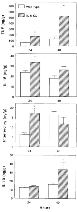
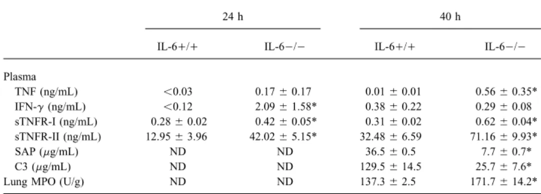
Inflammatory responses in IL-60/0 and IL-6/// mice (fig-ure 3 and table 1). As expected, IL-6 could not be detected in plasma or lungs of IL-60/0 mice during pneumonia. In addition, no IL-6 mRNA was found in the lungs of these mice after administration of S. pneumoniae (data not shown). In IL-6/// mice, plasma IL-6 levels were 0.59 { 0.10 ng/mL after 24 h and 3.76{ 1.50 ng/mL after 40 h; lung IL-6 concentra-tions were 50.74{ 4.91 and 228.37 { 41.57 ng/g, respectively. Lung TNF levels were 3- to 4-fold higher in IL-60/0 mice than in IL-6/// mice (P õ .05). In plasma, TNF remained undetectable (õ0.03 ng/mL) in all but 1 IL-6/// mouse. By contrast, all IL-60/0 mice had detectable plasma TNF levels at 40 h after inoculation (Põ .01 vs. IL-6/// mice). Lung IL-1b and IFN-g levels were higher in IL-60/0 mice at 24 h after inoculation (Põ .05). In plasma, IL-1b remained unde-tectable (õ0.12 ng/mL) in all but 2 mice. Plasma IFN-g was higher in IL-60/0 mice at 24 h after induction of pneumonia (P õ .05). Lung levels of the antiinflammatory cytokine IL-10 were higher in IL-60/0 mice at 40 h after inoculation
(Põ .05), while IL-10 remained undetectable (õ0.12 ng/mL) Figure 3. Mean ({ SE) lung concentrations of tumor necrosis factor (TNF)-a, IL-1b, interferon (IFN)-g, and IL-10 at 24 and 40 h after in plasma in all mice. Also, plasma concentrations of soluble
intranasal inoculation with 106
cfu of Streptococcus pneumoniae in IL-TNFR-I and IL-TNFR-II were higher in IL-60/0 mice than in
60/0 (IL-6 KO) and IL-6/// (wild type) mice. There were 5 or 6 IL-6/// mice (P õ .05).
mice/group/time point. * Põ .05, Wilcoxon test, vs. IL-6/// mice. The acute-phase protein response was strongly attenuated in
IL-60/0 mice (P õ .01 vs. IL-6/// mice). Lung MPO activity
after inoculation than did IL-6/// mice (608 { 346 1 107
was higher in IL-60/0 mice than in IL-6/// mice (P Å .01).
and 112 { 59 1 107 cfu, respectively; P õ .05). From all
Bacterial clearance. IL-60/0 mice had Ç6-fold more S.
Table 1. Inflammatory responses during pneumococcal pneumonia in IL-60/0 and IL-6/// mice.
24 h 40 h
IL-6/// IL-60/0 IL-6/// IL-60/0
Plasma TNF (ng/mL) õ0.03 0.17{ 0.17 0.01{ 0.01 0.56{ 0.35* IFN-g (ng/mL) õ0.12 2.09{ 1.58* 0.38{ 0.22 0.29{ 0.08 sTNFR-I (ng/mL) 0.28{ 0.02 0.42{ 0.05* 0.31{ 0.02 0.62{ 0.04* sTNFR-II (ng/mL) 12.95{ 3.96 42.02{ 5.15* 32.48{ 6.59 71.16{ 9.93* SAP (mg/mL) ND ND 36.5{ 0.5 7.7{ 0.7* C3 (mg/mL) ND ND 129.5{ 14.5 25.7{ 7.6*
Lung MPO (U/g) ND ND 137.3{ 2.5 171.7{ 14.2*
NOTE. Data are mean{ SE. At time 0, mice were inoculated with 50 mL (106
cfu) of S. pneumoniae; 5 or 6 mice/group/time point vs. IL-1b was undetectable in plasma in all but 2 mice. IL-10 could not be detected in plasma of any mouse. ND, not determined; TNF, tumor necrosis factor; IFN, interferon; sTNFR, soluble TNF receptor; SAP, serum amyloid P; MPO, myeloperoxidase.
* Põ .05 vs. IL-6/// mice by Wilcoxon test.
the mean number of colonies was much higher in IL-60/0 injection of endotoxin, inhibition of proinflammatory cytokines mice, the difference with IL-6/// mice was not significant may be beneficial [17, 18]. In such models, high levels of due to large interindividual variation (182 { 109 1 104and proinflammatory cytokines appear in the circulation, and
inhibi-13{ 3 1 104/mL, respectively; nonsignificant). tion of their systemic effects confers protection against tissue
Survival (figure 4). Survival studies were performed in 16 injury and lethality. However, these models of systemic chal-IL-60/0 and 16 IL-6/// mice. Mortality was assessed every lenges do not provide insight into the potential beneficial effects 12 h. IL-60/0 mice succumbed significantly earlier from pneu- of proinflammatory cytokines at the site of an infection. We mococcal pneumonia than did IL-6/// mice (P õ .05). Eight recently found that local production of TNF within the lung is IL-60/0 mice were dead at 48 h after inoculation versus 2 important for host defense in this murine model of pneumococ-IL-6/// mice; all IL-60/0 mice had died by 60 h, at which cal pneumonia [11], while IL-10 produced in lungs hampers time 6 IL-6/// mice were still alive. host defense [10]. In accord, neutralization of IL-10 in mice with pneumonia caused by Klebsiella pneumoniae was associ-ated with a decrease in Klebsiella colonies in lungs and an Discussion
increase in survival [19].
During overwhelming immune activation, especially in the In models of systemic inflammation induced by bolus admin-absence of a localized infectious source such as after bolus istration of endotoxin, TNF is a mediator of toxicity and death [17], while IL-10 is protective [18] (i.e., completely the oppo-site of their respective roles in models of gram-negative [19, 20] and gram-positive [10, 11] pneumonia, even though, in such models, blood cultures are positive). It should be noted, however, that in endotoxin-challenge models, lipopolysaccha-ride is injected directly into the circulation, while during pneu-monia, bacteria likely are shed gradually or intermittently from the source of the infection, which apparently does not result in detectable cytokine levels in the circulation [10, 11] (present data). Hence, animal studies of pneumonia and systemic endo-toxin effects represent two completely different entities, the former being more clinically relevant.
Although IL-6 is not an important mediator of endotoxinduced inflammatory responses [9, 21 – 23], recent studies in-dicate that this cytokine may have a significant role in host Figure 4. IL-60/0 mice show impaired defense against pneumo- defense against bacterial infection. IL-6 appears to be essential coccal pneumonia. Survival after intranasal inoculation with 106
cfu
for defense against Listeria monocytogenes, since IL-6 gene – of Streptococcus pneumoniae of IL-60/0 (IL-6 KO) and IL-6///
deficient mice succumb early from this gram-positive infection (wild type) mice. There were 16 mice/group. IL-60/0 mice died
mortality after intraperitoneal administration of live Esche- creased number of bacterial colonies in IL-60/0 mice was associated with reduced survival time in these animals. It has richia coli, which is associated with increased bacterial
num-bers in organs during the course of this infection [23]. The been postulated that the absence of an adequate neutrophilic response in IL-6 – deficient mice contributes to the increased objective of the present study was to determine the role of
endogenous IL-6 in the pathogenesis of pneumococcal pneu- susceptibility to L. monocytogenes and E. coli infection [23, 24]. In our study, IL-60/0 mice had enhanced pulmonary MPO monia, the most frequent manifestation of community-acquired
pneumonia. activity, as determined 40 h after inoculation with S.
pneumo-niae. Although neutrophil numbers were not actually counted The absence of an IL-6 response in IL-60/0 mice was
asso-ciated with enhanced induction of both agonist and antagonist in our study, it has previously been shown that MPO activity corresponds well with the number of neutrophils in tissue sec-members of the cytokine network. Some of these findings were
anticipated from previous studies. Indeed, IL-6 has been con- tions of inflamed lungs [30]. Conceivably, the elevated lung TNF levels in IL-60/0 mice contributed to increased neutrophil sidered an antiinflammatory cytokine by virtue of its ability to
inhibit endotoxin-induced TNF and IL-1 production by mono- influx, since TNF is considered to play a significant role in this characteristic inflammatory response [20].
nuclear cells in vitro [25, 26] and to reduce TNF release in
endotoxemic mice in vivo [25]. In accord, endotoxin-treated In comparison with other cytokines, IL-6 has been reported most consistently in the circulation of septic patients [31]. Until IL-60/0 mice produce 3 times more TNF than wild type
con-trols [22]. However, administration of IL-6 to cancer patients recently, IL-6 was merely considered a marker for the severity of the bacterial challenge, rather than a significant player in results in an increase in the plasma levels of soluble TNFR-I
[27]. Therefore, the enhanced release of soluble TNFR-I and the pathogenesis of bacterial infection. This and other studies [9, 23, 24] indicate that the role of IL-6 in host defense against TNFR-II in IL-60/0 mice was unexpected. Interestingly
enough, IL-60/0 mice also had increased pulmonary levels of bacteria is more important than previously recognized. Our study does not elucidate the mechanisms by which IL-6 con-IL-10, a pleiotropic cytokine known to inhibit the production
of proinflammatory cytokines under various in vitro and in tributes to antibacterial mechanisms or activation of inflamma-tory responses. Considering the limited (direct) effects of IL-vivo conditions [18]. Hence, it should be noted that IL-60/0
mice had elevated proinflammatory cytokine levels despite con- 6 in various in vitro systems, it is conceivable that IL-6 is involved indirectly and influences the activity of mediators currently elevated IL-10 concentrations.
acting more directly on inflammatory processes. Nonetheless, The mechanism by which the absence of an IL-6 response
these results indicate that neutralization of IL-6 activity in resulted in enhanced release of soluble TNF receptors and
humans, as has been propagated for some diseases [32, 33], IL-10 remains to be elucidated. Possibly, the elevated TNF
may hamper a specific resistance against bacterial infections. levels in IL-60/0 mice contributed to these exaggerated
antiin-Pneumococcal pneumonia is the most common community-flammatory responses, since TNF can induce release of its
acquired pneumonia. In our study, IL-6 was produced locally receptors and of IL-10 in humans, and anti-TNF treatment of
within the lung during pneumonia caused by S. pneumoniae. endotoxemic primates abrogates the appearances of soluble
Endogenous IL-6 played a major role in the induction of the TNF receptors and IL-10 [28, 29].
cytokine network within the lung, controlling the activation of TNF was not detectable in plasma of wild type mice during
both agonist and antagonist mediators during pneumonia. The pneumonia. Although we cannot exclude the possibility that
net effect of IL-6 was protective, reducing the growth of pneu-TNF was detectable in plasma at early time points after
inocula-mococci and prolonging survival. Thus, IL-6 was an important tion, we consider this unlikely, since during pneumonia TNF
mediator of inflammation within lungs during pneumococcal is produced within lungs, and the highest lung TNF levels are
pneumonia. According to our findings and those of earlier stud-found after§24 h [11]. Rather, these data suggest that TNF
ies addressing the role of cytokines in the pathogenesis of production is compartmentalized within lungs and that plasma
bacterial pneumonia [10, 11, 19, 20], it becomes increasingly TNF levels are a weak reflection of lung TNF concentrations.
clear that local inflammation, facilitated directly or indirectly The acute-phase protein response was markedly reduced in
by proinflammatory cytokines, is essential for local defense IL-60/0 mice with pneumococcal pneumonia. These data
ex-against bacteria. Neutralization of proinflammatory cytokines tend previous studies with IL-60/0 mice, demonstrating that
in patients with bacterial infections may therefore be potentially IL-6 is not required to generate an acute-phase protein response
hazardous and may be one reason, among others, why clinical to endotoxin but is an important mediator of acute-phase
pro-trials with strategies directed against TNF, based primarily tein release during sterile inflammation induced by
subcutane-on animal models in which bacteria or their products were ous injection of turpentine and during infection with L.
monocy-administered as a bolus, have not been successful [17, 34]. togenes [9, 22].
Significantly more pneumococci were recovered from lungs
References
of IL-60/0 mice than from lungs of IL-6/// mice 40 h after 1. Garibaldi RA. Epidemiology of community-acquired respiratory tract in-inoculation. Although a large interindividual variation existed, fection in adults: incidence, etiology, and impact. Am J Med1985; 78:
S32 – 7. we consider this difference clinically relevant, since the
in-2. American Thoracic Society. Guidelines for the initial management of 19. Greenberger MJ, Strieter RM, Kunkel SL, Danforth JM, Goodman RE, Standiford TJ. Neutralization of IL-10 increases survival in a murine adults with community-acquired pneumonia: diagnosis, assessment of
severity, and initial antimicrobial therapy. Am Rev Respir Dis1993; model of Klebsiella pneumonia. J Immunol1995; 155:722 – 9.
20. Kolls JK, Lei D, Nelson S, Summer WR, Greenberg S, Beutler B. Adenovi-148:1418 – 26.
3. Marrie TJ, Durant H, Yates L. Community-acquired pneumonia requiring rus-mediated blockade of tumor necrosis factor in mice protects against endotoxic shock yet impairs pulmonary host defense. J Infect Dis1995;
hospitalization: a 5 year prospective study. Rev Infect Dis1989; 11:
586 – 99. 171:570 – 5.
21. Libert C, Vink A, Coulie P, et al. Limited involvement of interleukin-6 4. Torres A, Serra-Batlles J, Ferrer A, et al. Severe community-acquired
pneumonia: epidemiology and prognostic factors. Am Rev Respir Dis in the pathogenesis of lethal septic shock as revealed by the effect of monoclonal antibodies against interleukin-6 or its receptor in various
1991; 144:312 – 8.
5. Friedland IR, McCracken GH. Management of infections caused by antibi- murine models. Eur J Immunol1992; 22:2625 – 30.
22. Fattori E, Cappelletti M, Costa P, et al. Defective inflammatory response otic-resistant Streptococcus pneumoniae. N Engl J Med1994; 331:377 –
82. in interleukin-6 – deficient mice. J Exp Med1994; 180:1243 – 50.
23. Dalrymple SA, Slattery R, Aud DM, Krishna M, Lucian LA, Murray R. 6. Hack CE, de Groot ER, Felt-Bersma RJF, et al. Increased plasma levels
of interleukin-6 in sepsis. Blood1989; 74:1704 – 10. Interleukin-6 is required for a protective immune response to systemic Escherichia coli infection. Infect Immun1996; 64:3231 – 5.
7. Dehoux MS, Boutten A, Ostinelli J, et al. Compartmentalized cytokine
production within the human lung in unilateral pneumonia. Am J Respir 24. Dalrymple SA, Lucian LA, Slattery R, et al.Interleukin-6 – deficient mice are highly susceptible to Listeria monocytogenes infection: correlation Crit Care Med1994; 150:710 – 6.
8. Puren AJ, Feldman C, Savage N, Becker PJ, Smith C. Patterns of cytokine with inefficient neutrophilia. Infect Immun1995; 63:2262 – 8.
25. Aderka D, Le J, Vilcek J. IL-6 inhibits lipopolysaccharide-induced tumor expression in community-acquired pneumonia. Chest1995; 107:1342 –
9. necrosis factor production in cultured human monocytes, U937 cells
and mice. J Immunol1989; 143:3517 – 23.
9. Kopf M, Baumann H, Freer G, et al. Impaired immune and acute-phase
responses in interleukin-6 – deficient mice. Nature1994; 368:339 – 42. 26. Schindler R, Mancilla J, Endres S, Ghorbani R, Clark R, Dinarello CA. Correlations and interactions in the production of interleukin-6 (IL-6), 10. Van der Poll T, Marchant A, Keogh CV, Goldman M, Lowry SF.
Interleu-kin 10 impairs host defense in murine pneumococcal pneumonia. J IL-1, and tumor necrosis factor (TNF) in human blood mononuclear cells: IL-6 suppresses IL-1 and TNF. Blood1990; 75:40 – 7.
Infect Dis1996; 174:994 – 1000.
11. Van der Poll T, Keogh CV, Buurman WA, Lowry SF. Passive immuniza- 27. Tilg H, Trehu E, Atkins MB, Dinarello CA, Mier JW. Interleukin-6 (IL-6) as an antiinflammatory cytokine: induction of circulating IL-1 receptor tion against tumor necrosis factor a impairs host defense during
pneu-mococcal pneumonia in mice. Am J Respir Crit Care Med1997;155: antagonist and soluble tumor necrosis factor receptor p55. Blood1994;
83:113 – 8. 603 – 8.
12. Espevik T, Nissen-Meyer J. A highly sensitive cell line, WEHI 164 clone 28. Jansen J, van der Poll T, Levi M, et al. Inhibition of the release of soluble tumor necrosis factor receptors in experimental endotoxemia by an anti – 13, for measuring cytotoxic factor/tumor necrosis factor from human
monocytes. J Immunol Methods1986; 95:99 – 109. tumor necrosis factor-a antibody. J Clin Immunol1995; 15:45 – 50.
29. Van der Poll T, Jansen J, Levi M, ten Cate H, ten Cate JW, van Deventer 13. Bemelmans MHA, Gouma DJ, Buurman WA. Tissue distribution and
clearance of soluble murine TNF receptors in mice. Cytokine1994; 6: SJH. Regulation of interleukin 10 release by tumor necrosis factor in humans and chimpanzees. J Exp Med1994; 180:1985 – 8.
608 – 15.
14. Oldenburg HSA, Rogy MA, Lazarus DD, et al. Cachexia and the acute- 30. Goldblum SE, Wu KM, Jay M. Lung myeloperoxidase as a measure of pulmonary leukostasis in rabbits. J Appl Physiol1985; 59:1978 – 85.
phase protein response in inflammation are regulated by interleukin-6.
Eur J Immunol1993; 23:1889 – 94. 31. Lowry SF, Calvano SE, van der Poll T. Measurement of inflammatory mediators in clinical sepsis. In: Sibbald WJ, Vincent JL, eds. Clinical 15. Koike K, Moore FA, Moore EE, Read RA, Carl VS, Banerjee A. Gut
ischemia mediates lung injury by a xanthine oxidase – dependent neutro- trials for the treatment of sepsis. Berlin: Springer Verlag,1995:86 – 105.
32. Klein B, Wijdenes J, Zhang XG, et al. Murine anti – interleukin-6 mono-phil mechanism. J Surg Res1993; 54:469 – 73.
16. Chiu CP, Moulds C, Coffman RL, Rennick D, Lee F. Multiple biological clonal antibody therapy for a patient with plasma cell leukemia. Blood
1991; 78:1198 – 204.
activities are expressed by a mouse interleukin 6 cDNA clone isolated
from bone marrow stromal cells. Proc Natl Acad Sci USA1988; 85: 33. Emilie D, Wijdenes J, Gisselbrecht C, et al. Administration of an anti – interleukin-6 monoclonal antibody to patients with acquired immunode-7099 – 103.
17. Van der Poll T, Lowry SF. Tumor necrosis factor in sepsis: mediator of ficiency syndrome and lymphoma: effect on lymphoma growth and on B clinical symptoms. Blood1994; 84:2472 – 9.
multiple organ failure or essential part of host defense? Shock1995; 3:
1 – 12. 34. Van der Poll T, van Deventer SJH. The role of tumour necrosis factor a in sepsis revisited. From good, to bad, to worse. Exp Opin Invest Drugs 18. Moore KW, O’Garra A, de Waal Malefyt R, Vieira P, Mosmann TR.

