DOI 10.1007/s00221-002-1175-9 R E S E A R C H A R T I C L E
U. Leonards · V. Ibanez · P. Giannakopoulos
The role of stimulus type in age-related changes
of visual working memory
Received: 20 July 2001 / Accepted: 24 May 2002 / Published online: 5 July 2002 3 Springer-Verlag 2002
Abstract Aging is accompanied by increasing difficulty
in working memory associated with the temporary storage and processing of goal-relevant information. Face recog-nition plays a preponderant role in human behavior, and one might therefore suggest that working memory for faces is spared from age-related decline compared to socially less important visual stimulus material. To test this hypothesis, we performed working memory (n-back) tasks with two different visual stimulus types, namely faces and doors, and compared them to tasks with primarily verbal material, namely letters. Age-related reaction time slowing was comparable for all three stimulus types, supporting hypotheses on general cogni-tive and motor slowing. In contrast, performance sub-stantially declined with age for faces and doors, but little for letters. Working memory for faces resulted in significantly better performance than that for doors and was more sensitive to on-line manipulation errors such as the temporal order. All together, our results show that even though face perception might play a specific role in visual processing, visual working memory for faces undergoes the same age-related decline as it does for socially less relevant visual material. Moreover, these results suggest that working memory decline cannot be solely explained by increasing vulnerability in prefrontal cortex related to executive functioning, but indicate an age-related decrease in a visual short-term buffer, possi-bly located in the temporal cortex.
Keywords Visual working memory · Aging · Stimulus
type · Face perception
Introduction
If we are engaged in an active discussion, are planning our next holidays, are solving complex mathematical equations or philosophical problems, or are simply following the news on TV, we are continuously relying on our working memory, the capacity to store information in some kind of short-term memory buffer and to simultaneously manipulate this information on-line (Bad-deley 1986; Shallice 1988). Visual working memory can be thought of as consisting of several stages. These include the formation of a perceptual image originating from an external stimulus (sensory processing), the storing of this image in some kind of domain-specific, short-term buffer, and the activation of executive pro-cesses, such as on-line manipulation, inhibition, and response co-ordination that operate on the contents of the incoming information. In terms of Baddeley’s working memory model (Baddeley 1986), the first two stages would correspond to a modality-specific, limited short-term buffer or slave system, in case of the visual system the so-called “visuospatial sketchpad”, located in primary and secondary visual cortices. The third stage would correspond to Baddeley’s “central executive” in the prefrontal cortex, which is responsible for allocation of processing abilities to the visuospatial sketchpad. Other models of working memory are based on distributed cortical networks (for reviews see, for example, Fuster 1998; Mesulam 1998). In these latter models, the three processes can be interpreted as those that are automatic, primarily driven by the external stimulus (“bottom-up”, pre-attentive processing), and those that are under volun-tary control (“top-down”, attention-demanding) (see, for example, Desimone et al. 1995; Hasegawa et al. 1999). Bottom-up processes consist of a passive cortical flow of information from primary over secondary visual cortices, i.e., in occipitotemporal regions if primarily concerned with working memory for visual objects, toward the prefrontal cortex. Top-down processing as the active part of working memory originates in prefrontal cortex and influences via feedback projections lower processing U. Leonards ()) · V. Ibanez · P. Giannakopoulos
Division de Neuropsychiatrie, DHpartement de Psychiatrie, HIpitaux Universitaires de GenJve – Belle IdHe, 2,
Chemin du Petit-Bel-Air, 1225 ChÞne-Bourg GE, Switzerland e-mail: [email protected]
Tel.: +41-22-3055379 Fax: +41-22-3055350
levels such as posterior association cortices (see, for example, Hasegawa et al. 1999; Tomita et al. 1999), presumably even down to primary sensory cortices (see, for example, Desimone et al. 1995; Ungerleider et al. 1998).
Today, it is generally accepted that working memory performance declines with age (for reviews see Craik and Jennings 1992; Grady and Craik 2000). However, the reasons for this decline are still a matter of debate, and the proposed mechanisms vary substantially. For example, the elderly were said to have decreased limits in storage resources (see, for example, Foos 1989; Foos and Wright 1992), indicating deficits in bottom-up processing. Other findings point toward deficits in executive control (Craik 1990; Morris et al. 1988; Salthouse and Skovronek 1992; Van der Linden et al. 1992, 1994), presumably provoked by decreased functioning of the frontal lobe or of top-down control. Recent neuropsychological and functional imaging studies support a hypothesis of frontal aging (Albert and Kaplan 1980; Hochanadel and Kaplan 1984; Mittenberg et al. 1989), showing that activation differ-ences in the dorsolateral prefrontal cortex correlate with working memory decline in normal aging (Rypma and D’Esposito 2000; see also Reuter-Lorenz et al. 2000).
In recent years, there has been increasing evidence for the idea that aging is associated with numerous alterations of brain structure and functions (for recent reviews see BussiJre and Hof 2001; Giannakopoulos et al. 1997; Kemper 1994; Raz 2000). Presumably, aging affects the total volume of the brain, and the integrity of the white matter is supposed to predict the global decline of cognitive functions (see, for example, Gunning-Dixon and Raz 1999). In particular, substantial neurofibrillary tangle formation occurs in the hippocampal formation and Brodmann’s area 20 in cognitively intact elderly people, suggesting that midtemporal structures are particularly vulnerable to the degenerative power of normal brain aging (see, for example, Giannakopoulos et al. 1997). The prefrontal cortex also shows pronounced signs of aging (see, for example, Esiri 1994), while primary sensory regions remain largely spared (see, for example, Gian-nakopoulos et al. 1995; Hof et al. 1996).
On the behavioral level, there are also several findings which indicate that cognitive age effects cannot be restricted to the executive control of frontal cortex (Mencl et al. 2000). For example, a profound loss in visual acuity and contrast sensitivity is often observed that could easily explain large parts of age-related variance in cognition (Baltes and Lindenberger 1997; Kline et al. 1983; Lindenberger and Baltes 1994; Sekuler et al. 1982). Visual processing speed (Briggs et al. 1999) and iconic memory (see, for example, DiLollo et al. 1982) have been shown to slow with age, and increased stimulus com-plexity affected the performance of older subjects more than that of younger controls (Adamowicz 1976; Adamowicz and Hudson 1978). As a consequence, in the elderly the stored information is incomplete and hampers any cognitive judgment based on such informa-tion (Oscar-Berman and Bonner 1985; Oscar-Berman et
al. 1992). Such observations led to the development of models based on general cognitive slowing with age (see, for example, Babcock and Salthouse 1990; Cerella 1990; Cerella et al. 1980; Greenwood 2000; Salthouse et al. 1996), as opposed to the frontal aging hypothesis (Albert and Kaplan 1980; Hochanadel and Kaplan 1984; Mitten-berg et al. 1989; Van der Linden et al. 1994). They were supported by findings of cortical activation differences not only in the frontal, but also the extrastriate cortex and temporal lobe in young compared to older observers during memory tasks (see, for example, Cabeza et al. 1997; Grady et al. 1998).
To date, little attention has been paid to the fact that age-related decline in visual working memory might depend on the stimulus material used and its direct relevance to human social behavior. For instance, human faces seem to be a special class of objects with particular biological and social significance (Carey 1992; GrOsser and Landis 1991), suggesting an evolutionary advantage for individuals who have a preserved ability to process such images. In patients with left spatial neglect, face images can be spared from visual extinction observed for other visual images (Vuilleumier 2000), and normal subjects perceive human faces more easily than other stimuli under conditions of inattention or divided atten-tion (Mack and Rock 1998). Further, the ability to detect and orient to faces appears very early in human babies, suggesting that face perception may rely in part on prewired neural mechanisms (Braddick et al. 1986; Goren et al. 1975). On the other hand, face memory can be specifically disturbed, leading to the clinical symptom of prosopagnosia (Giannakopoulos et al. 2000; Whiteley and Warrington 1977). Thus, faces take a dominant role in visual processing and recognition (see, for example, Farah 1996; Haxby et al. 1995, 2000), perhaps even to the degree that they are processed by face-specific cortical networks (Kanwisher et al. 1997, 1999; McCarthy et al. 1997). Moreover, activation studies with PET, functional MRI, and event-related potentials indicate that specialized neural substrates for visual memory processes might be accessed by different object categories (see, for example, Gur et al. 1997; Ishai et al. 1999; Kim et al. 1999; McDermott et al. 1999; Owen et al. 1999; Ruchkin et al. 1997). It is therefore easy to imagine that these separate substrates may undergo differential changes with increas-ing age. Indications for such differential age-related changes in processing of faces compared to other visual material come from working memory tasks requiring recall of the temporal order of images, for example, older subjects lacked the typical U-shaped performance curve for face images, while their performance curves for abstract shapes were similar to those of young subjects (Bruyer and Vanberten 1998). Furthermore, elderly individuals showed more false recognition of new faces than younger subjects (Bartlett and Fulton 1991; Bartlett et al. 1989; Fulton and Bartlett 1991). Both findings suggest that memory for faces might be even more sensitive to age-related decline than memory to other visual material.
However, the specific role of faces in visual processing and memory has recently been questioned (see, for example, Gauthier et al. 1999), and rather than catego-ry-specific visual face modules, a continuous representa-tion of informarepresenta-tion about object form was proposed with a “featurotopic” organization (Ishai et al. 1999; see Gau-thier 2000 for review).
Here, we investigated whether working memory for socially relevant visual material was differentially pro-tected from age-related decline as compared to that of socially less important visual material. Therefore, perfor-mance and reaction times of intellectually preserved individuals, aged between 20 and 69 years, were compared in a paradigm including faces as socially relevant items and doors, already used before by Badde-ley et al. (1994; see also Davis et al. 1999), as socially non-relevant but nevertheless common, everyday visual material. Different components involved in visual work-ing memory were controlled, such as sensory processwork-ing and short-term memory. To distinguish between age-related decline of visual working memory functions and general executive control, data were compared to those obtained in the same subjects with a primarily verbal stimulus type, namely letters.
Materials and methods
SubjectsFifty right-handed adult subjects (20–69 years) participated in this study. They had normal or corrected-to-normal visual acuity and normal contrast sensitivity. Subjects were grouped in five age groups of 10 subjects each (mean average and standard deviation in years: 24.7€1.49; 34.4€2.88; 44.28€3.05; 53.67€2.84; 64.55€2.74). Subjects were healthy and free of any medication that might influence attention or motor responses. Volunteers gave their informed written consent in accordance with the Declaration of Helsinki, and experiments were approved by the Ethics Committee of the University Hospital of Geneva.
Tasks and stimuli Experiment 1
Black and white photographs of 27 different faces (AT&T Laboratories Cambridge) or their scrambled version, both subtend-ing 6RS7R of visual angle, were displayed at the center of a computer-controlled 21-inch high-resolution monitor (IIyama Vi-sion Master Pro 510) with a refresh rate of 100 Hz. The viewing distance was fixed at 90 cm.
Subjects performed two variants of a working memory task (1back and 2back; Fig. 1a, b) that have been used previously in a number of behavioral and neuroimaging experiments (see, for example, Braver et al. 1997; Owen et al. 1999; Smith et al. 1996). A simple detection task (Fig. 1c) served as control for subjects’ capacities to visually process complex stimuli, to take decisions based on this sensory information, and to give a motor response.
In all three tasks, subjects indicated the presence of a target by pressing as fast as possible, a computer-controlled push-button with the index finger of their right (dominant) hand. For non-target stimuli, no response was required. In the detection task, subjects responded whenever a complete face was presented in a sequence of scrambled face images. For the two working memory tasks, only
complete face images were used. In the 1back task, the target was any face that was identical to the one immediately preceding it. In the 2back condition, the target was any face identical to the one presented two trials back. Stimuli were presented in pseudorandom order of 90 images per sequence, with a stimulus duration of 500 ms and an interstimulus interval (onset–onset) of 5 s. In 26.7% of the trials, a target was present. Every series was preceded by up to ten warm-up trials to ensure that subjects had understood the instruc-tions for the new condition. Every subject performed each of the three tasks in pseudorandom order. In addition, 34 of the 50 subjects were tested with a 2back letter task, presenting white letters out of a pool of 24 letters that are common in the French alphabet on a gray background. Letters subtended 3.5RS2R of visual angle on a gray background of 6RS7R.
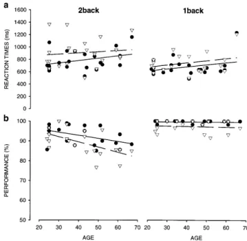
Fig. 1 Diagrams of the two memory conditions [1back (a) and 2back (b) task] for a sequential working memory task for faces, and of the control condition, a simple face detection task (c). x Correct target positions
Experiment 2
Of the 50 subjects from experiment 1, 24 participated in experi-ment 2. Subjects were tested with 1back and 2back tasks, using the same experimental set-up as described for experiment 1. Images consisted of 27 different doors, taken from the “Doors and People Memory Test” battery of Baddeley et al. (1994). All images were presented as gray-level images.
Error analysis for errors related to temporal order
Errors occurring during the 2back task were classified into false alarms, i.e., responses to non-targets, and misses, i.e., missing responses. False alarms were defined relative to temporal order, if subjects responded to images that were identical to the one just seen before (1back) or three images (3back) before. Other types of false alarms, as for example, feature similarities between faces or a developing familiarity of the images due to repeated presentation of the same stimuli, should be independent of the exact sequence order and would be expected to depend on deficits in the memory buffer. We defined missing responses as temporal order-related if subjects did not respond to a target that followed one or two trials after a prevailing event (button-press).
Results
Experiment 1
In the first experiment, we investigated age effects on the visual short-term buffer and on-line manipulation of complex visual material with social relevance, specifi-cally human faces. Results were compared to those for letters requiring little visual but mostly verbal working memory resources. We reasoned that age-related decline in executive functioning should be independent of whether the information to be treated was primarily handled by the visuospatial sketchpad or the phonological loop (Baddeley 1986).
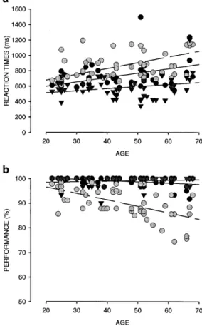
Reaction times for working memory using face images As shown in Fig. 2a for face images, subjects responded most rapidly for the face detection task and most slowly for the face 2back task (Table 1). In addition, reaction times significantly increased with age for the 2back task (Table 1; Kendall tau correlation analysis of performance and age).
A 5 (age group) S 3 (task) ANOVA with repeated measures revealed significant main effects of age
[F(4,45)=2.71; P<0.05] and task [F(2,90)=58.32;
P<0.0001]. Post hoc analysis showed that reaction times significantly increased from detection to 1back (ScheffH P<0.0001), and further from 1back to 2back task (ScheffH P<0.0001). However, there was no significant difference in reaction time slowing with age between the three tasks, as controlled by a two-way interaction analysis between age S task [F(8,90)=1.67; n.s.].
It is therefore most likely that reaction time slowing with age, even though not significant for face detection and 1back task, is not exclusively related to the 2back
task, but reflects a more general age-related slowing of motor and/or cognitive components.
Performance for working memory using face images Subjects’ performance is shown in Fig. 2b. Performance significantly declined with age for 1back and 2back task, but not for face detection (see Table 1 for Kendall correlations). A 5 (age) S 3 (task) ANOVA with repeated measures indicated that, similarly to reaction times, performance was affected by tasks [F(2,90)=123.53; P<0.0001]. Moreover, performance for face detection and 1back task did not differ significantly (ScheffH n.s.; Fig. 2b). Despite the fact that the 1back task requires additional cognitive processes, i.e., short-term storage and image comparison, the two tasks revealed similar perfor-mance values of more than 90% correct responses. Compared to 1back and detection task, all subjects performed significantly worse in the 2back task (ScheffH: 1back versus 2back P<0.0001; face detection versus 2back P<0.0001). Pronounced age-related performance decline was only observed for the 2back task, as confirmed by a two-way interaction between age and Fig. 2 Individual reaction times (a) and performance values (b) plotted against subjects’ age for 1back (black circles), 2back (gray circles), and detection task (triangles), using faces as stimulus material. Lines correspond to linear regression lines (corresponding statistics in Table 1)
task [F(8,90)=4.992; P<0.0001; ScheffH (2back) P<0.007 (age group 1 versus group 3) to P<0.0001 (age group 1 versus group 5)].
Faces versus letters: visual versus verbal working memory
As summarized in Fig. 3a, reaction times for the 2back task for letters increased significantly with age (see also Table 1), in a similar way as for faces. Even though the two stimulus types differed remarkably with respect to their visual complexity, and thus sensory processing, as well as in the degree of involvement of a verbal component, no significant difference was found between reaction times for letters and faces. This was confirmed by a 2 (stimulus type) S 5 (age) ANOVA with repeated
measures, revealing a significant effect of age
[F(4,30)=2.75; P<0.046], but not of stimulus type [F(1,30)=3.71; n.s.; interaction: F(4,30)=0.281; n.s.].
In contrast to reaction times, the stimulus type strongly influenced subjects’ performance levels (Fig. 3b; Table 1), leading to a better performance for letters than for faces, and less performance decrease with age for the prior (see Table 1 for Kendall tau correlations). Results of a 2 Fig. 3 Individual reaction times (a) and performance values (b) plotted against subjects’ age for the 2back task for face images (filled black circles) or letters (open triangles). Regression lines: faces (solid line), letters (broken line); corresponding statistics in Table 1 Table 1 Mean reaction times (in milliseconds) and mean performance (in percent) with standard errors for experiment 1, and corresponding statistics for the correlation analyses with age. (r 2 Regression coefficient, t Kendall tau, PP level for Kendall, n.s. non-significant, n number of subjects) Age group (years) Reaction times (ms) Performance (%) Face detection (n =50) Face 1back (n =50) Face 2back (n =50) Letter 2back (n =34) Face detection (n =50) Face 1back (n =50) Face 2back (n =50) Letter 2back (n =34) 20–29 605€24 662€25 742€51 635€34 99.3€0.7 99.7€0.2 94.4€1.4 97.4€1.3 30–39 620€22 727€31 854€49 760€49 97.0€0.9 99.2€0.4 92.8€1.3 97.8€0.8 40–49 551€18 643€22 813€63 735€62 100 99.5€0.3 91.2€1.5 96.2€1.7 50–59 661€24 776€69 862€42 869€63 99.4€0.6 99.0€0.4 88.2€1.7 98.7€0.9 60–69 723€60 787€58 943€57 890€67 99.3€0.5 97.2€0.9 84.3€2.0 90.9€0.9 r 2 =0.084 r 2 =0.146 r 2 =0.227 r 2 =0.312 r 2 =0.018 r 2 =0.158 r 2 =0.343 r 2 =0.264 t=0.044 t=0.174 t=0.244 t=0.272 t=0.115 t=–0.204 t=–0.442 t=–0.282 P =n.s. P =n.s. P <0.01 P <0.05 P =n.s. P <0.05 P <0.001 P <0.05
(stimulus type) S 5 (age) ANOVA with repeated mea-sures indicated significant effects of age [F(4,30)=5.024; P<0.003] and stimulus type [F(1,30)=58.709; P<0.0001]. The significant two-way interaction between age and stimulus type [F(4,30)=4.646; P<0.0048] was due to the performance decline with age for faces [ScheffH: P<0.0129 (age group 1 versus group 3) to P<0.004 (age group 1 versus group 5)], but not for letters (ScheffH: n.s.).
Experiment 2: visual working memory for face and door images
In the second experiment, we investigated age effects on the working memory of a visual stimulus type without social relevance, namely doors. Similar to faces, doors build a common and complex everyday-object category. We chose doors instead of house images common in neuroimaging studies on working memory (see, for example, Haxby et al. 1999, 2000), or other living images with little or no social relevance (i.e., animals or flowers), since these doors are used in one of the major clinical tests on recognition memory in the elderly, the “Doors and People Memory Test” of Baddeley et al. (1994).
Results for door images (experiment 2) were directly compared to those for faces (experiment 1) for the same subjects (Table 2). As shown in Fig. 4a, subjects required more time to respond to door than to face images for both 1back (right panel), and 2back tasks (left panel). Further, there was a decrease in overall performance for doors compared to faces (Fig. 4b), indicating that basic visual
Table 2 Mean reaction times (in milliseconds) and mean performance (in percent) with standard errors for experiment 2 (n =24), and corresponding statistics for the correlation analyses with age (same conventions as in Table 1) Age group (years) Reaction times (ms) Performance (%) Faces 1back Doors 1back Faces 2back Doors 2back Faces 1back Doors 1back Faces 2back Doors 2back 20–29 651€39 699€37 746€83 895€125 99.7€0.3 97.€1.4 94.5€2.3 91.6€1.7 30–39 704€50 786€67 809€52 941€120 100€0 98.8€0.7 94.1€2.2 93.5€2.4 40–49 610€36 647€66 710€67 809€102 99.2€0.5 97.1€1.3 91.4€2.9 86.7€3.2 50–59 626€21 769€85 826€98 969€110 100€0 97.2€2.8 92.2€2.8 88.2€3.7 60–69 858€190 880€216 908€134 982€179 99.4€0.6 96.7€2.4 89.1€2.3 82.7€3.2 r 2 =0.095 r 2 =0.092 r 2 =0.103 r 2 =0.013 r 2 =0.007 r 2 =0.009 r 2 =0.197 r 2 =0.392 t=0.092 t=0.114 t=0.210 t=0.102 t=–0.039 t=–0.041 t=–0.289 t=–0.398 P =n.s. P =n.s. P =n.s. P =n.s. P =n.s. P =n.s. P =n.s. (0.07) P <0.01
Fig. 4 Comparison of individual reaction times (a) and perfor-mance values (b) plotted against subjects’ age for faces (black circles) versus doors (open triangles), for 2back (first column) and 1back (second column) task. Lines correspond to linear regression lines; corresponding statistics in Table 2
information processing and/or short-term buffering were more difficult for doors than for faces, despite eventual task-related training effects for 1back and 2back tasks (note that all subjects tested with doors had already passed the tasks with letters and faces in experiment 1). This was confirmed by separated 5 (age) S 2 (image) ANOVAs (repeated measures) for 1back and 2back tasks,
revealing significant effects of the type of image for both tasks (Table 3). Larger difficulty corresponds well to subjective observations that door images seemed to “fade” shortly after presentation.
Even though the age-related performance decline for the face 2back task was only marginal for subjects selected for experiment 2, it was significant for the door 2back task (see Table 2 for Kendall tau correlations between age and performance).
Error analysis for the 2back tasks for faces and doors
In Fig. 5a, mean error rates for the face 2back task are plotted against age for false alarms and misses (the number of errors for the letter task was not sufficient for statistical analysis). Despite large interindividual vari-ability and a non-normal distribution, both types of error significantly increased with age (see regression coeffi-cients and Kendall tau correlations for errors and age in legend of Fig. 5). The percentage of temporal order-related false alarms compared to the total number of false alarms is shown in Fig. 5b (see Materials and methods for definition of temporal order-related errors). Both young and old subjects gave false alarms significantly more often due to temporal order than would have been expected by chance, as evidenced by a Wilcoxon signed pair test (see asterisks in Fig. 5b). In contrast to expectations, the ratio of temporal order-related versus unrelated false alarms tended to decrease with age.
In Fig. 5c, the percentage of temporal order-related misses relative to the total number of misses is plotted for the different age groups. Similar to false alarms, such temporal order-specific misses occurred more often than by chance (Wilcoxon a<0.05).
Thus, for both younger and older subjects, the analysis of false alarms and misses in the 2back face task revealed that the majority of erroneous events was related to the temporal order of a given stimulus sequence.
As plotted in the left panel of Fig. 6a, false alarms for faces (black circles) significantly increased with age, while false alarms for doors (open triangles) did not (see Kendall tau correlations in legend of Fig. 6). Furthermore, false alarms for doors were less related to temporal order than those for faces (Fig. 6b left panel; Wilcoxon: doors n.s., faces a<0.05, calculated over all subjects, see Materials and methods).
Performance differences between doors and faces were due to a higher number of misses for door targets than for faces (Fig. 6a right panel; Wilcoxon a<0.01). Note that misses for door targets, but not for face targets, signif-Table 3 Statistical comparison
of performance and reaction times for working memory tasks in dependence on the visual material (faces versus doors; n=24)
Reaction time Performance
F(1,14) P ScheffH (P) F(1,14) P ScheffH (P)
1back 15.05 0.0017 0.0014 8.758 0.0103 0.0092
2back 9.22 0.0089 0.0079 7.459 0.0162 0.0146
Fig. 5a–c Errors for the 2back task, using face images, plotted against age (n=50). a Percentages of the total amount of false alarms (black circles) and misses (gray triangles). Lines correspond to linear regression lines; corresponding statistics for false alarms: r2=0.304; t=0.39; P<0.0001; misses: r2=0.158; t=0.292; P<0.005. Ratios of temporal order-related errors relative to the total number of false alarms (FA; b) or misses (c). Horizontal lines marked by arrows indicate chance levels. ** a<0.01; * a<0.05 for values above chance
icantly increased with age (see Kendall tau correlations in legend of Fig. 6). As for false alarms, misses for doors were not related to temporal order (Wilcoxon n.s., calculated over all subjects) but could occur at any given time in the sequence (Fig. 6b right panel). Misses for faces, in contrast, were significantly related to temporal order (Wilcoxon a<0.05, calculated over all subjects).
Discussion
In this study, we investigated the influence of the type of stimulus material and its social relevance (faces, doors, and letters) on age-related changes in working memory. Working memory for human faces showed substantial performance decline with age, even though not as much as working memory for doors. Performance decline for verbal working memory (letters) was less pronounced. Thus, while complex visual images led to performance decline, the verbal stimulus type was relatively spared. This indicates that visual functions implicated in working memory were specifically sensitive to changes with age. Reaction times, in contrast, slowed with age for letters as much as for faces or doors, and relative intraindividual reaction time changes due to stimulus type were of the same magnitude in younger and older subjects. Therefore, reaction time slowing seemed quite unspecific for the
type of stimulus material used, reflecting rather general slowing (see, for example, Cerella 1990; Cerella et al. 1980; Salthouse 1996; Swearer and Kane 1996).
Consistent with previous observations (for review see Grady and Craik 2000), we found that performance decline with age correlated with the complexity of the performed task (2back>1back>detection), the 2back task with its highest working memory demands provoking the worst performance. Furthermore, performance and reac-tion time was likely to depend on the social relevance of the visual stimulus used, being worse for doors than for faces. While errors during working memory for faces were primarily related to on-line manipulation, errors for less socially relevant working memory (doors) were most likely to be caused by limits in the visual short-term buffer. We therefore suggest that, even though our data favor a processing advantage of socially important face material in all subjects independently of their age, memory processing of this material is as sensitive to aging effects as that of less socially relevant door material.
The stimulus type as indicator for a possible cortical site of age-related working memory decline
Observed effects on performance decline with age were strongly dependent on the stimulus type used. Age-related performance decline for face material was substantial for the 2back task, leaving face detection unaffected and showing little effect on the 1back task. According to Baddeley’s model, 2back and 1back tasks differ primarily in their memory load and executive control functions such as on-line manipulation based on prefrontal cortex activity (see, for example, Braver et al. 1997). Thus, these results, taken on their own, might easily be interpretable as favoring of a frontal decline in the elderly (see, for example, Van der Linden et al. 1994). Neu-roimaging studies have shown that working memory for different stimulus types (here faces, doors, and letters) is based on overlapping prefrontal regions (see, for example, Gauthier et al. 2000; Puce et al. 1996; but see Gruber and Von Cramon 2001; Reuter-Lorenz et al. 2000), suggesting that frontal cortex decline should affect visual (faces and doors) and verbal (letter) working memory to similar extents. However, our results revealed that, in contrast to face performance, performance for the 2back letter task showed far less decline with age. Since the task was identical to the one for faces, executive control functions attributed to the frontal cortex could not be the dominant factor for age-related decline, but the visual aspects of the stimulus material must have played a bigger role. In fact, a larger decline for faces than for letters suggests storage or even sensory processing as the most important cause of vulnerability in working memory in the elderly (see also Babcock and Salthouse 1990). This idea is strengthened by the results for door images. Door images weakened not only subjects’ performance in general, but showed even Fig. 6 aErrors in percent during the 2back task for faces (black
circles) compared to doors (open triangles) plotted against age (n=24). Lines correspond to linear regression lines; corresponding statistics false alarms (left column): faces: r2=0.249; t=0.364; P<0.01; doors: r2=0.020; t=0.167; P n.s.; misses (right column): faces r2=0.000; t=0.066; P n.s.; doors: r2=0.126; t=0.293; P<0.05. b Ratios of temporal order-related errors relative to the total number of errors for false alarms (left column) and misses (right column). Gray bars Faces, white bars doors, horizontal lines marked by arrows chance levels
larger effects on age-related decline in the 2back task than faces, as if storage and sensory processing for doors had been even more hindered in our older subjects.
Differences in age-related performance decline for faces/doors and letter tasks point toward processing in separate regions, primarily related to storage or sensory processing. This makes the temporal cortex a likely region for the observed age effects, given that it plays a critical role in object discrimination and object-related memory and involves at least partially separated cortical processing regions for working memory of different stimulus types (see, for example, Courtney et al. 1997; Mishkin 1982; Miyashita 1993; Rolls 1991; Ungerleider et al. 1998). Indeed, working memory decline in temporal cortex would match well with neuroanatomical findings of age-related changes in this region (see, for example, Giannakopoulos et al. 1995; Hof et al. 1996).
It thus seems likely that it was the bottom-up processing (or the visuospatial sketchpad) that was mainly responsible for the observed age-related decline in working memory. Top-down processes (or executive control), in contrast, probably only played a secondary role. Of course, we cannot exclude the possibility that this interpretation holds only for the specific working memory task used here, partially masking signs of prefrontal cortex age-related decline. Further experiments will be needed to control for this possibility.
The stimulus type biases interactions between top-down and bottom-up processing
An alternative to the “localized” view of age-related decline in working memory mentioned above is that the type of stimulus might influence the balance of top-down (or central executive) and bottom-up (visuospatial sketch-pad) driven processes in working memory, with the temporal cortex, which is assumed to strongly depend on top-down control through feedback projections from prefrontal cortex (Hasegawa et al. 1999; Tomita et al. 1999), playing the role of a “relay” station.
Higher age-related vulnerability for executive func-tioning or top-down control should have been reflected in a higher susceptibility for a typical temporal order-related problem of on-line manipulation, i.e., the “shuffling” effect. Shuffling is based on the ability to correctly update the short-term buffer to the last few observed images, by keeping the temporal order of the images intact. As an alternative to shuffling, subjects might have had difficul-ties in inhibiting the response to an image similar to the one seen one or three images back, since it seemed more familiar than other images. Also such a lack of inhibition would indicate difficulties in top-down control.
During the 2back task with face material, both missing responses and false alarms were significantly related to their temporal order within the presented stimulus sequence, indicating high sensitivity for executive or top-down control. However, nothing in our data obtained with faces pointed toward higher susceptibility for such
errors in our older subjects, even though this could have been expected from the literature; memory for the temporal order of items in the reconstruction of a list of words, pictures, or activities (Kausler et al. 1985; Kinsbourne 1973; Naveh-Benjamin 1990), or in judging the relative recency of two items (McCormack 1982) had been found to worsen with age, specifically for human face material (Bartlett et al. 1991; Bruyer and Vanberten 1998). Moreover, in experiments affecting both short-term buffer and temporal order, elderly subjects were more susceptible to temporal order memory loss (Cabeza et al. 2000; Korsnes and Magnussen 1996; Maylor et al. 1999).
In contrast to errors for face material, errors for complex non-facial material (doors) did not depend on the temporal order or the familiarity of the stimuli; irrespec-tive of its temporal order in the sequence, elderly subjects missed significantly more often when responding to a target than younger subjects. This rather indicates limi-tations in the short-term buffer of task-relevant visual cues, strengthening the hypothesis of an age-related decline of bottom-up processing.
Taken together, the comparison of errors for faces and doors suggest that errors based on working memory for faces were more influenced by top-down processing such as an inhibition of responses due to familiarity or difficulties with shuffling. Errors based on working memory for doors, in contrast, were more likely to be influenced by bottom-up processes. Thus, the stimulus type seems to determine the processing level at which age-related changes in working memory are observed.
The relationship of social relevance of stimulus material and working memory
All subjects, irrespective of their age, had better general performance and missed less targets after having given a response based on working memory for faces, rather than for doors. We suggested that this advantage for processing face images was due to their high social relevance; the more socially relevant an image, the easier it would be handled in working memory. Surprisingly, working memory for face images was not specifically protected against decline with age, as if the evolutionary advantage for face processing (see, for example, Carey 1992; GrOsser and Landis 1991; Vuilleumier 2000) would not resist a general age-related decline. Speculations along these lines could open interesting new fields in cognitive research on aging, combining observations from social sciences with those from the neurosciences, physiology, and genetics. We should, however, note that instead of social relevance or belonging to verbal or visual working memory, a number of other differences between face, door, and letter stimuli might have contributed to the performance differences observed in this study. Differ-ences could have been due to living versus non-living, holistic versus feature-based analysis, and so forth. Moreover, it might have been the computational
com-plexity of the visual stimulus material that had caused the observed performance decrease from letters to faces and to doors. Since, however, computational complexity is not simply defined by the physical complexity of a stimulus that should have been comparable in face and door images, but also by the speed in which the visual system can handle the image (Tsotsos 2001), a stimulus category relevant for human observers will automatically be less complex in computational terms than any other natural visual stimulus category.
Conclusion
Our results for visual working memory based on different types of stimuli strengthen the importance of the stimulus material used when investigating higher cognitive func-tions. If we had based conclusions on age-related decline in working memory only on the face 2back task, we might have interpreted our data in favor of frontal aging alone. Instead, integrating evidence from several stimulus types, our results favor a network-based theory of cognitive aging with the temporal cortex as an important relay station. While socially relevant visual material, such as human faces, seems to have advantages for working memory processing in younger subjects, processing of such material tends to be similarly affected by age as that of any other visual information.
Acknowledgements We thank Tom Troscianko for his helpful comments on the manuscript. This study was supported by the Swiss National Science Foundation (SNSF grant 31–059321.99).
References
Adamowicz JK (1976) Visual short-term memory and aging. J Gerontol 31:39–46
Adamowicz JK, Hudson BR (1978) Visual short-term memory, response delay, and age. Percept Mot Skills 46:267–270 Albert MS, Kaplan E (1980) Organic implications of
neuropsy-chological deficits in the elderly. In: Poon lw, Fozard jl (eds) New directions in memory and aging. Erlbaum, Hillsdale, NJ, pp 403–432
Babcock RL, Salthouse TA (1990) Effects of increased processing demands on age differences in working memory. Psychol Aging 3:421–428
Baddeley AD(1986) Working memory. Oxford University Press, London
Baddeley AD, Emslie H, Nimmo-Smith I (1994) The doors and people test: a test of visual and verbal recall and recognition. Thames Valley Test Company, Bury St. Edmunds
Baltes PB, Lindenberger U (1997) Emergence of a powerful connection between sensory and cognitive function across the adult life span: a new window to the study of cognitive aging? Psychol Aging 12:12–21
Bartlett JC, Fulton A (1991) Familiarity and recognition of faces in old age. Mem Cogn 19:229–238
Bartlett JC, Leslie JE, Tubbs A, Fulton A (1989) Aging and memory for pictures of faces. Psychol Aging 4:276–283 Bartlett JC, Strater L, Fulton A (1991) False recency and false fame
of faces in young adulthood and old age. Mem Cogn 19:177– 188
Braddick OJ, Wattam-Bell J, Atkinson J (1986) Orientation-specific cortical responses develop in early infancy. Nature 320:617–619
Braver TS, Cohen JD, Nystrom LE, Jonides J, Smith EE, Noll DC (1997) A parametric study of prefrontal cortex involvement in human working memory. Neuroimage 5:49–62
Briggs SD, Raz N, Marks W (1999) Age-related deficits in generation and manipulation of mental images. I. The role of sensorimotor speed and working memory. Psychol Aging 14:427–435
Bruyer R, Vanberten M (1998) Short-term memory for faces: aging and the serial position effect. Percept Mot Skills 87:323–327 BussiJre T, Hof PR (2001) Morphological changes in human
cerebral cortex during normal aging. In: Hof PR, Mobbs CV (eds) Functional neurobiology of aging. Academic Press, New York, pp 77–84
Cabeza R, Grady CL, Nyberg L, McIntosh AR, Tulving E, Kapur S, Jennings JM, Houle S, Craik FI (1997) Age-related differences in neural activity during memory encoding and retrieval: a positron emission tomography study. J Neurosci 17:391–400 Cabeza R, Anderson ND, Houle S, Mangels JA, Nyberg L (2000)
Age-related differences in neural activity during item and temporal-order memory retrieval: a positron emission tomo-graphy study. J Cogn Neurosci 12:197–206
Carey S(1992) Becoming a face expert. Philos Trans R Soc Lond B 335:95–103
Cerella J (1990) Aging and information processing rate. In: Birren JE, Schaie KW (eds) Handbook of the psychology of aging, 3rd edn. Academic Press, San Diego, CA, pp 201–221 Cerella J, Poon LW, Williams DM (1980) Age and the complexity
hypothesis. In: Poon LW (ed) Aging in the 1980s. American Psychological Association, Washington, DC, pp 332–340 Courtney SM, Ungerleider LG, Keil K, Haxby JV (1997) Transient
and sustained activity in a distributed neural system for human working memory. Nature 386:608–611
Craik FIM (1990) Changes in memory with normal aging: a functional view. Adv Neurol 51:201–205
Craik FIM, Jennings JM (1992) Human memory. In: Craik FIM, Salthouse TA (eds) The handbook of aging and cognition. Erlbaum, Hillsdale, NJ, pp 51–110
Davis C, Bradshaw CM, Szabadi E (1999) The doors and people memory test: validation of norms and some new correction formulae. Br J Clin Psychol 38:305–314
Desimone R, Miller EK, Chelazzi L, Lueschow A (1995) Multiple memory systems in the visual cortex. In: Gazzaniga MS(ed) The cognitive neurosciences. MIT Press, Cambridge, MA, pp 475–486
Di Lollo V, Arnett JL, Kruk RV (1982) Age-related changes in rate of visual information processing. J Exp Psychol Hum Percept Perform 8:225–237
Esiri M (1994) Dementia and normal aging: neuropathology. In: Huppert FA, Brayne C, O’Conner DV (eds) Dementia and normal aging. Cambridge University Press, Cambridge, UK, pp 385–436
Farah MJ (1996) Is face recognition ’special’? Evidence from neuropsychology. Behav Brain Res 76:181–189
Foos PW (1989) Adult age differences in working memory. Psychol Aging 4:269–275
Foos PW, Wright L (1992) Adult age differences in the storage of information in working memory. Exp Aging Res 18:51–57 Fulton A, Bartlett JC (1991) Young and old faces in young and old
heads: the factor of age in face recognition. Psychol Aging 6:623–630
Fuster JM (1998) Distributed memory for both short and long term. Neurobiol Learn Mem 70:268–274
Gauthier I (2000) What constraints the organization of the ventral temporal cortex? Trends Cogn Sci 4:1–2
Gauthier I, Behrmann M, Tarr MJ (1999) Can face recognition really be dissociated from object recognition? J Cogn Neurosci 11:349–370
Gauthier I, Tarr MJ, Moylan J, Skudlarski P, Gore JC, Ander-son AW (2000) The fusiform “face area” is part of a network
that processes faces at the individual level. J Cogn Neurosci 12:495–504
Giannakopoulos P, Hof PR, Vallet PG, Giannakopoulos AS, Charnay Y, Bouras C (1995) Quantitative analysis of neu-ropathologic changes in the cerebral cortex of centenarians. Prog Neuropsychopharmacol Biol Psychiatry 19:577–592 Giannakopoulos P, Hof PR, Michel JP, Guimon J, Bouras C (1997)
Cerebral cortex pathology in aging and Alzheimer’s disease: a quantitative survey of large hospital-based geriatric and psychiatric cohorts. Brain Res Brain Res Rev 25:217–245 Giannakopoulos P, Gold G, Duc M, Michel JP, Hof PR, Bouras C
(2000) Impaired processing of famous faces in Alzheimer’s disease is related to neurofibrillary tangle densities in the prefrontal and anterior cingulate cortex. Dement Geriatr Cogn Disord 11:336–341
Goren CC, Sarty M, Wu RWK (1975) Visual following and pattern discrimination of face-like stimuli by newborn infants. Pediat-rics 56:544–549
Grady CL, Craik FI (2000) Changes in memory processing with age. Curr Opin Neurobiol 10:224–231
Grady CL, McIntosh AR, Bookstein F, Horwitz B, Rapoport SI, Haxby JV (1998) Age-related changes in regional cerebral blood flow during working memory for faces. Neuroimage 8:409–425
Greenwood PM (2000) The frontal aging hypothesis evaluated. J Int Neuropsychol Soc 6:705–726
Gruber O, Cramon DY von (2001) Domain-specific distribution of working memory processes along human prefrontal and parietal cortices: a functional magnetic resonance imaging study. Neurosci Lett 297:29–32
GrOsser OJ, Landis T (1991) Visual agnosia and other disturbances of visual perception and cognition. MacMillan, London Gunning-Dixon FM, Raz N (1999) The cognitive correlates of
white matter abnormalities in normal aging: a quantitative review. Am J Neuroradiol 19:1501–1507
Gur RC, Ragland JD, Mozley LH, Mozley PD, Smith R, Alavi A, Bilker W, Gur RE (1997) Lateralized changes in regional cerebral blood flow during performance of verbal and face recognition tasks: correlations with performance and “effort”. Brain Cogn 33:388–414
Hasegawa I, Hayashi T, Miyashita Y (1999) Memory retrieval under the control of the prefrontal cortex. Ann Med 31:380–387 Haxby JV, Ungerleider LG, Horwitz B, Rapoport SI, Grady CL (1995) Hemispheric difference in neural systems for face working memory: a PET-rCBF study. Hum Brain Mapp 3:68– 82
Haxby JV, Ungerleider LG, Clark VP, Schouten JL, Hoffman EA, Martin A (1999) The effect of face inversion on activity in human neural systems for face and object perception. Neuron 22:189–199
Haxby JV, Hoffman EA, Gobbini MI (2000) The distributed human neural system for face perception. Trends Cogn Sci 4:223–233 Hochanadel G, Kaplan E (1984). Neuropsychology of normal aging. In: Albert ML (ed) Clinical neurology of aging. Oxford University Press, New York, pp 231–244
Hof PR, Giannakopoulos P, Bouras C (1996) The neuropatholog-ical changes associated with normal brain aging. Histol Histopathol 11:1075–1088
Ishai A, Ungerleider LG, Martin A, Schouten JL, Haxby JV (1999) Distributed representation of objects in the human ventral visual pathway. Proc Natl Acad Sci U S A 96:9379–9384 Kanwisher N, McDermott J, Chun MM (1997) The fusiform face
area: a module in human extrastriate cortex specialized for face perception. J Neurosci 17:4302–4311
Kanwisher N, Stanley D, Harris A (1999) The fusiform face area is selective for faces not animals. Neuroreport 10:183–187 Kausler DH, Lichty W, Davis TM (1985) Temporal memory for
performed activities: intentionality and adult age differences. Dev Psychol 211:1132–1138
Kemper TL (1994) Neuroanatomical and neuropathological chang-es during aging and in dementia. In: Albert ML, Knopefel EJE
(eds) Clinical neurology of aging, 2nd edn. Oxford University Press, New York, pp 3–67
Kim JJ, Andreasen NC, O’Leary DS, Wiser AK, Ponto LL, Watkins GL, Hichwa RD (1999) Direct comparison of the neural substrates of recognition memory for words and faces. Brain 122:1069–1083
Kinsbourne M (1973) Age effects on letter span related to rate and sequential dependency. J Gerontol 28:317–319
Kline DW, Schieber F, Abusamra LC, Coyne AC (1983) Age, the eye, and the visual channels: contrast sensitivity and response speed. J Gerontol 38:211–216
Korsnes MS, Magnussen S (1996) Age comparisons of serial position effects in short-term memory. Acta Psychol (Amst) 94:133–143
Lindenberger U, Baltes PB (1994) Sensory functioning and intelligence in old age: a strong connection. Psychol Aging 9:339–355
Mack A, Rock I (1998) Inattentional blindness. MIT Press, Cambridge, MA
Maylor EA, Vousden JI, Brown GD (1999) Adult age differences in short-term memory for serial order: data and a model. Psychol Aging 14:572–594
McCarthy G, Puce A, Gore J, Allison T (1997) Face-specific processing in the fusiform gyrus. J Cogn Neurosci 9:605–610 McCormack PD (1982) Temporal coding and study-phase retrieval
by young and elderly adults. Bull Psychon Soc 20:242–244 McDermott KB, Buckner RL, Petersen SE, Kelley WM, Sanders AL
(1999) Set- and code-specific activation in frontal cortex: an fMRI study of encoding and retrieval of faces and words. J Cogn Neurosci 11:631–640
Mencl WE, Pugh KR, Shaywitz SE, Shaywitz BA, Fulbright RK, Constable RT, Skudlarski P, Katz L, Marchione KE, Lacadie C, Gore JC (2000) Network analysis of brain activations in working memory: behavior and age relationships. Microsc Res Tech 51:64–74
Mesulam MM (1998) From sensation to cognition. Brain 121:1013–1052
Mishkin M.(1982) A memory system in the monkey. Philos Trans R Soc Lond B Biol Sci 298:83–95
Mittenberg W, Seidenberg M, O’Leary DS, DiGiulio DV (1989) Changes in cerebral functioning associated with normal aging. J Clin Exp Neuropsychol 11:918–932
Miyashita Y (1993) Inferior temporal cortex: where visual percep-tion meets memory. Annu Rev Neurosci 16:245–263
Morris RG, Gick ML, Craik FI (1988) Processing resources and age differences in working memory. Mem Cogn 16:362–366 Naveh-Benjamin M (1990) Coding of temporal order information:
an automatic process? J Exp Psychol Learn Mem Cogn 16:117– 126
Oscar-Berman M, Bonner RT (1985) Matching- and delayed matching-to-sample performance as measures of visual pro-cessing, selective attention, and memory in aging and alcoholic individuals. Neuropsychologia 23:639–651
Oscar-Berman M, Hutner N, Bonner RT (1992) Visual and auditory spatial and nonspatial delayed-response performance by Kor-sakoff and non-KorKor-sakoff alcoholic and aging individuals. Behav Neurosci 106:613–622
Owen AM, Herrod NJ, Menon DK, Clark JC, Downey SP, Carpenter TA, Minhas PS, Turkheimer FE, Williams EJ, Robbins TW, Sahakian BJ, Petrides M, Pickard JD (1999) Redefining the functional organization of working memory processes within human lateral prefrontal cortex. Eur J Neurosci 11:567–574
Puce A, Allison T, Asgari M, Gore JC, McCarthy G (1996) Differential sensitivity of human visual cortex to faces, letterstrings, and textures: a functional magnetic resonance imaging study. J Neurosci 16:5205–5215
Raz N (2000) Aging of the brain and its impact on cognitive performance: integration of structural and functional findings. In: Craik FIM, Salthouse TA (eds) The handbook of aging and cognition, 2nd edn. Erlbaum, Hillsdale, NJ, pp 1–90
Reuter-Lorenz PA, Jonides J, Smith EE, Hartley A, Miller A, Marshuetz C, Koeppe RA (2000) Age differences in the frontal lateralization of verbal and spatial working memory revealed by PET. J Cogn Neurosci 12:174–187
Rolls ET (1991) Neural organization of higher visual functions. Curr Opin Neurobiol 1:274–278
Ruchkin DS, Johnson R Jr, Grafman J, Canoune H, Ritter W (1997) Multiple visuospatial working memory buffers: evidence from spatiotemporal patterns of brain activity. Neuropsychologia 35:195–209
Rypma B, D’Esposito M (2000) Isolating the neural mechanisms of age-related changes in human working memory. Nat Neurosci 3:509–515
Salthouse TA (1996) The processing-speed theory of adult age differences in cognition. Psychol Rev 103:403–428
Salthouse TA, Skovronek E (1992) Within-context assessment of age differences in working memory. J Gerontol 47:P110–P120 Salthouse TA, Hancock HE, Meinz EJ, Hambrick DZ (1996) Interrelations of age, visual acuity, and cognitive functioning. J Gerontol B Psychol Sci Soc Sci 51:P317–P330
Sekuler R, Owsley C, Hutman L (1982) Assessing spatial vision of older people. Am J Optom Physiol Optics 59:961–968 Shallice T (1988) From neuropsychology to mental structure.
Cambridge University Press, Cambridge, UK
Smith EE, Jonides J, Koeppe RA (1996) Dissociating verbal and spatial working memory using PET. Cereb Cortex 6:11–20
Swearer JM, Kane KJ (1996) Behavioral slowing with age: boundary conditions of the generalized slowing model. J Geron-tol B Psychol Sci Soc Sci 51B:P189–P200
Tomita H, Ohbayashi M, Nakahara K, Hasegawa I, Miyashita Y (1999) Top-down signal from prefrontal cortex in executive control of memory retrieval. Nature 401:699–703
Tsotsos JK (2001) Complexity, vision, and attention. In: Jenkins M, Harris L (eds) Vision and attention. Springer, Berlin Heidelberg New York, pp 105–128
Ungerleider LG, Courtney SM, Haxby JV (1998) A neural system for human visual working memory. Proc Natl Acad Sci U S A 95:883–890
Van der Linden M, Coyette F, Seron X (1992) Selective impair-ment of the “central executive” component of working memory: a single case study. Cogn Neuropsychol 9:301–326 Van der Linden M, Bredart S, Beerten A (1994) Age-related
differences in updating working memory. Br J Psychol 85:145– 152
Vuilleumier P (2000) Faces call for attention: evidence from patients with visual extinction. Neuropsychologia 38:693–700 Whiteley AM, Warrington EK (1977) Prosopagnosia: a clinical,
psychological, and anatomical study of three patients. J Neurol Neurosurg Psychiatry 40:395–403
![Fig. 1 Diagrams of the two memory conditions [1back (a) and 2back (b) task] for a sequential working memory task for faces, and of the control condition, a simple face detection task (c)](https://thumb-eu.123doks.com/thumbv2/123doknet/14875955.642081/3.892.457.815.81.764/diagrams-memory-conditions-sequential-working-control-condition-detection.webp)