An assembly model for the autophagy initiation
complex
by
Daniel Fernando Ramirez Montero Sc.B. Biochemistry and Molecular Biology
Brown University, 2016
SUBMITTED TO THE DEPARTMENT OF BIOLOGY IN PARTIAL FULLFILLMENT OF THE REQUIREMENTS FOR THE DEGREE OF
MASTER OF SCIENCE IN BIOLOGY AT THE
MASSACHUSETTS INSTITUTE OF TECHNOLOGY SEPTEMBER 2019
© 2019 Massachusetts Institute of Technology. All rights reserved.
Signature of Author: _____________________________________________________________ Daniel Fernando Ramirez Montero Department of Biology August 8, 2019 Certified by: ___________________________________________________________________ Joseph H. Davis Assistant Professor Accepted by: ___________________________________________________________________ Stephen P. Bell Uncas and Helen Whitaker Professor of Biology
Investigator, Howard Hughes Medical Institute Co-Director, Biology Graduate Committee
An assembly model for the autophagy initiation
complex
by
Daniel Fernando Ramirez Montero
Submitted to the Department of Biology on August 8, 2019 in partial fulfillment of the requirements for the degree of Master of Science in Biology at the
Massachusetts Institute of Technology
Abstract
Autophagy is a highly conserved eukaryotic homeostasis process that facilitates degradation of intracellular components. During times of starvation, autophagy is vital in replenishing pools of biosynthetic precursors through degradation of these cytosolic components, and it also plays key roles in responding to cytotoxic stress as it acts to specifically degrade damaged organelles, aggregated proteins, and pathogens. This is achieved by the formation of an autophagosome, a double membrane organelle that engulfs cytoplasmic components and then fuses with the vacuole or the lysosome leading to degradation of the engulfed components. Of note, defects in autophagy have been genetically linked with cancer, neurodegeneration, inflammation and aging, highlighting the importance of deeply understanding this process.
Although several genetic screens have enumerated the proteins required for autophagy, our mechanistic understanding of how these proteins interact and function in autophagy is very limited. In this work, I focus on the most upstream autophagy protein complex, called the Atg1 complex or autophagy initiation complex (AIC), which binds high curvature lipid vesicles and is thought to catalyze their fusion to initiate autophagosome biogenesis. In chapters 1-4 of this work, I review what is currently known about different steps of the mechanism of AIC formation, its interactions with lipid vesicles and its putative functional role in initiating autophagosome formation, highlighting outstanding questions throughout. Finally, in Chapter 5, I describe my efforts to develop a new single-molecule approach to study the mechanism of AIC assembly, and discuss the important unanswered questions that this new approach may allow us to address.
Thesis Supervisor: Joseph H. Davis Title: Assistant Professor of Biology
Acknowledgements
First of all, I would like to thank my advisor, Joey Davis, for his support and mentoring during my time in his group, for always gently pushing me to become a better scientist and for believing in me from the beginning. Most importantly, I want to thank Joey for his friendship, which I will always cherish.
I would also like to thank everyone in the Davis Laboratory for always brightening up my day with a joke, a smile or a friendly bark, and for having made me feel welcomed and cared for from the very first day. In particular, I would like to thank Andrew Grassetti for his friendship and for teaching me by example that kindness is the best quality a scientist can have, Sam Webster for constantly brightening up my day with her fun facts and jokes, and Bertina Telusma for the impromptu salsa lessons. I will miss you guys dearly.
I also want to sincerely thank professors Steve Bell and Amy Keating for their support during this time, for constantly reminding me that I was not alone, and for helping me discover my love for biophysics.
I also thank my parents and siblings for their love and support during this time and for teaching me that no ocean or border can separate us. Your constant text messages and phone calls helped me keep going when I did not think that I could.
Finally, I want to thank my partner and best friend, Diego López Barreiro, for all his love and support during this time, without which I could not have reached this point. Thank you for constantly inspiring me to become a better scientist and person, and for always bringing happiness to my life, Dieguín.
Table of contents
Abstract _____________________________________________________________________ 2 Acknowledgements ____________________________________________________________ 3 Table of figures _______________________________________________________________ 6 Preface ______________________________________________________________________ 7 Chapter 1: Interactions between Atg1 and Atg13 ____________________________________ 10 Atg1: A conserved kinase and scaffolding protein. ________________________________ 11 Atg13: A highly flexible scaffolding protein. _____________________________________ 12 Starvation-induced assembly of the Atg1/Atg13 sub-complex. _______________________ 13 Atg1 kinase activation. ______________________________________________________ 15 Open questions on Atg1/Atg13 complex formation. _______________________________ 17 A model for Atg1/Atg13 complex formation. ____________________________________ 18 Chapter 2: Interactions between Atg17, Atg29 and Atg31 _____________________________ 19 Atg17: An AIC organizer. ____________________________________________________ 19 Atg29: A poorly characterized, weakly conserved Atg1 scaffolding component. _________ 21 Atg31: A key facilitator of Atg17 and Atg29 association. ___________________________ 22 Atg29/Atg31: An obligate dimer. ______________________________________________ 23 The Atg17/Atg29/Atg31 trimer: a putative vesicle scaffold. _________________________ 24 Outstanding questions and a path forward. _______________________________________ 25 A model for Atg17/Atg29/Atg31 assembly. ______________________________________ 26 Chapter 3: Assembly of the pentameric AIC _______________________________________ 27 Atg13 is a key protein in the formation and activation of the pentameric AIC. ___________ 28 Atg13 physically links the components of the pentameric AIC. ______________________ 28 Atg13 mediates the supramolecular assembly of AICs. _____________________________ 29 A proposed role of liquid-liquid phase separation in AIC formation and activation. _______ 30 Atg1 phosphorylation causes the dissociation of the AIC. ___________________________ 31 A model for pentameric AIC assembly. _________________________________________ 33 Assembly of the pentameric AIC: open questions and future directions. ________________ 36 Chapter 4: interactions between the pentameric the AIC and Atg9-loaded vesicles _________ 39 Atg9: a transmembrane protein essential for initial phagophore formation. _____________ 39 The HORMA domain of Atg13 is a strong recruiter of Atg9 to the PAS. _______________ 40
Atg1 phosphorylates Atg9 and may define the selectivity for high-curvature vesicles. ____ 41 Atg9-loaded vesicle binding to Atg17 requires the displacement of the Atg29/Atg31 dimer. 42 A role for Atg9 self-association in vesicle recruitment to the PAS. ____________________ 44 Cooperative recruitment of Atg9 to the PAS may facilitate AIC-mediated tethering of Atg9-loaded vesicles. ____________________________________________________________ 44 Open questions on the interaction between Atg9 and the AIC. _______________________ 45 A model for Atg9-loaded vesicle PAS recruitment and fusion. _______________________ 45 Chapter 5: A single-molecule approach to study the AIC _____________________________ 48 A single-molecule fluorescence microscopy assay to study Atg17 dimerization. _________ 49 Generating protein constructs for initial proof-of-concept experiments. ________________ 50 Protein labeling and separation of free ligands. ___________________________________ 52 Atg17 may not be a constitutive dimer in solution. ________________________________ 52 Data analysis. _____________________________________________________________ 53 Concluding remarks on single-molecule assay development and path forward. __________ 54 Experimental methods ________________________________________________________ 57 Construct generation. _______________________________________________________ 57 Protein expression and purification. ____________________________________________ 57 Analytical gel filtration. _____________________________________________________ 57 Protein labeling and separation of free dye. ______________________________________ 57 TIRF microscopy. __________________________________________________________ 57 Buffers. __________________________________________________________________ 58 References __________________________________________________________________ 59
Table of figures
Figure 1: Overview of starvation-induced autophagy 10
Figure 2: Architecture of Saccharomyces cerevisiae Atg1 11
Figure 3: Architecture of Saccharomyces cerevisiae Atg13 13
Figure 4: Carbon and nitrogen sensing by Atg1 and Atg13 14
Figure 5: Conformations of inactive and active Atg1 kinase 16
Figure 6: Model for the assembly of the Atg1/Atg13 complex 18
Table 1: Genetic epistasis experiments suggesting that Atg17 is a central organizer of the AIC 19 Figure 7: Binding sites for other AIC components on the Atg17 dimer 20
Figure 8: Architecture of the Atg29/Atg31 complex 21
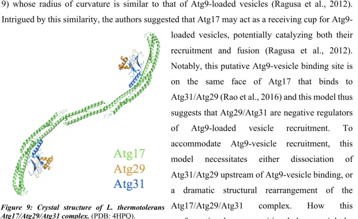
Figure 9: Crystal structure of L. thermotolerans Atg17/Atg29/Atg31 complex 24
Figure 10: A model for Atg17/Atg29/Atg31 complex formation 27
Figure 11: Atg13 crosslinks AICs 29
Figure 12: A model for the assembly of the pentameric AIC 35
Figure 13: A model for a potential Atg13-mediated control of Atg-9-loaded vesicle gap
distancing and fusion 38
Figure 14: Architecture of S. cerevisiae Atg9 39
Figure 15: Atg9 oligomerization may block spontaneous lipid vesicle fusion 40
Figure 16: A model for Atg9 binding to Atg17 43
Figure 17: A proposed role for Atg1-mediated Atg9 phosphorylation 45
Figure 18: A model for AIC-mediated Atg9-loaded vesicle fusion 47
Figure 19: A single-molecule assay to study Atg17 dimerization 49
Figure 20: Initial constructs used in assay validation 50 50
Figure 21: SNAP-Atg17ΔC1 is a monomer in solution 51
Figure 22: Labeling of SNAP-Atg17 with SNAP ligands 51
Figure 23: Two possible interpretation of appearance and disappearance of fluorescent
spots in single-molecule experiments 52
Figure 24: SNAP-Atg17 and SNAP-Atg17ΔC1 bind to the microscope slide
non-specifically 53
Preface
Autophagy is a highly conserved (Mizushima, 2010; Mizushima et al., 2011; Nakatogawa et al., 2009) homeostasis process whereby cells degrade intracellular proteins, organelles, or pathogens. During times of starvation, autophagy is vital in replenishing pools of biosynthetic precursors through degradation of these cytosolic components; it also plays key roles in responding to cytotoxic stress as it acts to specifically degrade damaged organelles, aggregated proteins, and pathogens (Morishita and Mizushima, 2019). Highlighting the importance of this process, defects in autophagy have been linked to neurodegeneration, cancer, infection and aging (Galluzzi et al., 2015; Gomes and Dikic, 2014; Menzies et al., 2015; Mizushima and Komatsu, 2011). Much of our molecular understanding of autophagy comes from seminal genetic screens carried out in the budding yeast Saccharomyces cerevisiae in the 1990s, which lead to a nearly complete inventory of 42 autophagy-related (Atg) proteins (Kabeya et al., 2007; Kawamata et al., 2005; Klionsky et al., 2003; Mizushima et al., 2011; Morishita and Mizushima, 2019; Tsukada and Ohsumi, 1993). In contrast, the human autophagy protein inventory is incomplete with new components constantly being uncovered. Because our understanding of human autophagy is more limited (Bento et al., 2016), I will focus on yeast autophagy in this work.
Autophagy can be triggered by multiple cellular stresses (Hurley and Young, 2017; Klionsky, 2007; Mizushima et al., 2011) including starvation-induced autophagy, which is the best characterized (Fujioka et al., 2014; Kabeya et al., 2009; Kamada et al., 2000; Nakatogawa et al., 2009) and the focus of this work. In yeast, nutrient starvation induces the formation of a protein complex known as the Atg1 complex or autophagy initiation complex (AIC) which assembles in a punctate structure at a perivacuolar location known as the phagophore assembly site (PAS) (Kawamata et al., 2008; Noda and Inagaki, 2015; Suzuki et al., 2001, 2007). There, the AIC recruits small membrane vesicles (Rao et al., 2016; Reggiori et al., 2005; Sekito et al., 2009; Suzuki et al., 2007, 2015) and is thought to catalyze their fusion (Rao et al., 2016; Yamamoto et al., 2012), which leads to formation of a cup-shaped membrane structure known as the phagophore. The growing phagophore then engulfs cytoplasmic cargo, forming a double-membrane vesicle known as the autophagosome (Hurley and Young, 2017). Next, the autophagosomal outer membrane fuses with the vacuole exposing the inner membrane and the engulfed cargo to the degradative capacity
of vacuolar hydrolases, which degrade these components to their biosynthetic precursors (e.g. amino acids, nucleotides, lipids). Release of these biosynthetic precursors replenishes their cytosolic pools and acts to repress autophagy through a negative feedback loop (Kaur and Debnath, 2015; Morishita and Mizushima, 2019; Onodera and Ohsumi, 2005). Given the efficacy of the lysosomal system in degrading autophagic cargo, the pathway is highly regulated (He, 2010) and many factors are dedicated to substrate selection (Morishita and Mizushima, 2019).
Starvation-induced autophagic substrate selection can be divided into three categories: non-selective, in which autophagosomes engulf cargos irrespectively of their identity; exclusive, in which an autophagosome specifically and exclusively engulfs a single cargo using adaptor proteins (Lynch-Day and Klionsky, 2010; Morishita and Mizushima, 2019); and selectively, in which a single autophagosomes contains both non-selective cargoes as well as specific targets that have been selected using dedicated adaptors proteins (Lynch-Day and Klionsky, 2010). These subdivisions of starvation-induced autophagy, however, have not been thoroughly studied in the existing starvation-induced autophagy literature and, for this reason, I will not differentiate between them here. Importantly, though I aim to detail common features of these three types of substrate selection in starvation-induced autophagy, further experimental work is required to confirm that the observations that I highlight here are in fact shared by the three categories of substrate selection.
Of the 42 known autophagy-related proteins, 18 are involved in autophagosome formation and they have been classified into six functional groups (Noda and Inagaki, 2015) that are thought to arrive at the PAS in a specific order (Suzuki et al., 2007). These groups include: the AIC; the transmembrane protein Atg9; the autophagy-specific phosphatidylinositol 3-kinase (PI3K) complex; the Atg2-Atg18 complex; and the Atg8 and Atg12 conjugation systems. In this work, I will specifically focus on the AIC and on our current mechanistic understanding of its assembly and function.
The AIC is made up of 5 proteins: Atg1, Atg13, Atg17, Atg29 and Atg31 (Köfinger et al., 2015). While few mechanistic biochemical studies have been performed, the AIC is generally thought to assemble from two pre-assembled subcomplexes, Atg1/Atg13 and Atg17/Atg29/Atg31
Chapter 2, I focus on interactions within the Atg1/Atg13 and the Atg17/Atg29/Atg31 subcomplexes, respectively. In Chapter 3, I describe the interactions between these two subcomplexes that give rise to the active pentameric AIC. In Chapter 4, I discuss the recruitment of lipid vesicles and their potential fusion by the AIC. Finally, in Chapter 5, I describe the experimental progress that I have made towards developing a new single-molecule approach to study the AIC and discuss the important questions that this new approach may answer.
Chapter 1: Interactions between Atg1 and Atg13
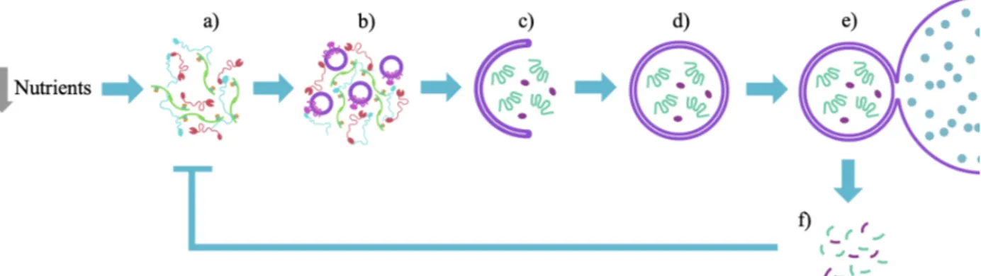
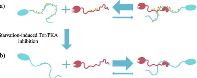
The Atg1 complex, also known as the autophagy initiation complex (AIC), is the first autophagy-related protein complex that assembles upon starvation and it is composed of five proteins: Atg1, Atg13, Atg17, Atg29 and Atg31 (Chew LH et al., 2015; Fujioka et al., 2014; Köfinger et al., 2015; Rao et al., 2016; Stjepanovic et al., 2014). In yeast, the AIC assembles at a perivacuolar punctate structure known as the phagophore assembly site (PAS) (Kawamata et al., 2008; Noda and Inagaki, 2015; Suzuki et al., 2001, 2007) (Figure 1a). Once assembled, the pentameric AIC recruits high-curvature membrane vesicles containing the only essential transmembrane autophagy protein, Atg9 (Noda et al., 2000; Rao et al., 2016; Reggiori et al., 2005; Sekito et al., 2009; Suzuki et al., 2007, 2015) (Figure 1b). The observation that these Atg9-loaded vesicles fuse at the PAS has led to the suggestion that the AIC catalyzes the fusion of Atg9-loaded vesicles and thereby initiates the formation of a cup-shaped membrane structure known as the phagophore (Rao et al., 2016; Yamamoto et al., 2012) (Figure 1c). The growing phagophore then engulfs cytoplasmic cargo, forming a double-membrane structure known as the autophagosome (Hurley and Young, 2017) (Figure 1d). Next, the autophagosomal outer membrane fuses with the vacuole (Figure 1e) exposing the inner membrane and the engulfed cargo to the degradative capacity of vacuolar hydrolases, which degrade these components to biosynthetic precursors such as amino acids, nucleotides, or lipids (Figure 1f). Release of these biosynthetic precursors
Figure 1: Overview of starvation-induced autophagy. Diagram shows key steps in starvation-induced autophagy. Starvation induces AIC assembly (a). Upon assembly, the AIC recruits Atg9-loaded vesicles (b) and is thought to catalyze their fusion to start to form a phagophore, which elongates and engulfs a cytoplasmic cargo (c). The phagophore eventually closes and forms an autophagosome (d). Once the autophagosome is formed, its outer membrane fuses with the vacuole (e) and the vacuolar hydrolases degrade the autophagosome’s cargo and inner membrane, releasing important biosynthetic precursors (f) that negatively feedback on the entire process.
replenishes their cytosolic pools and acts to repress autophagy through a negative feedback loop (Kaur and Debnath, 2015; Morishita and Mizushima, 2019; Onodera and Ohsumi, 2005). Given the efficacy of the lysosomal system in degrading autophagic cargo, this pathway is highly regulated (He, 2010) and many factors are dedicated to substrate selection (Morishita and Mizushima, 2019). Notably, whether the pentameric AIC is sufficient to mediate Atg9-loaded vesicle fusion remains an open question of debate. Irrespectively, proper AIC assembly at the PAS plays a vital role in initiating autophagy, which highlights the importance of understanding how the AIC assembles and how this assembly is regulated.
While few mechanistic studies have been performed, the AIC is thought to be assembled from two pre-assembled subcomplexes, Atg1/Atg13 and Atg17/Atg29/Atg31 (Stjepanovic et al., 2014). In this chapter, I will discuss the known interactions between Atg1 and its regulator, Atg13, and how these interactions give rise to the Atg1/Atg13 subcomplex. Finally, at the end of the chapter, I will briefly discuss Atg17, another regulator of Atg1 that cooperates with Atg13 in the activation of Atg1 (Yeh et al., 2010, 2011).
Atg1: A conserved kinase and scaffolding protein.
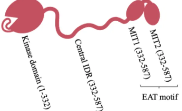
The Atg1 kinase is essential in autophagy and the only component of the AIC with known catalytic activity in addition to scaffolding functions (Chang and Neufeld, 2009; Cheong et al., 2008; Rao et al., 2016; Reggiori et al., 2004a). This dual catalytic/scaffolding role is of interest as the kinase domain could serve as a prime target for the development of broadly acting autophagy modulators with potential therapeutic benefit. This kinase domain resides at Atg1’s N-terminus and is connected through a putative intrinsically disordered region (IDR) to an early autophagy targeting/tethering (EAT) motif (Ragusa et al., 2012) made up of two tandem microtubule-interacting and transport domains (tMIT) named Figure 2: Architecture of Saccharomyces cerevisiae
Atg1. Atg1 consists of an N-terminal kinase
domain (including auto-inhibitory loop), followed by a central putatively disordered region and a C-terminal early autophagy targeting/tethering (EAT) motif, made up of two tandem microtubule-interacting and transport motifs (MIT1 and MIT2). Ovals represent structured regions and squiggles represent putatively disordered regions.
MIT1 and MIT2 (Fujioka et al., 2014) (Figure 2). The scaffolding and kinase functions of Atg1 are thought to be involved at distinct stages of autophagy (Cheong et al., 2008; Sekito et al., 2009). Indeed, the scaffolding functions of Atg1 are required for the recruitment of other AIC components to the PAS whereas the kinase domain is dispensable for PAS formation (Cheong et al., 2008; Kawamata et al., 2008). Instead, kinase activity is required for the recycling of other early autophagy components from the PAS, as evidenced by the fact that Atg9, Atg17 and Atg29 aberrantly accumulate at the PAS in the presence of Atg1 variants bearing catalytically inactive kinase domains (Cheong et al., 2008; Kawamata et al., 2008; Sekito et al., 2009; Shintani and Klionsky, 2004). Additionally, Atg1 kinase activity is essential for autophagy progression (Kamada et al., 2000) and it has been suggested to be responsible for the dissociation of the AIC into subcomplexes by phosphorylating other AIC components (Rao et al., 2016), suggesting an important role of Atg1 in auto-regulation of AIC formation. Thus, understanding Atg1 kinase activation might prove critical in understanding how several diseases and aging cause defects in autophagy (Galluzzi et al., 2015; Gomes and Dikic, 2014; Menzies et al., 2015; Mizushima and Komatsu, 2011). The activation of the Atg1 kinase activity requires other Atg proteins (Kabeya et al., 2005) that I will discuss in the following sections.
Atg13: A highly flexible scaffolding protein.
Atg13 consists of a Hop/Rev7/Mad2 (HORMA) domain at the N-terminus (Jao et al., 2013) and a putative C-terminal IDR (Figure 3). Atg13 is a conserved regulator of Atg1 kinase activity, is essential for autophagy, and is the only AIC component reported to directly bind Atg1 (Chew LH et al., 2015; Stephan et al., 2009). Although Atg13’s HORMA domain is thought to be more structured than its IDR, the HORMA domain can readily undergo order-disordered transitions in physiological conditions (Yamamoto et al., 2016), suggesting that it may need a binding partner to stabilize the fold. Of note, HORMA domains in other proteins bind other HORMA domains (Qi et al., 2015), but the binding partner for S. cerevisiae Atg13 is not known. Finding this partner might be crucial in understanding how the HORMA domain is stabilized, which, as I will soon discuss, could play a role in recruiting Atg9-loaded vesicle to the PAS (Suzuki et al., 2015).
Vital autophagy initiation functions have been mapped throughout the Atg13 sequence. First, the N-terminal HORMA domain is thought to directly interact with Atg9 and to be primarily responsible for Atg9 recruitment to the PAS (Suzuki et al., 2015) (discussed in Chapter 4). Second, short sequences within the C-terminal IDR bind to conserved sites on the Atg17 scaffold protein (Chew LH et al., 2015; Fujioka et al., 2014;
Yamamoto et al., 2016). Finally, additional contacts in the C-terminal IDR bind to Atg1 (Fujioka et al., 2014; Stjepanovic et al., 2014). Taken together, these Atg13 binding sites effectively link the Atg17/Atg29/Atg31 subcomplex to Atg9-loaded vesicles and to the Atg1 kinase (Stjepanovic et al., 2014). Moreover, Atg13 is thought to mediate crosslinking of multiple pentameric AICs (Yamamoto et al., 2016). As I will discuss in Chapter 3, this Atg13-mediated crosslinking
increases the local concentrations of all the components of the AIC, which likely facilitates AIC assembly and Atg9-loaded vesicle fusion to start phagophore formation (further discussed in Chapter 4). In this chapter, however, I focus on the Atg1/Atg13 interactions, leaving the interactions between Atg13 and other AIC components for subsequent chapters.
Starvation-induced assembly of the Atg1/Atg13 sub-complex.
The formation of the AIC is a highly regulated step in autophagy (Chang and Neufeld, 2009; Kamada et al., 2000; Stephan et al., 2009), suggesting that it is an important decision point in the cell. Therefore, I predict that understanding which interactions within the AIC are regulated will provide us with important insights toward the development of therapeutic agents to modulate autophagy. Within Atg13’s C-terminal IDR are two tandem MIT-interacting motifs (tMIM) named MIM-N and MIM-C, that interact with the tMIT motifs of Atg1 (Fujioka et al., 2014; Stjepanovic et al., 2014). The Atg1/Atg13 interaction is essential for autophagy (Kawamata et al., 2008) and it is mediated by binding of these motifs in an anti-parallel fashion: MIM-N (Atg13) binds MIT2 (Atg1) and MIM-C (Atg13) binds MIT1 (Atg1) (Fujioka et al., 2014). Of note, in vitro
hydrogen-Figure 3: Architecture of Saccharomyces cerevisiae Atg13. HORMA domain contains Atg9 binding site; 17BR1, 17BR2 and 17LR are Atg17 binding motifs; MIM-N and MIM-C are binding Atg1 binding sites. 17BR1, 17BR2, 17LR, MIM-N and MIM-C contain serines thought to be phosphorylated by Tor.
deuterium exchange (HDX) experiments showed that Atg1’s tMIT motif is highly dynamic in isolation but becomes more rigid upon binding to the Atg13’s tMIMs (Stjepanovic et al., 2014), suggesting a potential role of this binding event in the stabilization of the Atg1tMIT motif. This
putative stabilization of Atg1tMIT motif would be important because, as I will further discuss in
Chapter 4, the Atg1tMIT motif has been suggested to play a role in the selective recruitment of high
curvature Atg9-loaded vesicles to the PAS (Ragusa et al., 2012; Rao et al., 2016). Additionally, having linked binding of two tandem motifs allows for tighter overall affinity through avidity effects and through the effects of tethering on the effective concentration of the individual motifs, while potentially allowing for more facile regulation, as both motifs could exchange and expose themselves to regulatory elements, such as the kinases and phosphatases that I will discuss in the following sections. Concordantly, isothermal titration calorimetry (ITC) experiments showed that, when compared in isolation, Atg13MIM-N is a stronger binding site for Atg1tMIT than Atg13MIM-C;
however, adding Atg13MIM-C to Atg13MIM-N greatly enhances the overall binding for Atg1tMIT
(Fujioka et al., 2014).
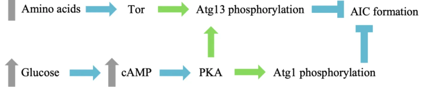
Both Atg1 and Atg13 are phosphoproteins whose phosphorylation states depend on the nutritional state of the cell, suggesting that they may be primary starvation sensors (Budovskaya et al., 2005; Stephan et al., 2009) (Figure 4). Under fed conditions, residues within Atg1’s central IDR are phosphorylated by the (cyclic AMP)-dependent protein kinase (PKA) (Budovskaya et al., 2005). Upon carbon starvation, PKA is inactivated (Thevelein et al., 2000) and Atg1 is readily dephosphorylated by cellular phosphatases, which, through an unknown mechanism, results in recruitment of Atg1 to the PAS (Budovskaya et al., 2005). Similarly, Atg13 is phosphorylated by nutrient-dependent kinases PKA and Tor (target of rapamycin) under fed conditions and becomes
Figure 4: Carbon and nitrogen sensing by Atg1 and Atg13. Blue arrows represent activation, green arrows represent phosphorylation, blue Ts represent inhibition and gray arrows represent increases in intracellular concentrations.
hypo-phosphorylated upon nutrient starvation (Stephan et al., 2009). Of note, Tor is inactivated upon amino acid starvation (Yuan et al., 2017), which, in addition to PKA would make Atg13 an important sensor for carbon and nitrogen starvation. Although phosphomimetic substitutions in Atg13’s tMIM are known to decrease the affinity for Atg1’s tMIT (Fujioka et al., 2014), it is still controversial whether the Atg1/Atg13 subcomplex forms exclusively under starvation conditions or whether it is a constitutive complex independent of the cells nutritional state (Fujioka et al., 2014; Kamada et al., 2000; Kraft et al., 2012). Indeed, initial evidence arguing for a constitutive interaction was based on observed coimmunoprecipitation of Atg1 and Atg13 from fed cells (Kraft et al., 2012), but more recent observations argue that Atg13 is readily dephosphorylated during lysis in the absence of phosphatase inhibitors, suggesting that the apparent constitutive interaction was an experimental artifact (Fujioka et al., 2014). Nevertheless, the MIMs of Atg13 contain several Tor-target serine residues within MIM-C and within the flexible loop between MIM-N and MIM-C (Fujioka et al., 2014), suggesting that phosphorylation and dephosphorylation may play a regulatory role in this interaction, whether or not Atg1 and Atg13 are constitutively bound or not. Determining whether the Atg1/Atg13 is dynamic is important as it will allow us to better understand the key autophagy initiation interactions that cells have evolved to regulate, and could highlight key interactions to target therapeutically.
Atg1 kinase activation.
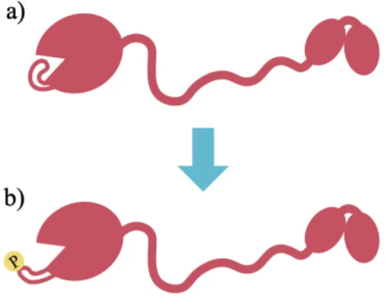
The Atg1 kinase is the most upstream catalytic component of autophagy and is essential to transduce signals in response to cellular stress (Papinski et al., 2014; Reggiori et al., 2004a; Stephan et al., 2009; Yeh et al., 2010). Thus, understanding how this kinase is activated could provide useful insights into how disease and aging affect autophagy (Galluzzi et al., 2015; Gomes and Dikic, 2014; Menzies et al., 2015; Mizushima and Komatsu, 2011). Under fed conditions, Atg1 kinase activity is auto-inhibited through an intrinsic activation loop that blocks the active site (Figure 5a); however, upon starvation, Atg1 auto-activates in an Atg13-dependent manner by phosphorylating its activation loop, resulting in displacement of this loop and exposure of the active site (Kamada et al., 2000; Kijanska et al., 2010; Yeh et al., 2010) (Figure 5b). This Atg1 activation is essential for autophagy progression (Yeh et al., 2010) and Atg1 is known to play critical roles in phosphorylating some autophagy proteins downstream of the phagophore formation process (Papinski et al., 2014). Additionally, Atg1 is reported to phosphorylate two AIC
components (Atg13 and Atg29), which occurs concomitantly with dissociation of the AIC and, presumably a cessation of autophagic initiation (Rao et al., 2016). How these seemingly opposing kinase activities – positive regulation of the downstream components and negative-feedback regulation of the AIC – are temporally regulated to support AIC disassembly is an open area of research. A possible mechanism is that all these protein substrates of Atg1 evolved to have different association constants with Atg1, which favors a specific order of Atg1-mediated phosphorylation.
Mechanistically, this critical Atg1 activation reaction could occur either by auto-phosphorylation in cis through direct auto-phosphorylation of one kinase domain’s own activation loop, or in trans, with one kinase domain phosphorylating the activation loop of a second Atg1 kinase domain. This distinction is important as the in trans
model necessitates assembly of higher order AICs, and motivates our goal to understand how such Atg1-Atg1 multimers might assemble, disassemble, interact with other autophagy proteins, and respond to cellular stimuli. In support of the in trans model, addition of an exogenous dimerization domain to Atg1 increases autophosphorylation in the absence of starvation (Yeh et al., 2011), which suggests that endogenous autophagy proteins could facilitate Atg1 dimerization and transphosphorylation in a nutrient-dependent manner. Indeed, chemical
induction of autophagy using the Tor inhibitor rapamycin increases Atg1 self-interaction (Yeh et al., 2011) and activation loop phosphorylation in an Atg13-dependent manner in vivo (Kabeya et al., 2005; Yeh et al., 2011). These data suggest that Atg13 is at least partially responsible for bringing two Atg1 molecules in close proximity and allowing for autophosphorylation. Of note, these co-immunoprecipitation (co-IP) studies cannot exclude a model in which Atg13 forms a complex with several Atg1 molecules and induces autophosphorylation of each Atg1 kinase in cis. A final observation supporting the in trans model is that the isolated Atg1EAT motif is a dimer in
Figure 5: Conformations of inactive and active Atg1 kinase. a) Auto-inhibited conformation of Atg1 in which the activation loop is unphosphorylated and blocking the active site. b) Active conformation of Atg1 in which the phosphorylated activation loop can no longer block the active site.
solution irrespectively of the presence of Atg13 (Ragusa et al., 2012; Stjepanovic et al., 2014), suggesting that Atg1 might have an intrinsic mechanism for self-assembly that could be facilitated by other AIC components. Nonetheless, full length Atg1 was reported to be monomeric in solution (Rao et al., 2016), suggesting that full-length Atg1 self-interactions may require additional factors.
In addition to Atg13, maximal activation of Atg1 kinase activity requires Atg17, another regulator of the starvation-induced Atg1 kinase activity (Yeh et al., 2010, 2011). While I will detail the structure and known interactions of Atg17 in subsequent chapters, I highlight a few key observations of its role in Atg1 activation here. In vivo co-IP experiments (Kabeya et al., 2005) and in vitro cross-linking mass spectrometry experiments done on the pentameric AIC (Chew LH et al., 2015) suggest that Atg13 is the only AIC component that directly interacts with Atg1. These observations suggest that Atg17 may contribute to Atg1 kinase activation indirectly. Of note, the described crosslinking experiments could not conclusively exclude direct Atg1-Atg17 interactions as they relied on a truncated version of Atg1 and such co-IP and XL-MS experiments are generally prone to false-negatives. While evidence of a direct Atg1-Atg17 interaction is lacking, it is clear that Atg17 plays some role in activating the kinase. Specifically, knocking out Atg17 reduces Atg1-mediated phosphorylation of myelin basic protein in an in vitro phosphorylation assay (Kamada et al., 2000). Interestingly, it was also noted that knocking out Atg17 does not have an effect on Atg1 self-association (Yeh et al., 2011), suggesting that Atg17 contributes to Atg1 kinase activation by a different and unknown mechanism.
Open questions on Atg1/Atg13 complex formation.
An important open question on the formation of the Atg1/Atg13 complex is the true role of the Atg1EAT motif in the overall process (Ragusa et al., 2012). Indeed, as discussed above, its
oligomeric state is disputed (Ragusa et al., 2012; Rao et al., 2016) and, as I will discuss in Chapter 4, the Atg1EAT motif was reported to bind and tether high curvature lipid vesicles in vitro, which
could be important in what is thought to be the key activity of the AIC – recruiting and potentially fusing Atg9-loaded vesicles at the PAS (Ragusa et al., 2012; Rao et al., 2016). Reconciling these apparently contradicting evidence surrounding the oligomeric state(s) of Atg1 at the PAS could provide both important insights into the Atg1 auto-phosphorylation mechanism, and also help us better understand how the pentameric AIC tethers and potentially fuses high-curvature lipid
vesicles to start autophagosome biogenesis. Despite the controversy, henceforth I will assume that full-length Atg1 is monomeric and that the Atg1EAT motif does not tether Atg9-loaded vesicles
based on my analysis of the existent data.
A model for Atg1/Atg13 complex formation.
Based on the described data, I propose the following model for the formation of the Atg1/Atg13 (Figure 6): Under fed conditions, Atg1 is phosphorylated at its central IDR by PKA and Atg13 is phosphorylated at its C-terminal IDR by PKA and Tor. In their phosphorylated forms, Atg1/Atg13 complex formation is disfavored (Figure 6a). Upon nutrient starvation, however, the Tor and PKA kinases are inactivated, leading to phosphatase-mediated dephosphorylation of Atg1 and Atg13. According to my model, dephosphorylation of Atg1 and Atg13 then induces conformational changes in these proteins that allow them to bind through a rigid interface in a 1:1 stoichiometry that stabilizes the Atg1EAT motif. In Chapter 3, I will also argue that Atg17 facilitates
Atg13 self-interactions that in turn bring two Atg1 molecules into close proximity to allow for transphosphorylation.
Figure 6: Model for the assembly of the Atg1/Atg13 complex. a) Under fed conditions, the IDRs of Atg13 and Atg1 are phosphorylated, which disfavors Atg1/Atg13 complex formation. b) The starvation-induced inhibition of Tor and PKAs results in the phosphatase-mediated dephosphorylation of Atg1 and Atg13, allowing the proteins to adopt a conformation that allows them to bind through a rigid interface that stabilizes the Atg1EAT motif. Arrows represent chemical equilibria.
Chapter 2: Interactions between Atg17, Atg29 and Atg31
Upon starvation, the Atg1/Atg13 complex described above is thought to interact with the starvation-specific Atg17/Atg29/Atg31 subcomplex (Kabeya et al., 2009; Kawamata et al., 2008) giving rise to a pentameric AIC (Stjepanovic et al., 2014). As I will discuss in Chapter 4, this pentameric AIC thought to play a crucial role in the starvation-induced recruitment and fusion of Atg9-loaded vesicles required for autophagosome formation (Ragusa et al., 2012; Rao et al., 2016). Despite the known importance of the Atg17/Atg29/Atg31 complex, the mechanism of formation of this subcomplex is poorly understood and is ripe for exploration. In this chapter, I will discuss what is known mechanistically about the formation of the Atg17/Atg29/Atg31 subcomplex and what open questions remain.
Atg17: An AIC organizer.
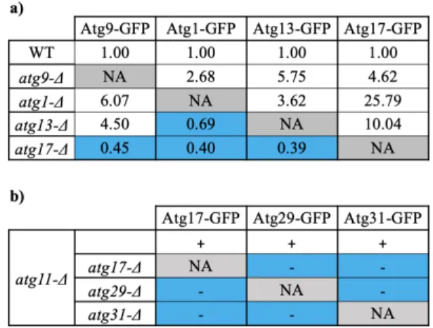
In addition to facilitating Atg1 activation (Yeh et al., 2010, 2011), Atg17 is thought to be the main organizer of the PAS (Rao et al., 2016; Suzuki et al., 2007), and one of the first proteins to arrive there (Kawamata et al., 2008; Suzuki et al., 2007). In support of this organizational role, genetic epistasis experiments showed that Atg17 was required for rapamycin-dependent PAS localization of Atg1, Atg13 and most of the other early autophagy Atg proteins, but that Atg17 itself could localize to the PAS in the absence of these other components (Table 1a). Importantly, as I will discuss below, the only two exceptions found (Atg29 and Atg31) are thought to form a constitutive complex with Atg17 and arrive at the PAS as a complex (Kabeya et al., 2009; Kawamata et al., 2008). In further support of Atg17’s organizational role, quantitative fluorescence
Table 1: Genetic epistasis experiments suggesting that Atg17 is a central organizer of the AIC. a) Relative fluorescence microscopy quantification of the PAS localization of the proteins specified in the columns in the strains specified in the rows. Blue cells indicate combinations in which the PAS localization of the protein specified in the column was disrupted by the genetic ablation of the gene indicated in the row. (Adapted from Suzuki et al, 2007). b) Qualitative analysis of PAS localization of the proteins specified in the columns in the strains specified in the rows; + indicates PAS localization and - indicates no PAS localization; blue cells indicate combinations in which the PAS localization of the protein specified in the column was disrupted by the genetic ablation of the gene indicated in the row. Of note, experiments in b) were carried out in an atg11-Δ background to discard effects of non-starvation-induced autophagy (see Lynch-Day and Klionsky, 2010).
microscopy experiments suggest that Atg17 is constitutively present at the PAS (Köfinger et al., 2015), and artificial targeting of Atg17 to the cell membrane by an exogenous transmembrane domain results in other Atg proteins forming a PAS-like structure at the membrane (Suzuki et al., 2007). Furthermore, as I will further discuss in Chapter 4, Atg17 has also been suggested to have an Atg9-binding activity (Rao et al., 2016). Taken together, these observations highlighting the importance of studying Atg17 interactions with other AIC components in understanding autophagy initiation.
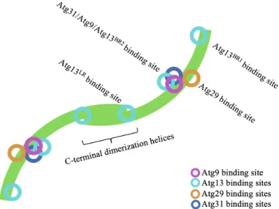
Structural studies have shown that Atg17 homodimerizes via a C-terminal helix (Ragusa et al., 2012) (Figure 7) and this dimerization is essential for PAS organization and for autophagy progression (Ragusa et al., 2012; Rao et al., 2016). While Atg17 is generally thought to be a constitutive dimer (Chew et al., 2013; Köfinger et al., 2015; Rao et al., 2016), neither the dimerization kinetic constants nor the dimerization equilibrium constant has been reported, raising the possibility that Atg17 dimers may in fact rapidly exchange with monomers under physiological conditions. As I will discuss in Chapters 3 and 4, Atg13 binds at the Atg17 dimer-dimer interface, and Atg17 dimerization has been suggested to play a role in regulating the Atg9-binding activity of Atg17 (Rao et al., 2016). Thus, figuring out whether the dimers are constitutive could provide important mechanistic insights into how the AIC is assembled and how it recruits and fuses Atg9-loaded vesicles.
As evidenced by crystal structures, Atg17 forms a complex with the Atg29 and Atg31 members of the AIC (Kawamata et al., 2008; Köfinger et al., 2015; Ragusa et al., 2012; Stjepanovic
Figure 7: Binding sites for other AIC components on the Atg17 dimer. Schematic of the Atg17 dimer (green) indicating the location of the binding sites for Atg9, Atg13, Atg29 and Atg31. Binding sites shown are based on the crystal structure of the Atg17/Atg29/Atg31 trimeric complex (PDB: 4HPQ), which has been abstracted for simplicity.
et al., 2014), which have been suggested to regulate the activity of Atg17 (Rao et al., 2016). While I will further discuss this putative regulation in chapters 3 and 4, here I discuss what is known about the mechanism of assembly of the Atg17/Atg29/Atg31 trimeric complex.
Atg29: A poorly characterized, weakly conserved Atg1 scaffolding component.
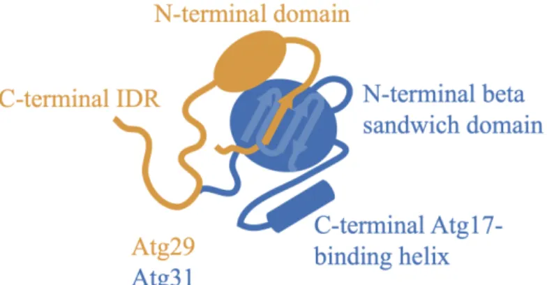
As I will discuss in Chapter 4, Atg29 is a putative regulator of Atg17’s ability to bind to Atg9 and, like Atg17, Atg29 is required for full starvation-induced autophagic activity (Kawamata et al., 2005; Mao et al., 2013). Atg29 is composed of two distinct regions (Figure 8): a folded N-terminal domain that associates with Atg31 through a beta sandwich formed via strand exchange between the both proteins (Ragusa et al., 2012) (Figure 8), and a large C-terminal region that is predicted to be intrinsically disordered and is thought to somehow regulate autophagy in a phosphorylation-dependent manner
(Mao et al., 2013). Specifically, the starvation-induced phosphorylation of the three most C-terminally located serine residues was shown to be essential for full autophagic activity, but neither the responsible kinase(s), nor the mechanistic role of these phosphorylation events was reported (Mao et al., 2013). Of note, at separate study (Rao et al., 2016) reported that Atg1 phosphorylates the C-terminus of
Atg29 in vitro upon addition of ATP to the pentameric AIC, suggesting that Atg1 is responsible for these key Atg29 phosphorylation events. It remains an open question, however, whether there is another kinase with partial redundancy with Atg1 that may also phosphorylate the C-terminal IDR of Atg29 and thus regulate autophagy progression, and which of the 23 serine residues in the C-terminal IDR of Atg29 (Mao et al., 2013) are important for this regulation. Answering these important questions may not only provide us with a new potential kinase target for small molecule drugs, but also help understand what appears to be an essential step in autophagy progression.
Figure 8: Architecture of the Atg29/Atg31 complex. Schematic of the Atg29/Atg31 dimer based on the crystal of the trimeric Atg17/Atg29/Atg31 complex (PDB: 4HPQ). Atg29 consists of an N-terminal folded domain followed by a putative C-terminal IDR. Atg31 consists of an N-terminal beta sandwich consisting of beta strands from Atg29 and Atg31, and a C-terminal helix that interacts with Atg17.
While a specific mechanistic role of Atg29 phosphorylation in autophagic progression has not yet been elucidated, it is clear that Atg29 phosphorylation is dispensable for formation of the Atg29/Atg31 or the Atg17/Atg29/Atg31 subcomplexes (Mao et al., 2013). Indeed, I and other groups have expressed and purified Atg29/Atg31 in E. coli as a complex (Chew et al., 2013; Chew LH et al., 2015; Fujioka et al., 2014; Köfinger et al., 2015; Ragusa et al., 2012; Rao et al., 2016; Stjepanovic et al., 2014), presumably in their unphosphorylated forms, and this dimer can be assembled with Atg17 also produced in E. coli, suggesting that Atg29 phosphorylation is dispensable for binding to Atg17. Nevertheless, in Chapter 4, I argue that Atg29 phosphorylation could play a role in the activation of the Atg9-binding activity of Atg17. In the subsequent chapters, I discuss other critical AIC interactions that motivate and provide context for this activation model.
Atg31: A key facilitator of Atg17 and Atg29 association.
The last component of the trimeric complex discussed in this chapter, Atg31, is also necessary for full starvation-induced autophagic activity (Kabeya et al., 2007) and it contains two predicted IDRs that span roughly 50% of the protein (Feng et al., 2015) (Figure 8). Like the Atg13 and Atg29 IDRs, these Atg31 IDRs contain several mapped phosphorylation sites. While the cognate kinases are not-yet-identified, Atg1 has been excluded as these sites are phosphorylated in an Atg1-null background (Feng et al., 2015; Kabeya et al., 2009). In contrast to Atg29, Atg31 is reported to be constitutively phosphorylated in vivo (Kabeya et al., 2009), which has been interpreted to imply a structural role for the marks as opposed to a regulatory function. Notably, phosphorylation of serine 174 is essential for full autophagic activity, and introduction of a S174A phospho-null mutant results in aberrant accumulation of Atg9-loaded vesicles at the PAS (Feng et al., 2015). These data suggest that Atg31 phosphorylation plays a role in Atg9 recycling, however, how this mark acts structurally or functionally is unclear (Feng et al., 2015). As detailed for Atg29 above, these Atg31 phosphorylation marks must be dispensable for Atg29/Atg31 and Atg17/Atg29/Atg31 subcomplex assembly as we and others have reconstituted these complexes from components expressed and purified in E. coli (Chew et al., 2013; Chew LH et al., 2015; Köfinger et al., 2015; Ragusa et al., 2012; Rao et al., 2016; Stjepanovic et al., 2014), suggesting that pS174 phosphorylation may be required in an autophagy step downstream of Atg17/Atg29/Atg31 assembly. It should be noted that no mass spectrometry data has been reported on these purified complexes, so we cannot discard potential E. coli phosphorylation sites. Notably,
Atg31 S174 is located at the Atg17 binding interface, and molecular dynamic simulations suggested that pS174 aids in formation of an additional Atg31 alpha helix that interacts with Atg17 and further stabilizes the complex (Feng et al., 2015). As I will discuss in Chapter 4, the Atg29/Atg31 complex has been suggested to inhibit the Atg9-loaded vesicle binding activity of Atg17 (Rao et al., 2016), raising the possibility that pS174 may be important in Atg17 activation. Furthermore, S174 could be subject to phosphatases and kinases and thus modulate Atg31/Atg17 association and downstream AIC assembly.
Atg29/Atg31: An obligate dimer.
Atg29 and Atg31 assemble as a stable dimeric complex (Rao et al., 2016) and, consistent with an obligate dimer, each protein is highly aggregation prone when expressed in isolation in E.
coli, but stable when co-expressed (Kabeya et al., 2009; Mao et al., 2013; Rao et al., 2016). This
initial observation that Atg29 and Atg31 might help each other fold was substantiated by a crystal structure of the Atg17/Atg29/Atg17 complex from the thermophilic yeast Lachancea
thermotolerans, which is closely related to S. cerevisiae (Ragusa et al., 2012). This structure
showed that Atg29 and Atg31 interact via a sandwich beta sheet domain in which one beta sheet uses strands from both Atg29 and Atg31 (Ragusa et al., 2012). While it has not been reported whether these proteins can adopt alternative folds in isolation or if they can exchange with monomers, based on the structural data, I will treat them as an obligate heterodimer for the remainder of this discussion.
Recent epistasis experiments showed that Atg17, Atg29 and Atg31 are mutually essential for their PAS localization upon rapamycin treatment (Kawamata et al., 2008), suggesting that the three proteins arrive at the PAS as a complex (Table 1b). This evidence, together with the suggestion that Atg17 is constitutively present at the PAS (Köfinger et al., 2015), raises the possibility that Atg29 and Atg31 are also constitutively present at the PAS, but this Atg29/Atg31 localization assay has not been directly performed. The genetic data described above recapitulate biochemical observations consistent with a constitutive complex, as whole-cell lysate gel filtration experiments showed that the three proteins form a stable complex independent of the nutritional state of the cell (Kabeya et al., 2009). Nevertheless, no dimerization kinetic or thermodynamic parameters of complex formation have been reported and it remains possible that formation of the
Atg17/Atg29/Atg31 complex could be dynamic and regulated. As I will discuss in Chapter 4, the Atg29/Atg31 dimer has been suggested to compete with Atg13 and Atg9 for binding to Atg17. Testing this model, which could provide us with important clues of how Atg9-loaded vesicles are recruited to the PAS and subsequently fused, will require further understanding of the dynamics of Atg17/Atg29/Atg31 complex formation.
The Atg17/Atg29/Atg31 trimer: a putative vesicle scaffold.
As I will further discuss in Chapter 4, the Atg17/Atg29/Atg31 complex is suggested to act as a vesicle scaffold and to have an important role in the AIC-mediated fusion of Atg9-loaded vesicles (Ragusa et al., 2012; Rao et al., 2016). The crystal structure of the Atg17/Atg29/Atg31 trimeric complex showed that Atg17 adopts an interesting S-shaped double crescent shape (Figure 9) whose radius of curvature is similar to that of Atg9-loaded vesicles (Ragusa et al., 2012). Intrigued by this similarity, the authors suggested that Atg17 may act as a receiving cup for
Atg9-loaded vesicles, potentially catalyzing both their recruitment and fusion (Ragusa et al., 2012). Notably, this putative Atg9-vesicle binding site is on the same face of Atg17 that binds to Atg31/Atg29 (Rao et al., 2016) and this model thus suggests that Atg29/Atg31 are negative regulators of Atg9-loaded vesicle recruitment. To accommodate Atg9-vesicle recruitment, this model necessitates either dissociation of Atg31/Atg29 upstream of Atg9-vesicle binding, or a dramatic structural rearrangement of the Atg17/Atg29/Atg31 complex. How this conformational or compositional change might be regulated is completely unexplored and motivates the biochemical experiments described in Chapter 5. Interestingly, negative stain electron micrographs of Atg17 imaged in isolation showed that while the C-terminal dimerization interface is stable, the N-terminus of Atg17 is flexible, which enables the dimer to expand and contract like a spring (Chew LH et al., 2015). In contrast, addition of Atg29/Atg31 stabilizes this spring-like motion and generally rigidifies the complex. Figure 9: Crystal structure of L. thermotolerans
These data suggest that Atg29 and Atg31 might play a role in stabilizing the rigid S-shape that Atg17 adopts in the trimeric complex (Chew et al., 2013). Despite these structural studies, whether Atg29/Atg31 affect Atg17 dimerization and whether the Atg29/Atg31 ‘shape lock’ is necessary for binding to other AIC components remain open questions.
Outstanding questions and a path forward.
The role of Atg31 phosphorylation in formation of this trimeric complex is entirely unexplored and warrants detailed biochemical investigation. We need to determine basic kinetic and thermodynamic parameters for assembly of this complex in the presence of phospho-null and phospho-mimetic mutants to begin untangling how these modifications might regulate the assembly process.
Another outstanding question in the formation of the Atg17/Atg29/Atg31 dimer of trimers is the allowed binding order(s). It has been shown that Atg17 can dimerize on its own (Chew LH et al., 2015), that pre-made Atg29/Atg31 dimer can bind Atg17 in vitro (Rao et al., 2016), and that a monomeric mutant of Atg17 can bind Atg29/Atg31 (Ragusa et al., 2012). These data suggest one possible binding order (Atg29/Atg31 dimer binding Atg17 monomers before Atg17 dimerization). Nonetheless, whether other binding orders are possible and the frequency with which each path is utilized remains an open question. For instance, we do not know whether Atg29/Atg31 dimer can rigidify Atg17 monomers into a curved shape, and whether this curved monomeric Atg17 is capable of dimerizing to give rise to a full double crescent. The current biochemical and biophysical approaches to study the Atg17/Atg29/Atg31 complex have not been able to answer these important questions.
Finally, it remains unclear whether Atg29 directly contacts Atg17. Although the crystal structure of the trimeric Atg17/Atg29/Atg31 complex suggests that the folded domain of Atg29 lacks direct contacts with Atg17 (Ragusa et al., 2012), this structure could not resolve Atg29’s C-terminal IDR, and could not exclude contacts between Atg29 and Atg17. In fact, Atg29 can co-immunoprecipitate Atg17 in an E. coli host, and crosslinking-mass spectrometry experiments done on the pentameric Atg1complex implied that the C-terminal tail of Atg29 binds to the concave face of Atg17 (Chew LH et al., 2015). Although it is unknown whether this putative Atg17/Atg29
interaction also occurs in the isolated tetrameric Atg17/Atg29/Atg31 complex, the fact that this putative interaction between Atg17 and Atg29 seems to be mediated by the part of Atg29 that becomes phosphorylated upon starvation (Mao et al., 2013) suggests a new potential in vivo-regulated step in autophagy initiation. I propose that tight binding of Atg29/Atg31 to Atg17 is driven by multiple dynamic interactions between Atg31 and Atg29 with Atg17. This way, cells can simultaneously have very tight binding between Atg17 and the Atg29/Atg31 dimer while allowing for easy access of kinases and phosphatases that may regulate this complex. In Chapter 4, I will discuss a model that I propose in which Atg1-mediated phosphorylation of the C-terminal IDR of Atg29 reduces the binding affinity of the Atg29/Atg31 dimer, helping to displace the Atg29/Atg31 dimer from the Atg9 binding site on Atg17 and thus activate the Atg9-loaded vesicle binding activity of the AIC.
A model for Atg17/Atg29/Atg31 assembly.
Based on the data described above, I propose the following model for Atg17/Atg29/Atg31 formation (Figure 10): Atg17, Atg29 and Atg31 are present at the PAS constitutively and they are in a dynamic equilibrium. Atg17 exchanges between monomers and dimers (Figure 10a) while constitutive Atg29/Atg31 dimers quickly alternate between several binding sites for Atg17 (Figure 10b) without affecting the Atg17 monomer-dimer equilibrium. Notably, although this dynamic equilibrium model for the binding of the Atg29/Atg31 dimer to Atg17 implies the existance of a population of dissociated Atg29/Atg31 dimer, I propose that the equilibrium strongly favors the trimeric Atg17/Atg29/Atg31 complex and that the population of unbound Atg29/Atg31 is negligible. For this reason, I will treat Atg17/Atg29/Atg31 as a constitutive complex in the subsequent chapters. Finally, as I will discuss in Chapter 3, I propose that the starvation-induced binding of Atg13 at the Atg17 dimer interface (Fujioka et al., 2014) stabilizes the Atg17 dimer (Figure 10c) which, as I will discuss in Chapter 4, may be important for the putative Atg9-vesicle fusion activity of the AIC.
Figure 10: A model for Atg17/Atg29/Atg31 complex formation. Atg17, Atg29 and Atg31 are in a dynamic equilibrium: a) Atg17 exchanges between monomers and dimers and b) the constitutive Atg29/Atg31 dimers quickly alternate between several binding sites for Atg17 without affecting the Atg17 monomer-dimer equilibrium. Of note, I propose that there is a small population of dissociated Atg29/Atg31 dimer, but that the equilibrium strongly favors the trimeric complex and this population is thus negligible. c) Upon starvation, Atg13 binds to the Atg17 dimer interface, stabilizing the Atg17 dimer and thus driving the Atg17 monomer-dimer equilibrium towards the dimeric form.
Chapter 3: Assembly of the pentameric AIC
In the previous chapters, I discussed the isolated assembly of the Atg1/Atg13 and Atg17/Atg29/Atg31 subcomplexes, but these two subcomplexes must come together to produce an active AIC able to recruit Atg9-loaded vesicles (Rao et al., 2016), phosphorylate and regulate downstream autophagy components, and, possibly, catalyze Atg9-vesicle fusion to initiate formation of the phagophore. This AIC assembly process is an essential step for autophagy and is thought to be a critical decision point, effectively committing the cell to undergo starvation-induced autophagy (Kabeya et al., 2005; Kamada et al., 2000; Stephan et al., 2009; Yamamoto et al., 2016). Thus, a deep mechanistic understanding of AIC formation is important for understanding cellular regulation of this potentially destructive pathway and will be vital in the development of therapeutics that regulate autophagy. In this chapter, I discuss our current mechanistic understanding of the formation and activation of the pentameric AIC.
Atg13 is a key protein in the formation and activation of the pentameric AIC.
Atg13 is a crucial protein in the assembly of the complete and active AIC. First, it is the physical link between Atg1 and the Atg17/Atg29/Atg31 complex (Fujioka et al., 2014; Köfinger et al., 2015; Stjepanovic et al., 2014) as no direct contacts have been mapped between Atg1 and any of the Atg17/Atg29/Atg31 components. Second, through an unknown mechanism, Atg13 increases the binding affinity of Atg17 for Atg9 (Rao et al., 2016), which is an essential step in phagophore formation. Finally, Atg13 is thought to crosslink multiple pentameric AICs into higher order oligomers (Yamamoto et al., 2016) and thereby locally concentrates AIC components, which may be crucial for AIC assembly and Atg9-vesicle fusion. Below, I discuss each of these important Atg13 functions separately before discussing a model for pentameric AIC formation.
Atg13 physically links the components of the pentameric AIC.
As discussed in Chapter 1, although both Atg13 and Atg17 (Kabeya et al., 2005; Kamada et al., 2000) are required for Atg1 kinase activation, only Atg13 is thought to directly contact Atg1 (Chew LH et al., 2015). Atg17 and Atg1 are instead thought to be linked through Atg13, as short regions in Atg13’s C-terminal IDR are known to bind Atg17 (Fujioka et al., 2014; Yamamoto et al., 2016). This suggests that the important interactions between Atg1 and the Atg17/Atg29/Atg31
complex, such as Atg29 phosphorylation (Mao et al., 2013; Rao et al., 2016), are driven by binding of Atg13 and facilitated by the intrinsic flexibility of this important linker protein. Similar to the regulated Atg1:Atg13 binding discussed in Chapter 1 (Fujioka et al., 2014), this Atg13 linking activity is likely regulated as the Atg17 binding regions on Atg13 contain putative Tor phosphorylation sites that are phosphorylated under basal conditions and become dephosphorylated upon rapamycin treatment (Chew LH et al., 2015; Fujioka et al., 2014; Yamamoto et al., 2016). Consistent with these sites acting as nutrient sensors, phospho-mimetic and phospho-null substitutions affect Atg13/Atg17 binding affinity as expected: phospho-mimetic substitutions generally decrease the KD and phospho-null substitutions generally increase the KD
(Chew LH et al., 2015; Fujioka et al., 2014; Yamamoto et al., 2016). As I discuss below, Atg13 binding to Atg17 is also thought to play an essential role in activating the AIC’s ability to recruit Atg9-loaded vesicles (Rao et al., 2016), which further highlights the importance of tightly regulating these interactions to limit spurious autophagy initiation.
Atg13 mediates the supramolecular assembly of AICs.
In addition to linking Atg1 with the Atg17/Atg29/Atg31 complex, Atg13 may crosslink AICs by simultaneously binding to multiple Atg17 molecules (Yamamoto et al., 2016). Mutational analysis showed that Atg13’s C-terminal IDR includes
at least two Atg17 binding regions – named 17BR1 (for Atg17 binding region 1) and 17LR (for Atg17-linking region) (Yamamoto et al., 2016) (Figure 11). Of note, the Atg1317LR motif binds Atg17 in hydrophobic pocket
at the Atg17 dimer interface (Yamamoto et al., 2016), suggesting that Atg13:Atg17 association could also modulate the Atg17 monomer-dimer equilibrium (see Chapter 2), with dimers favored upon Atg13 binding. Additionally, the observed 17LR binding site suggests that Atg17 dimerization is essential for the supramolecular assembly of AICs, which might explain why C-terminally truncated monomeric Atg17 mutants cannot support starvation-induced autophagy (Ragusa
Figure 11: Atg13 crosslinks AICs. Atg13 binds crosslinks Atg17 dimers through at least 2 Atg17 binding sites on Atg13. Atg17 binding sites are named on diagram. Atg1, Atg29 and Atg31 were omitted for clarity.
et al., 2012). Testing and validation of this model will require measuring thermodynamic and kinetic parameters of Atg17 dimerization and analyzing how the binding of other AIC components affects them. Such a detailed understanding of these interactions is important as Atg17 dimerization is necessary for the Atg9-loaded vesicle binding activity of the pentameric AIC in
vitro (Rao et al., 2016), suggesting that Atg13-mediated stabilization of the Atg17 dimer could be
a crucial step leading to Atg9-loaded vesicle fusion in vivo.
In addition to the two Atg17 binding sites on Atg13 mentioned above, a third Atg17 binding site has been reported, called 17BR2 (Chew LH et al., 2015) (Figure 11). This binding site appears to be dispensable for the supramolecular assembly of AICs and it is weaker than the other two Atg17 binding motifs on Atg13 (Yamamoto et al., 2016). Nevertheless, as I will further discuss in Chapter 4, Atg1317BR2 might play a crucial role in activating the AIC’s Atg9-vesicle binding
activity.
A proposed role of liquid-liquid phase separation in AIC formation and activation.
Using isothermal titration calorimetry (ITC), the Hurley group (Stjepanovic et al., 2014) measured a ~10 μM KD for binding between the preformed Atg1tMIM/Atg13tMIT and
Atg17/Atg29/Atg31 subcomplexes: 2(Atg1: Atg13) + 2(Atg17: Atg29: Atg31) ↔ 2(Atg1: Atg13: Atg17: Atg29: Atg31). Given the extremely low intracellular concentrations of these proteins (~1-100 nM) (Belle et al., 2003), this relatively weak binding affinity is surprising and implies that the Atg1/Atg13/Atg17/Atg29/Atg31 components must be localized and concentrated at the PAS or exist in a tighter binding form (through post-translational modification, for example) to assemble as an active pentameric AIC in vivo. Based on recent preliminary experiments performed by Samantha Webster (personal communication), I will argue that the local concentrations of AIC components are increased via liquid-liquid phase separation and that the PAS is a dynamic, phase-separated condensate of Atg proteins.
Liquid-liquid phase separation (LLPS) is a reversible, dynamic protein assembly phenomenon whereby multivalent interactions between intrinsically disordered proteins separate and concentrate intracellular components in a droplet-like condensate (Boeynaems et al., 2018; Li et al., 2012). All these requirements are fulfilled by the components of the AIC. First, Atg1, Atg9,
Atg13, Atg29 and Atg31 have long predicted intrinsically disordered regions (Feng et al., 2015; Mao et al., 2013; Meia et al., 2014; Yamamoto et al., 2016). Second, as discussed above, Atg13 interacts with Atg1 and Atg17 via multivalent interactions (Yamamoto et al., 2016) and there are likely additional as-yet-uncharacterized interactions between the poorly characterized IDRs of other AIC components. Finally, gel filtration experiments showed that upon dilution, the AIC dissociates into subcomplexes, demonstrating that the AIC assembly is reversible (Yamamoto et al., 2016). For all these reasons, I propose that PAS is a phase-separated condensate that increases the local concentrations of the AIC components and facilitates the formation of the aforementioned ordered contacts in the pentameric AIC. Further, I propose that this behavior is regulated by the starvation-induced dephosphorylation of Atg13 (Stephan et al., 2009) and that by increasing the local concentration of Atg1 at the PAS, LLPS also helps to bring Atg1 kinases in close proximity, allowing for trans-phosphorylation and activation of Atg1 kinase (Figure 12a-h) activity. In Chapter 1, I discussed that Atg13 was essential for the self-assembly and auto-phosphorylation of Atg1 (Yeh et al., 2010, 2011). Based on the LLPS model described above, I propose that upon starvation, dephosphorylation of Atg13 induces not only the formation of the Atg1/Atg13 complex, but also the LLPS of the IDRs of Atg13, Atg1, Atg31 and Atg29 at the PAS. According to my model, this LLPS increases the local concentration of both the Atg1/Atg13 and the Atg17/Atg29/Atg31 complexes at the PAS, which facilitates pentameric AIC formation.
Atg1 phosphorylation causes the dissociation of the AIC.
Above, I have focused on the regulated, rapid assembly of the pentameric AIC, which enables autophagic degradation. Clearly, it is also vital that cells can inhibit this pathway to rapidly stop degrading cytoplasmic components once nutrients become available. Given the putative role of the AIC in cellular decision making discussed in the previous chapters, disassembly of the AIC seems a likely place to downregulate autophagic flux upon nutrient availability. Thus, understanding the disassembly of the AIC is also of great importance. As discussed, nutrient-induced activation of Tor and the subsequent phosphorylation of Atg13 drives dissociation of Atg13 from Atg17/Atg29/Atg31 (Chang and Neufeld, 2009; Kamada et al., 2000; Kaur and Debnath, 2015; Stephan et al., 2009; Thevelein et al., 2000). Additionally, in vitro studies showed that the addition of ATP to a pentameric AIC assembled from purified components results in Atg1-mediated phosphorylation of Atg13 and Atg29 and the concomitant dissociation of the pentameric
AIC into Atg1/Atg13 and Atg17/Atg29/Atg31 subcomplexes (Rao et al., 2016), suggesting that the active Atg1 kinase could negatively feedback on AIC formation. Interestingly, several of the Atg1 phosphorylation sites on Atg13 are serine residues that become dephosphorylated upon rapamycin treatment and map to the tMIM, 17BR1 and 17BR2 motifs of Atg13 (Rao et al., 2016), which are binding sites for Atg1 and Atg17. This apparent partial redundancy of Atg1 and Tor could be beneficial to the cell, as the effective concentration of Atg1 in the pentameric AIC is likely much higher than that of cytosolic Tor, and high local Atg1 concentrations could allow for faster phosphorylation and dissociation of the AIC upon nutrient addition. Moreover, these data also suggest that constant autophagic flux would require continuous signaling and continuous assembly of AIC, which would allow the cell to rapidly stop starvation-induced autophagy once it is no longer needed, consistent with the AIC disassembling within 10 minutes of nutrient re-addition in vivo (Kawamata et al., 2008). While both the activatory and inhibitory roles of Atg1 have been reported, it remains unclear how the cell balances and regulates these disparate functions.
The role of Atg1-mediated phosphorylation of Atg29 on AIC disassembly is unknown. Nonetheless, knocking out the Atg1 kinase activity results in the aberrant accumulation of Atg29 at the PAS, suggesting that Atg29 phosphorylation might play a role in the shuttling Atg29 from the PAS to the cytoplasm (Kawamata et al., 2008). Notably, most of the Atg1 phosphorylation sites on Atg29 are in its C-terminal IDR (Rao et al., 2016), a region of Atg29 that is phosphorylated upon nutrient deprivation and whose phosphorylation was shown to be essential for starvation-induced autophagic activity (Mao et al., 2013). I propose that Atg1 phosphorylates the C-terminal IDR of Atg29 in the context of an assembled AIC and that this modification decreases the binding affinity of the Atg29/Atg31 dimer for Atg17. According to my model, phosphorylation-mediated dissociation of the Atg29/Atg31 dimer could then activate Atg17 for Atg9-loaded vesicle binding. In support of my model, three of the Atg1 phosphorylation sites on the C-terminal tail of Atg29 map to a region of Atg29 that was shown by XL-MS to crosslink to the convex face of Atg17 in the context of the pentameric AIC (Chew LH et al., 2015; Rao et al., 2016).
The evidence described above suggests that there is exquisite regulation of AIC disassembly, which could be a likely place for disease mutations to act. Thus, understanding the