HAL Id: hal-02636301
https://hal.inrae.fr/hal-02636301
Submitted on 27 May 2020
HAL is a multi-disciplinary open access
archive for the deposit and dissemination of
sci-entific research documents, whether they are
pub-lished or not. The documents may come from
teaching and research institutions in France or
abroad, or from public or private research centers.
L’archive ouverte pluridisciplinaire HAL, est
destinée au dépôt et à la diffusion de documents
scientifiques de niveau recherche, publiés ou non,
émanant des établissements d’enseignement et de
recherche français ou étrangers, des laboratoires
publics ou privés.
models of colonic hypersensitivity
Mathieu Meleine, Ludivine Boudieu, Agathe Gelot, Emilie Muller, Amandine
Lashermes, Julien Matricon, Celine Silberberg, Vassilia Theodorou, Alain
Eschalier, Denis Ardid, et al.
To cite this version:
Mathieu Meleine, Ludivine Boudieu, Agathe Gelot, Emilie Muller, Amandine Lashermes, et al..
Com-parative effects of alpha 2 delta-1 ligands in mouse models of colonic hypersensitivity. World
Jour-nal of Gastroenterology, Baishideng Publishing Group Co. Limited, 2016, 22 (31), pp.7111-7123.
�10.3748/wjg.v22.i31.7111�. �hal-02636301�
Mathieu Meleine, Ludivine Boudieu, Agathe Gelot, Emilie Muller, Amandine Lashermes, Julien Matricon, Celine Silberberg, Alain Eschalier, Denis Ardid, Frederic A Carvalho, Clermont Université, Université d'Auvergne, INSERM 1107 Neuro-Dol, F-63000 Clermont-Ferrand, France
Mathieu Meleine, Ludivine Boudieu, Agathe Gelot, Emilie Muller, Amandine Lashermes, Julien Matricon, Celine Silberberg, Alain Eschalier, Denis Ardid, Frederic A Carvalho, Université d’Auvergne, INSERM 1107 Neuro-Dol,
F-63001 Clermont-Ferrand, France
Vassilia Theodorou, INRA, EI-Purpan, UMR 1054
Neuro-Gastroenterology and Nutrition, F-31000 Toulouse, France Author contributions: Meleine M and Boudieu L contributed equally to this work; Meleine M, Boudieu L, Gelot A, Muller E, Ardid D and Carvalho FA designed the research; Meleine M, Boudieu L, Muller E and Lashermes A performed the research; Matricon J, Silberberg C, Theodorou V and Eschalier A contributed new analytic tools; Meleine M, Boudieu L, Gelot A, Muller E, Ardid D and Carvalho FA analyzed the data; Meleine M, Boudieu L, Ardid D and Carvalho FA wrote the paper.
Supported by ANR VISCERALGY.
Institutional review board statement: The study was reviewed
and approved by the Auvergne University Institutional review board.
Institutional animal care and use committee statement: All animal experiments were performed following the ethical guidelines set out by the International Association for the Study of Pain (IASP), with EU guidelines and the regulations of the French Agriculture and Forestry Ministry (decree 874848) and with approval from local ethical committee (N° CE08-10 and N° CE09-10).
Conflict-of-interest statement: The authors have no conflict of
interest to declare.
Data sharing statement: There are no additional data available.
Open-Access: This article is an open-access article which was selected by an in-house editor and fully peer-reviewed by external reviewers. It is distributed in accordance with the Creative Commons Attribution Non Commercial (CC BY-NC 4.0) license, which permits others to distribute, remix, adapt, build upon this work non-commercially, and license their derivative works on different terms, provided the original work is properly cited and the use is non-commercial. See: http://creativecommons.org/ licenses/by-nc/4.0/
Manuscript source: Invited manuscript
Correspondence to: Frederic A Carvalho, PhD, Université d’
Auvergne, INSERM 1107 Neuro-Dol, 28 Place Henri Dunant, BP 38, F-63001 Clermont-Ferrand,
France. frederic.carvalho@inserm.fr Telephone: +33-473-178103
Fax: +33-473-274621
Received: April 19, 2016
Peer-review started: April 21, 2016
First decision: May 27, 2016
Revised: June 9, 2016
Accepted: July 6, 2016 Article in press: July 6, 2016 Published online: August 21, 2016
Abstract
AIM: To investigate anti-hypersensitive effects of
α2δ-1 ligands in non-inflammatory and
inflammation-associated colonic hypersensitivity (CHS) mouse models.
METHODS: To induce an inflammation-associated
CHS, 1% dextran sulfate sodium (DSS) was admi-nistered to C57Bl/6J male mice, in drinking water, for 14 d. Regarding the non-inflammatory neonatal maternal separation (NMS) -induced CHS model,
wild-DOI: 10.3748/wjg.v22.i31.7111 © 2016 Baishideng Publishing Group Inc. All rights reserved.
ORIGINAL ARTICLE
Comparative effects of α2δ-1 ligands in mouse models of
colonic hypersensitivity
Basic Study
Mathieu Meleine, Ludivine Boudieu, Agathe Gelot, Emilie Muller, Amandine Lashermes, Julien Matricon,
Celine Silberberg, Vassilia Theodorou, Alain Eschalier, Denis Ardid, Frederic A Carvalho
type C57BI/6J pups were isolated from their mother from day 2 to day 14 (P2 to P14), three hours per day (from 9:00 a.m. to 12:00 p.m.). Colorectal distension was performed by inflating distension probe from 20 µL to 100 µL by 20 µL increment step every 10 s. After a first colorectal distension (CRD), drugs were administered subcutaneously, in a cumulative manner, (Gabapentin at 30 mg/kg and 100 mg/kg; Pregabalin at 10 mg/kg and 30 mg/kg; Carbamazepine at 10 mg/kg and 30 mg/kg) and a second CRD was performed one hour after each injection.
RESULTS: The visceromotor response (VMR) to
CRD was increased by our NMS paradigm protocol in comparison to non-handled (NH) mice, considering the highest distension volumes (80 µL: 0.783 ± 0.056 mV/s
vs
0.531 ± 0.034 mV/s,P
< 0.05 and 100 µL: 1.087 ± 0.056 mV/svs
0.634 ± 0.038 mV/s,P
< 0.05 for NMS and NH mice, respectively). In the inflammation-associated CHS, DSS-treated mice showed a dramatic and significant increase in VMR at 60 and 80 µL distension volumes when compared to control mice(60 µL: 0.920 ± 0.079 mV/s
vs
0.426 ± 0.100 mV/sP
< 0.05 and 80 µL: 1.193 ± 0.097 mV/svs
0.681 ± 0.094 mV/sP
< 0.05 for DSS- and Water-treated mice, respectively). Carbamazepine failed to significantly reduce CHS in both models. Gabapentin significantly reduced CHS in the DSS-induced model for both subcutaneous injections at 30 or 100 mg/kg. Pregabalin significantly reduced VMR to CRD in the non-inflammatory NMS-induced CHS model for the acute subcutaneous administration of the highest cumulative dose (30 mg/kg) and significantly reduced CHS in low-dose DSS-treated mice in a low-dose-dependent manner. Finally, the percent decrease of AUC induced by acute GBP or Pregabalin treatment were higher in the inflammatory DSS-induced CHS model in comparison to the non-inflammatory NMS-induced CHS model.CONCLUSION: This preclinical study demonstrates
α2δ-1 ligands efficacy on inflammation-associated CHS,
highlighting their potential clinical interest in patients with chronic abdominal pain and moderate intestinal inflammation.
Key words: Neonatal maternal separation; Dextran
sulfate sodium; Colonic hypersensitivity mouse models; Colorectal distension; α2δ-1 ligands
© The Author(s) 2016. Published by Baishideng Publishing
Group Inc. All rights reserved.
Core tip: Chronic abdominal pain is commonly
re-ported by patients suffering from gastrointestinal disorders. These patients form a very heterogeneous group in regards to their colonic inflammation status. Such heterogeneity may explain the current lack of satisfactory options regarding pain management in these patients. This preclinical study shows that α2δ-1
ligands are effective against colonic hypersensitivity, especially when they are used in the case of moderate
intestinal inflammation. Thus, our results pointed out the need for assessing the clinical benefits of α2δ-1
ligands on chronic abdominal pain based on the level of intestinal inflammation observed in patients.
Meleine M, Boudieu L, Gelot A, Muller E, Lashermes A, Matricon J, Silberberg C, Theodorou V, Eschalier A, Ardid D, Carvalho FA. Comparative effects of α2δ-1 ligands in mouse
models of colonic hypersensitivity. World J Gastroenterol 2016; 22(31): 7111-7123 Available from: URL: http://www.wjgnet. com/1007-9327/full/v22/i31/7111.htm DOI: http://dx.doi. org/10.3748/wjg.v22.i31.7111
INTRODUCTION
Visceral pain is a diffuse and stabbing sensation, which may result from functional visceral disorders or from inflammation of a visceral organ. Irrita ble bowel syndrome (IBS), a common functional gastrointestinal disorder characterized by Rome
Ⅲ criteria[1], is characterized by changes in bowel
habits and development of colonic hypersensitivity (CHS) in the absence of macroscopic organic lesions. In contrast, inflammatory bowel diseases (IBD), including ulcerative colitis (UC) and Crohn’s disease (CD), are characterized by active phases with active mucosal inflammation ultimately leading to gut wall alterations[2]. These active phases are interspersed
with remission periods, characterized by lowgrade inflammation and IBS-like symptoms[3,4].
Patients suffering from IBS and IBD during active or remission phases often complain about abdominal pain[5,6]. This common symptom is a crucial feature
because of his strong impairments on patient’s quality of life, in the absence of efficient therapies[3]. The
modulation of intestinal sensory neurotransmission has been considered as a therapeutic approach in IBS patients. For many years, the ability pain of many potential targets expressed in intestinal sensory afferents to modulate abdominal has been tested in several preclinical models and in different clinical conditions[7,8].
Notably, tricyclic antidepressants and selective serotonin reuptake inhibitors have proved to be effective[9],
but were associated with strong adverse effects such as sedation, central nervous system dysfunction or anticholinergic symptoms[10]. Inflammatory Bowel
Diseases treatments mostly target inflammation either
via administration of aminosalicylates compounds,
glucocorticoids or antitumor necrosis factorα antibodies strategy[11], or by reducing the immune response
using immunosuppressive thiopurines azathioprine or 6mercaptopurine[12,13]. However, to date, no therapy
aiming to relieve CHS associated with IBD is currently available. In this context, development of innovative treatments is needed to alleviate abdominal pain in IBS patients and in IBD patients during the quiescent periods.
Anticonvulsants, initially designed to modulate GABAergic or glutamatergic systems, target neuronal excitability by modulating ion channels, receptors, and intracellular signaling pathways[14,15]. Among them,
carbamazepine (CBZ) induces a decrease of sodium (Na+) channel conductance, whereas gabapentin (GBP)
and pregabalin (PGB), a new class of anticonvulsants called α2δ1 ligands, act on voltagegated calcium
(Ca2+) channels (Ca(v)) to inhibit presynaptic glu
tamate release[14,16,17]. Drugs designed as α2δ1
ligands have provided new options for pain treatment, notably for neuropathic pain management[18,19].
Concerning colonic pain, all studies were focused on IBS pathology, which share some features with neuropathic pain, such as an increase in sensitivity, without clear macroscopic organic dysfunction. Some clinical studies have highlighted the beneficial effect of GBP[20] and PGB[21] on IBSassociated symptoms, and
their analgesic activity was confirmed in rat models of noninflammatory visceral pain[2224]. Surprisingly,
although their antihyperalgesic impact on somatic inflammatory pain has been widely described[2528], the
effects of these compounds have never been evaluated either in specific inflammatory colonic pain models or in IBD patients, among which 35% exhibit IBS-like symptoms[3].
In this context, this preclinical study investigates α2δ1 ligand (GBP and PGB) beneficial effects on
CHS, in different mouse models, associated or not to intestinal inflammation. They will be compared to those receiving another anticonvulsant drug, i.e., CBZ. The main question addressed by this preclinical study will be to describe the effect of α2δ1 ligand
compounds, in a CHS mouse model associated with a colonic inflammation induced by dextran sulfate sodium (DSS) administration compared those obtained in a non-inflammatory IBS-like model, in-duced by neonatal maternal separation (NMS).
MATERIALS AND METHODS
Drugs
Gabapentin (2[1(aminomethyl)cyclohexyl]acetic acid; Zhejiang Chiral Medicine Chemicals Co, Ltd, Hanghzou, China) and Pregabalin [(S)(+)3 (aminomethyl)5methylhexanoic acid; Pierre Fabre Laboratories, Castres, France] were dissolved in 0.9% saline. Carbamazepine [5carbamoyl5H dibenzo(b,f)azépine; SigmaAldrich, Saint Quentin Fallavier, France] was suspended in 0.9% NaCl and sonicated for 45 min, to allow proper suspension.
Animal models of CHS
Regarding the noninflammatory NMSinduced CHS model, C57Bl/6J pregnant mice were purchased from Janvier laboratories (Le Genest Saint Isle, France) and singlehoused, up to birth of the pups. Briefly, wild type C57BI/6J pups were isolated from their mother
from day 2 to day 14 (P2 to P14), three hours per day (from 9:00 a.m. to 12:00 p.m.) and placed in individual temperatureregulated boxes (37 ℃) in a separate room set up with similar environmental conditions, as previously described[29]. These mice,
named NMS, were compared to nonhandled (NH) mice, used as control (Figure 1A). Pups were then left with their mothers up to weaning (P21). All experiments were then performed on nine-week-old male mice.
Regarding the inflammatory DSSinduced CHS model, C57Bl/6J male mice weighing 2024 g were purchased from Janvier laboratories (Le Genest Saint Isle, France), housed 8 per cage and allowed to acclimatize to the animal care facility for at least one week after their arrival and before any experiment. Briefly, inflammationassociated CHS induction in mice was adapted from Okayasu et al[30]
by administering 1% DSS (MP Biomedicals, Illkirch, France) in drinking water for 14 days. Colorectal distension test was performed on the last day of treatment at D14 (Figure 1B).
All animals were housed in a temperature controlled environment with a 12/12 h light/dark cycle and ad libitum access to food and water. All experiments were performed following the ethical guidelines set out by the International Association for the Study of Pain (IASP)[31], with EU guidelines and
the regulations of the French Agriculture and Forestry Ministry (decree 874848) and with approval from local ethical committee (N° CE0810 and N° CE0910).
Colorectal distension and CHS measurement
Prior to colonic sensitivity measurement, mice were anaesthetized with an intraperitoneal injection (0.1 mL/10 g) of a Ketamine and Xylazine 2:1 (v/v) mixture diluted in 0.9% NaCl, the left abdominal external musculature was exposed by skin incision and two nickel-chromium electrodes (Nikrothal 80, Kanthal, Sweden) were implanted into the abdominal oblique muscle. Electrodes were secured with 50 sutures (Ethicon, Somerville, NJ, United States). The opposite electrode ends were externalized subcutaneously through an incision at the back of the neck and wrapped around a polyethylene catheter attached to the skin with three 3-0 sutures (Ethicon, Somerville, NJ, United States). Mice were allowed to recover at least five days before experiments.
On experimental day, mice were accustomed to the restraint device for 3 h before colorectal distension (CRD). They were then briefly anaesthetized (2% Isoflurane) in order to insert the distension probe (Fogarty Arterial Embolectomy Catheter 4, Edward Lifescience, CA, United States) at 1 cm from the anal margin, and a nickel-chromium reference electrode (Nikrothal 80, Kanthal, Sweden) was inserted subcutaneously in the tail. Animals were placed in homemade restriction cages, tapemaintained on
cumulative manner (Figure 1). Briefly, after basal assessment of visceral sensitivity, mice were sub cutaneously injected with a first dose of anticonvulsants (GBP 30 mg/kg, PGB 10 mg/kg and CBZ 10 mg/kg) and their effects on colonic sensitivity were assessed 1h later concerning the two distension volumes displaying the highest significant differences in VMR, between control and sensitized groups (80 and 100 µL for NMS model and 60 and 80 µL for DSS model). A second subcutaneous injection was performed to reach the second dose of anticonvulsants (GBP 100 mg/kg, PGB 30 mg/kg and CBZ 30 mg/kg) and colonic sensitivity was assessed again 1h later with the same distension volume.
Tissue myeloperoxidase assay
A piece of colon (around 50 mg) was thoroughly washed in PBS and homogenized (50 mg/mL) in 0.5% hexadecyltrimethylammonium bromide (Sigma) in 50 mmol/L PBS, (pH 6.0), freezethawed 3 times, sonicated and centrifuged. Myeloperoxidase (MPO) was assayed in the supernatant by adding 1mg/mL of dianisidine dihydrochloride (Sigma) and 5 × 10%5 × 4% H2O2 and the change in optical density measured
the tail and allowed to recover for 30 min prior CRD. Electrodes were connected to a BioAmp® recording
system (AD Instruments, Oxford, United Kingdom) linked to an acquisition interface device (Power-Lab®, AD Instruments, Oxford, United Kingdom).
Electromyographic signal was amplified, filtered at 100 Hz, integrated and smoothed. Colorectal distension was performed by inflating the distension probe from 20 µL to 100 µL, by 20 µL increment step (Figure 1). A 5 min interval period was observed between stimulations. Recording was performed 10 s before stimulation (baseline) and 10 s during the stimulation period. Visceromotor responses were quantified as the area under the electromyogram activity curve during the 10 s stimulation period minus the area measured during the 10 s baseline. Treated mice displaying VMR values lower than the mean minus 2 SEM for all distension volumes were considered as nonsensitized and excluded from the analysis.
Effect of anticonvulsants on CHS
In order to reduce the number of animals included in the study, as recommended by ethical guidelines, all drugs were administered subcutaneously in a
Figure 1 Experimental design of anticonvulsant treatment assessment on colonic hypersensitivity. A: Non-inflammatory hypersensitivity was induced by
neonatal maternal separation from day 2 to day 14 (P2 to P14). At week seven, electrodes were surgically implanted in abdominal muscle. On week eight, mice were accustomed to restraint device for 3 h before probe insertion for the colorectal distension (CRD). Balloon was inflated from 20 to 100 µL to assess basal colonic sensitivity. Then mice were subcutaneously injected with a first dose of anticonvulsants [carbamazepine (CBZ) 10 mg/kg, gabapentin (GBP) 30 mg/kg or pregabalin (PGB) 10 mg/kg] or saline and colonic sensitivity was assessed 1 h later for the two distension volumes displaying the highest significant differences in visceromotor response (VMR) between control and sensitized groups (80 and 100 µL). A second subcutaneous injection was performed to reach the second dose of anticonvulsants (CBZ 30 mg/kg, GBP 100 mg/kg or PGB 30 mg/kg) or saline and the same protocol was repeated; B: A moderate intestinal inflammation was induced by replacing drinking water by 1% DSS at D1 for 14 d. At D10, electrodes were surgically implanted in abdominal muscle. On the last day of treatment (D14), mice were accustomed to restraint device for 3h before probe insertion for the CRD. Balloon was inflated from 20 to 100 µL to assess basal colonic sensitivity. Then mice were subcutaneously injected with a first dose of anticonvulsants (CBZ 10 mg/kg, GBP 30 mg/kg or PGB 10 mg/kg) or saline and colonic sensitivity was assessed 1h later for the two distension volumes displaying the highest significant differences in VMR between control and sensitized groups (60 and 80 µL). A second subcutaneous injection was performed to reach the second dose of anticonvulsants (CBZ 30 mg/kg, GBP 100 mg/kg or PGB 30 mg/kg) or saline and the same protocol was repeated. CRD: Colorectal distension.
Neonatal maternal separation or non-Handled Electrodes implantation Habituation CRD (volume in µL) CRD CRD P2 P14 W7 W8 10 s 10 s 10 s 10 s 10 s 5 5 5 5 min min min min
10 s 10 s 5 min 10 s 10 s 5 min PGB, GBP, CBZ or saline 1st injection PGB, GBP, CBZ or saline 2nd injection 3 h 20 40 60 80 100 1 h 80 100 1 h 80 100 DSS treatment or drinking water Electrodes implantation Habituation CRD (volume in µL) CRD CRD D1 D10 D14 10 s 10 s 10 s 10 s 10 s 5 5 5 5 min min min min
10 s 10 s 5 min 10 s 10 s 5 min PGB, GBP, CBZ or saline 1st injection PGB, GBP, CBZ or saline 2nd injection 3 h 20 40 60 80 100 1 h 60 80 1 h 60 80
A
B
at 450 nm. Human neutrophil MPO (Sigma) was used as standard. One unit of MPO activity was defined as the amount that degraded 1.0 µmol of peroxide/min at 25 ℃[32].
Histology
Colons were fixed for 24 h in 4% buffered formalin at 4 ℃ and then subjected to hematoxylin and eosin staining on tissue sections of 5 µm thickness. Stained slides were scored, blindly from the study protocol as previously described[33]. Slides were scored for the
presence/absence of active inflammation, the intensity of inflammation (average number of neutrophils and the number of fields that were involved), the extent of inflammation (mucosa, submucosa, or serosa), the presence or absence of ulceration, architectural disarray, and the pattern of involvement.
Statistical analysis
All data were expressed as mean ± SE. Statistical analyses were performed with GraphPad Prism software. For VMR analysis in model validation, a twoway (Volume and Treatment) ANOVA followed by Bonferroni post-hoc test for multiple comparisons were used. For the effects of anticonvulsants on VMR, a oneway (Treatment) ANOVA followed by Bonferroni
post-hoc test for multiple comparisons were used.
A p value less than 0.05 was considered statistically significant.
RESULTS
Effect of acute treatment with
α
2
δ
-1 ligands in a
non-inflammatory NMS-induced CHS model
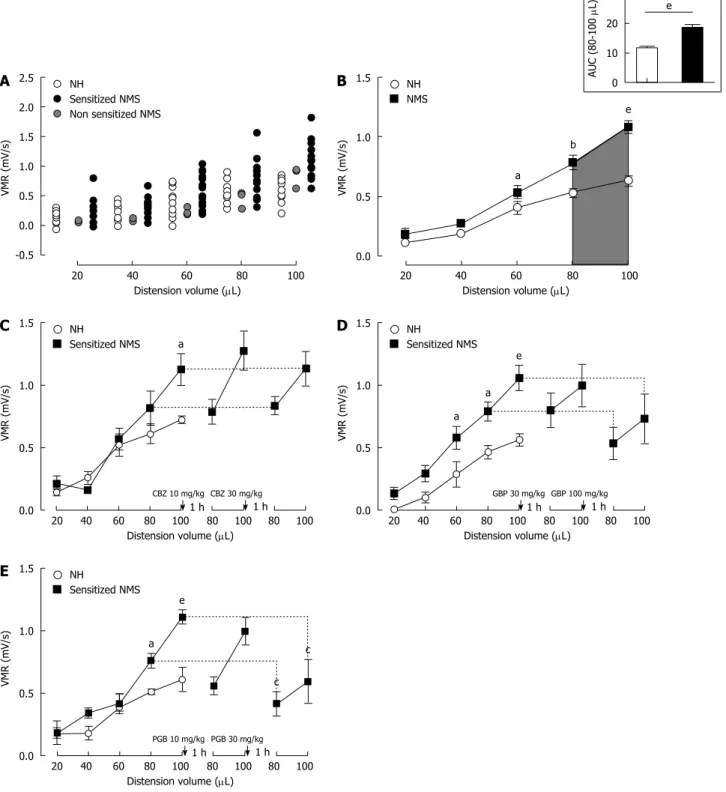
As shown in Figure 2A, the VMR to CRD of both mouse groups, NH (n = 17) and NMS (n = 26) mice, gradually increased in response to balloon inflation. Three mice were excluded after our NMS paradigm from the analysis because they were considered as nonsensitized (see material and methods) (Figure 2A, grey circles). Thus, among NMS mice used for model validation, 23 of the 26 mice (88.5%) were considered to develop CHS (sensitized NMS mice), by our NMS paradigm protocol, in comparison to NH mice (Figure 2A). When compared to NH mice, maternal deprivation induced a significant increase in VMR to CRD for the highest distension volumes reflecting CHS
(80 µL: 0.783 ± 0.056 mV/s vs 0.531 ± 0.034 mV/s,
p < 0.05 and 100 µL: 1.087 ± 0.056 mV/s vs 0.634 ± 0.038 mV/s, p < 0.05 for NMS sensitized and NH mice, respectively) (Figure 2B). The area under the curve (AUC) between 80 and 100 µL exhibited a significant (p
< 0.0001) 1.61fold increase of the colonic sensitivity
in sensitized NMS mice in comparison to NH mice (18.72 ± 1.02 and 11.65 ± 0.58, respectively) (Figure 2B, topright insert).
Using the noninflammatory NMSinduced CHS model, CBZ (n = 8) at 10 and 30 mg/kg failed to significantly reduce VMR in response to CRD (Figure 2C). Gabapentin (n = 8) at the highest cumulative dose of 100 mg/kg slightly reduces the VMR, but this effect is not significant (Figure 2D). Subcutaneous administration of Pregabalin also reduces VMR to CRD and this effect was significant for the highest cumulative dose of 30 mg/kg (1.111 ± 0.056 mV/s vs 0.595 ± 0.174 mV/s) (Figure 2E).
Effect of acute treatment with
α
2
δ
-1 ligands in an
inflammatory DSS-induced CHS model
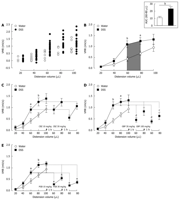
Among 1% DSStreated mice used for model validation, none of them was considered as non sensitized (see material and methods) (Figure 3A). Control mice (Water, n = 7) and DSStreated mice (DSS 1%, n = 22) exhibit an increased VMR to balloon inflation. DSStreated mice showed a dramatic and significant increase in VMR at 60 and 80 µL when compared to control mice (60 µL: 0.920 ± 0.079 mV/s
vs 0.426 ± 0.100 mV/s p < 0.05 and 80 µL: 1.193 ± 0.097 mV/s vs 0.681 ± 0.094 mV/s p < 0.05 for DSS and Watertreated mice, respectively) (Figure 3B). Unlike in NMS model, the most significant increase occurred for 60 and 80 µL of distension volumes (Figure 3B). The AUC between 60 and 80 µL is significantly (p = 0.0053) increased by 110.3% in DSStreated mice compared to water-drinking mice (23.30 ± 3.10 and 11.08 ± 1.84, respectively) (Figure 3B, topright insert). In addition, as expected, lowdose 1% DSS treatment induced moderate alterations of colonic mucosa characterized by a few focused epithelium disorganization and inflammatory cell infiltration (Figure 4). Thus, a slight but significant increase of the histological score was observed in the lowdose 1% DSStreated mice (Table 1). Moreover, MPO activity was not significantly modify in lowdose 1% DSStreated mice compared to control watertreated mice (Table 1). All together, these results tended to highlight a moderate colonic inflammatory impairment associated to CHS.
In the CHS model associated with intestinal low grade inflammation (1% DSS in drinking water), CBZ (n = 6) administration exhibited any significant antihypersensitive effect in mice sensitized with DSS (Figure 3C). In contrast, both α2δ1 ligands, GBP (n = 7)
and PGB (n = 9), showed a strong antihypersensitive effect. For a 60 µLdistension volume, s.c. adminis tration of GBP and PGB induced a dosedependent
Table 1 Inflammatory parameters in low dose 1% dextran sulfate sodium-induced colonic hypersensitivity model
Water DSS
Histological score 0.1 ± 0.1 4.0 ± 0.6b MPO activity 2.4 ± 0.3 4.0 ± 0.6 On the same mouse, intestinal inflammation has been assessed by measuring the colonic myeloperoxidase (MPO) activity and histological score. bP < 0.01 Water vs dextran sulfate sodium (DSS) treated ANOVA 1 Way followed by Tuckey’s post-hoc test for multiple comparisons.
Figure 2 Mouse model of non-inflammatory colonic hypersensitivity and assessment of pregabalin, gabapentin and carbamazepine treatments. A: After
colorectal distension (CRD) test, three mother-separated mice are considered non-sensitized (grey circles) as described in material and methods and are excluded from the analysis. Distribution of visceromotor response (VMR) values to CRD test highlights an increased response in sensitized neonatal mother-separated (sensitized NMS) (black circles n = 23) compared to non-handled (NH) (white circles n = 17) mice; B: Assessment of colonic sensitivity in sensitized NMS (black squares n = 23) and non-handled (white circles n = 17) mice displays significant differences between groups for highest distension volumes (60, 80 and 100 µL). Top right insert represents the area under the curve (AUC) between 80 µL and 100 µL; C: Carbamazepine (CBZ) administration (10 and 30 mg/kg s.c.) has no effect on VMR response in neonatal mother-separated mice (NMS : n = 8; NH : n = 6); D: Both doses (30 and 100 mg/kg s.c.) of gabapentin (GBP) have no effect on CHS induced by neonatal maternal separation (NMS: n = 8; NH: n = 6); E: Only the highest cumulative dose (30 mg/kg s.c.) of pregabalin (PGB) induces a VMR reduction in response to CRD in NMS mice (NMS : n = 8; NH : n = 6). aP < 0.05; bP < 0.01, eP < 0.001, Neonatal Mother-separated vs Non-handled ANOVA 2 Way (Animal housing; Volume) followed by Bonferroni post-hoc test for multiple comparisons and cP < 0.05 Pre-injection vs Post-injection; ANOVA 2 Way (Treatment; Volume) followed by Bonferroni post-hoc test for multiple comparisons. NMS: Neonatal maternal separation; NH: Non-handled.
2.5 2.0 1.5 1.0 0.5 0.0 -0.5 20 40 60 80 100 Distension volume (µL) NH Sensitized NMS Non sensitized NMS VMR (mV/s) 1.5 1.0 0.5 0.0 20 40 60 80 100 Distension volume (µL) NH NMS VMR (mV/s) a b e 30 20 10 0 AUC (80-100 µL) e 1.5 1.0 0.5 0.0 20 40 60 80 100 80 100 80 100 Distension volume (µL) NH Sensitized NMS VMR (mV/s) a CBZ 10 mg/kg CBZ 30 mg/kg 1.5 1.0 0.5 0.0 20 40 60 80 100 80 100 80 100 Distension volume (µL) NH Sensitized NMS VMR (mV/s) a GBP 30 mg/kg GBP 100 mg/kg a e 1 h 1 h 1 h 1 h 1.5 1.0 0.5 0.0 20 40 60 80 100 80 100 80 100 Distension volume (µL) NH Sensitized NMS VMR (mV/s) PGB 10 mg/kg PGB 30 mg/kg 1 h 1 h a e c c
A
B
C
D
E
Figure 3 Mouse model of inflammation-associated colonic hypersensitivity and assessment of pregabalin, gabapentin and carbamazepine treatments. A:
All animals were considered as sensitized by dextran sulfate sodium (DSS) treatment. Distribution of visceromotor response (VMR) values highlights an increased response in DSS-treated (black circles n = 22) compared to control (white circles n = 7) mice; B: Assessment of colonic sensitivity in DSS-treated (n = 22) and control mice (n = 7) displays significant differences between groups for 60 and 80 µL distension volumes. Top right insert represents the area under the curve (AUC) between 60 µL and 80 µL; C: Carbamazepine (CBZ) administration (10 and 30 mg/kg s.c.) has no significant effect on VMR response in DSS-treated mice (DSS n = 6; Water n = 7); D: Both doses (30 and 100 mg/kg s.c.) of gabapentin (GBP) induce a VMR reduction in DSS-treated mice (DSS n = 7; Water n = 7); E: Pregabalin (PGB) administration (10 and 30 mg/kg s.c.) induces a highly significant reduction of VMR in response to CRD in DSS-treated mice (DSS n = 9; Water n = 7).aP < 0.05; bP < 0.01, DSS-treated vs Water ANOVA 2 Way (Treatment; Volume) followed by Bonferroni post-hoc test for multiple comparisons; cP < 0.05; dP < 0.01, eP < 0.001, Pre-injection vs Post-Pre-injection ANOVA 2 Way (Treatment; Volume) followed by Bonferroni post-hoc test for multiple comparisons. CRD: Colorectal distension.
2.5 2.0 1.5 1.0 0.5 0.0 -0.5 20 40 60 80 100 Distension volume (µL) Water DSS VMR (mV/s) 2.0 1.5 1.0 0.5 0.0 20 40 60 80 100 Distension volume (µL) Water DSS VMR (mV/s) 30 20 10 0 AUC (60-80 µL) b 2.0 1.5 1.0 0.5 0.0 20 40 60 80 100 60 80 60 80 Distension volume (µL) Water DSS VMR (mV/s) CBZ 10 mg/kg CBZ 30 mg/kg 2.0 1.5 1.0 0.5 0.0 20 40 60 80 100 60 80 60 80 Distension volume (µL) Water DSS VMR (mV/s) GBP 30 mg/kg GBP 100 mg/kg 1 h 1 h 1 h 1 h 2.0 1.5 1.0 0.5 0.0 20 40 60 80 100 60 80 60 80 Distension volume (µL) Water DSS VMR (mV/s) PGB 10 mg/kg PGB 30 mg/kg 1 h 1 h
A
B
C
D
E
b a a b b a c d d a b e e e eVMR decrease in DSStreated mice (1.092 ± 0.160 mV/s vs 0.460 ± 0.099 mV/s and vs 0.390 ± 0.090 mV/s for GBP 30 and 100 mg/kg, respectively; 0.793 ± 0.082 mV/s vs 0.283 ± 0.033 mV/s and vs 0.236 ± 0.046 mV/s for PGB 10 and 30 mg/kg, respectively). For a 80 µLdistension volume, the highest cumulative dose of GBP (100 mg/kg) and both doses of PGB (10 and 30 mg/kg) induced a significant reduction of VMR in DSStreated mice (1.238 ± 0.157 mV/s vs 0.630 ± 0.153 mV/s for GBP 100 mg/kg; 1.123 ± 0.110 mV/s
vs 0.550 ± 0.087 mV/s and vs 0.373 ± 0.068 mV/s
for PGB 10 and 30 mg/kg, respectively) (Figure 3D and E).
Comparative effect of acute treatment with
α
2
δ
-1
ligands on CHS
To compare the effects of anticonvulsant drug (GBP, PGB and CBZ) treatment, the AUC corresponding to the two distension volumes displaying the highest significant
differences in VMR between control and sensitized groups (80 and 100 µL for NMS model and 60 and 80
µL for DSS model) were calculated. Carbamazepine failed to significantly reduce CHS in both models (Figure 5A and B). Gabapentin also failed to reduce CHS in NMS-induced model, but significantly reduced it in the DSSinduced model for both subcutaneous injections of 30 or 100 mg/kg (Figure 5C and D). Subcutaneous acute administration of the highest cumulative dose of PGB (30 mg/kg) significantly reduced VMR to CRD (Figure 5E), and significantly reduced CHS in low-dose DSStreated mice, since s.c. acute administration of PGB induced a dosedependent inhibition of abdominal contractile responses to CRD (Figure 5F). In addition, none of these drugs (CBZ, GBP and PGB) did modify the colonic sensitivity in control (NH or watertreated) animals.
Finally, the percent decrease of AUC induced by acute GBP or PGB treatment were higher in the inflammatory DSS-induced CHS model in comparison to the noninflammatory NMSinduced CHS model (Table 2). This decrease was significant for both doses of PGB.
DISCUSSION
The present study demonstrates the acute anti hypersensitive effect of α2δ1 ligands gabapentin
and pregabalin, compared to the nonα2δ1 ligand
anticonvulsant carbamazepine, on CHS models, in mice. A more effective effect of α2δ1 ligands was observed in
CHS associated with colonic inflammation compared to the non-inflammatory model.
The inflammatory animal model of CHS used in this work involved the administration of 1% DSS in drinking water during 14 consecutive days. Dextran sulfate sodium is widely used in rodents to induce colitis and generates quite similar symptoms to those of patients displaying IBD, such as inflammatory cell infiltration, cytokines production and fecal blood[34],
showing the relevancy of this model to study intestinal
Figure 4 Representative histologic examinations of low dose 1% dextran sulfate sodium-treated mice colons after hematoxylin and eosin-staining (scales bars = 100 µm). DSS: Dextran sulfate sodium.
Table 2 Comparison of the carbamazepine, gabapentin or pregabalin efficacy on neonatal mother-separated and dextran sulfate sodium induced colonic hypersensitivity models
NMS DSS CBZ 10 mg/kg 9 ± 8 6 ± 18 30 mg/kg 5 ± 13 -17 ± 15 GBP 30 mg/kg 15 ± 32 -45 ± 9 100 mg/kg -14 ± 37 -56 ± 9 PGB 10 mg/kg -16 ± 8 -54 ± 7a 30 mg/kg -47 ± 13 -66 ± 7b On the same mouse, the visceromotor response to gradual colorectal distensions (CRD) has been evaluated before and after the first and the second injection of CBZ, GBP or PGB. For each gradual CRD, the area under the curve (AUC) has been calculated between 80 µL and 100 µL in NMS model or between 60 µL and 80 µL for DSS model. Data represented the mean percent variation of the AUC calculated after the first or the second injection in comparison to the AUC calculated before any injection. aP < 0.05, bP < 0.01 NMS I DSS treated ANOVA 1 Way followed by Tuckey’s post-hoc test for multiple comparisons. CBZ: Carbamazepine; GBP: Gabapentin; PGB: Pregabalin; NMS: Neonatal maternal separation; DSS: Dextran sulfate sodium.
Water DSS
100 µm 100 µm
inflammation. A few studies investigated the ability of DSS to induce CHS and displayed discrepant results with either a hypersensitive effect[35,36] or no influence
of DSS administration on colonic sensitivity[37,38].
Comparing the data of those works is challenging since different protocols of visceral sensitivity mea surement and treatment time course were used. However, it appears that high doses of DSS (> 3%), despite inducing a severe colitis, fail to enhance
colonic sensitivity[37,38]. A possible explanation of
this phenomenon might be the impairment of nerve terminals due to tissue lesion that leads to primary afferents desensitization. A recent paper has shown that low-dose of DSS (0.5% to 1% DSS) in drinking water of mice did not induce marked inflammation but only signs of low-grade inflammation[39]. Thus, in
our study, lowdoses of DSS probably do not induce severe tissue lesion and subsequent desensitization,
Figure 5 Comparative effect of carbamazepine, gabapentin and pregabalin on visceral hypersensitivity in non-inflammatory and inflammation-associated colonic hypersensitivity mouse models. The area under the curve (AUC) (A, C and E) between 80 µL and 100 µL for neonatal maternal separation (NMS) model
and (B, D and F) between 60 µL and 80 µL for Dextran Sulfate Sodium (DSS) model has been calculated for both doses of (A and B) carbamazepine (CBZ), (C and D) gabapentin (GBP) and (E and F) pregabalin (PGB). aP < 0.05; bP < 0.01, eP < 0.001, ANOVA 2 Way (Treatment; Dose) followed by Bonferroni post-hoc test for multiple comparisons for each treatment in one CHS model. CHS: Colonic hypersensitivity; DSS: Dextran sulfate sodium.
30 25 20 15 10 5 0 NMS - - - + + + CBZ - 10 30 - 10 30 AUC (80-100 µL) 30 25 20 15 10 5 0 DSS - - - + + + CBZ - 10 30 - 10 30 AUC (60-80 µL) a 30 25 20 15 10 5 0 NMS - - - + + + GBP - 30 100 - 30 100 AUC (80-100 µL) a 30 25 20 15 10 5 0 DSS - - - + + + GBP - 30 100 - 30 100 AUC (60-80 µL) a e e b b 30 25 20 15 10 5 0 NMS - - - + + + PGB - 10 30 - 10 30 AUC (80-100 µL) a b 30 25 20 15 10 5 0 DSS - - - + + + PGB - 10 30 - 10 30 AUC (60-80 µL) b e e
A
B
C
D
E
F
but are sufficient to enhance colonic primary afferents response to colorectal distension.
Studying IBS-like non-inflammatory CHS is more challenging as mechanisms involved in IBS pathophysiology are poorly known. However, several studies have reported a close link between IBS development and early life traumatic events such as neglect or abuse[40]. Accordingly, Coutinho et al[41]
pointed out an altered viscerosomatic response of rats separated from their mother during neonatal life. These sensitive disturbances seem to be cor related with hypothalamicpituitaryadrenal axis dysfunctions[42] and are associated with plastic changes
at spinal cord level[43,44] but not with organic injuries of
colonic mucosa[45]. It was recently demonstrated that
the same separation paradigm led to the increase of VMR to CRD in mice as well[29]. Our results corroborate
this observation with a colonic sensitization of the vast majority of mice separated from their mother during postnatal period.
In DSSinduced model, we showed that α2δ1
ligands were able to decrease CHS at the two used experimental doses, while only PGB significantly reduced the CHS in NMS model. In contrast, CBZ failed to modify VMR scores in both models. Chosen doses of α2δ1 ligands have been shown to display an analge
sic effect up to 2 h on mechanical hypersensitivity developed after spinal cord injury in mice[46]. Carba
mazepine was administered at 10 and 30 mg/kg corresponding to doses reducing inflammatory somatic pain related behaviors[47]. Antihyperalgesic properties
of α2δ1 ligands are widely used in the management
of neuropathic pain and fibromyalgia[48,49]. Regarding
visceral pain, their ability to decrease colonic sensitivity has been demonstrated in two clinical trials in IBS patients[20,21]. At preclinical level, although GBP and
PGB efficiency is documented in IBS models, only few studies reported their potential beneficial impact in acute inflammatory models. Indeed, PGB has been shown to decrease visceral hypersensitivity induced by LPS[50]. Our study confirms the ability of an α2δ1
ligand (PGB) to relieve CHS in a noninflammatory model. Interestingly, the two α2δ1 ligands (GBP and
PGB) more potently reduce CHS in our inflammatory model. In addition, not all classes of anticonvulsants are suitable to alleviate CHS, as highlighted by CBZ treatment failure, suggesting a specific involvement of α2δ1 in the mechanism of action of GBP and PGB.
The first evoked action of GBP and PGB on GABAergic system does not seem to occur as conclude Taylor in his review[48]. Instead, their high affinity for
α2δ1 subunits of Ca(v) channels[51,52] could explain their
effect on chronic pain. Indeed, it is well documented that α2δ1 subunit stimulation increase both current
amplitude of high voltage activated Ca(v) channels
and their trafficking to the plasmatic membrane[53].
Consistent with this observation, mutation in the α2δ1
Ca2+ channel subunit preventing the binding of α2δ1
ligands, led to a decrease in their analgesic effects in
mouse pain models[54,55]. Regarding visceral sensitivity,
Liao et al[56] demonstrated the spinal upregulation of
α2δ-1 subunit and the efficacy of GBP in a pancreatitis
model in rats. Among Ca(v) channels, several data
demonstrate that all subtypes can be involved in chronic pain development including visceral pain[5759].
Their preponderant role is the increase of spinal synaptic transmission by promoting neurotransmitter containing vesicle release that may lead to long term potentiation phenomenon underlying chronic pain state[60]. Involvement of α2δ1 subunits was also
demonstrated in visceral pain conditions. Hence, in a rat model of CHS induced by uterine cross sensitization a spinal overexpression of α2δ1 subunit
was described[61]. It is, therefore, possible that CHS
in NMS mice is related to the upregulation of Ca(v)
channels and associated α2δ1 subunits at presynaptic
terminals of spinal sensory neurons and, in this model, antihyperalgesic effect of PGB is mainly due to spinal neurotransmission inhibition.
Besides spinal mechanisms of α2δ-1 ligands evoked
in NMSinduced model and that probably occur in inflammatory model as well, an effect on peripheral sensitization cannot be ruled out explaining a higher efficacy in DSSinduced CHS. In fact, α2δ1 subunit
are retrieved in intestine primary afferent neurons associated with Ntype calcium channels[62] and could
be overexpressed in an inflammatory context, as observed in somatic inflammatory pain models. In fact, α2δ1 and Ca(v)2.2 proteins are overexpressed
in the nerves arising from L4 and L5 ganglia ipsi lateral to the site of CFA administration[63]. In a rat
model of osteoarthritic pain an increase in α2δ1
mRNA levels was observed in ipsilateral L3 and L4 ganglia. In this model, pregabalin inhibit dorsal horn neuronal responses[64]. Other studies demonstrated
antinociceptive efficacy of α2δ1 ligands on behavioral
and neuronal responses in cutaneous models of acute inflammatory pain[65,66]. Thereby, with inhibition of
spinal synaptic transmission, α2δ1 ligands could also
target inflammation in DSSinduced CHS model, explaining their potent effect in this model.
To conclude, our results, combined with available data of literature, confirmed the beneficial effect of α2δ1 ligands in the management of visceral
hypersensitivity. Moreover, our findings highlight their efficacy in inflammationinduced CHS. Further studies are needed to investigate the mechanism of action of α2δ1 ligands on peripheral sensitization in
an inflammatory context. Finally, this work pointed out the interest to study the clinical relevance of α2δ1
ligands in IBD patients in remission to alleviate IBS like symptoms, particularly abdominal pain.
COMMENTS
Background
Colonic hypersensitivity is often reported in patients with abdominal pain as irritable bowel syndrome (IBS) patients or inflammatory bowel diseases
(IBD) patients during remission phases. Despite many potential tested targets expressed in intestinal sensory afferents to modulate abdominal pain in several preclinical models or under different clinical conditions, colonic hypersensitivity (CHS) is not targeted by any therapy as of today. In this context, the development of new treatments is needed to provide IBS patients and IBD patients during the quiescent periods with a therapeutic option in alleviating abdominal pain.
Research frontiers
Clinical studies highlighted the antinociceptive effect of alpha-2-delta-1 (α2δ-1) ligands, initially commercialized as anticonvulsant, in patients suffering from IBS. However, no investigation was done based on the intestinal inflammation state.
Innovations and breakthroughs
The goal of this study was to determine if the α2δ-1 ligands (gabapentin and pregabalin) have beneficial effects on CHS, using different mouse models of IBS and IBD. They will be compared to those of another anticonvulsant drug, carbamazepine. The main question addressed by this preclinical study will be the effect of α2δ-1 ligand compounds in a CHS mouse model associated with a colonic inflammation induced by dextran sulfate sodium administration and compare these effects with those obtained in a non-inflammatory IBS-like model, induced by neonatal maternal separation.
Applications
The present study suggests that α2δ-1 ligands may be beneficial for the management of visceral hypersensitivity. In particular, our findings show the efficacy of α2δ-1 ligands on inflammation-induced CHS and further studies on the mechanisms of action of α2δ-1 ligands on peripheral sensitization in an inflammatory context will pave the way for new CHS treatments for IBD patients. Finally, this work shows the clinical relevance of using α2δ-1 ligands also in IBD patients who are in remission in order to alleviate IBS-like symptoms, such as abdominal pain.
Terminology
Mouse CHS is evaluated in our different animal models using a colorectal distension procedure and by evaluating the animal discomfort reflected by changes in the visceromotor response, a pseudoaffective reflex response widely used as index of visceral pain in rodents, to colorectal distension.
Peer-review
This preclinical study demonstrates α2δ-1 ligands efficacy on inflammation-associated CHS, showing their potential clinical interest for patients with chronic abdominal pain and moderate intestinal inflammation.
REFERENCES
1 Drossman DA, Dumitrascu DL. Rome III: New standard for
functional gastrointestinal disorders. J Gastrointestin Liver Dis 2006;
15: 237-241 [PMID: 17013448]
2 Chassaing B, Darfeuille-Michaud A. The commensal microbiota
and enteropathogens in the pathogenesis of inflammatory bowel diseases. Gastroenterology 2011; 140: 1720-1728 [PMID: 21530738 DOI: 10.1053/j.gastro.2011.01.054]
3 Halpin SJ, Ford AC. Prevalence of symptoms meeting criteria for
irritable bowel syndrome in inflammatory bowel disease: systematic review and meta-analysis. Am J Gastroenterol 2012; 107: 1474-1482 [PMID: 22929759 DOI: 10.1038/ajg.2012.260]
4 Piche T, Ducrotté P, Sabate JM, Coffin B, Zerbib F, Dapoigny M,
Hua M, Marine-Barjoan E, Dainese R, Hébuterne X. Impact of functional bowel symptoms on quality of life and fatigue in quiescent Crohn disease and irritable bowel syndrome. Neurogastroenterol Motil 2010; 22: 626-e174 [PMID: 20403099 DOI: 10.1111/ j.1365-2982.2010.01502.x]
5 Stacher G, Christensen J. Visceral hypersensitivity in irritable bowel
syndrome: a summary review. Dig Dis Sci 2006; 51: 440-445 [PMID: 16619394]
6 Wagtmans MJ, Verspaget HW, Lamers CB, van Hogezand RA.
Crohn’s disease in the elderly: a comparison with young adults. J Clin Gastroenterol 1998; 27: 129-133 [PMID: 9754773]
7 Hicks GA. TRP channels as therapeutic targets: hot property, or time
to cool down? Neurogastroenterol Motil 2006; 18: 590-594 [PMID: 16918723 DOI: 10.1111/j.1365-2982.2006.00823.x]
8 Kirkup AJ, Brunsden AM, Grundy D. Receptors and transmission
in the brain-gut axis: potential for novel therapies. I. Receptors on visceral afferents. Am J Physiol Gastrointest Liver Physiol 2001;
280: G787-G794 [PMID: 11292585]
9 Bradesi S, Herman J, Mayer EA. Visceral analgesics: drugs with a
great potential in functional disorders? Curr Opin Pharmacol 2008; 8: 697-703 [PMID: 18786654 DOI: 10.1016/j.coph.2008.08.009] 10 Clouse RE. Antidepressants for irritable bowel syndrome. Gut 2003;
52: 598-599 [PMID: 12631677]
11 Rutgeerts P, Van Assche G, Vermeire S. Optimizing anti-TNF treatment in inflammatory bowel disease. Gastroenterology 2004;
126: 1593-1610 [PMID: 15168370]
12 Beattie RM, Croft NM, Fell JM, Afzal NA, Heuschkel RB. Inflammatory bowel disease. Arch Dis Child 2006; 91: 426-432 [PMID: 16632672 DOI: 10.1136/adc.2005.080481]
13 Rogler G. Gastrointestinal and liver adverse effects of drugs used for treating IBD. Best Pract Res Clin Gastroenterol 2010; 24: 157-165 [PMID: 20227029 DOI: 10.1016/j.bpg.2009.10.011]
14 Johannessen Landmark C. Antiepileptic drugs in non-epilepsy disorders: relations between mechanisms of action and clinical efficacy. CNS Drugs 2008; 22: 27-47 [PMID: 18072813]
15 Landmark CJ. Targets for antiepileptic drugs in the synapse. Med Sci Monit 2007; 13: RA1-RA7 [PMID: 17179916]
16 Lynch JJ, Honore P, Anderson DJ, Bunnelle WH, Mortell KH, Zhong C, Wade CL, Zhu CZ, Xu H, Marsh KC, Lee CH, Jarvis MF, Gopalakrishnan M. (L)-Phenylglycine, but not necessarily other alpha2delta subunit voltage-gated calcium channel ligands, attenuates neuropathic pain in rats. Pain 2006; 125: 136-142 [PMID: 16781071 DOI: 10.1016/j.pain.2006.05.012]
17 Macdonald RL, Kelly KM. Mechanisms of action of currently prescribed and newly developed antiepileptic drugs. Epilepsia 1994;
35 Suppl 4: S41-S50 [PMID: 7513639]
18 Rogawski MA, Löscher W. The neurobiology of antiepileptic drugs for the treatment of nonepileptic conditions. Nat Med 2004; 10: 685-692 [PMID: 15229516 DOI: 10.1038/nm1074]
19 Waszkielewicz AM, Gunia A, Słoczyńska K, Marona H. Evaluation of anticonvulsants for possible use in neuropathic pain. Curr Med Chem 2011; 18: 4344-4358 [PMID: 21861814]
20 Lee KJ, Kim JH, Cho SW. Gabapentin reduces rectal mecha-nosensitivity and increases rectal compliance in patients with diarrhoea-predominant irritable bowel syndrome. Aliment Pharmacol Ther 2005; 22: 981-988 [PMID: 16268973 DOI: 10.1111/j.1365-2036.2005.02685.x]
21 Houghton LA, Fell C, Whorwell PJ, Jones I, Sudworth DP, Gale JD. Effect of a second-generation alpha2delta ligand (pregabalin) on visceral sensation in hypersensitive patients with irritable bowel syndrome. Gut 2007; 56: 1218-1225 [PMID: 17446306 DOI: 10.1136/gut.2006.110858]
22 Diop L, Raymond F, Fargeau H, Petoux F, Chovet M, Doherty AM. Pregabalin (CI-1008) inhibits the trinitrobenzene sulfonic acid-induced chronic colonic allodynia in the rat. J Pharmacol Exp Ther 2002; 302: 1013-1022 [PMID: 12183658]
23 Million M, Wang L, Adelson DW, Roman F, Diop L, Taché Y. Pregabalin decreases visceral pain and prevents spinal neuronal activation in rats. Gut 2007; 56: 1482-1484 [PMID: 17872585 DOI: 10.1136/gut.2007.129304]
24 Ravnefjord A, Brusberg M, Larsson H, Lindström E, Martínez V. Effects of pregabalin on visceral pain responses and colonic compliance in rats. Br J Pharmacol 2008; 155: 407-416 [PMID: 18574457 DOI: 10.1038/bjp.2008.259]
25 Field MJ, Oles RJ, Lewis AS, McCleary S, Hughes J, Singh L. Gabapentin (neurontin) and S-(+)-3-isobutylgaba represent a novel class of selective antihyperalgesic agents. Br J Pharmacol 1997;
121: 1513-1522 [PMID: 9283683 DOI: 10.1038/sj.bjp.0701320]
Ugresić ND, Prostran MS, Bosković B. GABAergic mechanisms are involved in the antihyperalgesic effects of carbamazepine and oxcarbazepine in a rat model of inflammatory hyperalgesia. Pharmacology 2008; 82: 53-58 [PMID: 18434764 DOI: 10.1159/000127841]
27 Tomić MA, Vucković SM, Stepanović-Petrović RM, Ugresić N, Prostran MS, Bosković B. The anti-hyperalgesic effects of carbamazepine and oxcarbazepine are attenuated by treatment with adenosine receptor antagonists. Pain 2004; 111: 253-260 [PMID: 15363868 DOI: 10.1016/j.pain.2004.07.010]
28 Vucković SM, Tomić MA, Stepanović-Petrović RM, Ugresić N, Prostran MS, Bosković B. The effects of alpha2-adrenoceptor agents on anti-hyperalgesic effects of carbamazepine and oxcarbazepine in a rat model of inflammatory pain. Pain 2006; 125: 10-19 [PMID: 16777327 DOI: 10.1016/j.pain.2006.04.023]
29 Miquel S, Martín R, Lashermes A, Gillet M, Meleine M, Gelot A, Eschalier A, Ardid D, Bermúdez-Humarán LG, Sokol H, Thomas M, Theodorou V, Langella P, Carvalho FA. Anti-nociceptive effect of Faecalibacterium prausnitzii in non-inflammatory IBS-like models. Sci Rep 2016; 6: 19399 [PMID: 26775847 DOI: 10.1038/srep19399] 30 Okayasu I, Hatakeyama S, Yamada M, Ohkusa T, Inagaki Y,
Nakaya R. A novel method in the induction of reliable experimental acute and chronic ulcerative colitis in mice. Gastroenterology 1990;
98: 694-702 [PMID: 1688816]
31 Zimmermann M. Ethical guidelines for investigations of experimental pain in conscious animals. Pain 1983; 16: 109-110 [PMID: 6877845]
32 Carvalho FA, Barnich N, Sauvanet P, Darcha C, Gelot A, Darfeuille-Michaud A. Crohn’s disease-associated Escherichia coli LF82 aggravates colitis in injured mouse colon via signaling by flagellin. Inflamm Bowel Dis 2008; 14: 1051-1060 [PMID: 18338780 DOI: 10.1002/ibd.20423]
33 Onyeagocha C, Hossain MS, Kumar A, Jones RM, Roback J, Gewirtz AT. Latent cytomegalovirus infection exacerbates experimental colitis. Am J Pathol 2009; 175: 2034-2042 [PMID: 19815702 DOI: 10.2353/ajpath.2009.090471]
34 Sha T, Igaki K, Yamasaki M, Watanabe T, Tsuchimori N. Establishment and validation of a new semi-chronic dextran sulfate sodium-induced model of colitis in mice. Int Immunopharmacol 2013;
15: 23-29 [PMID: 23142502 DOI: 10.1016/j.intimp.2012.10.022]
35 Boué J, Basso L, Cenac N, Blanpied C, Rolli-Derkinderen M, Neunlist M, Vergnolle N, Dietrich G. Endogenous regulation of visceral pain via production of opioids by colitogenic CD4(+) T cells in mice. Gastroenterology 2014; 146: 166-175 [PMID: 24055279 DOI: 10.1053/j.gastro.2013.09.020]
36 Jain P, Hassan AM, Koyani CN, Mayerhofer R, Reichmann F, Farzi A, Schuligoi R, Malle E, Holzer P. Behavioral and molecular processing of visceral pain in the brain of mice: impact of colitis and psychological stress. Front Behav Neurosci 2015; 9: 177 [PMID: 26217204 DOI: 10.3389/fnbeh.2015.00177]
37 Larsson MH, Miketa A, Martinez V. Lack of interaction between psychological stress and DSS-induced colitis affecting colonic sensitivity during colorectal distension in mice. Stress 2009; 12: 434-444 [PMID: 19929445 DOI: 10.1080/10253890802626603] 38 Larsson MH, Rapp L, Lindström E. Effect of DSS-induced
colitis on visceral sensitivity to colorectal distension in mice. Neurogastroenterol Motil 2006; 18: 144-152 [PMID: 16420293 DOI: 10.1111/j.1365-2982.2005.00736.x]
39 Chassaing B, Srinivasan G, Delgado MA, Young AN, Gewirtz AT, Vijay-Kumar M. Fecal lipocalin 2, a sensitive and broadly dynamic non-invasive biomarker for intestinal inflammation. PLoS One 2012;
7: e44328 [PMID: 22957064 DOI: 10.1371/journal.pone.0044328]
40 Heitkemper MM, Cain KC, Burr RL, Jun SE, Jarrett ME. Is childhood abuse or neglect associated with symptom reports and physiological measures in women with irritable bowel syndrome? Biol Res Nurs 2011; 13: 399-408 [PMID: 21196423 DOI: 10.1177/1 099800410393274]
41 Coutinho SV, Plotsky PM, Sablad M, Miller JC, Zhou H, Bayati AI, McRoberts JA, Mayer EA. Neonatal maternal separation alters stress-induced responses to viscerosomatic nociceptive stimuli in
rat. Am J Physiol Gastrointest Liver Physiol 2002; 282: G307-G316 [PMID: 11804852 DOI: 10.1152/ajpgi.00240.2001]
42 Bravo JA, Dinan TG, Cryan JF. Alterations in the central CRF system of two different rat models of comorbid depression and functional gastrointestinal disorders. Int J Neuropsychopharmacol 2011; 14: 666-683 [PMID: 20860876 DOI: 10.1017/S1461145710000994] 43 Chung EK, Zhang XJ, Xu HX, Sung JJ, Bian ZX. Visceral
hyperalgesia induced by neonatal maternal separation is associated with nerve growth factor-mediated central neuronal plasticity in rat spinal cord. Neuroscience 2007; 149: 685-695 [PMID: 17913374 DOI: 10.1016/j.neuroscience.2007.07.055]
44 Tsang SW, Zhao M, Wu J, Sung JJ, Bian ZX. Nerve growth factor-mediated neuronal plasticity in spinal cord contributes to neonatal maternal separation-induced visceral hypersensitivity in rats. Eur J Pain 2012; 16: 463-472 [PMID: 22396076 DOI: 10.1016/ j.ejpain.2011.07.005]
45 O’Mahony SM, Marchesi JR, Scully P, Codling C, Ceolho AM, Quigley EM, Cryan JF, Dinan TG. Early life stress alters behavior, immunity, and microbiota in rats: implications for irritable bowel syndrome and psychiatric illnesses. Biol Psychiatry 2009; 65: 263-267 [PMID: 18723164 DOI: 10.1016/j.biopsych.2008.06.026] 46 Tanabe M, Ono K, Honda M, Ono H. Gabapentin and pregabalin
ameliorate mechanical hypersensitivity after spinal cord injury in mice. Eur J Pharmacol 2009; 609: 65-68 [PMID: 19285498 DOI: 10.1016/j.ejphar.2009.03.020]
47 Sahebgharani M, Hossein-Abad AA, Zarrindast MR. On the mechanism of carbamazepine-induced antinociception in the formalin test. Int J Neurosci 2006; 116: 1097-1113 [PMID: 16861171 DOI: 10.1080/00207450600808669]
48 Taylor CP. Mechanisms of analgesia by gabapentin and pregabalin--calcium channel alpha2-delta [Cavalpha2-delta] ligands. Pain 2009;
142: 13-16 [PMID: 19128880 DOI: 10.1016/j.pain.2008.11.019]
49 Tzellos TG, Toulis KA, Goulis DG, Papazisis G, Zampeli VA, Vakfari A, Kouvelas D. Gabapentin and pregabalin in the treatment of fibromyalgia: a systematic review and a meta-analysis. J Clin Pharm Ther 2010; 35: 639-656 [PMID: 21054455 DOI: 10.1111/ j.1365-2710.2009.01144.x]
50 Eutamene H, Coelho AM, Theodorou V, Toulouse M, Chovet M, Doherty A, Fioramonti J, Bueno L. Antinociceptive effect of pregabalin in septic shock-induced rectal hypersensitivity in rats. J Pharmacol Exp Ther 2000; 295: 162-167 [PMID: 10991974] 51 Cheng JK, Chiou LC. Mechanisms of the antinociceptive action of
gabapentin. J Pharmacol Sci 2006; 100: 471-486 [PMID: 16474201] 52 Gee NS, Brown JP, Dissanayake VU, Offord J, Thurlow R, Woodruff GN. The novel anticonvulsant drug, gabapentin (Neurontin), binds to the alpha2delta subunit of a calcium channel. J Biol Chem 1996;
271: 5768-5776 [PMID: 8621444]
53 Gurnett CA, De Waard M, Campbell KP. Dual function of the voltage-dependent Ca2+ channel alpha 2 delta subunit in current stimulation and subunit interaction. Neuron 1996; 16: 431-440 [PMID: 8789958]
54 Bian F, Li Z, Offord J, Davis MD, McCormick J, Taylor CP, Walker
LC. Calcium channel alpha2-delta type 1 subunit is the major binding protein for pregabalin in neocortex, hippocampus, amygdala, and spinal cord: an ex vivo autoradiographic study in alpha2-delta type 1 genetically modified mice. Brain Res 2006; 1075: 68-80 [PMID: 16460711 DOI: 10.1016/j.brainres.2005.12.084]
55 Field MJ, Cox PJ, Stott E, Melrose H, Offord J, Su TZ, Bramwell S, Corradini L, England S, Winks J, Kinloch RA, Hendrich J, Dolphin AC, Webb T, Williams D. Identification of the alpha2-delta-1 subunit of voltage-dependent calcium channels as a molecular target for pain mediating the analgesic actions of pregabalin. Proc Natl Acad Sci USA 2006; 103: 17537-17542 [PMID: 17088553 DOI: 10.1073/ pnas.0409066103]
56 Liao XZ, Zhou MT, Mao YF, Xu H, Chen H, Sun JH, Xiong YC. Analgesic effects of gabapentin on mechanical hypersensitivity in a rat model of chronic pancreatitis. Brain Res 2010; 1337: 104-112 [PMID: 20417627 DOI: 10.1016/j.brainres.2010.04.035]
57 Marger F, Gelot A, Alloui A, Matricon J, Ferrer JF, Barrère C, Pizzoccaro A, Muller E, Nargeot J, Snutch TP, Eschalier A,
Bourinet E, Ardid D. T-type calcium channels contribute to colonic hypersensitivity in a rat model of irritable bowel syndrome. Proc Natl Acad Sci USA 2011; 108: 11268-11273 [PMID: 21690417 DOI: 10.1073/pnas.1100869108]
58 McGivern JG. Targeting N-type and T-type calcium channels for the treatment of pain. Drug Discov Today 2006; 11: 245-253 [PMID: 16580601 DOI: 10.1016/S1359-6446(05)03662-7]
59 Swayne LA, Bourinet E. Voltage-gated calcium channels in chronic pain: emerging role of alternative splicing. Pflugers Arch 2008; 456: 459-466 [PMID: 18389277 DOI: 10.1007/s00424-007-0390-4] 60 Youn DH, Gerber G, Sather WA. Ionotropic glutamate receptors
and voltage-gated Ca2+ channels in long-term potentiation of spinal dorsal horn synapses and pain hypersensitivity. Neural Plast 2013;
2013: 654257 [PMID: 24224102 DOI: 10.1155/2013/654257]
61 Wang Y, Zhang M, Xie F, Li X, Bao M, Yang N, Shi R, Wang Z, Wu A, Guan Y, Yue Y. Upregulation of α2δ-1 Calcium Channel Subunit in the Spinal Cord Contributes to Pelvic Organ Cross-Sensitization in a Rat Model of Experimentally-Induced Endometriosis. Neurochem Res 2015; 40: 1267-1273 [PMID: 25935199 DOI: 10.1007/ s11064-015-1592-3]
62 Needham K, Bron R, Hunne B, Nguyen TV, Turner K, Nash
M, Furness JB. Identification of subunits of voltage-gated calcium channels and actions of pregabalin on intrinsic primary afferent neurons in the guinea-pig ileum. Neurogastroenterol Motil 2010; 22: e301-e308 [PMID: 20618837 DOI: 10.1111/ j.1365-2982.2010.01567.x]
63 Lu SG, Zhang XL, Luo ZD, Gold MS. Persistent inflammation alters the density and distribution of voltage-activated calcium channels in subpopulations of rat cutaneous DRG neurons. Pain 2010; 151: 633-643 [PMID: 20884119 DOI: 10.1016/j.pain.2010.08.030] 64 Rahman W, Bauer CS, Bannister K, Vonsy JL, Dolphin
AC, Dickenson AH. Descending serotonergic facilitation and the antinociceptive effects of pregabalin in a rat model of osteoarthritic pain. Mol Pain 2009; 5: 45 [PMID: 19664204 DOI: 10.1186/1744-8069-5-45]
65 Stanfa LC, Singh L, Williams RG, Dickenson AH. Gabapentin, ineffective in normal rats, markedly reduces C-fibre evoked responses after inflammation. Neuroreport 1997; 8: 587-590 [PMID: 9106728]
66 Yoon MH, Yaksh TL. Evaluation of interaction between gabapentin and ibuprofen on the formalin test in rats. Anesthesiology 1999; 91: 1006-1013 [PMID: 10519504]
P- Reviewer: Elpek GO S- Editor: Ma YJ L- Editor: A E- Editor: Wang CH