HAL Id: hal-01459767
https://hal.archives-ouvertes.fr/hal-01459767
Submitted on 20 Sep 2018
HAL is a multi-disciplinary open access archive for the deposit and dissemination of sci-entific research documents, whether they are pub-lished or not. The documents may come from teaching and research institutions in France or abroad, or from public or private research centers.
L’archive ouverte pluridisciplinaire HAL, est destinée au dépôt et à la diffusion de documents scientifiques de niveau recherche, publiés ou non, émanant des établissements d’enseignement et de recherche français ou étrangers, des laboratoires publics ou privés.
Distributed under a Creative Commons Attribution| 4.0 International License
Tick Vectors in Cote d’Ivoire
Cyrille Bile Ehounoud, Kouassi Patrick Yao, Mustapha Dahmani, Yaba
Louise Achi, Nadia Amanzougaghene, Adele Kacou N’Douba, Jean David
N’Guessan, Didier Raoult, Florence Fenollar, Oleg Mediannikov
To cite this version:
Cyrille Bile Ehounoud, Kouassi Patrick Yao, Mustapha Dahmani, Yaba Louise Achi, Nadia Aman-zougaghene, et al.. Multiple Pathogens Including Potential New Species in Tick Vectors in Cote d’Ivoire. PLoS Neglected Tropical Diseases, Public Library of Science, 2016, 10 (1), pp.e0004367. �10.1371/journal.pntd.0004367�. �hal-01459767�
Multiple Pathogens Including Potential New
Species in Tick Vectors in Côte d
’Ivoire
Cyrille Bilé Ehounoud1,2,3, Kouassi Patrick Yao3, Mustapha Dahmani1, Yaba Louise Achi4, Nadia Amanzougaghene1, Adèle Kacou N’Douba5, Jean David N’Guessan3,
Didier Raoult1,2, Florence Fenollar1,2, Oleg Mediannikov1,2*
1 Aix-Marseille Université, URMITE, UM63, CNRS 7278, IRD 198, Inserm U1095, Faculté de médecine, Marseille cedex 05, France, 2 Campus International UCAD-IRD, Dakar, Senegal, 3 Felix Houphouet Boigny Université, UFR Biosciences, Côte D’Ivoire, 4 Ecole de spécialisation en Elevage de Bingerville, Côte D’Ivoire, 5 Felix Houphouet Boigny Université, UFR Sciences médicales, Côte D’Ivoire
*olegusss1@gmail.com
Abstract
Background
Our study aimed to assess the presence of different pathogens in ticks collected in two regions in Côte d’Ivoire.
Methodology/Principal Findings
Real-time PCR and standard PCR assays coupled to sequencing were used. Three hun-dred and seventy eight (378) ticks (170 Amblyomma variegatum, 161 Rhipicepalus micro-plus, 3 Rhipicephalus senegalensis, 27 Hyalomma truncatum, 16 Hyalomma marginatum rufipes, and 1 Hyalomma impressum) were identified and analyzed. We identified as patho-genic bacteria, Rickettsia africae in Am. variegatum (90%), Rh. microplus (10%) and Hya-lommaspp. (9%), Rickettsia aeschlimannii in Hyalomma spp. (23%), Rickettsia massiliae in Rh. senegalensis (33%) as well as Coxiella burnetii in 0.2%, Borrelia sp. in 0.2%, Anaplasma centralein 0.2%, Anaplasma marginale in 0.5%, and Ehrlichia ruminantium in 0.5% of all ticks. Potential new species of Borrelia, Anaplasma, and Wolbachia were detected. Candi-datusBorrelia africana and Candidatus Borrelia ivorensis (detected in three ticks) are phylo-genetically distant from both the relapsing fever group and Lyme disease group borreliae; both were detected in Am. variegatum. Four new genotypes of bacteria from the Anaplas-mataceaefamily were identified, namely Candidatus Anaplasma ivorensis (detected in three ticks), Candidatus Ehrlichia urmitei (in nine ticks), Candidatus Ehrlichia rustica (in four ticks), and Candidatus Wolbachia ivorensis (in one tick).
Conclusions/Significance
For the first time, we demonstrate the presence of different pathogens such as R. aeschli-mannii, C. burnetii, Borrelia sp., A. centrale, A. marginale, and E. ruminantium in ticks in Côte d’Ivoire as well as potential new species of unknown pathogenicity.
OPEN ACCESS
Citation: Ehounoud CB, Yao KP, Dahmani M, Achi YL, Amanzougaghene N, Kacou N’Douba A, et al. (2016) Multiple Pathogens Including Potential New Species in Tick Vectors in Côte d’Ivoire. PLoS Negl Trop Dis 10(1): e0004367. doi:10.1371/journal. pntd.0004367
Editor: Joseph M. Vinetz, University of California, San Diego School of Medicine, UNITED STATES
Received: November 9, 2015
Accepted: December 15, 2015
Published: January 15, 2016
Copyright: © 2016 Ehounoud et al. This is an open access article distributed under the terms of the
Creative Commons Attribution License, which permits unrestricted use, distribution, and reproduction in any medium, provided the original author and source are credited.
Data Availability Statement: All relevant data are within the paper.
Funding: This study was funded by the IHU Méditerranée Infection ( http://en.mediterranee-infection.com/), UEMOA (PASRES Project) (http:// www.csrs.ch/pasres/) and the association APRI (Marseille, France). The funders had no role in study design, data collection and analysis, decision to publish, or preparation of the manuscript.
Competing Interests: The authors have declared that no competing interests exist.
Author Summary
The management of febrile illnesses represents a veritable challenge in sub Saharan-Africa. Until recently most of them were considered as malaria. However, it was showed that a large part of non-malarial febrile diseases in African rural regions (for instance, in Senegal) may be caused by tick-borne infections. Unfortunately, no data exist about the prevalence and incidence of tick-borne diseases in Côte d'Ivoire and their role in public health. We aimed to search for different pathogenic bacteria in ticks in order to understand if there is the background for tick-borne diseases. We detected pathogenic bacteria responsible for many infectious diseases like Rickettsia (spotted fevers), Borrelia (relapsing fevers), Ana-plasma, Ehrlichia (ehrlichiosis and anaplasmosis) and Coxiella burnetii (Q fever). These finding suggested that, as in others sub-Saharan African countries, tick-borne disease may be considered as a health care problem in Cote d'Ivoire.
Introduction
Ticks are important vectors of many pathogens and are considered as the second biggest vec-tors of human and animal diseases after mosquitoes [1,2]. Many tick-borne bacterial emerging diseases such as spotted fevers, borrelioses, anaplasmoses, ehrlichioses, and Q fever have been described worldwide [3,4,5]. It was recently shown that in many tropical countries tick- and acari-borne infections play important role in human pathology. In Senegal, for instance, arthropod-borne borreliosis and rickettsiosis were identified in 16.3% of acute fevers recorded by rural dispensaries [6]. Acari-borne tsutsugamushi fever is one of the major causes of acute febrile morbidity in South-Eastern Asia [7]. Investigations of the vectors of tick-borne diseases are one of the main keys to controlling related morbidity [8].
Rickettsioses, caused by bacteria belonging to the spotted fever group (SFG) of the genus Rickettsia, are considered among the oldest known vector-borne zoonotic diseases [9]. The most common rickettsia in Africa is Rickettsia africae, the etiological agent of African tick-borne fever [10]. This disease has been reported with high seroprevalence in sub-Saharan Afri-can countries including Cameroon (11.9% - 51.8%) and Senegal (21.4% - 51%) [11,12]. R. afri-cae has been detected by PCR in ticks in Mali, Niger, Burundi, and Sudan [13]. Amblyomma hebraeum and Amblyomma variegatum ticks are the main reservoirs and vectors of R. africae in Southeastern Africa and sub-Saharan Africa, respectively [9,14]. It was also reported in other species of Amblyomma such as Amblyomma lepidum in Djibouti [15] and Amblyomma compressum in the Democratic Republic of Congo and Liberia [16,17]. In Western Africa, R. africae has been detected in several Rhipicephalus ticks including Rhipicephalus annulatus in Guinea, Senegal, and Nigeria [12,16,18], Rhipicephalus evertsi evertsi in Senegal and Nigeria [12,18], Rhipicephalus decoloratus in Nigeria [19], Rhipicephalus geigyi in Liberia [16], and Hyalomma spp. ticks including Hyalomma impeltatum in Nigeria [18] and Hyalomma margin-atum rufipes in Guinea [16] but not in Côte d’Ivoire, where a strain of R. africae has been iso-lated from Am. variegatum [20].
Rickettsia aeschlimannii is an agent of spotted fever which was first identified in a patient returning from Morocco [21]. In this country, it was first isolated from Hyalomma margina-tum marginamargina-tum ticks [22]. R. aeschlimannii was also reported by PCR in other Hyalomma ticks including H. marginatum rufipes and Hyalomma truncatum ticks collected from camels and cows in Egypt, Algeria, Sudan, and Tunisia [23]. In Western Africa, R. aeschlimannii was also detected in 15% to 95% of H. marginatum rufipes from Mali, Niger, Senegal and Nigeria [12,13,24] and in 6% to 7% of H. truncatum from Senegal [12] but not in Côte d’Ivoire.
Rickettsia massiliae is another SFG rickettsia. Since its description in 2005, R. massiliae infec-tions in humans have been confirmed in Europe and South America [25,26,27]. It is associated with Rhipicephalus ticks. R. massiliae was found by PCR in Rhipicephalus spp. ticks including Rhipicephalus spp. from Côte d’Ivoire [28], Rhipicephalus guilhoni from Senegal [12], Rhipice-phalus senegalensis from Guinea [16], and Rhipicephalus eversti from Nigeria [18].
Different borrelioses are caused by bacteria from the Borrelia genus. They are traditionally classified into the Lyme disease group and the relapsing fever group. The former is ecologically associated with hard ticks and is mostly found in the temperate northern hemisphere [29]. Relapsing fever group borreliae are mostly associated with soft ticks and found in subtropical regions worldwide [30,31]. In endemic regions, borrelioses may play an important role, for example in Slovakia [32]. Relapsing fever is one of the most common diseases in several Afri-can regions including Senegal [33,34] and east African countries [35]. It is caused by different Borrelia species such as Borrelia hispanica, Borrelia duttonii, and Borrelia crocidurae. B. hispa-nica was recently detected in 11.6% to 20% of Ornithodoros ticks from northern Africa [31,36]. B. crocidurae is responsible for tick-borne relapsing fever in West Africa. Its distribution in the south is thought to be limited by the 750 mm isohyets [37]. Neither this borrelia nor any other from the relapsing group has been reported in Côte d’Ivoire. A controversial study, based on molecular data, reported 30 cases of borreliosis in Togo but its epidemiology was not identified [38] and studies in neighboring countries did not confirm the presence of borreliosis in west tropical sub-Saharan Africa. In Ethiopia, Borrelia sp. was recently identified by PCR in 7.3% of Amblyomma cohaerens [39]. Phylogenetically, this Borrelia sp. was placed in an intermediate position between Lyme disease and relapsing fever groups.
All bacteria from the Anaplasmataceae family are intracellular mammal parasites, arthro-pods nematodes, and trematodes [40]. Anaplasma centrale and Anaplasma marginale are two etiological agents of bovine anaplasmosis in ruminants [41]. These species are distributed in tropical and subtropical regions of Africa and naturally infect cattle [42]. They were previously found by molecular biology in ticks in neighboring Mali [43]. These bacteria are often found in Dermacentor, Rhipicephalus, and Amblyomma ticks throughout the world [44]. Ehrlichia rumi-nantium is responsible for cowdriosis in ruminants with the Amblyomma genus ticks as a vec-tor [45]. Cowdriosis induces mortality in ruminants in sub-Saharan Africa and in islands in the Caribbean where it causes serious losses to animal production [40]. E. ruminantium was previously identified in Am. variegatum in Burkina Faso but not in Côte d’Ivoire. No cases of human ehrlichiosis or anaplasmosis have been reported in Africa, but recently human patho-gens such as Anaplasma phagocytophilum have been reported in Senegal and Algeria [46,47]. Bacteria from the Wolbachia genus of the Anaplasmatacae family are associated with arthro-pods and filarial nematodes. They are responsible for reproductive alterations in arthroarthro-pods which are indirectly (via nematodes) associated with human pathogenesis [40].
Finally, Q fever is a zoonotic disease caused by Coxiella burnetii. This bacterium may cause severe infections such as chronic endocarditis and abortion [48,49]. It infects humans usually by a direct contact with domestic animals such as cattle, sheep, goats, and dogs [50]. It was pre-viously reported in Amblyomma, Rhipicephalus, and Dermacentor ticks [43]. In Senegal, C. burnetii was detected in 0.8% to 14.2% of ticks including Am. variegatum, Rhipicephalus spp., Hyalomma spp., and Ornithodoros sonrai [51] and may play a role in Q fever epidemiology. In Côte d’Ivoire, the seroprevalence was estimated at 3.4% [52].
Although these diseases have emerged in many African countries, they remain neglected. In Côte d’Ivoire, little information is available about these diseases and their epidemiology. To date, the existence and/or prevalence of tick-borne associated pathogens remain poorly under-stood. Our study provides the first data screening for multiple tick-borne associated pathogens in Côte d’Ivoire.
Materials and Methods
Ethics statement
To perform this study, an approval of Cote d'Ivoire Ethics committee was received under the number N°86/MSLS/CNERN-dkn.
Period, study area and tick collection
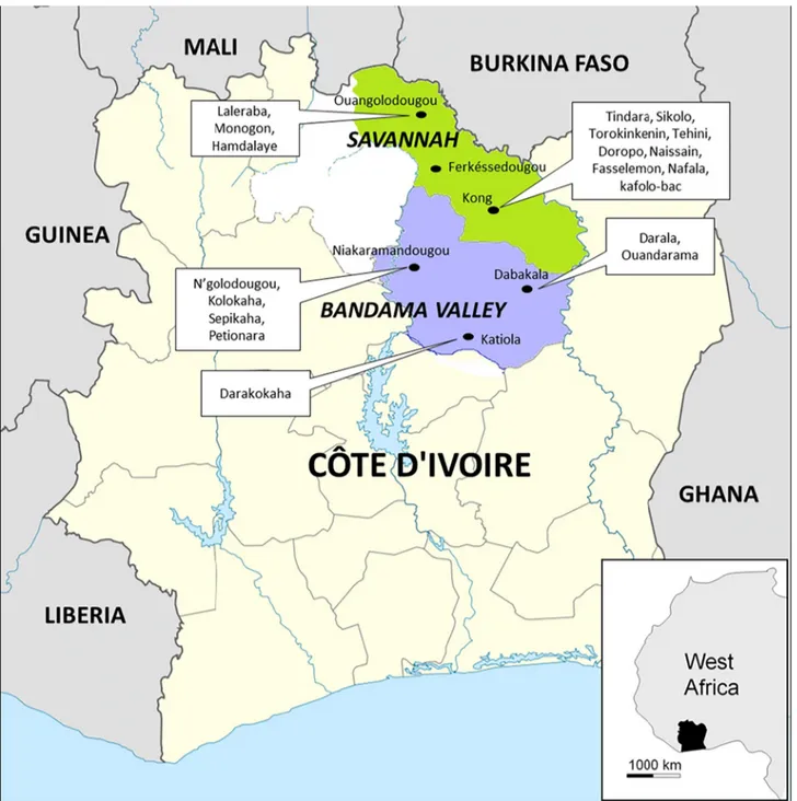
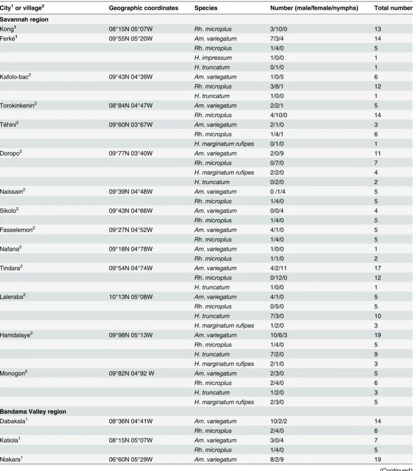
The tick collection was conducted over a period ranging from October 30 to November 8, 2014. Ticks were manually collected from cattle in two regions of Côte d'Ivoire: Savannah and Bandama Valley (Fig 1,Table 1). In total, 378 ticks (304 adults and 74 nymphs) were collected
Fig 1. Map of Côte d’Ivoire showing the regions, cities and villages where the ticks were collected for our study. doi:10.1371/journal.pntd.0004367.g001
Table 1. Geographic coordinates of tick collection sites.
City1or village2 Geographic coordinates Species Number (male/female/nymphs) Total number Savannah region
Kong1 08°15N 05°07W Rh. microplus 3/10/0 13
Ferké1 09°55N 05°20W Am. variegatum 7/3/4 14
Rh. microplus 1/4/0 5
H. impressum 1/0/0 1
H. truncatum 0/1/0 1
Kafolo-bac2 09°43N 04°39W Am. variegatum 1/0/5 6
Rh. microplus 3/8/1 12
H. truncatum 1/0/0 1
Torokinkenin2 08°84N 04°47W Am. variegatum 2/2/1 5
Rh. microplus 4/10/0 14
Téhini2 09°60N 03°67W Am. variegatum 2/1/0 3
Rh. microplus 1/4/1 6
H. marginatum rufipes 0/1/0 1
Doropo2 09°77N 03°40W Am. variegatum 2/0/9 11
Rh. microplus 0/7/0 7
H. marginatum rufipes 2/2/0 4
H. truncatum 0/2/0 2
Naissain2 09°39N 04°48W Am. variegatum 0 /1/4 5
Rh. microplus 1/4/0 5
Sikolo2 09°43N 04°66W Am. variegatum 0/0/4 4
Rh. microplus 1/4/0 5
Fasselemon2 09°27N 04°52W Am. variegatum 4/1/0 5
Rh. microplus 1/4/0 5
Nafana2 09°18N 04°78W Am. variegatum 1/0/0 1
Rh. microplus 1/1/0 2
Tindara2 09°54N 04°74W Am. variegatum 4/2/11 17
Rh. microplus 0/12/0 12
H. truncatum 1/0/0 1
Laleraba2 10°13N 05°08W Am. variegatum 4/1/0 5
Rh. microplus 0/5/0 5
H. truncatum 7/3/0 10
H. marginatum rufipes 1/2/0 3
Hamdalaye2 09°98N 05°13W Am. variegatum 10/6/3 19
Rh. microplus 1/4/0 5
H. truncatum 7/2/0 9
H. marginatum rufipes 2/1/0 3
Monogon2 09°82N 04°92 W Am. variegatum 2/3/0 5
Rh. microplus 2/4/0 6
H. truncatum 1/2/0 3
H. marginatum rufipes 2/3/0 5
Bandama Valley region
Dabakala1 08°36N 04°41W Am. variegatum 10/2/2 14
Rh. microplus 2/4/0 6
Katiola1 08°15N 05°07W Am. variegatum 3/0/4 7
Rh. microplus 1/4/0 5
Niakara1 06°60N 05°29W Am. variegatum 8/2/9 19
from three cities and 12 villages in the Savannah region and three cities and seven villages in the Bandama Valley region (Table 1). Ticks were stored in 70% ethanol until morphological and molecular analyses in laboratory of URMITE, Marseille (France). The species and sex of the ticks were identified according to standard taxonomic keys for adult ticks [2].
DNA extraction and real-time PCR
Total DNA from half of each tick was extracted using the EZ1 DNA tissue kit (Qiagen, Hilden, Germany) following the manufacturer’s instructions. DNA extracts were stored at +4°C until use. Bacterial DNA was initially detected using bacterial genus-specific or species-specific quantitative real-time PCRs (qPCRs) targeting: Rickettsia spp., R. africae, R. aeschlimannii, R. massiliae, Borrelia spp., Anaplasmataceae spp., A. phagocytophilum, Bartonella spp., C. burne-tii, and Spiroplasma spp. (Table 2). Samples with a high discordance in the cycle threshold number (Ct) for Rickettsia spp. and R. africae (in all cases, low Ct for Rickettsia spp. and high Ct for R. africae) were subjected to specific qPCRs for two other rickettsial species: R. aeschli-mannii and R. massiliae in order to identify possible co-infection. qPCRs were performed using a CFX 96 Real Time System (Bio-Rad, Marnes-la-Coquette, France) and the Eurogentec MasterMix Probe PCR kit (Eurogentec, Liège, Belgium). PCR tests were considered to be posi-tive when the Ct was lower than 35 Ct [22]. In addition, two different specific qPCRs targeting two different sequences had to be positive in order to confirm the presence of a bacterium in the ticks. Positive controls (bacterial DNA) and negative controls (master mix or water) were used to validate the PCR runs.
Table 1. (Continued)
City1or village2 Geographic coordinates Species Number (male/female/nymphs) Total number
Rh. microplus 3/2/0 5
Darala2 08°44N 04°35W Am. variegatum 2/1/0 3
Rh. microplus 1/4/0 5
Rh. senegalensis 1/1/0 2
Ouandarama2 08°68N 04°39W Am. variegatum 2/0/4 6
Rh. microplus 1/4/1 6
Rh. senegalensis 0/1/0 1
Darakokaha2 08°27N 05°16W Am. variegatum 1/0/0 1
Rh. microplus 1/4/0 5
N’golodougou2 09°15N 05°12W Am. variegatum 4/2/0 6
Rh. microplus 2/7/0 9
Kolokaha2 08°97N 05°21W Am. variegatum 2/2/0 4
Rh. microplus 1/4/0 5
Sépikaha2 08°91N 05°03W Am. variegatum 1/0/9 10
Rh. microplus 2/4/2 8 Petionara2 08°47N 05°03W Rh. microplus 1/4/0 5 Am. variegatum 72/29/69 170 Rh. microplus 34/122/5 161 Rh. senegalensis 1/2/0 3 H. truncatum 17/10/0 27 H. marginatum rufipes 7/9/0 16 H. impressum 1/0/0 1
Total of ticks for the two regions 132/172/74 378
Standard PCR and sequencing
Most of samples which were considered positive by qPCRs were subsequently subjected to standard PCR. All samples which were positive using Rickettsia genus-specific but negative with R. africae qPCR were subjected to standard PCR to amplify a portion of the ompA gene. We also chose two positive ticks for R. africae by species to confirm the presence of R. africae by standard PCR. The primers used (190.70, 190.180, and 190.701) amplified a 632-bp frag-ment of the Rickettsia ompA gene [60]. For the identification of Borrelia species, primers target-ing a portion of the flaB gene were used [33]. Anaplasmataceae spp. (Anaplasma spp.,
Ehrlichia spp., and Wolbachia spp.) were identified using Ana 212f and Ana 753r primers tar-geting a 500 bp portion of the 23S rRNA gene [47].
Table 2. Primers and probes used for real-time quantitative PCR in this study.
Microorganisms Targeted sequence Primers f, r (5’-3’) and Probes p (6FAM–TAMRA) References
Rickettsiaspp. gltA(RKNDO3) f_GTGAATGAAAGATTACACTATTTAT [53]
r_GTATCTTAGCAATCATTCTAATAGC p_CTATTATGCTTGCGGCTGTCGGTTC
R. africae poT15-dam2 f_TGCAACACGAAGCACAAAAC [6]
r_CCTCTTGCGAAACTCTACTT p_TGA CGTGTGGATTCGAGCACCGGA
R. aeschlimannii Intergenic spacer (RaescSca1) f_AAAGAAATGGATTTCACGGCGAA [12] r_ACCAAGTAAACGTCTCGTAC
p_TGGGGAAATATGCCGTATACGCAAGC
R. massiliae Hypothetical protein f_CCAACCTTTTGTTGTTGCAC [54]
r_TTGGATCAGTGTGACGGACT p_CACGTGCTGCTTATACCAGCAAACA
Anaplasmaspp. 23S rRNA (TtAna) f_TGACAGCGTACCTTTTGCAT [47]
r_TGGAGGACCGAACCTGTTAC p_GGATTAGACCCGAAACCAAG
Anaplasma phagocytophilum apaG f_TAAGCGCAGTTGGAAGATCA [55]
r_CGGCACATCCACATAAAACA p_TGATGAACGGCTGGTATCAG
Spiroplasma rpoB f_TGTTGGACCAAACGAAGTTG [55]
r_CCAACAATTGGTGTTTGTGG p_GCTAACCGTGCTTTAATGGG
Coxiella burnetii Insertion Sequence (IS1111) f_CAAGAAACGTATCGCTGTGGC [56]
r_CACAGAGCCACCGTATGAATC p_CCGAGTTCGAAACAATGAGGGCTG
(IS30A) f_CGCTGACCTACAGAAATATGTCC [57]
r_GGGGTAAGTAAATAATACCTTCTGG p_CATGAAGCGATTTATCAATACGTGTATG
Bartonellaspp. Internal transcribed spacer16S (BartoITS3) f_GATGCCGGGGAAGGTTTTC [58] r_GCCTGGGAGGACTTGAACCT
p_GCGCGCGCTTGATAAGCGTG
Borreliaspp Internal transcribed spacer 16S RNA (Bor ITS4) f_GGCTTCGGGTCTACCACATCTA [59] r_CCGGGAGGGGAGTGAAATAG p_TGCAAAAGGCACGCCATCACC (Bor_16S) f_AGCCTTTAAAGCTTCGCTTGTAG [34] r_GCCTCCCGTAGGAGTCTGG p_CCGGCCTGAGAGGGTGAACGG doi:10.1371/journal.pntd.0004367.t002
Standard PCR was performed on a ThermalCycler (Applied Biosystem, Paris, France). The reactions were carried out using the Hotstar Taq-polymerase (Qiagen), in accordance with the manufacturer’s instructions. The amplicons were visualized using electrophoresis on a 1.5% agarose gel stained with ethidium bromide and examined using an ultraviolet transilluminator. The PCR products were purified using a PCR filter plate Millipore NucleoFast 96 PCR kit fol-lowing the manufacturer’s recommendations (Macherey–Nagel, Düren, Germany). The ampli-cons were sequenced using the BigDye Terminator Cycle Sequencing Kit (Applied Biosystems) with an ABI automated sequencer (Applied Biosystems).The sequences which were obtained were assembled using ChromasPro software (ChromasPro 1.7, Technelysium Pty Ltd.,Tewan-tin, Australia) and compared with those available in GenBank by NCBI BLAST (http://blast. ncbi.nlm.nih.gov/Blast.cgi).
Phylogenetic analysis
DNA sequences alignment was carried out using MEGA 6 (http://www.megasoftware.net/ mega.php). We selected the Bayesian method [61] using TOPALi 2.5 software (Biomathemat-ics and Statist(Biomathemat-ics Scotland) to construct phylogenetic trees.
Results
Of the 378 ticks identified, 170 Am. variegatum, 161 Rh. microplus, 3 Rh. senegalensis, 27 H. truncatum, 16 H. marginatum rufipes, and one H. impressum were analyzed. No A. phagocyto-philum, Bartonella spp. and Spiroplasma spp. were detected in ticks. Rickettsia spp. was found in 187 of 378 ticks (49%); most of them, 174/378 (46%), were identified as R. africae with spe-cific qPCR (Table 3). R. africae was detected in 154/170 (90%) Am. variegatum, 16/161 (10%) Rh. microplus, 2/16 (12%) H. marginatum rufipes, 1/27 (4%) H. truncatum and 1/1 H. impres-sum (Table 3). To confirm the presence of R. africae, we performed standard PCR using two positive ticks per species. The BLAST search of the ompA gene sequences from ticks revealed 100% nucleotide identity with the ompA gene of R. africae detected in Am. variegatum col-lected in Antigua (GenBank EU622980). We amplified the ompA fragment in all ticks positive for Rickettsia spp. but negative for R. africae qPCR. The BLAST analyses showed that ompA sequences of R. aeschlimannii were detected in 7/16 (44%) H. marginatum rupifes and 3/27 (11%) H. truncatum. The sequences were 99% identical to those of R. aeschlimannii, previously detected in H. impeltatum collected in Egypt (GenBank HQ335157) and 100% identical to those detected in H. marginatum in Turkey (GenBank KF791251). R. massiliae was observed in 1/3 (33%) Rh. senegalensis with 100% similarity R. massiliae, previously detected in Rh. sene-galensis in Guinea (GenBank JN043508). Finally, these results were confirmed by a specific qPCR for R. aeschlimannii and R. massiliae (Table 2). We also performed these species-specific qPCR on three samples (two H. marginatum rufipes and one Rh. senegalensis) where we observed a high discordance (more than 5 Cts) between Rickettsia genus-specific qPCR (low Ct) and R. africae species-specific qPCR (higher Ct). We found that in all three cases, a co-infection by two rickettsia species: R. massiliae plus R. africae in Rh. senegalensis and R. aeschli-mannii plus R. africae in H. marginatum rufipes.
C. burnetii was detected in one tick (Table 3). Screening of all ticks for Borrelia spp. using qPCR, detected 16/378 (4%) positive ticks. We succeeded in amplifying a fragment of flaB gene and 16S rRNA sequence only in 4/378 (1%) ticks. A BLAST search showed that these sequences probably belong to an undescribed species, because only 87% (288/329 bp), 87% (287/328 bp), 97% (319/328 bp), and 87% (288/329 bp) similarities were observed with, respectively, the flaB gene of Borrelia duttonii (GenBank AB105132), Borrelia sp. IA-1 (GenBank EU492387), Borre-lia sp. BrFlab (GenBank EF141022), and BorreBorre-lia sp. IA-1 (GenBank EU492387). The
phylogenetic position of this Borrelia is shown inFig 2. Because these potentially new species had not previously been isolated, we propose the provisional names Candidatus Borrelia afri-cana for the genotype TCI22 and Candidatus Borrelia ivorensis for the genotypes TCI140 and TCI351. In a phylogenetic tree based on a 344 bp fragment of the Borreliae flaB gene, the sequences of Candidatus Borrelia africana and Candidatus Borrelia ivorensis are situated in the
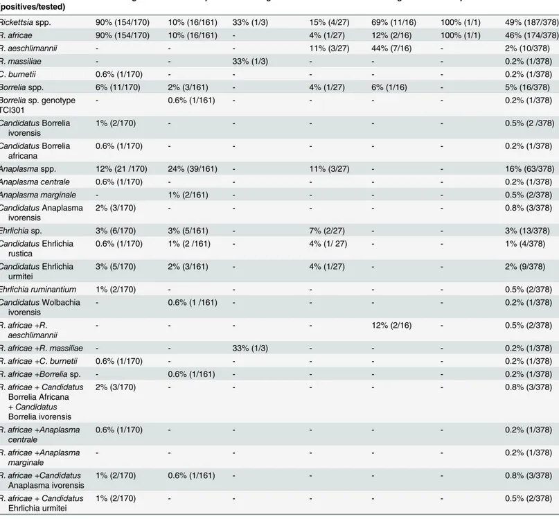
Table 3. Prevalence of positive ticks by PCR.
Bacterium% (positives/tested)
Am. variegatum Rh. microplus Rh. senegalensis H. truncatum H. marginatum H. impressum Total Rickettsiaspp. 90% (154/170) 10% (16/161) 33% (1/3) 15% (4/27) 69% (11/16) 100% (1/1) 49% (187/378) R. africae 90% (154/170) 10% (16/161) - 4% (1/27) 12% (2/16) 100% (1/1) 46% (174/378) R. aeschlimannii - - - 11% (3/27) 44% (7/16) - 2% (10/378) R. massiliae - - 33% (1/3) - - - 0.2% (1/378) C. burnetii 0.6% (1/170) - - - 0.2% (1/378) Borreliaspp. 6% (11/170) 2% (3/161) - 4% (1/27) 6% (1/16) - 5% (16/378) Borreliasp. genotype TCI301 - 0.6% (1/161) - - - - 0.2% (1/378) CandidatusBorrelia ivorensis 1% (2/170) - - - 0.5% (2 /378) CandidatusBorrelia africana 0.6% (1/170) - - - 0.2% (1/378) Anaplasmaspp. 12% (21 /170) 24% (39/161) - 11% (3/27) - - 16% (63/378) Anaplasma centrale 0.6% (1/170) - - - 0.2% (1/378) Anaplasma marginale - 1% (2/161) - - - - 0.5% (2/378) CandidatusAnaplasma ivorensis 2% (3/170) - - - 0.8% (3/378) Ehrlichiasp. 3% (6/170) 3% (5/161) - 7% (2/27) - - 3% (13/378) CandidatusEhrlichia rustica 0.6% (1/170) 1% (2 /161) - 4% (1/ 27) - - 1% (4/378) CandidatusEhrlichia urmitei 3% (5/170) 2% (3/161) - 4% (1/27) - - 2% (9/378) Ehrlichia ruminantium 1% (2/170) - - - 0.5% (2/378) CandidatusWolbachia ivorensis - 0.6% (1 /161) - - - - 0.2% (1/378) R. africae +R. aeschlimannii - - - - 12% (2/16) - 0.5% (2/378) R. africae +R. massiliae - - 33% (1/3) - - - 0.2% (1/378) R. africae +C. burnetii 0.6% (1/170) - - - 0.2% (1/378) R. africae +Borrelia sp. - 0.6% (1/161) - - - - 0.2% (1/378) R. africae + Candidatus Borrelia Africana + Candidatus Borrelia ivorensis 2% (3/170) - - - 0.8% (3/378) R. africae +Anaplasma centrale 0.6% (1/170) - - - 0.2% (1/378) R. africae +Anaplasma marginale - - - 0.2% (1/378) R. africae +Candidatus Anaplasma ivorensis 1% (2/170) 0.6% (1/161) - - - - 0.8% (3/378) R. africae + Candidatus Ehrlichia urmitei 1% (2/170) - - - 0.5% (2/378)
= 0%; the name‘Candidatus’ is employed here for the new species because they are not isolated doi:10.1371/journal.pntd.0004367.t003
Borrelia genus near Borrelia sp. from Ethiopian Amblyomma cohaerens (GenBank JX089967) and are closer to the relapsing fever group than to that of Lyme disease. As previously shown, Ethiopian Borrelia group together with these new genotypes to form a separate and
Fig 2. flaB gene-based phylogenetic analysis of the strains identified in this study. Phylogenetic tree highlighting the position of Borrelia sp. identified in the present study relative to borrelia type strains and uncultured borreliae. The flaB sequences were aligned using CLUSTALW, and phylogenetic inferences were obtained from a Bayesian phylogenetic analysis with the HKY+Γ; JC+ Γ and HKY+ Γ substitution models for the first, second and third codons respectively. The GenBank accession numbers are indicated at the end. Sequences obtained in the present study are in bold. The numbers at the nodes are the bootstrap values obtained by repeating the analysis 100 times to generate a majority consensus tree. There were a total of 300 positions in the final dataset. The scale bar indicates a 10% nucleotide sequence divergence.
well-supported (bootraps 100) branch on the phylogenetic tree situated between Lyme disease and relapsing fever clusters, albeit closer to the latter. We also identified Borrelia sp. (genotype TCI301) in Rh. microplus which was almost identical to Borrelia sp. previously identified in the same ticks in Brazil (GenBank EF141022).
Sixty-three ticks were positive using qPCR targeting the 23S rRNA of Anaplasmataceae. Only 39 DNA samples were positive using qPCR were successfully amplified in standard PCR. A possible explanation may consist of the lower sensitivity of standard PCR compared to qPCR. After sequencing, we obtained good quality sequences for only 22 samples (22/378; 6%). We suggest that the poor sequence quality may be explained by co-infection by two or more species belonging to the Anaplasmataceae family. We have identified one case of A. centrale in Am. variegatum (100% identity with the A. centrale strain Israel, NR_076686), and two cases of E. ruminantium in Am. variegatum (100% identity with the E. ruminantium strain Welgevon-den, NR_077000). We have identified A. marginale in two Rh. microplus (100% of homology with A. marginale strain Florida, NR_0765879). Finally, for all remaining sequences, Blast anal-ysis shows a homology score of under 92% which means that these sequences are likely to
Fig 3. 23S rRNA based phylogenetic analysis of strains identified in this study. Phylogenetic tree highlighting the position of Anaplasma sp, Ehrlichia sp and Wolbachia sp identified in the present study relative to Anaplama, Ehrlichia and Wolbachia type and uncultured strains. The 23S rRNA sequences were aligned using MEGA 6 and phylogenetic inferences were obtained from a Bayesian phylogenetic analysis with the HKY standard model.
correspond to new species. After the construction of a phylogenetic tree (Fig 3), we propose that the status of Candidatus is applied to an uncultured species but not formally recognized by the International Code of Nomenclature of Bacteria [62]. The result shows three cases of Anaplasma: Candidatus Anaplasma ivorensis related to A. phagocytophilum identified in ticks, two in Am. variegatum, and one in Rh. microplus. The three sequences have one to two SNP (single nucleo-tide polymorphism) between them. In one Rh. microplus, a potential new Wolbachia sp., Candi-datus Wolbachia ivorensis, was identified, closely related to the Wolbachia endosymbiont of Cimex lectularius (GenBank AP013028). We also identified two groups of sequences correspond-ing to new Ehrlichia spp. which cluster in two clades. Indeed, in four cases (one Am. variegatum, two Rh.microplus, and one H. truncatum), we identified Candidatus Ehrlichia rustica in the sub-group of Ehrlichia chaffeensis. In nine ticks (five Am. variegatum, three Rh. microplus and one H. truncatum), we detected Candidatus Ehrlichia urmitei that was previously observed by our team in Rh. bursa ticks collected in the Bacque area of France (M. Dahmani, personal communication) (Fig 3). Candidatus Ehrlichia urmitei forms an independent and well-supported clade situated between the E. ruminantium clade and that of Ehrlichia muris (Fig 3).
Finally, 15 co-infections (15/378; 4%) were detected by qPCR. All 15 co-infections involved the presence of R. africae. In Am. variegatum, ten co-infections (10/15; 66%) were observed with R. africae plus another pathogen such as Coxiella burnetii (1/170; 0.6%), A. centrale (1/170; 0.6%), A. marginale (1/170; 0.6%), Candidatus Borrelia Africana, Candidatus Borrelia ivorensis (3/170; 2%), Candidatus Anaplasma ivorensis (2/170; 1%), or Candidatus Ehrlichia urmitei (2/170; 1%) as well as H. marginatum rufipes with R. africae plus R. aeschlimannii (2/ 16; 12%) and in Rh. senegalensis with R. africae plus R. aeschlimannii (1/3; 33%) (Table 3). The access numbers of the sequences of all the potential new species deposited in GenBank are summarized inTable 4.
Table 4. New sequences amplified in this study and deposited in GenBank.
Sequences type Gene Ascension number
CandidatusBorrelia africana TCI22 flaB KT364343
CandidatusBorrelia ivorensis TCI140 flaB KT364344
CandidatusBorrelia ivorensis TCI351 flaB KT364346
Borreliasp. genotype TCI301 flaB KT364345
CandidatusBorrelia africana TCI22 16S rRNA KT364339
CandidatusBorrelia ivorensis TCI140 16S rRNA KT364340 CandidatusBorrelia ivorensis TCI351 16S rRNA KT364341
Borreliasp. TCI301 16S rRNA KT364342
CandidatusAnaplasma ivorensis TCI50 23S rRNA KT364326 CandidatusAnaplasma ivorensis TCI94 23S rRNA KT364327 CandidatusAnaplasma ivorensis TCI149 23S rRNA KT364328 CandidatusWolbachia ivorensis TCI113 23S rRNA KT364329 CandidatusEhrlichia rustica TCI141 23S rRNA KT364330 CandidatusEhrlichia rustica TCI145 23S rRNA KT364331 CandidatusEhrlichia rustica TCI167 23S rRNA KT364332 CandidatusEhrlichia rustica TCI238 23S rRNA KT364333 CandidatusEhrlichia urmitei TCI148 23Sr RNA KT364334 CandidatusEhrlichia urmitei TCI230 23S rRNA KT364335 CandidatusEhrlichia urmitei TCI106 23S rRNA KT364336 CandidatusEhrlichia urmitei TCI127 23S rRNA KT364337 CandidatusEhrlichia urmitei TCI166 23S rRNA KT364338
Discussion
Domestic animal resources supply some 30% of total human food and agricultural production requirements. They are particularly vital to subsistence and economic development in develop-ing countries as they continually provide essential food products, draught power and manure for crop production and generate income as well as employment for most of the rural poor [63]. However, livestock-associated ticks are often reservoirs or vectors of human vector-borne diseases [18]. Intensification of livestock farming is one cause of the abundance of various vec-tors and tick-borne diseases. In recent years, the spectrum of tick-borne diseases infecting ani-mals has increased; many of these diseases, such as rickettsioses, borrelioses, Q fever,
anaplasmoses, and ehrlichioses, are gaining increasing attention from clinicians and veterinar-ian [4]. Advances in the development of molecular biology tools facilitate the detection of new bacteria [4,64].
Rickettsioses have been identified in humans, animals and ticks which are considered to be the main vectors of such pathogens as R. africae, R. aeschlimannii, and R. massiliae in sub-Saharan Africa [9]. In our study, rickettsial DNA was found in 49% of ticks collected from cat-tle. For the first time, the presence of R. aeschlimannii in ticks in Côte d’Ivoire is shown. This study provides evidence of R. aeschlimannii infection in 23% of Hyalomma ticks including H. marginatum rufipes (44%) and H. truncatum (11%). R. aeschlimannii has not been observed in other tick species. These data support the theory that the Hyalomma genus is a main vector and reservoir of R. aeschlimannii. It was previously reported in 45% to 51% of H. marginatum rufipes and 6% to 7% in H. truncatum collected from cows, donkeys, sheep, goats and horses in Senegal [12]. These data are comparable to those of our study. The high prevalence of R. africae (90%) in Am. variegatum can be explained by the high transovarial and trans-stadial transmis-sion rates (100%) and a filial infection rate (93%) that was previously demonstrated in Am. var-iegatum [20]. This result shows that this tick species acts as a vector but also as a reservoir for R. africae in Côte d’Ivoire. R. africae was recently detected in other tick genera including Rhipi-cephalus and Hyalomma [12,18,19,57]. In our study, the prevalence of R. africae is 10% in Rh. microplus and 9% in Hyalomma spp., which is lower than in co-fed Am. variegatum, suggesting that these ticks are probably not the competent vectors for R. africae. This bacterium likely infects Rh. microplus and Hyalomma spp. during co-feeding. The first report of the presence of R. massiliae in Côte d’Ivoire was in Rhipicephalus spp. [28]; this is comparable to the detection of R. massiliae in a Rh. senegalensis tick found in our study.
C. burnetii infections have been also reported as being between 0.7% and 6.8% in ticks from cattle in western African countries [51] but not in Côte d’Ivoire where the seroprevalence of C. burnetii was estimated to be 3% [52]. Here, we show for the first time the presence of C. burne-tii in Côte d’Ivoire, although only in one tick. Most Borrelia species such as B. hispanica, B. dut-tonii, and B. crocidurae detected in Africa, are related to soft ticks. Their main vectors are Ornithodoros spp. [65]. To date, Borrelia sp. was identified only once in an African hard tick, Am. cohaerens, in Ethiopia [39]. It has been also reported that Rhipicephalus spp. transmits Borrelia theileri to cattle, causing bovine borreliosis [18]. In Côte d’Ivoire, we show that Am. variegatum were infected by three potential new Borrelia and Rh. microplus by one potential new Borrelia. The sequences of Borrelia sp. (genotype TCI301) were identical to 99% of those of Borrelia sp. found in engorged Rhipicephalus sp. ticks collected from horse in Brazil
(EF141022). Phylogenetic analysis showed that Borrelia sp. TCI301 is classified in the relapsing fever group, close to B. crocidurae and B. hispanica, two etiological agents of relapsing fever in Africa [26,31]. Blast analysis of the flaB gene showed that three new Candidatus Borrelia ivor-ensis and Candidatus Borrelia africana borreliae were significantly different to all other borre-liae, except for this Borrelia sp in Am. cohaerens in Ethiopia [39]. These potential new Borrelia
form a new clade between the clades of Lyme disease borreliae and relapsing fever borreliae. Thus, this is the first time that Borrelia species have been detected in Côte d’Ivoire and the first time their presence has been confirmed in hard ticks in Africa.
Bacteria from the Anaplasmataceae family were previously known to be pathogens of veteri-nary importance. However, in the three last decades, many human pathogens have been identi-fied in this family [66]. Recently, based on the rrl gene, our team developed new tools to identify most bacteria belonging to Anaplasmataceae family [47]. These tools combine a qPCR followed by a standard PCR then sequencing, and have been used successfully to amplify DNA from bacteria belonging to Anaplasma spp., Ehrlichia spp., Neorickettsia spp., and Wolbachia spp. available in our laboratory [67]. We have successfully amplified Anaplasmataceae DNA in 6% of our ticks. For the first time, we have demonstrated the presence of A. marginale, A. cen-trale, E. ruminantium, and potential novel Ehrlichia, Anaplasma, and Wolbachia spp. in ticks in Côte d’Ivoire. A. marginale was observed in Am. variegatum and Rh. microplus. To the best of our knowledge, A. marginale has never been reported in Africa in Rh. microplus. The first report of its presence in Côte d’Ivoire was in 2007, but the exact route of its introduction into this region has not yet been determined [68]. A recent study indicated that the majority of the Rhipicephalus (ex-Boophilus) spp. collected and identified from farms around Azaguié (Côte d’Ivoire) are Rh. microplus (96%) [69]. A. centrale is a species closely related to A. marginale; this naturally attenuated strain has been used as a live vaccine to prevent severe diseases due to A. marginale senso stricto strains for 100 years [70]. We identified this species in Am. variega-tum. To the best of our knowledge, A. centrale has never previously been detected in these ticks. The potential of Am. variegatum to transmit A. centrale needs further investigation. E. ruminantium was previously described in Am. variegatum which is invasive to cattle attaches to the hooves and cattle remain standing, particularly in the rainy season [71,72]. Recent phylo-genetic analyses of Am. variegatum from Kenya, Mali, Burkina Faso, Ethiopia and the Carib-bean show low genetic diversity within this population, suggesting an westward expansion of these ticks and supporting east-west genetic separation, with Caribbean genetic sequences being associated with and often identical to West African haplotypes. The data suggest that Am. variegatum reached West Africa from Zambia [73]. We have also identified three potential new species, Candidatus Anaplasma ivorensis, Candidatus Ehrlichia urmitei, and Candidatus Ehrlichia rustica. The detection of these potential new species has limitations, as not all previ-ously described species are already molecularly characterized. Indeed, such species as Ana-plasma caudatum, AnaAna-plasma bovis, and AnaAna-plasma mesaeterum [74] are incompletely characterized with no strain available and no or few genes sequenced, so the detection of a ‘new’ genotype may, in fact, be the re-discovery of an old, incompletely characterized species. Further studies are required to clarify whether these new genetic variants represent a new spe-cies. The other potential new ehrlichiae was closely related to Ehrlichia sp. amplified from Rh. bursa in France. Interestingly, this new species was amplified from two different regions in the world and from different species of ticks (Rhipicephalus, Amblyomma, and Hyalomma spp).
Finally, it is reported that ticks are often infected following a blood meal from a co-infected host [75,76]. Recently, mixed infections were reported for the first time in West Africa in feeding ticks and caused mainly by Rickettsia spp. and C. burnetii [18]. In Côte d’Ivoire, for the first time we show multiple co-infections in ticks. These co-infections systematically involved R. africae. To date, no human cases of anaplasmoses, ehrlichioses, borrelioses, rickett-sioses or co-infections have been reported in Côte d’Ivoire. However, these diseases are still lit-tle known by clinicians and laboratory diagnostic is lacking in most cases. It is important to continue to study the epidemiological data of such emerging pathogens which may be the source of disease complications in both animals and humans. We provide evidence and dem-onstrate the endemicity of these different bacteria in the studied regions that have the same
characteristics agro-ecological and climatic. Furthermore, these diseases could be a cause of death of unknown origin in rural areas in Côte d'Ivoire [77].
Acknowledgments
We thank Ange Goun (IRD, Abidjan) for his help in logistic issues.
Author Contributions
Conceived and designed the experiments: KPY FF OM. Performed the experiments: CBE KPY MD YLA NA AKN JDN OM. Analyzed the data: CBE KPY MD DR FF OM. Contributed reagents/materials/analysis tools: CBE KPY YLA AKN JDN. Wrote the paper: CBE DR FF OM.
References
1. Bechah Y, Socolovschi C, Raoult D. Identification of rickettsial infections by using cutaneous swab specimens and PCR. Emerg Infect Dis. 2011; 17(1):83–6. doi:10.3201/eid1701.100854PMID:
21192860.
2. Walker AR, Bouattour A, Camicas J, Estrada-Pena A, Horak IG, Latif AA, et al. Ticks of Domestic Ani-mals in Africa: A Guide to Identification of Species. Published by Bioscience Reports, Edinburgh, United Kingdom. 2003.http://www.biosciencereports.pwp.blueyonder.co.uk/.
3. Pérez-Eid C. Emergence of tick-borne diseases in temperate countries. Ann Biol Clin (Paris). 2004; 62 (2):149–54. PMID:15047466.
4. Dantas-Torres F, Chomel BB, Otranto D. Ticks and tick-borne diseases: a One Health perspective. Trends Parasitol. 2012; 28(10):437–46. doi:10.1016/j.pt.2012.07.003PMID:22902521.
5. Robyn MP, Newman AP, Amato M, Walawander M, Kothe C, Nerone JD, et al. Q Fever Outbreak Among Travelers to Germany Who Received Live Cell Therapy -United States and Canada, 2014. MMWR Morb Mortal Wkly Rep. 2015, 2; 64(38):107. doi:10.15585/mmwr.mm6438a3PMID:
26421460.
6. Sokhna C, Mediannikov O, Fenollar F, Bassene H, Diatta G, Tall A, et al. Point-of-care laboratory of pathogen diagnosis in rural Senegal. PLoS Negl Trop Dis. 2013; 7(1):e1999. doi:10.1371/journal.pntd. 0001999PMID:23350001.
7. Mayxay M, Sengvilaipaseuth O, Chanthongthip A, Dubot-Pérès A, Rolain JM, Parola P, et al. Causes of Fever in Rural Southern Laos. Am J Trop Med Hyg. 2015; 93(3):517–20. doi: 10.4269/ajtmh.14-0772PMID:26149859.
8. Ghosh L, Nagar G. Problem of tics and tick-borne diseases in India with a special emphasis on prog-ress in tick control research: a review. J Vector Borne. 2014; 51 (4): 259–70. PMID:25540956. 9. Parola P, Paddock CD, Socolovschi C, Labruna MB, Mediannikov O, Kernif T, et al. Update on
tick-borne rickettsioses around the world: a geographic approach. Clin Microbiol Rev. 2013; 26 (4):657– 702. doi:10.1128/CMR.00032-13PMID:24092850.
10. Kelly PJ, Beati L, Mason PR, Matthewman LA, Roux, Raoult D, et al. Rickettsia africae sp. nov., the etio-logical agent of African tick bite fever. Int J Syst Bacteriol.1996; 46 (2):611–4. PMID:8934912. 11. Ndip LM, Fokam EB, Bouyer DH, Ndip RN, Titanji VP, Walker DH, et al. Detection of Rickettsia africae
in patients and ticks along the coastal region of Cameroon. Am J Trop Med Hyg. 2004; 71:363–6. PMID:15381820.
12. Mediannikov O, Diatta G, Fenollar F, Sokhna C, Trape JF, Raoult D. Tick-borne rickettsioses, neglected emerging diseases in rural Senegal. PLoS Negl Trop Dis. 2010; 14; 4(9). pii: e821. doi:10. 1371/journal.pntd.0000821PMID:20856858.
13. Parola P, Inokuma H, Camicas JL, Brouqui P, Raoult D. Detection and identification of spotted fever group Rickettsiae and Ehrlichiae in African ticks. Emerg Infect Dis. 2001; 7(6):1014–7. PMID:
11747731.
14. Parola P. Rickettsioses in sub-Saharan Africa. Ann N Y Acad Sci. 2006; 1078:42–7. PMID:17114679. 15. Socolovschi C, Matsumoto K, Marie JL, Davoust B, Raoult D, Parola P. Identification of Rickettsiae,
Uganda and Djibouti. Emerg Infect Dis. 2007; 13 (10):1508–10. doi:10.3201/eid1310.070078PMID:
16. Mediannikov O, Davoust B, Socolovschi C, Tshilolo L, Raoult D, Parola P. Spotted fever group rickett-siae in ticks and fleas from the Democratic Republic of the Congo. Ticks Tick Borne Dis. 2012; 3(5– 6):371–3. doi:10.1016/j.ttbdis.2012.10.015PMID:23137572.
17. Mediannikov O, Diatta G, Zolia Y, Balde MC, Kohar H, Trape JF, et al. Tick-borne rickettsiae in Guinea and Liberia. Tick Borne Dis. 2012; 3(1):43–8. doi:10.1016/j.ttbdis.2011.08.002PMID:22309858. 18. Reye AL, Arinola OG, Hubschen JM, Muller CP. Pathogen prevalence in ticks collected from the
vege-tation and livestock in Nigeria. Appl Environ Microbiol. 2012; 78(8):2562–8. doi: 10.1128/AEM.06686-11PMID:22327584.
19. Ogo NI, de Mera IG, Galindo RC, Okubanjo OO, Inuwa HM, Agbede RI, et al. Molecular identification of tick-borne pathogens in Nigerian ticks. Vet Parasitol. 2012; 187(3–4):572–7. doi:10.1016/j.vetpar. 2012.01.029PMID:22326937.
20. Socolovschi C, Huynh TP, Davoust B, Gomez J, RaoulT D, Parola P. Transovarial and trans-stadial transmission of Rickettsiae africae in Amblyomma variegatum ticks. Clin Microbiol Infect. 2009; 15 Suppl 2:317–8. doi:10.1111/j.1460691.2008.02278.xPMID:19456811.
21. Raoult D, Fournier PE, Abboud P, Caron F. First documented human Rickettsia aeschlimannii infection. Emerg Infect Dis. 2002; 8:748–9. PMID:12095451.
22. Parola P, Paddock CD., Raoult D. Tick-borne rickettsioses around the world: emerging diseases chal-lenging old concepts. Clin Microbiol Rev.2005, 18(4): 719–56. PMID:16223955.
23. Kernif T, Djerbouh A, Mediannikov O, Ayach B, Rolain JM, Raoult D, et al. Rickettsia africae in Hya-lomma dromedariiticks from sub-Saharan Algeria. Ticks Tick Borne Dis. 2012; 3(5–6):377–9. doi:10. 1016/j.ttbdis.2012.10.013PMID:23164496.
24. Kamani J, Baneth G, Apanaskevich DA, Mumcuoglu KY, Harrus S. Molecular detection of Rickettsia aeschlimanniiin Hyalomma spp. ticks from camels (Camelus dromedarius) in Nigeria, West Africa. Med Vet Entomol. 2015; 29(2):205–9. doi:10.1111/mve.12094PMID:25565180.
25. Parola P, Socolovschi C, Jeanjean L, Bitam I, Fournier PE, Sotto A, et al. Warmer weather linked to tick attack and emergence of severe rickettsioses. PLoS Negl Trop Dis. 2008; 2(11):e338. doi:10.1371/ journal.pntd.0000338PMID:19015724.
26. Garcia-Garcia JC, Portillo A, Nunez MJ, Santibanez S, Castro B, Oteo JA. A patient from Argentina infected with Rickettsia massiliae. Am J Trop Med Hyg. 2010; 82(4):691–2. doi:10.4269/ajtmh.2010. 09–0662PMID:20348520.
27. Cascio A, Torina A, Valenzise M, Blanda V, Camarda N, Bombaci S, et al. Scalp escharand neck lymphadenopathy caused by Rickettsia massiliae. Emerg Infect Dis. 2013; 19(5):836–7. doi:10.3201/ eid1905.121169PMID:23697545.
28. Berrelha J, Briolant S, Muller F, Rolain JM, Marie JL, Pagés F, et al. Rickettsia felis and Rickettsia mas-siliaein Ivory Coast, Africa. Clin Microbiol Infect. 2009; 15 Suppl 2:251–2. doi:10.1111/j.1469-0691. 2008.02273.xPMID:19548990.
29. Barbour AG. Fall and rise of Lyme of disease and other Ixodes tick-borne infections in North America and Europe. Br Med Bull. 1998; 54(3):647–58. PMID:10326291.
30. Cutler SJ, Abdissa A, Trape JF. New concepts for the old challenge of African relapsing fever borrelio-sis. Clin Microbiol Infect. 2009; 15(5):400–6. doi:10.1111/j.1469-0691.2009.02819.xPMID:
19489922.
31. Trape JF, Diatta G, Arnathau C, Bitam I, Sarih M, Belghyti D, et al. The epidemiology and geographic distribution of relapsing fever borreliosis in West and North Africa, with a review of the Ornithodoros erraticuscomplex (Acari: Ixodida). PLoS One. 2013; 8(11):e78473. doi:10.1371/journal.pone. 0078473eCollection2013. PMID:24223812.
32. Svihrova V, Hudeckova H, Jesenak M, Schwarzova K, Kostanova Z, Ciznar I, et al. Lyme borreliosis-analysis of the trends in Slovakia, 1999–2008. Folia Microbiol (Praha). 2011; 56(3):270–5. doi:10. 1007/s12223-011-0036-yPMID:21607749.
33. Vial L, Diatta G, Tall A, Ba el H, Bouganali H, Durand P, et al. Incidence of tick-borne relapsing fever in West Africa: longitudinal study. Lancet. 2006; 368(9529):37–43. PMID:16815378.
34. Parola P, Ryelandt J, Mangold AJ, Mediannikov O, Guglielmone AA, Raoult D, et al. Relapsing fever Borrelia in Ornithodoros ticks from Bolivia. Ann Trop Med Parasitol. 2011; 105(5):407–11. doi:10. 1179/1364859411Y.0000000021PMID:21929883.
35. Cutler SJ, Bonilla EM, Singh RJ. Population structure of East African relapsing fever Borrelia spp. Emerg Infect Dis. 2010; 16(7):1076–80. doi:10.3201/eid1607.091085PMID:20587177.
36. Sarih M M Garnier, Boudebouch N, Bouattour A, Rihani A, Hassar M, et al. Borrelia hispanica relapsing fever, Morocco. Emerg Infect Dis. 10: 1626–1629. Emerg Infect Dis. 2009; 15(10):1626–9.
37. Trape JF, Godeluck B, Diatta G, Rogier C, Legros F, Albergel J, et al. The spread of tick-borne borrelio-sis in West Africa and its relationship to sub-Saharan drought. Am J Trop Med Hyg. 1996; 54(3):289– 93. PMID:8600768.
38. Nordstrand A, Bunikis I, Larsson C, Tsogbe K, Schwan TG, Nilsson M, et al.Tickborne relapsing fever diagnosis obscured by malaria, Togo. Emerg Infect Dis. 2007; 13(1):117–23. PMID:17370524. 39. Mediannikov O, Abdissa A, Socolovschi C, Diatta G, Trape JF, Raoult D, et al. Detection of a new
Bor-relia species in ticks taken from cattle in Southwest Ethiopia. Vector Borne Zoonotic Dis. 2013; 13 (4):266–9. doi:10.1089/vbz.2011.0874PMID:23421894.
40. Pruneau L, Moumène A, Meyer DF, Marcelino I, Lefrançois T, Vachiéry N, et al. Understanding Ana-plasmataceaepathogenesis using“Omics” approaches. Front Cell Infect Microbiol. 2014; 4:86. doi:10. 3389/fcimb.2014.00086. eCollection 2014PMID:25072029.
41. Rymaszewska. A, Grenda S. Bacteria of the genus Anaplasma–characteristics of Anaplasma and their vectors: a review. Veterinarni Medicina. 2008. 53 (11): 573–584.
42. Aubry P, Geale DW. A review of bovine anaplasmosis. Transbound Emerg Dis. 2011; 58(1):1–30. doi:
10.1111/j.1865-1682.2010.01173.xPMID:21040509.
43. Raoult D, Parola P. Molecular tools in the epidemiology of tick–borne bacterial diseases Ann Biol Clin (Paris). 2001. 59(2):177–82. PMID:11282521.
44. Georges M, Garrity. Bergey’s of Manual of Systematic Bacteriology: The proteobacteria. Second Edi-tion. Volume two; 2005. pp.120-122.
45. Allsopp BA. Natural history of Ehrlichia ruminantium. Vet Parasitol. 2010; 167(2–4):123–35. doi:10. 1016/j.vetpar.2009.09.014PMID:19836892.
46. Djiba ML, Mediannikov O, Mbengue M, Thiongane Y, Molez JF, Seck MT, et al. Survey of Anaplasma-taceaebacteria in sheep from Senegal. Trop Anim Health Prod. 2013; 45(7):1557–61. doi:10.1007/ s11250-013-0399-yPMID:23553260.
47. Dahmani M, Bernard D, Mohamed SB, Fenollar F, Raoult D, Mediannikov O. Development of a new PCR-based assay to detect Anaplasmataceae and the first report of Anaplasma phagocytophilum and Anaplasma platysin cattle from Algeria. Comp Immunol Microbiol Infect Dis. 2015; 39:39–45. doi:10. 1016/j.cimid.2015.02.002PMID:25748051.
48. Maurin M, Raoult D. Q fever. Clin Microbiol Rev. 1999; 12(4):518–53. PMID:10515901. 49. Kazar J. Coxiella burnetii infection. Ann N Y Acad Sci. 2005; 1063:105–14. PMID:16481501. 50. Cooper A, Hedlefs R, Ketheesan N, Govan B. Serological evidence of Coxiella burnetii infection in
dogs in a regional centre. Aust Vet J. 2011; 89(10):385–7. doi:10.1111/j.1751-0813.2011.00819.x
PMID:21933165.
51. Mediannikov O. Coxiella burnetii in humans and ticks in rural Senegal. PLoS Negl Trop Dis. 2010; 4(4): e654. doi:10.1371/journal.pntd.0000654PMID:20386603.
52. Tissot-Dupont H, Brouqui P, Faugere B, Raoult D. Prevalence of antibodies to Coxiella burnetii, Rick-ettsia conorii, and RickRick-ettsia typhi in seven African countries. Clin Infect Dis. 1995; 21(5):1126–33. PMID:8589132.
53. Rolain JM, Sthul L, Maurin M, Raoult D. Evaluation of antibiotic susceptibilities of three rickettsial spe-cies including Rickettsia felis by a quantitative PCR DNA assay. Antimicrob Agents Chemother. 2002; 46(9):2747–51. PMID:12183224.
54. Socolovschi C, Frederic P, Raoult D. Rickettsia felis in Aedes albopictus mosquitoes, Libreville, Gabon. Emerg Infect Dis. 2012; 18(10):1687–9. doi:10.3201/eid1810.120178PMID:23017437. 55. Subramanian G, Sekevova Z, Raoult D, Medianikov O. Multiple tick-associated bacteria in Ixodes
rici-nusfrom Slovakia. Ticks Tick Borne Dis. 2012; 3(5–6):406–10. doi:10.1016/j.ttbdis.2012.10.001
PMID:23182274.
56. Rolain JM, Raoult D. Molecular detection of Coxiella burnetii in blood and sera during Q fever. QJM. 2005; 98(8):615–7; author reply 617–20. PMID:16027172.
57. Mediannikov O, Trape JF, Diatta G, Parola P, Fournier PE, Raoult D. Rickettsia africae, Western Africa. Emerg Infect Dis. 2010; 16(3):571–3. doi:10.3201/eid1603.090346PMID:20202453.
58. Raoult D, Roblot F, Rolain JM, Besnier JM, Loulergue J, Bastides, et al. First isolation of Bartonella alsaticafrom a valve of a patient with endocarditis. J Clin Microbiol. 2006; 44(1):278–9. PMID:
16390990.
59. Bottieau E, Verbruggen E, Aubry C, Socolovschi C, Vlieghe E. Meningoencephalitis complicating relapsing fever in traveler returning from Senegal. Emerg Infect Dis. 2012; 18(4):697–8. doi:10.3201/ eid1804.111771PMID:22469185.
60. Mura A, Socolovschi C, Ginesta J, Lafrance B, Magnan S, Rolain JM, et al. Molecular detection of spot-ted fever group rickettsiae in ticks from Ethipia and Chad. Trans R Soc Trop Med Hyg. 2008; 102 (9):945–9. doi:10.1016/j.trstmh.2008.03.015PMID:18440576.
61. Ronquist F, Huelsenbeck JP. MrBayes 3: Bayesian phylogenetic inference under mixed models. Bioin-formatics. 2003; 19 (12):1572–4. PMID:12912839.
62. Murray RG, Stackebrandt E. Taxonomic note: implementation of the provisional status Candidatus for incompletely described procaryotes. Int J Syst Bacteriol. 1995; 45 (1):186–7 I. PMID:7857801. 63. Ayalew W, Rege JEO, Getahun E, Tibbo M, Mamo Y. Delivering Systematic Information on Indigenous
Animal Genetic Resources–the development and prospects of DAGRIS. Animal Genetic Resources Group, International Livestock Research Institute. P, O Box 5689. 2003. Addis Ababa, Ethiopia. 64. Doudier B, Olano J, Parola P, Brouqui P. Factors contributing to emergence of Ehrlichia and Anaplasma
spp. as human pathogens. Vet Parasitol. 2010; 167(2–4):149–54. doi:10.1016/j.vetpar.2009.09.016
PMID:19836890.
65. Parola P, Raoult D. Ticks and tick-borne bacterial diseases in humans: an emerging infectious threat. Clin Infect Dis. 2001; 32(6):897–928. PMID:11247714.
66. Inokuma H. Vectors and reservoir hosts of Anaplasmataceae. Rickettsial diseases, books.google. com. 2007, pp. 199–212.
67. Dahmani M, Loudahi A, Mediannikov O, Fenollar F, Raoult D, Davoust B. Molecular detection of Ana-plasma platysand Ehrlichia canis in dogs from Kabylie, Algeria. Ticks Tick Borne Dis. 2015; 6(2):198– 203. doi:10.1016/j.ttbdis.2014.12.007PMID:25583345.
68. Madder M, Thys E, Geysen D, Baudoux C, Horak I. Boophilus microplus ticks found in West Africa. Exp Appl Acarol. 2007; 43(3):233–4. PMID:17929178.
69. Madder M, Thys E, Achi L, Touré A, De Deken R. Rhipicephalus (Boophilus) microplus: A most suc-cessful invasive tick species in West-Africa. Exp Appl Acarol. 2011; 53(2):139–45. doi:10.1007/ s10493-010-9390-8PMID:20711801.
70. Herndon DR, Palmer GH, Shkap V, Knowles DP, Brayton K. Complete genome sequence of Ana-plasma marginale subsp centrale. J Bacteriol. 2010; 192(1):379–80. doi:10.1128/JB.01330-09PMID:
19854912.
71. Kifle G, Sori T. Molecular Detection of Ehrlichia Species in Amblyomma Ticks Collected from Rumi-nants in Abernosa Ranch, Ethiopia. Global J Mol Sci. 2014; 9 (2): 12–18. doi:10.5829/idosi.gjms.2014. 9.2.85213
72. Stachurski F. Invasion of West African cattle by the tick Amblyomma variegatum. Med Vet Entomol. 2000; 14(4):391–9. PMID:11129703.
73. Beati L, Patel J, Lucas-Williams H, Adakal H, Kanduma EG, Tembo-Mwase E, et al. Phylogeography and demographic history of Ambryomma variegatum (Fabricius) (Acari: Ixodidae), the Tropical Bont Tick. Vector Borne Zoonotic Dis. 2012; 12(6):514–25. doi:10.1089/vbz.2011.0859PMID:22448720. 74. Georges M, Garrity. Bergey’s of Manual of Systematic Bacteriology: The proteobacteria. Second
Edi-tion. Volume two; 2005. pp. 121–125.
75. Levin M L, Fish D. Acquisition of coinfection and simultaneous transmission of Borrelia burgdorferi and Ehrlichia phagocytophilaby Ixodes scapularis ticks. Infect Immun. 2000; 68(4):2183–6. PMID:
10722618.
76. Levin M L, Fish D. Interference between the agents of Lyme disease and human granulocytic ehrlichio-sis in a natural reservoir host. Vector Borne Zoonotic Dis. 2001; 1(2):139–48. PMID:12653144. 77. Koné S, Fürst T, Jaeger FN, Esso EL, Baïkoro N, Kouadio KA, et al. Causes of death in the Taabo
health and demographic surveillance system, Côte d'Ivoire, from 2009 to 2011. Glob Health Action. 2015; 8:27271. doi:10.3402/gha.v8.27271. eCollection2015PMID:25959772.