HAL Id: hal-03040357
https://hal.archives-ouvertes.fr/hal-03040357
Submitted on 4 Dec 2020
HAL is a multi-disciplinary open access
archive for the deposit and dissemination of
sci-entific research documents, whether they are
pub-lished or not. The documents may come from
teaching and research institutions in France or
abroad, or from public or private research centers.
L’archive ouverte pluridisciplinaire HAL, est
destinée au dépôt et à la diffusion de documents
scientifiques de niveau recherche, publiés ou non,
émanant des établissements d’enseignement et de
recherche français ou étrangers, des laboratoires
publics ou privés.
A 2D FEM for shear wave modeling applied to soft
tissues
M Bilasse, S Chatelin, Guillaume Altmeyer, J Vappou, I Charpentier
To cite this version:
M Bilasse, S Chatelin, Guillaume Altmeyer, J Vappou, I Charpentier. A 2D FEM for shear wave
mod-eling applied to soft tissues. EMI International Conference, Oct 2016, Metz, France. �hal-03040357�
A 2D FEM for shear wave modeling applied to soft tissues
M. Bilasse
∗1,2, S. Chatelin
1,3, G. Altmeyer
1,2, J. Vappou
1and I. Charpentier
1April 2016
Over the past two decades, dynamic elasticity imaging has emerged as a virtual palpation tool allowing for a qualitative investigation of the in vivo mechanical response of biological soft tissues.
From an experimental point of view, the three main steps [1] are (i) the generation of shear waves in the organ under sutdy, (ii) the measurement of the displacement field using medical imaging, then (iii) the identification of mechanical properties. Clearly, coupling wave physics and biomechanics in an appropriate mechanical model is key issue for such an identification.
Although most of shear wave inverse approaches for Magnetic Resonance Elastography (MRE) make the assumption of isotropic viscoelasticity, computer models should better describe the complexity of the computational domain to be of general use. In particular, boundary conditions, heterogeneity and anisotropy that strongly impact the wave propagation have to be accounted in a fair manner, whether set to estimated values in the modeling or identified during the course of the inverse problem solution.
MRE experiments have been developed since 5 years in the Icube laboratory [2]. Computational mechanics aspects are now considered in their full complexity to go further and to foresee, at a medium term, a clinical validation of the proposed methods. To that end, our modeling builds on a 2D FEM based on composite plate elements that moreover account for a vertical transverse displacement. Such a choice is motivated by a lower computational cost compared to a 3D modeling and the straightforward comparison with classical 2D MRI images. The nonlinear wave propagation is solved following [3].
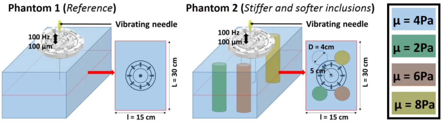
Figure 1: Left: Reference hydrogel phantom. Right: Hydrogel phantom with stiff and soft inclusions
The modeling will be evaluated with respect to MRE experiments realized on hydrogel phantoms with stiff and soft inclusions.
References
[1] Vappou J (2012) Magnetic resonance and ultrasound imaging-based elasticity imaging methods: a review, Biomedical Engineering 40:121–134.
[2] Corbin N, Vappou J, Breton E, Boehler Q, Barb´e L, Renaud P and de Mathelin M (2016) Interventional MR elastography for MRI-guided percutaneous procedures, Magnetic Resonance in Medicine 75:1110-8.
[3] Bilasse M, Charpentier I, Daya EM, Cherkaoui M (2009) A generic approach for the solution of nonlinear residual equations. Part II: Homotopy and complex nonlinear eigenvalue method, Computer Methods in Applied Mechanics and Engineering 198:3999–4004.
∗Corresponding Author [email protected];1Icube, CNRS & University of Strasbourg;2ECAM, Strasbourg;3IHU Strasbourg.