HAL Id: tel-01598656
https://tel.archives-ouvertes.fr/tel-01598656
Submitted on 29 Sep 2017HAL is a multi-disciplinary open access archive for the deposit and dissemination of sci-entific research documents, whether they are pub-lished or not. The documents may come from teaching and research institutions in France or abroad, or from public or private research centers.
L’archive ouverte pluridisciplinaire HAL, est destinée au dépôt et à la diffusion de documents scientifiques de niveau recherche, publiés ou non, émanant des établissements d’enseignement et de recherche français ou étrangers, des laboratoires publics ou privés.
Régulation de l’expression du brain-derived
neurotrophic factor par le récepteur des glucocorticoïdes
dans le neurone
Hui Chen
To cite this version:
Hui Chen. Régulation de l’expression du brain-derived neurotrophic factor par le récepteur des glu-cocorticoïdes dans le neurone. Biochemistry, Molecular Biology. Université Paris Saclay (COmUE), 2017. English. �NNT : 2017SACLS242�. �tel-01598656�
Neuronal Glucocorticoid Receptor
Regulation of Brain-Derived Neurotrophic
Factor Expression
Thèse de doctorat de l'Université Paris-Saclay préparée à l'Université Paris-SudÉcole doctorale n°568 Signalisation et Réseaux Intégratifs en Biologie Spécialité de Doctorat : Aspects Moléculaires et Cellulaires de la Biologie Thèse présentée et soutenue à Kremlin-Bicêtre, le 21 Septembre 2017, par
Madame Hui CHEN
Composition du Jury :
Monsieur Michael SCHUMACHER Président DR, INSERM (Université Paris-Sud-UMR-S 1195)
Madame Laurence LANFUMEY-MONGREDIEN Rapporteur DR, INSERM (Université Paris Descartes-UMR-S 894)
Monsieur Charbel MASSAAD Rapporteur
PU, INSERM (Université Paris Descartes-UMR-S 1124)
Madame Sophie PEZET Examinateur
MCU, CNRS (ESPCI Paris-UMR 8249)
Monsieur Marc LOMBES Directeur de thèse
DR, INSERM (Université Paris-Sud-UMR-S 1185)
Monsieur Damien LE MENUET Co-Directeur de thèse CR, INSERM (Université Paris-Sud-UMR-S 1185)
NNT : 201 7 SA CL S242
天行健,君子以自强不息; 地势坤,君子以厚德载物。 -易经
As heaven maintains vigor through movements, a gentle man should constantly strive for self-perfection. As earth's condition is receptive devotion, a gentle man should hold the outer world with broad mind. From I Ching, translated by Changquan Li
有志者事竟成。 -刘秀
Where there is a will, there is a way. By Xiu Liu
ACKNOWLEDGEMENTS
I would like to express my heartfelt gratitude to all the persons who helped me during my PhD study and research.
Foremost, my deepest gratitude first goes to my supervisor, Dr. Marc Lombès, who is so gentle and humorous. Thanks to him for giving me an opportunity to work in the lab of Inserm UMR1185. He sets a good example to us due to the expert suggestions, detailed interpretations and patient attitude on the experimental designs and performances as well as article and theses writing. What moves me very much is his continuous support and encouragement during all the time of my PhD study.
I would like to express my sincerest appreciation to my co-supervisor, Dr. Damien Le Menuet, who deserves my enormous esteem and gratitude. Thanks to him for teaching me all the techniques I need, paying great patience and enthusiasm, and always showing me his optimism and honesty. His guidance, valuable suggestions, encouragement, as well as his well-rounded character will benefit me throughout my entire life. Besides, he also helps me a lot and gives me much understanding in the daily life. I appreciate all of these very much.
I extend my sincere thanks to all the members of the jury. I would like to thank Dr. Michael Schumacher for accepting to be the president of my thesis committee. I am grateful to Dr. Laurence Lanfumey-Mongredien and Prof. Charbel Massaad for reviewing this thesis carefully and helping me ameliorate the quality of the thesis. I am also thankful to the examiner, Dr. Sophie Pezet for accepting to examine and evaluate my work.
Special thanks to China Scholarship Council for the financial support for my PhD studies in France.
I would like to thank my colleagues in our lab, which is just like a family, for the enthusiastic assist on my experiments, numerous stimulating discussions, insightful suggestions and kind patience. I warmly thank Dr. Say Viengchareun for his regular care and warm gifts for me and my family. I am pleased to thank Dr. Larbi Amazit for picking up my husband and me at the airport when we arrived in Paris for the first time four years ago. I am grateful to Dr. Jérôme Fagart for driving me home when there was a strike of public transport system. Thanks to Dr. Laëtitia Martinerie for offering me useful information on the registration in hospital when I
was pregnant. I greatly appreciate Dr. Stéphanie Chauvin for providing us the commercial N2a cell line, and thank Dr. Isabelle Beau and Dr. Justine Bouilly for the Renilla plasmid. Special thanks to Dr. Nadine Binart for her Doliprane when I had a serious backache. Thanks to Dr. Bruno Francou for his help on plasmid sequencing.
I want to express my big thanks to Florian Le Billan for his help on the inscription in the doctoral school and translation because of my poor French. I am grateful to Ingrid Lema for her explanation on the thesis defence procedure. I also owe a special debt of gratitude to Mirella Hage for inviting me to the dinners in restaurant and lunches on the grass. I would like to express gratitude to Mohsen Ayrout for his help on Western Blot and to Géraldine Vitellius for her help on bacterial transformation. Thanks to Geoffrey Boulate for his help on answering telephone when I had communication trouble with people just speaking French. Very big thanks to Simon Travers for his help on measuring steroid concentrations using LC/MS-MS method and special thanks to Fanny Jung for her preliminary data support for my thesis. I am thankful to Christophe Lhadj for aiding me settling down the troubles with my computer. I appreciate to the happy crepes hour that Vianney Demeocq brought us.
I want to express my warm gratitude for their moral support: Dr. Anne Guiochon, Dr. Jérôme Bouligand, Dr. Séverine Trabado and Dr. Peter Kamenicky. I also would like to thank the girls, Emmanuelle Kuhn, Segolène Hescot, Anne-Lise Lecoq, Khadija Lamribet, Valérie Bernard, Charlotte Sonigo, Laurence Dumeige, Sophie Lamothe, Nelly Ramos, Thi-An Vu, Clémence Delcour, Laura Bessiene and Françoise Magnin. I appreciate greatly for their accompanying and helping in my life bit by bit. I feel so happy and lucky to have met them at UMR 1185 in Paris. I really wish them the best future.
Thanks to secretaries, Madame Claudia Grangetaud, Madame Esther Kingue-Tiki and Madame Cindy De Biase, for their efficient help in facilitating my lab life. I am thankful to Meriem Messina for providing us clean and organized working environment.
I want to thank my friends with whom I spent such many happy hours in Paris and always feel deep care and precious happiness. I am grateful to Xiaoyan Zhu and Jianguo Wang for every call we made for information communication, every delicious and warm dinner we cooked, every trip we accompanied each other, and every talk from our hearts. I am often moved by your painstaking care, valuable advice and pervasive assist. I hope your baby grow up with consummate love and health. I also owe big thanks to my friends Yueyi Wang and Yongchao
Xu, for helping us on registration in hospital when I was pregnant; for preparing the documents and driving us back home from the hospital after I gave birth to a baby; for communicating with the director of creche for applying a position for my daughter. Thanks for Xiaoqing Liu and Xiubin Li for the care and help during the travels and greeting us when I was pregnant. I also want to thank Zhenyu Wang and Jing Hao for visiting my baby and me in hospital and their kind gifts. Thanks to Yuxiang Song, Liheng Yin, Wenwen Gao, Lin Lei, and Dawei Liu for their selfless help and regular care to my family. I will never forget these good memories constituted with all of you and I hope we could continue composing our friendship movement until the last day of our lives.
Very special thanks to our landlord, the Thomas family, for providing us kind help whatever we need and warm presents on every Christmas Day and my daughter’s birthday. I will never forget you called the doctor for my daughter who had a serious fever at 2:00 a.m. before dawn.
I would like to express my deepest gratitude and love to my husband Liang Zhang. I am thankful for his inclusive love like the vast sea, his understanding on my every word and deed, his encouragement and support in the hard moments, and his general heart which allows me to be a free-and-easy child. “It is the best day when I met the best you. What could I repay to
such a good person like you?” 今夕何夕,见此良人。子兮子兮,如此良人何!
I appreciate to my daughter, Hezhuo Zhang, whose arrival brings me so much happiness and power. Even though we encountered many difficulties and challenges taking care of her, every small smile from her cures all the discomforts. I wish my little sweet baby growing with health, love, sunshine and courage.
Last but not the least, I would like to give my most sincere gratitude to my parents, my parents-in-law, my younger brother and his family, my younger sister and her family, my elder sister-in-law and her family. It is all of them who give us the unconditional love, constant encouragement and spiritual support.
游子吟
孟郊
慈母手中线,游子身上衣。
临行密密缝,意恐迟迟归。
谁言寸草心,报得三春晖。
Hymn of the Traveller
By Jiao Meng; Translated by Chris Pereira
Threads adeptly brandished by a loving mother, sewed into garments for a son so soon to depart. Her sewing picks up pace as the date approaches, worries of belated return echoing in her heart.
Who dare claim that the green grass might somehow repay the sun for its warm hearth?
Régulation de l’expression du Brain Derived Neurotrophic Factor
par le récepteur des glucocorticoï
des dans le neurone
Résumé
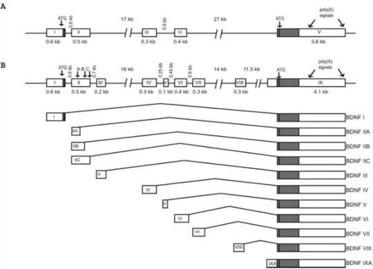
Le facteur de croissance brain-derived neurotrophic factor (BDNF) est un acteur clé de la fonction neuronale. BDNF est fortement exprimé dans tout le cerveau, mais son niveau d'expression le plus élevé se trouve dans l'hippocampe, une structure limbique d'importance majeure pour les fonctions cognitives, telles que la mémorisation, l'apprentissage, le comportement, le stress, les émotions et l'humeur. Dans le système nerveux central (SNC), le BDNF régule la survie neuronale, la différenciation et la croissance. Une quantité importante de donnée indique que le BDNF est également impliqué dans l'homéostasie neuronale et les processus liés à la plasticité du cerveau tels que la mémoire, l'apprentissage et l’addiction, ainsi que la potentialisation à long terme. Les modifications des niveaux d'expression de BDNF dans des sous-populations spécifiques de neurones ont été associées à diverses pathologies, y compris la dépression, l'épilepsie, la maladie d'Alzheimer, les maladies de Huntington et de Parkinson. Le BDNF fonctionne principalement en se liant à son récepteur à haute affinité, la kinase B (TrkB) associée à la tropomyosine, activant plusieurs voies telles que MAP kinase, PI3 kinase et Phospholipase C. Le gène Bdnf murin présente une structure génomique complexe comprenant au moins 9 exons (I À IX), qui sont alternativement épissés pour générer des variantes de transcription BDNF spécifiques sur un exon IX commun et unique à l'extrémité terminale 3 'codant pour la protéine. La génération d'un grand nombre d'isoformes de transcription est probablement d'importance biologique car dans les cultures neuronales d’hippocampe de rat, il a été démontré que les variants d'ARNm de BDNF sont distribués différemment dans des compartiments dendritiques spécifiques afin de réguler la disponibilité locale de la protéine BDNF. Dans l'ensemble, la complexité des événements régulant quantitativement et qualitativement l'expression de BDNF mettent en évidence sa contribution cruciale à la fonction du SNC en physiologie et pathologie.
Les hormones glucocorticoïdes (GC) exercent également des actions pleiotropiques sur les neurones en se liant et en activant le récepteur des glucocorticoïdes (GR, NR3C1), ainsi que le récepteur des minéralocorticoïdes (MR, NR3C2). Ce dernier témoigne d'une grande affinité du ligand et, par conséquent, il est presque toujours occupé par les GC, tandis que le GR est activé principalement sous des concentrations élevées de GC circulant, comme dans les
conditions de stress ou au pic circadien de l’hormone. Les deux récepteurs sont fortement exprimés dans l'hippocampe, agissant de concert ou de manière opposée pour réguler divers processus physiologiques et neurologiques tels que les réponses au stress, la survie, l'apoptose et la potentialisation à long terme. GR associé à son ligand fonctionne comme un facteur de transcription, se liant directement aux éléments de réponse des glucocorticoïdes sur l’ADN des séquences régulatrices ou alternativement il interagit indirectement avec d'autres facteurs de transcription, pour activer ou réprimer la transcription des gènes cibles. La régulation de l'expression de BDNF par le stress a des conséquences importantes sur la pathophysiologie des troubles de l'humeur et dans le mécanisme d'action des agents antidépresseurs. Comme l'exposition au stress aigu ou chronique déclenche une augmentation des concentrations circulantes de GC, un rôle de ces hormones dans la modulation de l'expression de BDNF a souvent été suggéré, mais la plupart de ces études sont basées sur des preuves indirectes et sont parfois contradictoires en fonction du modèle, de la dose et de la durée du traitement. Dans l'ensemble, les mécanismes moléculaires par lesquels les GC régulent l'expression de BDNF ne sont pas encore clairement définis à ce jour.
Dans le présent travail, nous avons démontré que les niveaux élevés de GC répriment l'expression de l'ARNm totale de Bdnf via GR dans divers modèles cellulaires neuronaux in
vitro par des analyses qPCR. Dans des cultures primaires neuronales d'hippocampe de souris
et la lignée immortalisée BZ, l’expression des transcrits contenant les exons I, IV et IV de
Bdnf est réprimé par ce mécanisme de régulation, mais ceux qui sont épissés à partir des
exons VII et VIII ne le sont pas, indiquant une régulation spécifique de certains exons par GR. En outre, par transfection transitoire réalisée dans des cellules N2a, nous avons montré que les activités de transcription du promoteur 1 et 4 étaient diminuées par GR au niveau transcriptionnel. De plus, grâce à des expériences de mutagenèse dirigée et d’immunoprécipitation de chromatine, nous avons révélé que la répression induite par GR sur la transcription de Bdnf se produit en se liant à, au moins en partie, une petite région de promoteur en amont de l'exon IV. En ce qui concerne la (les) séquence (s) d'ADN répondant au recrutement de GR sur le promoteur 4 de BDNF, alors que deux sites de liaison pour AP1 et deux autres pour CREB ont été exclus, d'autres éléments de réponse sur l’ADN pour AhR et NeuroD sont des candidats potentiels. Dans l'ensemble, nous proposons que l'un des mécanismes responsables de la répression de l'expression de Bdnf par DEX soit la liaison de GR juste en amont de l'exon IV, à travers des complexes ternaires avec des facteurs de transcription qui doivent encore être déterminés. Il convient de noter que cette région en
amont de l'exon IV présente un degré élevé d'homologie entre les espèces de mammifères mettant l'accent sur leur importance biologique.
En résumé, ces résultats contribuent à une meilleure compréhension des mécanismes sur la façon dont GR régule l'expression de Bdnf, ce qui apporte de nouveaux éléments sur les interactions moléculaires entre les voies de signalisation des glucocorticoïdes et celles des neurotrophines dans les neurones, les deux voies étant cruciales en physiologie et pathologie du système nerveux central.
TABLE OF CONTENTS
LIST OF FIGURES AND TABLES ... 1
LIST OF ABBREVIATIONS ... 3
FOREWORD ... 5
INTRODUCTION ... 7
1. Brain-Derived Neurotrophic Factor (BDNF) in the CNS ... 12
1.1 BDNF expression and signaling pathways ... 12
1.2 Pathophysiological functions of BDNF and related diseases ... 14
1.2.1 Physiological functions of BDNF ... 14
a. Role of BDNF on neurogenesis ... 14
b. BDNF and LTP associated with memory and learning ... 15
c. BDNF and exercise ... 17
1.2.2 BDNF and related diseases ... 18
a. BDNF and Alzheimer’s disease (AD) ... 19
b. BDNF and Huntington’s disease (HD) ... 20
c. BDNF and Parkinson’s disease (PD) ... 20
d. BDNF and depression ... 21
e. BDNF and epilepsy ... 22
f. BDNF and neuropathic pain ... 23
g. BDNF and drug addiction ... 24
1.2.3 Conclusion ... 26
1.3 Bdnf gene and protein ... 27
1.3.1 Bdnf gene structure ... 27
1.3.2 BDNF protein processing ... 30
1.3.3 BDNF Val66Met variant ... 31
2. Glucocorticoid receptor (GR) in the brain ... 36
2.1 Glucocorticoids (GC) act in the brain by binding to GR and MR ... 36
2.2 GR signaling pathways ... 40
2.3 Physiological functions and pathological association of GR and MR in the brain ... 45
2.3.1 GR roles in stress response and exercise ... 46
2.3.2 Synaptic and behavioral effects of GR ... 47
2.3.3 GR effects on neuronal survival and neurogenesis ... 50
2.3.4 GR and neurodegenerative disorders ... 51
2.3.5 Conclusion ... 53
2.4 Summary ... 53
3. Interaction between BDNF and GC in the CNS ... 55
3.1 GC regulate BDNF expression via GR ... 55
3.1.1 Regulation of BDNF transcript expression by GR ... 55
3.1.2 Regulation of BDNF signaling by GC ... 57
3.2 BDNF influences GR signaling ... 58
OBJECTIVES ... 61
RESULTS ... 65
Part I : Characterization on several in vitro neuron-like cell models and preliminary analysis on GC effects on BDNF expression ... 67
1. Introduction ... 69
2. Preliminary results ... 69
2.1. Neuronal differentiation of embryonic stem (ES) cells to study the regulation of BDNF by GC ... 69
2.2. Primary cultures of mouse hippocampal neurons as a good in vitro cell model ... 73
2.3. BZ cells as a model for studying kinetic mechanisms ... 76
2.4. N2A cell line as a model to analyze Bdnf promoter sequence activities79 3. Conclusion ... 82
Part II : Glucocorticoid receptor represses brain-derived neurotrophic factor
expression in neuron-like cells ... 83
1. Introduction ... 85
2. Supplementary results and discussion ... 87
2.1. Potential GR-binding DNA sequence and other transcription factors within the small region upstream of exon IV ... 87
2.2. Promoter 1 activity was repressed by GR ... 90
3. Conclusion and Perspectives ... 92
GENERAL DISCUSSION AND PERSPECTIVES ... 95
1. BDNF expression is regulated by GR at the transcriptional level ... 97
2. GR and BDNF promoters are involved in neuronal activity ... 98
3. Exon-specificity of BDNF expression in brain pathophysiology ... 99
4. Spatial distribution of BDNF transcripts ... 99
5. Conservation of BDNF SP4 sequence between human and mouse ... 101
GENERAL CONCLUSIONS ... 105
1
LIST OF FIGURES AND TABLES
Figure 1: Hippocampal structure……….12
Figure 2: Signaling pathways evoked by neurotrophins and their receptors…………...13
Figure 3: Schematic of long term potentiation (LTP) formation………16
Figure 4: Exon/intron structure and alternative transcripts of mouse and rat Bdnf genes…...28
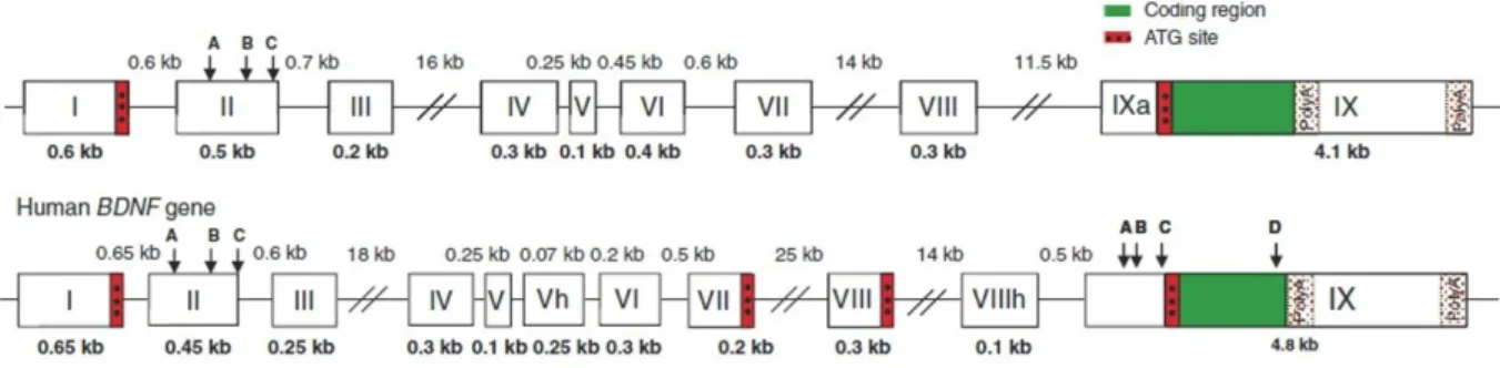
Figure 5: BDNF gene structures in the rodent and human………..29
Figure 6: Regulation of glucocorticoid hormone secretion by the hypothalamic-pituitary-adrenal (HPA) axis………37
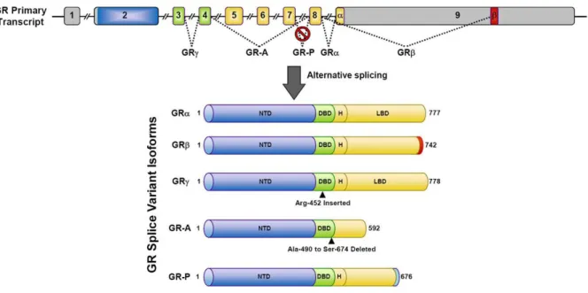
Figure 7: GR splice variants………39
Figure 8: GR signaling pathways………41
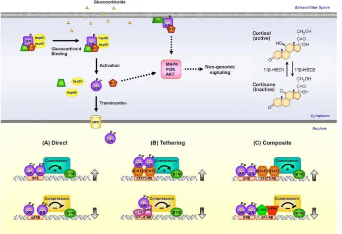
Figure 9: Simplified illustration of the mechanisms of glucocorticoid actions on the CNS...42
Figure 10: The neuronal differentiation of ES cells………70
Figure 11: BDNF (a), MR (b) and GR (c) mRNA expression in mouse brain and ES-derived neurons (ES)………..…71
Figure 12: Corticosteroid effects on BDNF mRNA expression in ES cell-derived neurons...72
Figure 13: Isolation of hippocampal neurons from prenatal mice, adapted from Beaudoin et al………73
Figure 14: Primary cultures of hippocampal neurons from prenatal mice………..74
Figure 15: Aldo effects on BDNF mRNA expression in PCN expressing MR………...75
Figure 16: DEX effects on BDNF mRNA expression in PCN………76
Figure 17: BZ cell culture………77
Figure 18: BDNF and MAP2 mRNA expression in BZ cells increase with time…………...77
Figure 19: FK effects (5 x 10⁻⁶ M) on total BDNF mRNA expression in BZ cells………….78
Figure 20: N2A cell neuronal differentiation………...79
Figure 21: Corticosterone effect on total BDNF mRNA expression in N2A cells…………..80
2 Figure 23: The arrangement of functional cis-elements and the corresponding transcription factors in Bdnf promoter IV, according to Zheng et al. ………...88
Figure 24: GR-dependent inhibitory effects on the activity of a small Bdbf promoter construct SP4.74………89
Figure 25: Aryl hydrocarbon receptor response element (AhR.RE) and Neuro/oligo response element (Neuro/oligo. RE) in SP4.74, as well as two CREs………90
Figure 26: GR-dependent inhibitory effects on LP1 activity………..91 Figure 27: The analysis on homology of mouse and human SP4 DNA sequence, as well as the similarity of potential response elements of AP1, CREB, AhR and Neuro/oligo family.102
Figure 28: Schematic drawing of interplay between GC and BDNF signaling pathways in the neurons………107
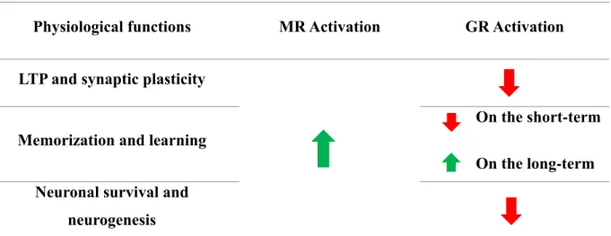
Table 1: Comparison of physiological functions of activated MR and GR on LTP and synaptic plasticity; memorization and learning; neuronal survival and neurogenesis………..49
3
LIST OF ABBREVIATIONS
11βHSD2: 11β-hydroxysteroid dehydrogenase type 2 Aβ: amyloid-β
ACTH: adrenocorticotropic hormone ADX: adrenalectomized
AhR: aryl hydrocarbon receptor
AhR.RE: aryl hydrocarbon receptor response element Aldo: aldosterone
AMPA: alpha-amino-3-hydroxy-5-methyl-4-isoxazolepropionic acid
AMPAR: alpha-amino-3-hydroxy-5-methyl-4-isoxazolepropionic acid receptor BDNF: brain-derived neurotrophic factor
bHLH: basic helix-loop-helix CA: Cornus Ammonis
CaRE: calcium response elements
CaRF: calcium-responsive transcription factor
CARM1: coactivator-associated arginine methyltransferase ChIP: Chromatin Immunoprecipitation
ChIP-seq: Chromatin Immunoprecipitation and sequencing CNS: central nervous system
Cort: corticosterone CPE: carboxy peptidase E
CREB: cAMP response element-binding CRH: corticotropin releasing hormone DBD: DNA-binding domain
DCC: dextran charcoal-coated DEX: dexamethasone
DG: dentate gyrus
DRG: dorsal root ganglion EBs: embryoid bodies
ERK: extracellular signal-regulated kinase ESC: embryonic stem cells
FBS: fetal bovine serum FK: Forskolin
FKBP5: FK506-binding protein 5 GABA: γ- aminobutyric acid GC: glucocorticoids
GFAP: glial fibrillary acidic protein GR: glucocorticoid receptor
GRE: glucocorticoid response element HDAC: histone deacetylase
hMR: human mineralocorticoid receptor HPA: hypothalamic-pituitary-adrenal HTT: huntingtin
JNK: c-Jun N-terminal kinase KA: kainic acid
KIV: knockout of exon IV LBD: ligand binding domain LIF: leukemia inhibitory factor
4 LTD: long-lasting synaptic depression
LTP: long term potentiation
L-VGCC: L type voltage gated calcium channel MAP2: Microtubule-Associated Protein 2 MAPK: mitogen-activated protein kinase
mBDNF: mature brain-derived neurotrophic factor MDD: major depressive disorder
MEM: non-essential amino acids
mGluR: metabotropic glutamate receptor MR: mineralocorticoid receptor
N2A: Neuro-2A cells
NCoR: nuclear receptor corepressor NFκB: nuclear factor-κB
NGF: nerve growth factor
nGRE: negative glucocorticoid response element NMDA: N-methyl-D-aspartate
NMDAR: N-Methyl-D-aspartic Acid receptor NP: neuropathic pain
NSCs: neural stem cells NT3: neurotrophin-3 NT4/5: neurotrophin-4/5 NTD: N-terminal domain
P75NTR: P75 neurotrophin receptor
PCN: primary cultures of hippocampal neurons PEPCK: phosphoenolpyruvate carboxykinase PER1: Period 1
PI3K-Akt: phosphatidylinositol-3-kinase Akt PKAc: catalytic subunit of protein kinase A PLCγ: phospholipase Cγ
PNS: peripheral nervous system
PSG: penicillin, streptomycin, glutamine PVN: paraventricular nucleus
RACE: 5‟ rapid amplification of cDNA ends RRS: repetitive restrain stress
RT-qPCR: real-time quantitative PCR RU: RU486 (Mifepristone)
SAGE: serial analysis of gene expression
SGK1: serum- and glucocorticoid- inducible kinase 1 SGZ: subgranular zone
Shc: Src homologous and collagen-like protein
SMRT: silencing mediator of retinoid and thyroid hormone receptors SNP: single nucleotide polymorphism
SRC: steroid receptor coactivator SVZ: subventricular zone
TRH: thyrotrophin-releasing hormone TrkB: tropomyosin-related kinase B TSA: trichostatin A
USF1/2: upstream stimulatory factor 1/2 UTR: 3‟ untranslated region
5
FOREWORD
When I obtained the master degree in Jilin University in China four years ago, a PhD program under convention between China Scholarship Council (CSC) and University Paris-SUD attracted me. Therefore, I contacted Dr. Marc Lombès‟ team, whose research projects were very interesting, asking for a PhD position in University Paris-SUD. Fortunately, they accepted my request and helped me successfully to pass the strict PhD candidate selection. Being financed by CSC, now I have finished my PhD project with the help of Dr. Marc Lombès and Dr. Damien Le Menuet.
As part of Inserm UMR 1185 lab, “Hormone Signaling, Endocrine and metabolism Physiopathology”, belonging to the University Paris-Sud and University Paris-Saclay, Marc Lombès‟ team which main interests is to decipher the effects and molecular mechanisms of mineralocorticoid receptor (MR, NR3C2) and glucocorticoid receptor (GR, NR3C1) signaling in various cellular contexts, to identify new molecular targets of MR and GR, and to study the mechanisms regulating their expression. Brain-derived neurotrophic factor (BDNF), a main neurotrophin, was suspected to be a potential GR target gene in the conditions of stress for several reasons. First, glucocorticoid (GC) receptors MR and GR as well as BDNF are co-expressed in hippocampal neurons. Second, BDNF exerts many overlapping but sometime opposite actions with those of GR. Third, the cellular events of BDNF effect, including the expression levels, activation of its receptor as well as many cascade proteins involved in BDNF signaling pathways, are mediated tightly by GR activation. However, few have been discovered on the molecular mechanisms involved in GR regulation of BDNF expression.
The aim of this thesis was to study the effects of GR on BDNF expression, as well as the underlying regulatory molecular mechanisms, in various in vitro neuron-like cellular models, which is important to understand the contribution of GR and BDNF to function and pathology of the central nervous system (CNS). Here, I present the principal results we have gotten during my doctoral period.
Before describing and discussing the results of our work, I begin with a bibliographic introduction including three parts: 1) BDNF in the CNS, 2) GR in the brain, and 3) interaction between BDNF and GC in the CNS. The introduction is followed by our data concerning the GR effects on BDNF expression. Using essentially cellular and molecular biology approaches, we demonstrated that GR was responsible for the repression of BDNF acting on a specific
6 DNA sequence. In the manuscript, the experimental data are presented in part as an original article, „Glucocorticoid receptor represses brain-derived neurotrophic factor expression in neuron-like cells‟, published in Molecular Brain in 2017, with additional data presented before and after the article. Finally, a short general discussion allows to put in perspective our results with the current bibliographic context, which is followed by a general conclusion and the bibliography.
7
INTRODUCTION
9 General introduction
As the center of the nervous system, the mammalian brain is a crucial organ that could integrate internal and external information, respond to various stimuli, and finally control the endocrine status and behavior of the organism. The brain is composed of neurons, glial cells, neural stem cells and blood vessels, protected by the cover of meninges. Neurons are the basic functional units of the brain, spinal cord, central nervous system (CNS), and the ganglia of the peripheral nervous system (PNS), acting by receiving, integrating and processing information. A typical neuron is composed of a cellular body (soma), multiple dendrites arising from the soma and often extending for hundreds of micrometers, and a unique cellular extension termed an axon. All neurons are electrically excitable and connect to each other
via synapses to form neural networks, which carry trains of electrical and chemical signals to
distant parts of the brain or body targeting specific recipient cells (Arbib, Érdi et al. 1998).
Dysfunctions and injuries of the human brain cause major deficits in intelligence, memory, personality, and coordination. Except for the physical damage and infectious factors, the brain is also susceptible to neurodegenerative disorders, such as Alzheimer‟s disease (AD), Parkinson‟s disease (PD) and Huntington‟s disease (HD), as well as motor neuron diseases caused by the gradual death of individual neurons, leading to diminution in movement control (ataxia), memory, and cognition. Moreover, numerous psychiatric conditions, such as clinical depression, schizophrenia, bipolar disorders and post-traumatic stress disorders, are thought to be associated with brain dysfunctions (Skerrett, Malm et al. 2014).
Synaptic plasticity is thought to be critical for the processing and encoding of information by neuronal networks (Bliss and Collingridge 1993) and it appears to be the leading mechanism involved in learning and memory. Of note, synaptic plasticity reinforces functional connections by a positive feedback, which is of importance for dynamic brain activity. In the CNS, the limbic system encompassing the hippocampus, the amygdala, and the prefrontal cortex, is one of the most crucial structure for cognitive functions such as behavior, memorization, learning, mood and emotions (McEwen 2007). The hippocampus is located under the cerebral cortex and in the medial temporal lobe in humans and other mammals, forming a bilateral structure on each side of the brain. Actually, the hippocampus is a structure of major importance for cognitive processes (McEwen 2007, Joels, Krugers et al. 2009), the stress response and brain structural and functional plasticity.
10 The discovery of stress hormone receptors acting in the hippocampus has been the starting point to the investigation of the mechanisms of stress and adrenal steroid action. In the brain, adrenal corticosteroid hormones such as glucocorticoids (GC) and mineralocorticoids are known to exert important effects on the neurocognitive processes, binding to intracellular or membrane receptors with several distinct but possibly additive mechanisms (Barnes 1998, Sapolsky, Romero et al. 2000, de Kloet, Joels et al. 2005, Viengchareun, Le Menuet et al. 2007). The two corticosteroid receptors, mineralocorticoid receptor (MR, NR3C2) and glucocorticoid receptor (GR, NR3C1), are expressed in hippocampal neurons and are involved in various physiological processes and psychiatric diseases. Both neuronal GR and MR in the hippocampus are involved in the stress response (de Kloet, Joels et al. 2005, Scheuer 2010, Hawkins, Gomez-Sanchez et al. 2012), memorization (Zhou, Bakker et al. 2010), learning, mood, as well as neuroprotection (Abraham, Harkany et al. 2001) and neurogenesis (Kim, Ju et al. 2004, Munier, Law et al. 2012). The large spectrum of activity of corticosteroids as well as their cognate receptors on the brain cells (neurons and glial cells) might exert deep influences on the cognitive processes both directly, by affecting the different phases of learning and memory, and indirectly, by promoting neuronal functioning and survival.
Beside adrenal glucocorticoids, there is a class of mediators implicated in the stress, brain plasticity, and hippocampal neurogenesis, called neurotrophins. Neurotrophin family, comprising nerve growth factor (NGF), brain-derived neurotrophic factor (BDNF), neurotrophin-3 (NT3), and neurotrophin-4/5 (NT4/5). Neurotrophins are peptidic growth factors that bind to specific receptors to promote survival, growth, and plasticity of neural networks. The discovery of BDNF provided important insights into the formation of neuronal communication during the development of the nervous system and into synaptic plasticity, memory, learning, stress response and neurogenesis in the adult brain (Tyler, Alonso et al. 2002, Yamada, Mizuno et al. 2002, Lipsky and Marini 2007, Bernd 2008).
Some studies suggest that both neuronal GR and MR are involved in regulating BDNF expression in the hippocampus (Hansson, Cintra et al. 2000, Kino, Jaffe et al. 2010). Exerting many overlapping actions with GR and MR, BDNF could be a potential target gene of GR and MR, as it has been recently proposed in rat cortical neuron primary cultures (Hansson, Cintra et al. 2000). Therefore, considering the importance of glucocorticoid and neurotrophin signaling pathways in physiology and pathology, we intend to determine whether BDNF is a
11 new molecular target gene of GR and/or MR in neurons, and to study further the underlying regulatory mechanisms.
12 1. Brain-Derived Neurotrophic Factor (BDNF) in the CNS
1.1 BDNF expression and signaling pathways
As described above, BDNF is a member of the neurotrophin family that includes NGF, NT3, and NT4/5. Over 20 years after the discovery of NGF, BDNF was discovered by Barde et al. in 1982, isolated from the pig brain as a factor able to promote neuronal survival (Barde, Edgar et al. 1982). In 1989, the full primary structure of BDNF and its sites of expression in the brain have been identified (Leibrock, Lottspeich et al. 1989). Of note, the four neurotrophins and their genes showed a marked homology in terms of DNA and peptidic sequences as well as a similar structure (Lessmann, Gottmann et al. 2003).
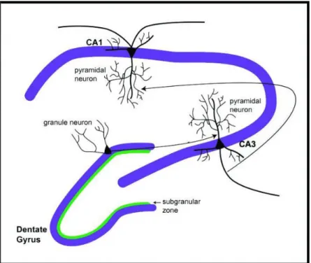
Fig. 1 Hippocampal structure. The hippocampus is composed of the dentate gyrus (DG) including the subgranular zone (SGZ), and three Cornus Ammonis (CA) subfields that are called CA1, CA2 and CA3 (Joels, Krugers et al. 2009). The information flow in hippocampus is generally unidirectional, from the axons of granule neurons in DG, to the dendrites of pyramidal neurons in CA3, through axons to the dendrites of pyramidal neurons in CA1 (Colgin, Denninger et al. 2009).
In the developing and mature mammalian brains, BDNF is strongly and widely expressed throughout the cerebral structures (Ernfors, Wetmore et al. 1990, Hohn, Leibrock et al. 1990), and its strongest expression is found within the hippocampus both at the mRNA and protein levels (Kawamoto, Nakamura et al. 1996, Yan, Rosenfeld et al. 1997). Beside the CNS, BDNF is also expressed in peripheral nervous tissues (Timmusk, Palm et al. 1993, Bishop, Mueller et al. 1994), as well as in several non-neural tissues at low levels, including testis,
13 lung, thymus, heart, liver and spleen (Aid, Kazantseva et al. 2007) where its function is less characterized. The hippocampus is constituted of several structures such as Cornus Ammonis (CA) subdivided in CA1, CA2 and CA3 subregions and the dentate gyrus (DG) (Joels, Krugers et al. 2009), presenting a specific neuronal organization which is described in Figure 1.
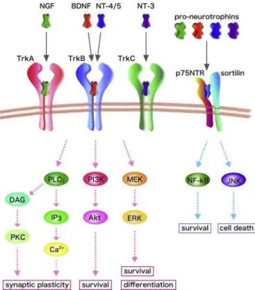
Fig. 2 Signaling pathways evoked by neurotrophins and their receptors (Numakawa, Adachi et al. 2013). Mammalian neorotrophins (NGF, BDNF, NT-3, and NT-4/5) stimulate intracellular signaling through binding to Trk receptors and a common receptor p75NTR. The mature BDNF binds to its specific TrkB receptor with high affinity, while pro-BDNF interacts with p75NTR with high affinity. TrkB activation stimulates three main signaling cascades including PLCγ, MAPK/ERK, and PI3K-Akt to exert beneficial effects on neurons. Activation of p75NTR is capable of inducing cell death or survival.
BDNF could bind to two types of receptors, the higher affinity receptor, tropomyosin-related kinase B (TrkB) (Chao 2003), and the lower affinity P75 neurotrophin receptor (P75NTR) (Fayard, Loeffler et al. 2005). BDNF functions mainly by binding to TrkB, activating downstream signaling cascades, predominant among which are mitogen-activated protein kinase (MAPK) /extracellular signal-regulated kinase (ERK), phospholipase Cγ (PLCγ), and the phosphatidylinositol-3-kinaseAkt (PI3K-Akt) pathways (Reichardt 2006) (Fig. 2). Briefly, a BDNF homodimer is captured by two TrkB receptors, which results in auto-phosphorylation of the intracellular domain of TrkB. The phosphorylation of TrkB initiates signal transduction of MAPK/ERK, PI3K-Akt, and PLCγ pathways, with the recruitment and phosphorylation of
14
Src homologous and collagen-like protein (Shc) (Chao 2003, Huang and Reichardt 2003,
Yoshii and Constantine-Paton 2010). Activation of BDNF/TrkB-dependent intracellular signaling contributes to neuronal survival and differentiation, morphological changes, and synaptic plasticity in the CNS.
In conclusion, as the most abundant and widely distributed neurotrophin in the mammalian CNS, BDNF binds to its receptors, activating different signaling pathways, leading to distinct functions.
1.2 Pathophysiological functions of BDNF and related diseases
1.2.1 Physiological functions of BDNF
In the CNS, BDNF controls various neural processes during development and in adulthood, especially related to synaptic formation and functions. BDNF regulates neuronal homeostasis and survival (Lipsky and Marini 2007), differentiation and growth (Bernd 2008) of the neurons. Growing evidence indicates that BDNF is also involved in several other functions, including brain plasticity-related processes such as memory (Yamada, Mizuno et al. 2002) and learning (Tyler, Alonso et al. 2002, Yamada, Mizuno et al. 2002) which are associated to long term potentiation (LTP) (McAllister, Katz et al. 1999) in the hippocampus. In this chapter, we discuss the importance of BDNF in all these processes.
a. Role of BDNF on neurogenesis
Much attention has been paid to the ability of BDNF and its receptors to modulate adult neurogenesis. In adulthood, the neural stem cells (NSCs), which maintain their capacity to proliferate and generate new neurons via a series of intermediate progenitor cells (Lledo, Alonso et al. 2006), are found in the subgranular zone (SGZ) in the DG of the hippocampus (Fig. 1) and the subventricular zone (SVZ) of the lateral ventricles. Both high levels of BDNF and TrkB expression in the SVZ niche were detected (Galvao, Garcia-Verdugo et al. 2008), and many studies showed that BDNF and BDNF/TrkB signaling pathways were tightly linked to the neuroblast migration along the rostral migratory stream, a special route by which the precursor neurons migrate from the SVZ to the main olfactory bulb (Snapyan, Lemasson et al. 2009, Bagley and Belluscio 2010). Meanwhile, it has been suggested that BDNF signaling may participate to the maturation of specific interneurons within the olfactory bulb in both
15 mice and rats (Berghuis, Agerman et al. 2006, Galvao, Garcia-Verdugo et al. 2008). Significantly, knockdown of BDNF in the DG and conditional deletion of TrkB in rat NSCs inhibited neurogenesis, while an increased neurogenesis was shown in response to BDNF injection (Scharfman, Goodman et al. 2005, Taliaz, Stall et al. 2010). Moreover, BDNF heterozygous (BDNF+/-) mice also exhibited reduced survival of new cells in the adult hippocampus (Sairanen, Lucas et al. 2005). Besides, BDNF may also facilitate differentiation of progenitor cells into granule neurons (Tozuka, Fukuda et al. 2005), which compose the middle or granular layer of the DG, and their maturation in the SGZ by enhancing -aminobutyric acid (GABA, the inhibitory neurotransmitter) release from parvalbumin-expressing GABA-ergic interneurons (Waterhouse, An et al. 2012). Furthermore, consistent to the results above, decreased levels of central BDNF in CNS-specific BDNF knockout mice induce deficits in terminal differentiation, dendritic complexity, migration of new granule neurons and GABA-mediated effects on neurogenesis (Chan, Cordeira et al. 2008). Taken together, BDNF promotes several neurogenic processes, such as migration, differentiation, maturation and survival, in progenitor cells from both SVZ and SGZ.
b. BDNF and LTP associated with memory and learning
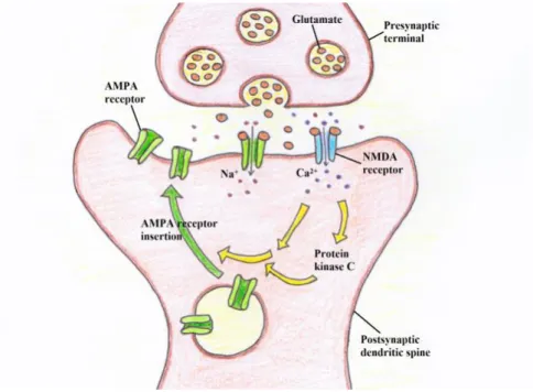
In addition to its roles in neuronal proliferation, neurogenesis and differentiation, BDNF exerts its critical function by regulating activity-dependent structural and functional modification of the synapses, which is the major mechanism promoting a crucial phenomenon called long-term potentiation (LTP) and involved in memorization. Synaptic plasticity refers to any lasting up-regulation or down-regulation of synaptic strength and is thought to be of much importance for the processing and encoding of information by neuronal networks (Bliss and Collingridge 1993). Activity-dependent plasticity seems to be the dominant mechanism involved in learning and memory. There is a general consensus that LTP, in which a brief high frequency synaptic stimulation triggers a long-lasting increase in synaptic strength, results in a reinforcement of the neuronal connection. This has been considered as the major mechanism involved in memory coding and the acquisition of learning (Nicoll and Roche 2013). The constitution of LTP depends on synaptic plasticity, partly on high-frequency pre-synaptic release of glutamate (usually from an axon termination) that activates receptors on post-synaptic dendritic spine (small membranous protrusion from a dendrite) (Fig. 3). Glutamate receptors, including N-methyl-D-aspartate (NMDA) and non-NMDA receptors, exert distinct roles in synaptic plasticity. The opening of NMDAR channel triggers a calcium
16 influx and activation of Protein kinase C that results in long-lasting potentiation of alpha-amino-3-hydroxy-5-methyl-4-isoxazolepropionic acid receptor (AMPAR, non-NMDA receptor) excitatory post-synaptic current (Fig. 3). Both NMDAR and AMPAR are ionotropic receptors.
Fig. 3 Schematic of long term potentiation (LTP) formation. Pre-synaptic release of glutamate activates receptors NMDAR on post-synaptic dendritic spine (Nicoll and Roche 2013). The opening of NMDAR channel triggers a calcium influx and activation of Protein kinase C, causing long-lasting potentiation of AMPAR excitatory post-synaptic current (Nicoll and Roche 2013).
Another important mechanism involved in synaptic plasticity and in memory formation is long-lasting synaptic depression (LTD) (Steele and Mauk 1999). Contrary to LTP, LTD reduces the efficacy of neuronal synapses, mediated typically by metabotropic glutamate receptors (mGluRs, that do not form ionic channels), in several areas of the brain including the hippocampus (Collingridge, Peineau et al. 2010). Taken together, LTP and LTD are forms of activity-dependent synaptic plasticity that increase or decrease, respectively, the strength of synaptic transmission and memory formation (Malenka and Bear 2004).
BDNF effects on both memory formation and LTP have been demonstrated previously (Malenka and Bear 2004). Several decades ago, two independent studies showed that BDNF knockout (BDNF-/-) mice, which presented with altered sensory neuron development and usually died between 2 and 4 weeks after birth (Ernfors, Lee et al. 1994, Jones, Farinas et al. 1994), displayed severe impairments in LTP at the CA1 synapses in hippocampus (Korte, Carroll et al. 1995, Patterson, Abel et al. 1996). Furthermore, this phenotype was rescued by
17 either acute administration of exogenous recombinant BDNF or virus-mediated BDNF gene transfer (Korte, Griesbeck et al. 1996, Patterson, Abel et al. 1996). Consistently, BDNF heterozygous knockout (BDNF+/-) mice, which presented with approximately half the level of BDNF protein in the brain than wild type animals, also exhibited LTP impairments and learning deficits in the hippocampus-dependent paradigms (Linnarsson, Bjorklund et al. 1997, Liu, Lyons et al. 2004). More directly, it was reported later that by activating its signaling pathways, BDNF may induce LTP and further promote neurophysiological foundation for learning and memory (Rex, Lin et al. 2007, Kemppainen, Rantamaki et al. 2012). Other animal studies showed that inhibition of BDNF signaling by hippocampus-specific gene knockout or infusion of antisense BDNF impairs spatial learning and memory (Heldt, Stanek et al. 2007). Along the same line, another group found severe spatial memory defects in forebrain-specific BDNF homozygous knockout mice (Gorski, Balogh et al. 2003). Moreover, infusion of BDNF antisense oligonucleotides or anti-BDNF antibodies also induced impaired spatial memory (Ma, Wang et al. 1998, Mu, Li et al. 1999).
As a key mediator of memory processing, BDNF exerts its effects at different molecular levels, by regulating cation channels including several subtypes of Na+ and K+ channels, by modulating ligand-gated channels like NMDA and AMPA receptors (Cunha, Brambilla et al. 2010), and by greatly affecting protein synthesis by transcriptional and translational mechanisms. From another aspect, the potential mechanism by which BDNF supports activity-dependent modification of synapses and brain function has been suggested by several studies. One of them hypothesized that the formation and occurrence of LTP require BDNF release from the presynaptic vesicles (Zakharenko, Patterson et al. 2003, Jia, Gall et al. 2010). Furthermore, BDNF may facilitate vesicle docking at the presynaptic active zone (Pozzo-Miller, Gottschalk et al. 1999). The postsynaptic involvement has also been suggested by one study showing that postsynaptic BDNF secretion is required for the long-term structural change of synaptic dendritic spines, important structures for LTP generation (Tanaka, Horiike et al. 2008). Taken together, presynaptic and postsynaptic BDNF release may exert their distinct actions, facilitating the induction and the maintenance phase of LTP, respectively.
c. BDNF and exercise
There is a growing body of evidence showing that exercise improves both cognition (Smith, Blumenthal et al. 2010, Roig, Nordbrandt et al. 2013) and mood (Rethorst and Trivedi 2013,
18 Josefsson, Lindwall et al. 2014), as well as much evidence suggesting that BDNF activity may be involved in these effects (Vaynman, Ying et al. 2004, van Praag, Shubert et al. 2005, Erickson, Miller et al. 2012, Heyman, Gamelin et al. 2012). Numerous studies on both human and animal models (Zoladz and Pilc 2010, Huang, Larsen et al. 2014) provide strong evidence that BDNF levels increase following exercise in humans and in rodents. For instance, enhanced BDNF mRNA expression in motoneurons in rats is observed after voluntary exercise (Gomez-Pinilla, Ying et al. 2002) or treadmill training (Neeper, Gomez-Pinilla et al. 1996, Wilhelm, Xu et al. 2012). Conversely, in neuron-specific knockout for BDNF or TrkB in mice, the effect of exercise is completely lost (Wilhelm, Xu et al. 2012, English, Liu et al. 2013). Using sensitive assays, it was shown that BDNF protein levels in serum were elevated after exercise protocols, such as moderate daily exercise for 2 weeks in mice in a nerve repair model (Park and Hoke 2014).
Altogether, exercise could be considered as a potential strategy for inducing BDNF activity to improve mood or cognition, due to the beneficial effect of BDNF on numerous cognitive processes, as well as on synaptic plasticity, neurogenesis and neuronal survival. It was hypothesized by Gordon and coworkers (Al-Majed, Tam et al. 2004) that exercise could promote axon regeneration in the peripheral nervous system (PNS) by a BDNF signaling mechanism in mice, thereby treating peripheral nerve injury (Wilhelm, Xu et al. 2012). Moreover, animal experiments provide support for various mechanisms by which exercise-stimulated BDNF increases result in improved cognition. For example, as little as one week of exercise may improve subsequent learning in animals, but this effect was abolished by BDNF blockade in the hippocampus using a TrkB-IgG chimera (Vaynman, Ying et al. 2004). Actually, some studies suggested that exercise could enhance cognitive abilities and quality of life of dementia patients (Forbes, Forbes et al. 2015, Ojagbemi and Akin-Ojagbemi 2017). Therefore, regular exercise provides beneficial effects not only on mood (Asmundson, Fetzner et al. 2013, Josefsson, Lindwall et al. 2014) and cognition (Smith, Blumenthal et al. 2010, Erickson, Miller et al. 2012) but also on extensive physical health (Alford 2010).
1.2.2 BDNF and related diseases
There are a large number of studies to suggest that changes of BDNF levels or function in specific neuron subpopulations may result in neuronal dysfunction, which contribute to various pathologies, including AD, HD, PD, depression, epilepsy, pain and drug addiction (Bibel and Barde 2000, Murer, Yan et al. 2001, Binder and Scharfman 2004, Bolanos and
19 Nestler 2004, Castren 2004, Cattaneo, Zuccato et al. 2005, Russo-Neustadt and Chen 2005). Subsequently, due to the wide expression of BDNF and TrkB in the CNS and their robust neuroprotective effects, exogenous administration of this neurotrophin, such as gene delivery and protein administration, could be considered as an appropriate therapeutic strategy for treating certain CNS disorders. However, a key and unavoidable challenge in the field of BDNF therapy is drug delivery into the CNS, because as a moderately sized and charged protein, BDNF cannot easily cross the blood-brain barrier via peripheral administration. Potential approaches including intraparenchymal protein infusion and gene delivery using viral vectors may solve this issue, and other methods to increase BDNF bioavailability (i.e., epigenetics, drug-induced endogenous BDNF modulation, peptide mimetics of BDNF, exercise and diet) will be effective (Nagahara and Tuszynski 2011).
a. BDNF and Alzheimer’s disease (AD)
AD is a neurodegenerative disease characterized by a progressive dementia that occurs in middle or late life (Querfurth and LaFerla 2010). Initially, this disease is linked to decreased levels of cholinergic synapses in the cerebral regions such as basal forebrain, the cortex and hippocampus. The key pathologic changes are increased levels of amyloid-β (Aβ) peptide in the form of extracellular senile plaques, as well as hyperphosphorylated tau protein as the intracellular neurofibrillary network tangles in some regions of brain (Reitz, Brayne et al. 2011).
BDNF was found to be associated with neuropsychiatric disorders (Angelucci, Brene et al. 2005, Autry and Monteggia 2012, Li, Chang et al. 2016) and neurodegenerative (Zuccato and Cattaneo 2009) progressions of AD (Rohe, Synowitz et al. 2009, Doi, Takeuchi et al. 2013). The expression level of BDNF is lower in AD patients than that in healthy controls (Connor, Young et al. 1997, Hock, Heese et al. 2000, Peng, Wuu et al. 2005). Similar results were also found in AD animal models (Francis, Kim et al. 2012, Naert and Rivest 2012, Meng, He et al. 2013). Both decreased mRNA and protein levels of BDNF were observed in several regions such as in the hippocampus and the parietal cortex (Holsinger, Schnarr et al. 2000, Garzon, Yu et al. 2002). As described above, numerous studies have shown that BDNF could stimulate survival and genesis of several types of neurons in the CNS, including those from the basal forebrain, hippocampus and cortex (Murer, Yan et al. 2001). Therefore, BDNF could help to prevent neuronal death in AD, and central local administration of exogenous
20 BDNF might be beneficial for the treatment of AD, by stopping or delaying the progression of neuronal loss and its cognitive consequences (Nagahara and Tuszynski 2011).
b. BDNF and Huntington’s disease (HD)
HD, an autosomal dominant inheritable and neurodegenerative disorder, is characterized by dramatic motor dysfunction, cognitive decline, and psychiatric symptoms (Ross and Tabrizi 2011), which leads to progressive dementia and death approximately 15-20 years after the onset (Landles and Bates 2004). The pathogenesis of HD is due to mutations (expanded CAG repeats) in the huntingtin (HTT) gene, leading to an increased number of polyglutamine repeats in the encoded protein HTT (McMurray and McMurray 2001). The molecular mechanisms by which mutant HTT protein leads to neuronal dysfunction and neurodegeneration remained to be identified, and no therapy is currently available for patients to date, beside some symptomatic treatments.
A large number of laboratories have shown that BDNF expression, trafficking, and signaling have been strongly reduced in numerous HD mouse models (Zuccato and Cattaneo 2009, Plotkin, Day et al. 2014). Because the mutant HTT protein causes transcriptional downregulation of the Bdnf gene (Buckley, Johnson et al. 2010), both cortical and striatal BDNF levels were shown to be reduced in postmortem HD brain (Her and Goldstein 2008, Wu, Fan et al. 2010). Moreover, it has been reported that the expression and trafficking of BDNF and TrkB are significantly altered in human HD tissues and HD mouse models (Zuccato, Liber et al. 2005, Gines, Bosch et al. 2006). Importantly, overexpressing BDNF rescues alterations of neuron structure and function in HD model mice (Zuccato, Liber et al. 2005, Lu, Nagappan et al. 2014), while BDNF knockout mice recapitulate atrophy phenotype of the striatum, the striped mass of white and grey matter controlling movement and balance, observed in HD patients (Antoniades and Watts 2013). Therefore, previous studies have suggested BDNF to be a prime putative candidate for the treatment of HD by preventing the underlying neuronal loss seen in HD patients (Alberch, Perez-Navarro et al. 2004, Fink, Deng et al. 2015).
c. BDNF and Parkinson’s disease (PD)
PD is also a progressive neurodegenerative disorder, characterized by difficulty in initiating movements, slowness of movement, rigidity, resting tremor, postural instability and gait
21 changes (Lang and Lozano 1998, Fahn 2003), as well as multiple cognitive impairments in visuospatial, attentional, executive and memory functions (Robbins and Cools 2014). The etiology of PD is still not well understood, however, several neuroinflammatory mechanisms, such as nitric oxide, oxidative stress and mitochondrial dysfunction, were proposed to be involved in the pathophysiology of this disease (Dauer and Przedborski 2003).
A large body of literature has shown alterations in BDNF levels in the blood or brain of PD patients. For instance, reduced levels of BDNF mRNA and protein expression were reported in the substantia nigra of PD patients, as well as in the putamen, a structure involved in motor skills (Mogi, Togari et al. 1999, Parain, Murer et al. 1999). Besides, low levels of serum BDNF were also observed in newly diagnosed PD patients (Scalzo, Kummer et al. 2010). Of note, it has been demonstrated that treatment with anti-parkinsonian drugs increases BDNF levels (Gyarfas, Knuuttila et al. 2010). These findings suggest that BDNF may be implicated in the pathogenic mechanisms of PD. Because of its important role in the survival and maintenance of neurons improving motor performance (Cohen, Tillerson et al. 2003), BDNF could be a useful agent in the treatment of PD in humans.
d. BDNF and depression
Depression is a neuropsychiatric disorder, with high lifetime prevalence, a low mood state and a high rate of suicide. The causes of depression are very diverse, including life events such as childhood abuse, personality, medical treatments, as well as psychiatric syndromes and non-psychiatric illnesses like PD, stroke and diabetes. It seems that the pathophysiology of depression is not due to neuron loss but rather to neuronal atrophy and monoaminergic neurotransmitter deficiency leading to a reduced synaptic transmission (Duman 2004) and signal transduction (Duman 2002).
A considerable number of studies have demonstrated that BDNF plays a key role in the pathophysiology and treatment of depression (Shelton 2007, Krishnan and Nestler 2008). The tight relationship between BDNF expression/signaling and mood disorders has been extensively investigated in both human and rodent models (Duman and Monteggia 2006, Martinowich, Manji et al. 2007, Boulle, van den Hove et al. 2012), and BDNF deficits are thought to contribute to depression. Numerous papers have indicated that BDNF and TrkB expression was significantly reduced in the hippocampus as well as in the circulating peripheral blood of depressed patients, suggesting that BDNF could be considered as a
22 valuable biomarker in severe depressive conditions (Dwivedi, Rizavi et al. 2003, Schmidt and Duman 2007, Hashimoto 2010, Martinotti, Pettorruso et al. 2016). On the other hand, analyses of postmortem tissues determined that hippocampal BDNF concentrations were much less reduced in patients treated with antidepressants, compared to those without antidepressant treatments (Chen, Dowlatshahi et al. 2001). Furthermore, BDNF injections in the hippocampus or the midbrain region in rodents resulted in similar effectiveness than antidepressants, as reviewed by Castren (Castren 2004), showing that BDNF downregulation may be causal in depression. Previous studies have indicated that rats exposed to chronic stress showed a reduced hippocampal level of BDNF mRNA, as well as a downregulation of its signaling pathways. This effect was counteracted by antidepressant treatment (Roceri, Hendriks et al. 2002, Blendy 2006, Castren and Rantamaki 2010, Razzoli, Domenici et al. 2011). Furthermore, a growing body of evidence demonstrated that rodents with BDNF signaling deficiency were more prone to depression, while injection of BDNF into the hippocampus reduces depression-like behavior and mimics the effects of antidepressants (Castren and Rantamaki 2010, Castren and Kojima 2017). Additional published studies supported the theory that antidepressant effects were mediated, at least in part, through an increase in hippocampal BDNF levels (Shirayama, Chen et al. 2002, Duman and Monteggia 2006, Tsankova, Berton et al. 2006, Taliaz, Stall et al. 2010).
e. BDNF and epilepsy
Epilepsy is a disease characterized by periodic and unpredictable occurrence of transient symptoms due to spontaneous abnormal cerebral activity, which are called seizures (Fisher, Acevedo et al. 2014). In humans and in animal models of epilepsy, decrease in memory (Helmstaedter and Kockelmann 2006) and learning abilities (Bell, Lin et al. 2011), inflammation, gliosis, neuronal loss in the hippocampus (Pitkanen and Sutula 2002, Lopim, Vannucci Campos et al. 2016) have been observed.
A large number of studies with animal models as well as analyses of postmortem brains from patients with epilepsy have shown that BDNF mRNA and protein expression levels were increased after epileptic seizures, indicating that epileptic activity may upregulate the BDNF protein level via a transcriptional activation. For example, increased levels of BDNF mRNA and protein have been detected in patients suffering epilepsy, in the DG and the CA1-CA3 pyramidal cell layers of the hippocampus (Murray, Isackson et al. 2000, Wang, Li et al. 2011).
23 In neuron-specific BDNF-/- mice, a moderate prevention in epileptogenesis was observed using the kindling model, consisting with a repeated electric stimulation of the brain (He, Kotloski et al. 2004). Moreover, using the heterozygous BDNF+/- mice, it was shown that the seizure-induced increase in BDNF concentration was reduced, and the kindling effect on seizure was lower compared to wild-type mice (Kokaia, Ernfors et al. 1995). Conversely, kainic acid-induced BDNF overexpressing mice have more severe seizures and display spontaneous seizures occasionally after treatment (Croll, Suri et al. 1999). Meanwhile, in mice, BDNF levels increased by transgene overexpression or acute infusion into the hippocampus could also enhance seizure severity (Croll, Suri et al. 1999, Scharfman, Goodman et al. 2002). However, it is worth noting that a decreased BDNF level in serum has been found in adult epileptic patients (LaFrance, Leaver et al. 2010). It is clear that BDNF may have a role in epileptogenesis, perhaps as a result of its positive regulatory function on neuronal excitability within the hippocampus (Binder, Croll et al. 2001). As a whole, BDNF has been thought to be a pro-epileptogenic factor (Rivera, Voipio et al. 2004), possibly because its expression was stimulated after seizure as an appropriate and physiological response to epilepsy in order to refrain neuronal death. Conversely, it was reported that delivery of intrahippocampal BDNF could attenuate or postpone the development of epilepsy in animal models, unraveling its complex role in the development of this pathology that is still a matter of debate (Koyama and Ikegaya 2005, Paradiso, Marconi et al. 2009).
f. BDNF and neuropathic pain
Pain is a very complex and subjective phenomenon generally caused by intense and damaging stimuli that accompanies a large variety of clinical syndromes. Neuropathic pain (NP) (Treede, Jensen et al. 2008) is associated either with various PNS lesions or injuries in relay structures of CNS. While the former is mainly characterized by intense alterations of gene expression pattern and protein interaction network, including inflammatory molecules such as leukocyte adhesion molecules (Max and Stewart 2008, Belfer and Dai 2010), the latter arises from metabolic disorders, traumatic injury or neurotoxicity (Calvino and Grilo 2006). Generally, NP is caused by acute or chronic trauma, neurotoxins, diabetes, tumor compression, viral infections or side effects of chemotherapy.
Changes in BDNF expression levels in the nervous system associated with numerous animal models for pain, including peripheral inflammation and neuropathic pain paradigms, have
24 been widely reported. Generally speaking, upregulated endogenous BDNF levels have been reported following nerve injury or inflammation in peripheral tissues (Shu, Llinas et al. 1999, Narita, Yajima et al. 2000) as well as in the spinal cord (Cho, Kim et al. 1997, Geng, Liao et al. 2010, Lin, Ro et al. 2011). This increase in BDNF quantity is responsible for synaptic remodeling and thus the development of chronic pain (Obata and Noguchi 2006). Moreover, several other groups have also demonstrated that BDNF expression levels were dramatically increased in the dorsal root ganglion (DRG) under distinct experimental conditions, including after spinal nerve ligation in rats (Fukuoka, Kondo et al. 2001) and in models of inflammatory pain where BDNF may function as a neuromodulator (Pezet, Malcangio et al. 2002, Zhang, Wang et al. 2016). Conversely, in neuropathic pain models where partial injury to peripheral nerves was provoked, BDNF was decreased in injured sensory neurons while increased in uninjured ones (Cho, Kim et al. 1998, Zhou, Chie et al. 1999, Fukuoka, Kondo et al. 2001). Additionally, enduring tactile allodynia (sensitization by normally non-painful stimuli) and thermal hyperalgesia (overaction to a painful stimulus) could be induced by intrathecal BDNF injections in mouse vertebra, indicating a direct link between BDNF and neuropathic pain (Yajima, Narita et al. 2005). Some other groups found an opposite effect of BDNF on pain, pointing out that exogenous BDNF could exert analgesic effects in neuropathic pain (Eaton, Blits et al. 2002). Even though the role of BDNF in neuropathic pain is contentious, previous evidence obtained from several animal models has proposed that BDNF exerts a crucial role in neuropathic pain pathways, acting as a neuromodulator when released from the nociceptive neurons (Kerr, Bradbury et al. 1999, Thompson, Bennett et al. 1999, Pezet, Malcangio et al. 2002, Zhang, Wang et al. 2016). In models of neuropathic pain, due to the downregulation of BDNF in injured fibers, it seems reasonable that exogenous BDNF administration with proper dose and time would exert beneficial anti-nociceptive effect, as already suggested previously (Miki, Fukuoka et al. 2000, Eaton, Blits et al. 2002).
g. BDNF and drug addiction
Drug addiction is a complex behavioral disorder with a large variety of causes, including genetic and environmental influences, as well as drug-induced changes in the brain (Kreek, Nielsen et al. 2005). Developed drug addiction is characterized by a loss of control over a substance use or a behavior (for instance, gambling), a concern with substance or behavior, a compulsive drug use despite adverse consequences, and relapses even after long periods of withdrawal (Moss, Chen et al. 2014, Sharma, Bruner et al. 2016). Laboratory experiments on