HAL Id: hal-02060389
https://hal.archives-ouvertes.fr/hal-02060389
Submitted on 25 May 2021
HAL is a multi-disciplinary open access
archive for the deposit and dissemination of sci-entific research documents, whether they are pub-lished or not. The documents may come from teaching and research institutions in France or abroad, or from public or private research centers.
L’archive ouverte pluridisciplinaire HAL, est destinée au dépôt et à la diffusion de documents scientifiques de niveau recherche, publiés ou non, émanant des établissements d’enseignement et de recherche français ou étrangers, des laboratoires publics ou privés.
A shear-induced network of aligned wormlike micelles in
a sugar-based molecular gel. From gelation to
biocompatibility assays
Juliette Fitremann, Barbara Lonetti, Emiliano Fratini, Isabelle Fabing, Bruno
Payre, Christelle Boulé, Isabelle Loubinoux, Laurence Vaysse, Luis Oriol
To cite this version:
Juliette Fitremann, Barbara Lonetti, Emiliano Fratini, Isabelle Fabing, Bruno Payre, et al.. A shear-induced network of aligned wormlike micelles in a sugar-based molecular gel. From gelation to biocompatibility assays. Journal of Colloid and Interface Science, Elsevier, 2017, 504, pp.721-730. �10.1016/j.jcis.2017.06.021�. �hal-02060389�
1
This document is the author version of a work published in Journal of Colloid and Interface
1Science, copyright ©Elsevier after peer review and technical editing by the publisher. The
2final edited and published work is available at:
3https://www.sciencedirect.com/science/article/abs/pii/S002197971730677X
4Citation: Fitremann, J.; Lonetti, B.; Fratini, E.; Fabing, I.; Payré, B.; Boulé, C.; Loubinoux, I.;
5Vaysse, L.; Oriol, L. A Shear-Induced Network of Aligned Wormlike Micelles in a Sugar-Based
6Molecular Gel. From Gelation to Biocompatibility Assays. Journal of Colloid and Interface
7Science 2017, 504, 721–730.
https://doi.org/10.1016/j.jcis.2017.06.021
.
89
A shear-induced network of aligned wormlike micelles in a sugar-based
10
molecular gel. From gelation to biocompatibility assays
11
Juliette Fitremann*a, Barbara Lonettia, Emiliano Fratinib, Isabelle Fabingc, Bruno Payréd, Christelle Boulée,
12
Isabelle Loubinouxf, Laurence Vayssef, Luis Oriolg
13
a CNRS - Université de Toulouse III Paul Sabatier, Laboratoire des Interactions Moléculaires et Réactivité Chimique et 14
Photochimique (IMRCP, UMR 5623), Bat 2R1, 118 Route de Narbonne, 31062 Toulouse Cedex 9, France. 15
b Department of Chemistry “Ugo Schiff” and CSGI, University of Florence, via della Lastruccia 3-Sesto Fiorentino, I-16
50019, Florence, Italy. 17
c CNRS UMR 5068, LSPCMIB, Université de Toulouse, Université Paul Sabatier, 118 Route de Narbonne, 31062 18
Toulouse cedex 9, France. 19
d Centre de Microscopie Electronique Appliquée à la Biologie (CMEAB), Faculté de Médecine Rangueil, Université de 20
Toulouse III Paul Sabatier, Bâtiment A5, R.D.C., 133 Route de Narbonne, 31400 Toulouse, France. 21
e Université Claude Bernard UCBL Lyon1, Service de Prestations CTµ EZUS, Bâtiment Darwin B, 5 rue Raphaël 22
Dubois, 69622 Villeurbanne Cedex, France. 23
f TONIC, Toulouse NeuroImaging Center, Université de Toulouse, Inserm, UPS, France. 24
g Instituto de Ciencia de Materiales de Aragon (ICMA),Universidad de Zaragoza -CSIC, Dpto. Quimica Organica, 25
Facultad de Ciencias, Pedro Cerbuna 12, 50009 Zaragoza, Spain. 26
27
A new low molecular weight hydrogelator with a saccharide (lactobionic) polar head linked by azide-28
alkyne click chemistry was prepared in three steps. It was obtained in high purity without 29
chromatography, by phase separation and ultrafiltration of the aqueous gel. Gelation was not obtained 30
reproducibly by conventional heating-cooling cycles and instead was obtained by shearing the aqueous 31
solutions, from 2wt% to 0.25 wt%. This method of preparation favored the formation of a quite unusual 32
network of interconnected large but thin 2D-sheets (7 nm-thick) formed by the association side-by-side 33
of long and aligned 7 nm diameter wormlike micelles. It was responsible for the reproducible gelation 34
at the macroscopic scale. A second network made of helical fibres with a 10-13 nm diameter, more or 35
less intertwined was also formed but was scarcely able to sustain a macroscopic gel on its own. The gels 36
were analysed by TEM (Transmission Electronic Microscopy), cryo-TEM and SAXS (Small Angle X-37
Ray Scattering). Molecular modelling was also used to highlight the possible conformations the 38
hydrogelator can take. The gels displayed a weak and reversible transition near 20°C, close to room 39
temperature, ascribed to the wormlike micelles 2D-sheets network. Heating over 30°C led to the loss of 40
the gel macroscopic integrity, but gel fragments were still observed in suspension. A second transition 41
near 50°C, ascribed to the network of helical fibers, finally dissolved completely these fragments. The 42
2
gels showed thixotropic behaviour, recovering slowly their initial elastic modulus, in few hours, after 43
injection through a needle. Stable gels were tested as scaffold for neural cell line culture, showing a 44
reduced biocompatibility. This new gelator is a clear illustration of how controlling the pathway was 45
critical for gel formation and how a new kind of self-assembly was obtained by shearing. 46
47
Keywords: supramolecular; gel; self-assembly; carbohydrate amphiphile; saccharide; triazole; fiber;
48
fibre; cylindrical micelle; shear; cell culture; biomaterial; LMWG; low molecular weight gelator. 49
Electronic Supplementary Information: Experimental section, extra electronic microscopy data,
50 HLPC-MS chromatograms, NMR spectra. 51
Graphical abstract
52 53 54Introduction
55Low molecular weight (LMW) hydrogelators provide an alternative family of gelling agents compared 56
with polymers, leading to soft materials with possible applications in the field of wet materials, 57
switchable gels, controlled release or uptake, cell culture. LMW gelators belong to different structural 58
families (peptides, cholesteryl, nucleobase or sugar amphiphiles, etc…) and the now quite large amount 59
of work done on these self-assembling molecules has been well reported in several reviews [1–21]. We 60
are more especially interested in sugar-derived molecular gelators (see the recent review on this family 61
of gelators and the following references for the most recent works [22–50]). Sugar gelators provide 62
generally a neutral hydrophilic polar head, with a low sensitivity to temperature changes on the contrary 63
to PEG amphiphiles. In the context of biological applications, carbohydrate derived hydrogels will 64
interact differently with biomolecules or cells compared with PEG or peptide derived gelators, notably 65
by mimicking to some extent the saccharidic components of the glycocalix, composed of glycoproteins 66
and glycolipids [22]. 67
From a practical point of view, simple, rapid and cheap syntheses are essential when considering sugar-68
based LMW hydrogelators applications. In former results, a family of hydrogelators based on a 69
disaccharide head has been described. It has been shown that the presence of a triazole linker enhanced 70
gelation, but the synthetic pathway consisted of six steps [23–26]. Other amphiphilic molecules with 71
close structures (namely, a sugar head, triazole linkers, and a fatty chain) have been described as well, 72
including hydrogelators [27–29], organogelators [30] and micelles [31–33], all of them involving also 73
3
protection-deprotection multistep synthesis and purification by chromatography. In this work, a new 74
gelator inspired from these structures has been prepared with a simpler synthetic route with only three 75
steps starting from lactobionic acid as the polar head (Scheme 1). Another important aspect in the field 76
of LMW hydrogelators is to control in a precise manner the supramolecular structure sustaining the gel. 77
In the case of very flexible molecules many conformations are possible. It can give rise to 78
polymorphism. Polymorphism is the main cause for the lack of reproducible gelation [34–37]. 79
Accordingly, the importance of controlling the conditions of the self-assembly in order to reach a 80
reproducible final state, with the related macroscopic properties, has been well pointed out in several 81
papers. But it still remains underestimated, quite unexplored and not well controlled [38–42]. Compound 82
3 is a typical example of such a situation. Its gelation behaviour appeared more complex than expected
83
and gave the opportunity to explore the effect of different pathways for the gel preparation. The self-84
assembled structures have been elucidated by electronic microscopy and Small Angle X-Ray Scattering 85
(SAXS). The rheology and thermal transitions of the gel have also been studied. Finally, the results 86
related to the use of these gels for cell culture are briefly discussed. 87
88
Results and discussion
8990
1. Preparation of the gelator
91 92
An easy access to the sugar based gelator 3 is represented in Scheme 1. The gelator was obtained 93
in three synthetic steps and was purified without chromatography. The fatty chain 1 was purified 94
by recrystallization (yield 60%) while the product 2 was obtained nearly pure by extraction and 95
was used without further purification in the next step. After the azide-alkyne "click chemistry" 96
step, leading to the compound 3, the unreacted and sparingly soluble azide was discarded by 97
centrifugation. The cleared and concentrated reaction medium was diluted in water and allowed 98
to rest for several hours, until a gel was formed. The gel was purified by ultrafiltration or dialysis, 99
removing water soluble by-products. Analysis of the filtrate showed that about 5% of the gelator 100
went out through the membrane after four volumes and 24h of ultrafiltration, evidencing a low 101
proportion of free molecules. A last step of filtration in methanol enabled to get rid of traces of 102
the azide remaining after ultrafiltration. After this sequence, the gelator was pure (yield 76%, 103
freeze-dried), according to NMR, HPLC-MS chromatograms (see Fig. SI-11-13). Residual 104
copper was analysed and it did not exceed 300 ppm (0.3µg/mg) when ultrafiltration was 105
performed in the presence of EDTA. 106
107 108
109
Scheme 1. Synthetic scheme for the preparation of the gelator 3 110
4
2. Gelation
111
Gelation of the gelator 3 was at first quite puzzling. The crude product coming directly from the 112
reaction mixture and diluted in water (after solvent removal) provided directly the hydrogel 113
within 24 hours of standing. Conversely, the purified and dried samples gave a non-reproducible 114
gelation behaviour. The usual method consisting in heating the solution until solubilisation 115
followed by cooling did not lead to reproducible gelation, whatever the concentration (from 0.1 116
to 2%wt). It has been checked that this effect was not the result of the degradation of the 117
molecule. It was still intact after heating for 4 hours at 60°C (see SI-12). Heating may favour a 118
change of intra or intermolecular bonding leading to insoluble aggregates instead of gel. 119
Concentrated samples at 2% usually provided gels but sometimes, viscous turbid solutions or 120
suspensions are obtained instead of gels. And conversely, it happened that very diluted samples 121
(0,3 to 0.1%) formed spontaneously gels, but in a non-reproducible manner (see SI-4). 122
Sonication did not help gelation (see SI-7), and the purification method did not appeared either 123
as the key parameter for gelation. 124
125
Finally, it was observed that shearing was a key factor for triggering the gelation reproducibly 126
for this compound. A reliable gelation procedure was then set up. A 2% suspension in water was 127
prepared and the hydration and solubilisation of the solid was allowed for few hours (2 to 24h). 128
It provided a heterogeneous suspension containing non-cohesive white cotton-like gels 129
fragments. The sample was then sheared through a needle until an homogeneous opalescent 130
solution was obtained. Finally, the sample was let resting for gelation at room temperature (20°C 131
or lower). A turbid gel, macroscopically homogenous was formed within c.a. 2 hours. It can be 132
turned upside down (Figure 6 and SI-1 "Gelation"). Additional cycles of shear/rest were 133
sometimes applied to improve gelation and homogeneity. The gel was also diluted stepwise up 134
to 0.25% by shearing, ranking the molecule among super-hydrogelators. Time also appeared as 135
a key factor for gelation, notably because of the solubilisation kinetics, but also because the gel 136
appeared to be thixotropic, but with a slow recovery (see section E-rheology). 137
The benefit of shear on gelation has already been observed in few examples of organogels 138
[43,44], hydrogels [45,46] and micellar solutions [47]. Shearing can affect, first, the distribution 139
of the aggregates in the solution and can help to get a more homogeneous distribution of the 140
growing self-assemblies. The shear can also affect the shape of the self-assemblies, by 141
lengthening the aggregates or even by changing the growth pathway and thus, the polymorph 142
formed, as it has been observed in the examples cited above. Shearing can also provide foam, 143
namely a large air-water interface on which the amphiphilic molecules can absorb and organize 144
before coalescence. The walls of the needle used to shear may also act as an interface for 145
organizing the molecules. In our case, the effect of shear on the gels of 3 will be discussed in the 146
light of SAXS, electronic microscopy and rheology (see section D). 147
148
Alternative preparation with DMSO as cosolvent (DMSO/H2O 1/2 v/v) was less challenging. 149
Gels with good mechanical strength were obtained easily by the conventional heating-cooling 150
method (see SI-8 for TEM). They were rinsed several times to remove DMSO, providing purely 151
aqueous gels. The solvent change did not alter their mechanical properties and stability, during 152
and after rinsing. However, introducing a solvent for gel formation is often undesirable, notably 153
for biocompatibility assays. For this reason, unravelling the conditions for gel f ormation in pure 154
water, as described above, was more challenging, but necessary. 155
5 157
3. Thermal transitions
158
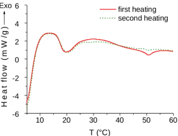
The thermal transitions have been studied by DSC and by visual inspection of the sample (Figure 159
1). For a gel at 2wt% prepared by shearing, two transitions were observed. A first transition 160
starting at 17°C and ending at 30°C (5 J/g with respect to the gelator; 0.2 J/g if considering the 161
entire gel) corresponded to the loss of the macroscopic gel integrity, but large gel domains were 162
still visible floating in a liquid phase. A second transition starting at 35°C and ending at 55°C 163
fitted with the complete disappearance of all macroscopic gelled fragments. Over 55°C, the 164
sample was completely liquid and clear. Both transitions were partly reversible, and t he gel was 165
recovered after 30 min at 5°C. However, repeated heating over 60°C caused the formation of a 166
precipitate and prevented any recovery of the gel state. 167
168 169
Figure 1. Two consecutive heating curves of hydrogel at 2wt% prepared by shearing (2°C/min). 170
171
4. Morphology
172
The morphology of the self-assembled aggregates has been observed with different microscopic 173
techniques and detailed by SAXS. 174
175
Microscopy. In a 2wt% gel obtained by shearing, two morphologies were mainly observed by
176
TEM and cryo-TEM: intertwined helical fibres (Figure 2-e-f-g), and large and flexible ribbons 177
or sheets (Figure 2-a-b-c). Both of them have a substructure. The wide helical fibres are made 178
by the intertwining of thinner 10-13 nm "single" helical fibres. These 10-13 nm fibres themselves 179
resulted from the stacking of structures with a 5-6 nm spacing. These structures are pairs of two 180
bright bands (≈2.5 nm) separated by a darker one, thus can be interpreted as bilayers (Figure 2-181
g and SAXS). Sheets are made of long structures with 7 nm width organized in parallel side-by-182
side on a long range, over several millimetres (Figure 2-b). These long structures typically look 183
like long cylindrical, wormlike micelles. Concentration played a role in the formation of the 184
sheets. At high concentration (2wt%), sheets are the main structures observed (and confirmed 185
by SAXS). Conversely, at lower concentration, helical fibres are the main ones. The latter are 186
generally not able to sustain a gel. In the 2 wt% and sheared gels of Figures 2-a, b, c, helical 187
fibres can however be seen (Figure 2-e), but they are a minority population. Shear also played a 188
role. Without shear, the structures formed at 2wt% looked like short platelets by TEM (SI-2). 189 10 20 30 40 50 60 -6 -4 -2 0 2 4 6 first heating second heating H e a t fl o w ( m W /g ) T (°C) Exo
6
They are too short to sustain a intertwined network and the sample often did not gel. After shear, 190
the intertwined long sheet-like structures are observed. These ones are able to form a persistent 191
network throughout the sample and are able to sustain a gel. The more the sample is sheared, the 192
more extended is this interconnected sheets structure. Besides, a population of short and 193
randomly arranged wormlike micelles with 6-7 nm width was also observed by cryo-TEM (this 194
population was the main one just after shearing but was still observed, to a lesser extent, after 195
resting as well) (Figure 2-d). 196
197
Figure 2. Morphologies observed in 2wt% gels in water: (a) Interconnected ribbons and flexible sheets (cryo-TEM, 198
bar = 500 nm). (b) close-up within the sheet-like assemblies: ribbons of thin 7 nm fibres assembled in parallel side-199
by-side (cryo-TEM, bar = 200 nm). (c) Interconnected ribbons and flexible sheets (TEM, bar = 1 µm). (d) random 200
wormlike micelles (6 nm width) observed in the background just after shearing (cryo-TEM, Bar = 200 nm). (e) single 201
and intertwined 10-13 nm helical fibres (cryo-TEM, bar = 200 nm). (f) Thick twin fibres (cryo-SEM, bar = 500 nm), 202
width of one single fibre = 21 nm. (g) close-up within the helical fibres revealing the twisted assembly of 5 nm thick 203
structures (bilayers) (TEM, negative staining, bar = 200 nm). 204
To better understand the effect of shear on those morphologies, cryo-TEM and TEM images 205
were also recorded just after hydration of the solid and just after shearing. The microscopy (and 206
also the SAXS, see below) of the solid hydrated for 16h at 2wt% in water and before shearing, 207
already revealed the presence of 2D-sheets (Figure SI-3). Just after shearing, they were no longer 208
observed. Instead, numerous small spots were seen on the whole grid, corresponding to dried 209
wormlike micelles fragments (Figure SI-3). By using cryo-TEM, that avoids the formation of 210
ambiguous drying figures, short and random 6-7 nm wide wormlike micelles were mainly 211 observed (Figure 2-d). 212 213
a
b
c
e
g
d
f
7
The shear thus mainly broke and dispersed the sheets into individual and short wormlike 214
micelles. After resting, these micelles re-assembled slowly into long wormlike micelles. 215
Shearing also favoured their assembly side-by-side instead of randomly. It has been already 216
shown that shear induced the formation of aligned wormlike micelles bundles in solutions of 217
ionic surfactants. This association led to the formation of gels (transient or stable gels)[47]. 218
However in the case of the gels of 3, the structures observed are much longer (more than several 219
microns long), and are organized in 7 nm thin sheets instead of bundles. They are able to sustain 220
a gel stable over the long term reproducibly. These structures are also reminiscent of the unusual 221
self-assembled lamellar plaques observed by Stupp et al. for amphiphilic peptides. These plaques 222
split under shear into bundles of parallel fibres made of long cylindrical micelles[48]. Another 223
example of long range and flexible 2D-self-assembly leading to gelation at relatively low 224
concentration are lamellar hydrogels. They are also quite scarce in the literature. They have a 225
purely lamellar organisation [49],[50]. This is not the case here. Besides, samples containing 226
mainly the intertwined 10-13 nm helical fibres often failed to gel. Probably the helical fibres 227
entanglements do not percolate through the whole sample or are not rigid, long and persistent 228
enough to sustain a macroscopic gelation [51]. 229
230
In contact with a surface (TEM grid), drying figures were formed. In the background of nearly 231
all the TEM images of gelled samples, spots, but also holes with a typical jagged shape were 232
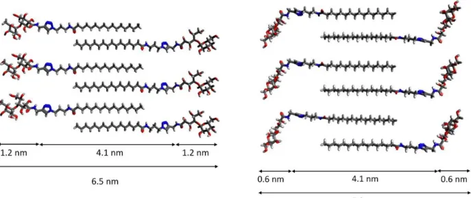
observed (Figure 3-b). By AFM, the same jagged shapes were observed. They looked like the 233
ones obtained for lipid bilayers dried on a surface. A constant thickness equal to 5-6 nm was 234
measured (Figure 3-a). It matches well with the thickness of a single bilayer of gelator molecules 235
with interdigitating alkyl chains and a polar head partly bended (Figure 5 and SI-15). Quite 236
intriguingly, stacked layers displaying the same mosaic of jagged holes with this typical shape 237
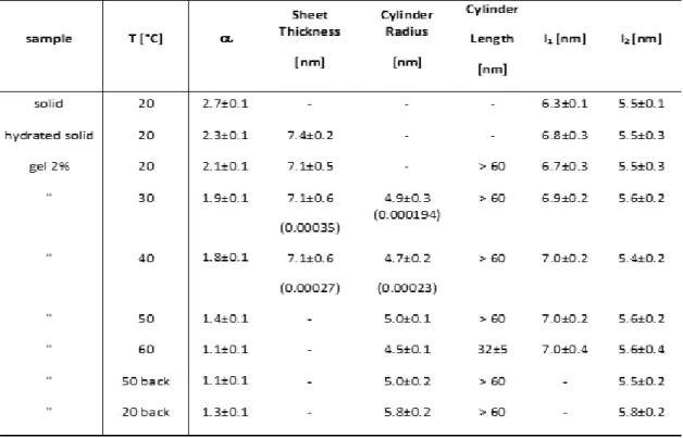
were observed also by cryo-SEM in addition to TEM and AFM (Figure 3-c). 238
239
Figure 3. Figures with "jagged holes" formed after drying. (a) bilayer membrane on mica (AFM height and profile), (b) same 240
membrane pattern on TEM grid, mixed with helical fibres (bar = 2 µm) (c) same pattern in cryo-SEM (bar = 1 µm). 241
Small Angle X-Ray Scattering (SAXS). SAXS investigation was performed to detail the
242
changes in the morphology at the nanometre scale. SAXS curves of a 2% gel from 20°C to 70°C 243
and the one of the freeze-dried solid are shown in Figure 4. The fit results are summarized in 244
Table 1. All curves displayed two main features as the scattering vector, q, changes: (i) a powe r 245
law trend, I(q) q-, in the low q region and, (ii) a broad interaction peak in the high q region.
246
In gel sample, at 20°C, the whole scattering curve can be described by isolated sheets with 7.1 247
nm thickness to which two Gaussian peaks must be added in order to fully describe the high q 248
region of the curve. These extra peaks correspond to a highly ordered state with a precise 249
a
b
8
correlation length very similar to a solid state. A progressive morphological change from sheets 250
to cylinders with a diameter of about 10 nm and a length higher than 60 nm was observed from 251
20°C to 60°C. This transition was witnessed by the change in the low q region of the curves, i.e. 252
the power law exponent went from about 2 to 1 as the temperature increased from 20°C to 60°C 253
(see Table 1). In this region, the exponent scales with the dimensionality proper of the scattering 254
object. 1-D morphologies (cylinders, rods, wires, etc.) give an exponent of 1, while 2-D 255
morphologies (disks, lamellae, etc.) result in an exponent of 2. In general values between 1 and 256
3 are usually associated to a mass fractal dimension where the limit of 3 is reached for full solids. 257
258
As already said, the high q region of the scattering curves is characterized by the presence of 259
interaction peaks. In the freeze-dried solid, these peaks were ascribed to the coexistence of two 260
polymorphs having two different organizations, one with a 6.3 nm spacing (l1, polymorph "L", 261
more abundant) and the other with a 5.5 nm spacing (l2, polymorph "C"). Given the flex ibility 262
of the polar head, it is very likely that the molecule can take two distinct conformations (at least) 263
giving rise to different bilayer packings (Figure 5). In modelling, the size of a fully extended 264
molecule is 4.2 nm (SI-15). According to the models, bilayer thicknesses should be within the 265
range from 8.4 nm (fully extended molecule and not interdigitated) to 4.3 nm (strongly bended 266
polar head and interdigitated). 267
268
Table 1. Results of fitting of SAXS curves: dry solid, solid after 24h hydration, gel 2wt%. = power-law index in the 269
q region between 0.009 Å-1 and 0.03 Å-1 for gel samples and 0.009 Å-1 and 0.04 Å-1 for the solid; l1 and l2 = spacing
270
distances obtained from the Gaussian interaction peaks positions from l = 2/q (Table SI-14-2). In brackets are 271
indicated the weight of each specie in the curve fits at 30 and 40°C. 272
9 274
Figure 4. SAXS curves of the freeze-dried solid at 20°C (in black, at the top) and of a 2wt% gel prepared by shearing, then 275
heated from 20°C to 60°C. L = sheets. C = cylinders 276
The dimensions measured by the interaction peaks are thus consistent with packings in which the alkyl 277
chains are interdigitated and the polar head is more or less bended. Two examples of such packings 278
providing a 6.5 nm (l1, extended) and a 5.3 nm (l2, partly bended) thickness are represented in Figure 279
5, keeping in mind that they are only illustrative [16,37,52]. The Gaussian interaction peaks were also 280
seen in the gel samples at 0.91 nm-1 and 1.14 nm-1. After hydration of the solid, without shear, the
281
interaction peaks correspond to 6.8 nm (l1) and 5.5 nm (l2) spacing and shearing had no main effect at 282
this microscopic scale (SI-14-1). The shift of l1 from 6.3 nm to 6.8 nm can be attributed to the 283
intercalation of water in inter-lamellar spaces in the gel. These interaction peaks are indicative of the 284
presence of solid-like inhomogeneities in the gel, after hydration. We assumed that the spacing measured 285
in these solid-like inhomogeneities is directly related to the molecular organization once the aggregates 286
are dispersed in the solution. 287
288
Figure 5. Illustration of possible conformations and packings of gelator 3 . Up: a bilayer model with molecules in a 289
fully extended conformation. Below: a bilayer model with molecules in a semi-bended conformation (molecular 290
modelling suite Avogadro 1.1.1. with the Universal Force Field [53]. Colour code: C=grey, O=red, N=blue and 291
H=white). 292
10
From the analysis of the high q region, the ratio polymorph "L" / polymorph "C" is high in the 293
solid. Conversely, in the gel sample this ratio is drastically reduced (almost five times less). This 294
decrease is indicative of the fact that polymorph "L" in its hydrated form is more easily 295
solubilised and provides the sheet-like aggregates at 20°C. Accordingly, the dimension of 296
polymorph "L", l1 = 6.8-7.0 nm, is consistent with the mean thickness of the sheets (7.1 nm). 297
And the dimension of the polymorph "C", l2 = 5.5 nm, appeared to be correlated to the cylinders 298
in the gel state. Temperature did not influence much the peaks position, but it affected their ratio. 299
Actually, the weight of the peak at 1.14 nm-1 increased with temperature and qualitatively the
300
spectra moved at higher q values. 301
302
Discussion. From the results of the different experiments we concluded that after hydration,
303
mainly two kinds of self-assembled objects are formed and dispersed in the solution: assemblies 304
of cylindrical shape with a mean diameter around 10 nm (polymorph "C") and sheet-like 305
assemblies with a sheet thickness of 7.1 nm (polymorph "L"). The sheet-like assemblies are the 306
prevalent objects in solution at 20°C and in concentrated samples (2 wt%). They are the only 307
ones detectable by scattering experiments. Their size is in good agreement with the one measured 308
by cryo-TEM in the sheet-like assemblies made of long 7 nm-wide wormlike micelles, 309
assembled parallel and side-by-side (Figure 2a-b). Besides, the 10-13 nm wide helical fibres 310
found in cryo-TEM images (Figure 2e) match well the 10 nm diameter cylindrical objects 311
detected in SAXS experiments above 30°C. The diameter of these cylinders is too big to 312
correspond to cylindrical micelles. It can instead be explained by the twisting of bilayers with a 313
5-6 nm width observed by TEM (Figure 2g) and corresponding to the correlation length l2 314
(polymorph "C")[16,36,54]. The relative ratio sheet / cylinder in the low q region of the curves 315
also decreases with temperature. The cylinders can be detected in scattering experiments only 316
above 30°C, as their concentration increases, even though a fraction already exists at room 317
temperature, as evidenced by the TEM images. The morphological transition from sheets to 318
cylinders with temperature may be induced by a change in the molecular conformation. It is 319
likely that the increase of temperature favours a bended conformation of the molecule, like in 320
the polymorph "C", more stable and characterized by a shorter molecular spacing. 321
322
These transitions agree with DSC results and macroscopic observations. In particular, in DSC 323
curves (Figure 1) a first peak at 20°C was observed, corresponding to the sheet-like structures 324
(polymorph "L"). This transition is over at 30°C. The sheet-like objects coming from the 325
polymorph "L" are responsible for the reproducible gelation after shearing, at the macroscopic 326
scale. Sheets are the main species in the 2% gel after shearing, at 20°C, but are based on weak 327
interactions. They disappear in favour of the more stable helical fibres, that exist in solution ti ll 328
60°C. This assumption fits well with the second transition observed in DSC, at 55°C. After 329
heating the sample over 30°C, gelled fragments were still observed floating in the solution: these 330
fragments are sustained by the helical fibres. In some cases, these assemblies sustained also the 331
macroscopic gelation, even in very diluted samples (0.5 %wt or less). Inthose samples only the 332
transition at 55°C was observed (see SI-4). By heating up to 70°C and cooling back to 20°C, 333
only the cylinders' signature was recovered, proving that this form corresponds to a 334
thermodynamically controlled self-assembly, while the sheet-like aggregates are not. Instead 335
they are closely related to the use of shear in concentrated samples. 336
11
Another point concerning the effect of heating is shown by the SAXS curve at 20°C relative to 338
the heated sample, which failed to gel and gave a precipitate (SI-5). The curve displays an 339
interaction peak corresponding to a 4.7 nm spacing (Figure SI-14-3), that is, lower compared 340
with l1 and l2. It can correspond to a more bended conformation producing big aggregates which 341
eventually precipitate. The presence of this third polymorph "P" is observed in the interaction 342
peaks of samples once they have been cooled down from 70°C (see Figure SI-14-3, curves “50°C 343
back” and “20°C back”). The formation of a third polymorph can explain the detrimental effect 344
of heating on gel formation. This is why heating cannot be chosen as a robust protocol for 345
producing the gels in a reproducible way. 346
Besides, low concentration gels at 0.5% were obtained in a reproducible manner by diluting and 347
shearing the 2% samples. Their SAXS curves only displayed the signature of cylindrical 348
structures (see Figure SI-14-2), indicating that the sheet-like structures did not withstand 349
dilution. Using this method, the network of helical fibres produced was able to sustain the 350
formation of a macroscopic gel. 351
352
E Rheology
353
Viscoelastic and thixotropic properties of gels at 2%wt have been measured. The frequency 354
sweep from 10Hz to 0.01Hz displays a typical rheogram of gel, with G' > G" over the whole 355
frequency range (Figure 6-a). G' is around 500 Pa, and G'' between 30-60 Pa, featuring a quite 356
fragile gel. The gel is thixotropic, namely after being sheared, it was able to recover its 357
viscoelastic properties after resting. This point is illustrated in Figure 6-b-c. In Figure 6b, a gel 358
(t=0, turned upside down) was vortexed for 15s. It flowed when turned upside down. One hour 359
later, it had recovered its gel state and did not flow when turned upside down. In Figure 6-c, a 360
gel (2%wt) was first sheared by passing through a needle, placed in the rheometer, and then G' 361
and G" were monitored for 4h. G' in the destructured gel started at around 10Pa and progressively 362
increased as the gel restructured up to 1000 Pa. The monitoring of the gel restructuration after 363
shearing both with TEM and rheology has showed that the gel is dynamic, but with a very slow 364
rate. According to macroscopic observations, this process of gel structuration with time kept on 365
over several days and even weeks, since it was observed that some weak gels finally did not flow 366
when turned upside down far later, after several weeks. From the microscopic point of view, the 367
shear broke down the self-assembled structures formed after hydration into small objects (see 368
section D "Microscopy", Figure 2d and SI-3). These fragments coalesced back slowly, into the 369
long wormlike micelles assembled side-by-side, providing again the cross-linking points 370
sustaining the gel network. 371
372
In relation to the slow dynamics of this restructuring, interestingly, it has been shown that the 373
introduction of a sugar polar head in a dipeptide gelator (cyclo-L-Tyr-L-Lys) led to a strong 374
slowing down of the gelation process[46]. But conversely, a nucleobase-derived gelator 375
appended with a monosaccharide group displayed a quick gelation[28]. These different examples 376
raise the question on how the introduction of sugar polar heads may affect more generally the 377
self-assembly kinetics. Besides, in the case of LMW gels, thixotropy is known to be affected by 378
the nature of self-assembled structures formed (e.g. spherulites versus fibres)[55]. In our case 379
too, possibly only one of the two networks may sustain this rheological property. The 380
viscoelastic changes with temperature have also been monitored by rheology, from 10 to 40°C. 381
An abrupt decrease of the elastic modulus G' was observed from 25 to 30°C (Figure 6-d). The 382
sharp increase of the phase from 5 to 70° in this temperature range evidenced the transition from 383
12
a gel to a sol. The elastic (G') and viscous (G") moduli crossed at 29°C. This measurement 384
showed clearly the loss of the gel integrity in this range of temperature. It is consistent with the 385
transition measured by DSC and SAXS and ascribed to the loss of the entangled sheets network 386 of the polymorph "L". 387 388 389 390 391 392 393 394 395 396 397 398 399 400 401 402 403 404 405 406 407 408 409 410 411 412 413 414 415 416 417 418 419 420 421 422 423 424 425 426 427 428 429 430
a
b
c
d
0 ,0 1 0 ,1 1 1 0 1 0 1 0 0 1 0 0 0 G ' o r G " ( P a ) fre q u e n c y (H z ) G ' G '' 1 0 1 5 2 0 2 5 3 0 3 5 4 0 0 ,0 1 0 ,1 1 1 0 1 0 0 1 0 0 0 1 0 0 0 0 G ' o r G " ( P a ) T (° C ) G ' G '' 0 1 0 2 0 3 0 4 0 5 0 6 0 7 0 8 0 9 0 d e lta d e lt a ( d e g r e e s ) 0 1 2 3 4 1 1 0 1 0 0 1 0 0 0 G ' o r G " ( P a ) tim e g lo b a l (h ) G ' G ''13
Figure 6. (a) Variation of G' (elastic modulus, squares) and G" (viscous modulus, circles) with frequency, at 1 Pa, 431
20°C. (b) Recovery of gel after shearing and resting for 1h. (c) Recovery of the elastic (G') and viscous (G") modulus 432
with time, after shearing, measured at 0.1 Pa and 1 Hz. (d) Variation of G', G'' and the phase (delta) with temperature, 433 measured at 0.8 Pa and 1 Hz. 434 435 F Biocompatibility 436
Several molecular gels have been already tested as scaffold for cell culture, most of them 437
belonging to the family of gelling peptides [2,56]. Compared with polymer gels, they can provide 438
a different environment to cells, including different stiffness, a looser internal structure and 439
higher clearance, that would help cells to grow through. Injectability, related to thixotropy, is 440
also expected [2,7]. 441
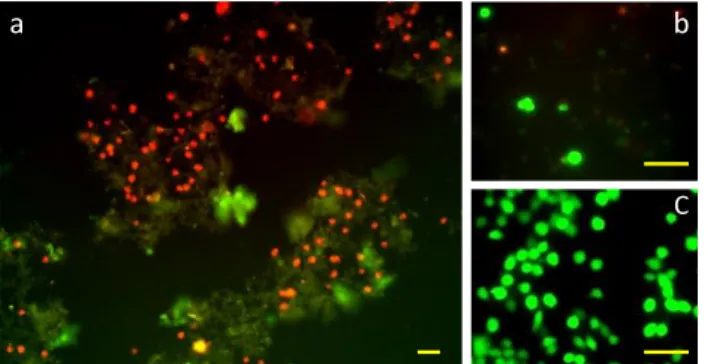
The gels of 3 have been tested as scaffold for cell culture with a neuronal cell line (Neuro 442
2A)[57]. After 3 days cultures, cell viability tests were performed (Figure 7). Cells adhered to 443
the gel but only a poor number of viable cells was observed (<10%) (Figure 7-b). The gels did 444
not appear suitable, whatever the method used for purifying the gelator or for preparing the gels. 445
This result is in contrast with other low molecular weight hydrogels with close chemical 446
structures on which cells were grown up to three days [58,59] or ten days [28]. Among the two 447
networks sustaining the gel, one of them was dissolved at 37°C. It may have provided a high 448
concentration of free gelator molecules, increasing the cytotoxicity. For comparison, the 449
cytotoxicity of surfactants with close structure (sugar-triazole-fatty chain) was studied in ranges 450
below 1 mmol/L, showing that surfactants with chains <6 carbons or >12 carbons had no toxicity 451
on mammalian cells in this range [60]. In our case, gels at 0.1% correspond to a concentration 452
of 1.4 mmol/L and gels at 1%, 14 mmol/L, far above the concentration range of this study. The 453
molecular gels differ strongly from polymer gels on this point, since there is no bioavailability 454
of small molecular units if biological degradation mechanisms do not provide fragments. This 455
difference in the mechanism of gelation may affect the biocompatibility. 456
457 458
Figure 7. Cell viability assay. Neuro 2A cells have been cultured for 3 days on a 0.25%wt gel. Red staining indicates 459
dead cells, green staining indicates live cells. The gel also appears by green autofluorescence. Fluorescence images 460
at (a) low magnification and (b) high magnification. (c) Control cells after 3 days on cell culture plate. Scale bar 30 461
µm. 462 463
In addition, the mechanical strength of the scaffold is known to affect the cell survival, and in 464
our gels, the dissolution of one of the two networks clearly decreased a lot the strength of the 465
gel. Accordingly, we observed that the surviving cells were exclusively aggregated on the 466
remaining gel fragments. It has been also demonstrated, in the case of amyloid peptides, that the 467
nature of the polymorph, namely the way in which the molecules self-assemble, affected the 468
cytotoxicity[40]. The same effects may occur in the case of supramolecular gelators. It points 469
a b
14
out that the cell death is related not only to the chemical nature of the gel, but also to its 470
mechanical properties and to subtle molecular organisation differences. 471
472 473
Conclusions
474This new sugar-based gelator, despite an easy synthetic access, displayed quite a complex 475
gelation behaviour in pure water due to polymorphism. The presence of two amides bonds, but 476
separated from each other by a rigid spacer (triazole), forces the molecule to bend and favours 477
the polymorphism. This observation may help the design of gelators. Without shearing, the 478
gelation was capricious. Starting from 2wt% solutions and applying a strong shear enabled to 479
get reproducible gelation. The resulting gels were diluted up to 0.25wt%. As a result of this 480
method, the gel was sustained by a double network, one network of thin helical fibres with 10 481
nm of diameter and an unusual network of large interconnected 2D-sheets made of thin 7 nm 482
wide wormlike micelles assembled side-by-side in parallel. The thermal transition of this second 483
network was within the room temperature range. The resulting gel was partly thermoreversible 484
and was thixotrope. Further heating, above 60°C, led to changes in the aggregation mode, giving 485
shorter aggregates which could not sustain gelation and a precipitate was formed. This work 486
illustrates that gelation can be highly dependent on the pathway used for the solubilisation and 487
the dispersion of the gelator molecules. In our case, it involved both the hydration and 488
solubilisation of the dry solid at room temperature in a range of concentration around 2wt%, 489
before shearing. This work illustrates also that full solubilisation by heating, followed by cooling 490
down can impair the self-assembly at the long range. It also showed that shearing induced the 491
formation of new self-assembled and dynamic structures able to sustain a stable gel. 492
Acknowledgements
493We acknowledge the following people for their technical assistance: TEM: I. Fourquaux, D. 494
Goudounèche (CMEAB); V. Sartor, S. Gineste (IMRCP); NMR: P. Lavedan, M. Vedrenne 495
(ICT). We acknowledge the European Union for funding (AFM, DSC) (FEDER 35477: "Nano-496
objets pour la biotechnologie") and The French National Research Agency (ANR) for financial 497
support (ANR Neuraxe). We thank the Integrated Screening Platform of Toulouse (PICT, 498
IBiSA) for providing access to HPLC equipment. LB and EF kindly acknowledge partial 499
financial support from Consorzio per lo sviluppo dei Sistemi a Grande Interfase (CSGI). Thanks 500
also to M. Mauzac for helpful discussions. 501
References
502[1] R.G. Weiss, P. Terech, eds., Molecular gels: materials with self-assembled fibrillar networks, 503
Springer, Dordrecht, 2006. 504
[2] X. Du, J. Zhou, J. Shi, B. Xu, Supramolecular Hydrogelators and Hydrogels: From Soft Matter 505
to Molecular Biomaterials, Chem. Rev. 115 (2015) 13165–13307. doi:10.1021/acs.chemrev.5b00299. 506
[3] N. Zweep, J.H. van Esch, CHAPTER 1. The Design of Molecular Gelators, in: B. Escuder, 507
J.F. Miravet (Eds.), RSC Soft Matter Ser., Royal Society of Chemistry, Cambridge, 2013: pp. 1–29. 508
http://ebook.rsc.org/?DOI=10.1039/9781849737371-00001 (accessed April 4, 2016). 509
[4] A. Sorrenti, O. Illa, R.M. Ortuño, Amphiphiles in aqueous solution: well beyond a soap 510
bubble, Chem. Soc. Rev. 42 (2013) 8200. doi:10.1039/c3cs60151j. 511
15
[5] M. de Loos, B.L. Feringa, J.H. van Esch, Design and Application of Self-Assembled Low 512
Molecular Weight Hydrogels, Eur. J. Org. Chem. 2005 (2005) 3615–3631. 513
doi:10.1002/ejoc.200400723. 514
[6] S.S. Babu, V.K. Praveen, A. Ajayaghosh, Functional π-Gelators and Their Applications, 515
Chem. Rev. 114 (2014) 1973–2129. 516
[7] K.J. Skilling, F. Citossi, T.D. Bradshaw, M. Ashford, B. Kellam, M. Marlow, Insights into low 517
molecular mass organic gelators: a focus on drug delivery and tissue engineering applications, Soft 518
Matter. 10 (2014) 237–256. doi:10.1039/C3SM52244J. 519
[8] J. Raeburn, D.J. Adams, Multicomponent low molecular weight gelators, Chem Commun. 51 520
(2015) 5170–5180. doi:10.1039/C4CC08626K. 521
[9] D.J. Adams, P.D. Topham, Peptide conjugate hydrogelators, Soft Matter. 6 (2010) 3707. 522
doi:10.1039/c000813c. 523
[10] C. Tomasini, N. Castellucci, Peptides and peptidomimetics that behave as low molecular 524
weight gelators, Chem. Soc. Rev. 42 (2013) 156. doi:10.1039/c2cs35284b. 525
[11] K. Araki, I. Yoshikawa, Nucleobase-Containing Gelators, in: Low Mol. Mass Gelator, 526
Springer Berlin Heidelberg, 2005: pp. 133–165. http://link.springer.com/chapter/10.1007/b107173 527
(accessed January 12, 2015). 528
[12] P. Dastidar, Supramolecular gelling agents: can they be designed?, Chem. Soc. Rev. 37 (2008) 529
2699. doi:10.1039/b807346e. 530
[13] L.E. Buerkle, S.J. Rowan, Supramolecular gels formed from multi-component low molecular 531
weight species, Chem. Soc. Rev. 41 (2012) 6089. doi:10.1039/c2cs35106d. 532
[14] Y. Lin, C. Mao, Bio-inspired supramolecular self-assembly towards soft nanomaterials, Front. 533
Mater. Sci. 5 (2011) 247–265. 534
[15] N.M. Sangeetha, U. Maitra, Supramolecular gels: Functions and uses, Chem. Soc. Rev. 34 535
(2005) 821. doi:10.1039/b417081b. 536
[16] L.A. Estroff, A.D. Hamilton, Water gelation by small organic molecules, Chem. Rev. 104 537
(2004) 1201–1218. 538
[17] F. Delbecq, Supramolecular gels from lipopeptide gelators: Template improvement and 539
strategies for the in-situ preparation of inorganic nanomaterials and for the dispersion of carbon 540
nanomaterials, Adv. Colloid Interface Sci. 209 (2014) 98–108. doi:10.1016/j.cis.2014.02.018. 541
[18] G. Fichman, E. Gazit, Self-assembly of short peptides to form hydrogels: Design of building 542
blocks, physical properties and technological applications, Acta Biomater. 10 (2014) 1671–1682. 543
doi:10.1016/j.actbio.2013.08.013. 544
[19] I.W. Hamley, Lipopeptides: from self-assembly to bioactivity, Chem Commun. 51 (2015) 545
8574–8583. doi:10.1039/C5CC01535A. 546
[20] E.K. Johnson, D.J. Adams, P.J. Cameron, Peptide based low molecular weight gelators, J 547
Mater Chem. 21 (2011) 2024–2027. doi:10.1039/C0JM03099F. 548
[21] X.-Q. Dou, C.-L. Feng, Amino Acids and Peptide-Based Supramolecular Hydrogels for 549
Three-Dimensional Cell Culture, Adv. Mater. (2017) 1604062. doi:10.1002/adma.201604062. 550
[22] T.K. Lindhorst, Essential of carbohydrate chemistry and biochemistry: [with 150 new 551
exercises], 3., completely rev. and enl. ed., 1. reprint, Wiley-VCH, Weinheim, 2008. 552
[23] M.J. Clemente, R.M. Tejedor, P. Romero, J. Fitremann, L. Oriol, Maltose-based gelators 553
having azobenzene as light-sensitive unit, RSC Adv. 2 (2012) 11419. doi:10.1039/c2ra21506c. 554
[24] M.J. Clemente, J. Fitremann, M. Mauzac, J.L. Serrano, L. Oriol, Synthesis and 555
Characterization of Maltose-Based Amphiphiles as Supramolecular Hydrogelators, Langmuir. 27 556
(2011) 15236–15247. doi:10.1021/la203447e. 557
[25] M.J. Clemente, P. Romero, J.L. Serrano, J. Fitremann, L. Oriol, Supramolecular Hydrogels 558
Based on Glycoamphiphiles: Effect of the Disaccharide Polar Head, Chem. Mater. 24 (2012) 3847– 559
3858. doi:10.1021/cm301509v. 560
[26] M.J. Clemente, R.M. Tejedor, P. Romero, J. Fitremann, L. Oriol, Photoresponsive 561
supramolecular gels based on amphiphiles with azobenzene and maltose or polyethyleneglycol polar 562
head, New J. Chem. (2015). doi:10.1039/C4NJ02012J. 563
[27] G. Godeau, P. Barthélémy, Glycosyl-Nucleoside Lipids as Low-Molecular-Weight Gelators, 564
Langmuir. 25 (2009) 8447–8450. doi:10.1021/la900140b. 565
[28] L. Latxague, M.A. Ramin, A. Appavoo, P. Berto, M. Maisani, C. Ehret, O. Chassande, P. 566
16
Barthélémy, Control of Stem-Cell Behavior by Fine Tuning the Supramolecular Assemblies of Low-567
Molecular-Weight Gelators, Angew. Chem. Int. Ed. 54 (2015) 4517–4521. 568
doi:10.1002/anie.201409134. 569
[29] H.P.R. Mangunuru, J.R. Yerabolu, D. Liu, G. Wang, Synthesis of a series of glucosyl triazole 570
derivatives and their self-assembling properties, Tetrahedron Lett. 56 (2015) 82–85. 571
doi:10.1016/j.tetlet.2014.11.013. 572
[30] E. Carretti, V. Mazzini, E. Fratini, M. Ambrosi, L. Dei, P. Baglioni, P. Lo Nostro, Structure 573
and rheology of gel nanostructures from a vitamin C-based surfactant, Phys Chem Chem Phys. 18 574
(2016) 8865–8873. doi:10.1039/C5CP07792C. 575
[31] A.G. Dal Bó, V. Soldi, F.C. Giacomelli, B. Jean, I. Pignot-Paintrand, R. Borsali, S. Fort, Self-576
assembled carbohydrate-based micelles for lectin targeting, Soft Matter. 7 (2011) 3453. 577
doi:10.1039/c0sm01411g. 578
[32] A.G. Dal Bó, V. Soldi, F.C. Giacomelli, C. Travelet, R. Borsali, S. Fort, Synthesis, 579
micellization and lectin binding of new glycosurfactants, Carbohydr. Res. 397 (2014) 31–36. 580
doi:10.1016/j.carres.2014.07.021. 581
[33] G.C. Feast, T. Lepitre, X. Mulet, C.E. Conn, O.E. Hutt, G.P. Savage, C.J. Drummond, The 582
search for new amphiphiles: synthesis of a modular, high-throughput library, Beilstein J. Org. Chem. 583
10 (2014) 1578–1588. doi:10.3762/bjoc.10.163. 584
[34] S. Díaz-Oltra, C. Berdugo, J.F. Miravet, B. Escuder, Study of the effect of polymorphism on 585
the self-assembly and catalytic performance of an L-proline based molecular hydrogelator, New J. 586
Chem. 39 (2015) 3785–3791. doi:10.1039/C5NJ00072F. 587
[35] V.J. Nebot, S. Díaz-Oltra, J. Smets, S. Fernández Prieto, J.F. Miravet, B. Escuder, Freezing 588
Capture of Polymorphic Aggregates of Bolaamphiphilic L-Valine-Based Molecular Hydrogelators, 589
Chem. – Eur. J. 20 (2014) 5762–5767. doi:10.1002/chem.201400346. 590
[36] J. Cui, A. Liu, Y. Guan, J. Zheng, Z. Shen, X. Wan, Tuning the Helicity of Self-Assembled 591
Structure of a Sugar-Based Organogelator by the Proper Choice of Cooling Rate, Langmuir. 26 (2010) 592
3615–3622. doi:10.1021/la903064n. 593
[37] B.P. Krishnan, K.M. Sureshan, A Molecular-Level Study of Metamorphosis and 594
Strengthening of Gels by Spontaneous Polymorphic Transitions, ChemPhysChem. 17 (2016) 3062– 595
3067. doi:10.1002/cphc.201600590. 596
[38] S. Pan, S. Luo, S. Li, Y. Lai, Y. Geng, B. He, Z. Gu, Ultrasound accelerated gelation of novel 597
l-lysine based hydrogelators, Chem. Commun. 49 (2013) 8045. doi:10.1039/c3cc44767g. 598
[39] A. Baral, S. Basak, K. Basu, A. Dehsorkhi, I.W. Hamley, A. Banerjee, Time-dependent gel to 599
gel transformation of a peptide based supramolecular gelator, Soft Matter. 11 (2015) 4944–4951. 600
doi:10.1039/C5SM00808E. 601
[40] A.T. Petkova, Self-Propagating, Molecular-Level Polymorphism in Alzheimer’s -Amyloid 602
Fibrils, Science. 307 (2005) 262–265. doi:10.1126/science.1105850. 603
[41] J. Raeburn, A. Zamith Cardoso, D.J. Adams, The importance of the self-assembly process to 604
control mechanical properties of low molecular weight hydrogels, Chem. Soc. Rev. 42 (2013) 5143. 605
doi:10.1039/c3cs60030k. 606
[42] V.J. Nebot, D.K. Smith, CHAPTER 2. Techniques for the Characterisation of Molecular Gels, 607
in: B. Escuder, J.F. Miravet (Eds.), RSC Soft Matter Ser., Royal Society of Chemistry, Cambridge, 608
2013: pp. 30–66. http://ebook.rsc.org/?DOI=10.1039/9781849737371-00030 (accessed April 4, 2016). 609
[43] J.T. van Herpt, M.C.A. Stuart, W.R. Browne, B.L. Feringa, Mechanically Induced Gel 610
Formation, Langmuir. 29 (2013) 8763–8767. doi:10.1021/la401286a. 611
[44] X. Cai, Y. Wu, L. Wang, N. Yan, J. Liu, X. Fang, Y. Fang, Mechano-responsive 612
calix[4]arene-based molecular gels: agitation induced gelation and hardening, Soft Matter. 9 (2013) 613
5807. doi:10.1039/c3sm50577d. 614
[45] A. Reddy M, A. Srivastava, Mechano-responsive gelation of water by a short alanine-615
derivative, Soft Matter. 10 (2014) 4863. doi:10.1039/c4sm00710g. 616
[46] Z. Xie, A. Zhang, L. Ye, X. Wang, Z. Feng, Shear-assisted hydrogels based on self-assembly 617
of cyclic dipeptide derivatives, J. Mater. Chem. 19 (2009) 6100. doi:10.1039/b912020c. 618
[47] J.J. Cardiel, A.C. Dohnalkova, N. Dubash, Y. Zhao, P. Cheung, A.Q. Shen, Microstructure 619
and rheology of a flow-induced structured phase in wormlike micellar solutions, Proc. Natl. Acad. Sci. 620
110 (2013) E1653–E1660. doi:10.1073/pnas.1215353110. 621
17
[48] S. Zhang, M.A. Greenfield, A. Mata, L.C. Palmer, R. Bitton, J.R. Mantei, C. Aparicio, M.O. 622
de la Cruz, S.I. Stupp, A self-assembly pathway to aligned monodomain gels, Nat Mater. 9 (2010) 623
594–601. doi:10.1038/nmat2778. 624
[49] H.E. Warriner, S.H.J. Idziak, N.L. Slack, P. Davidson, C.R. Safinya, Lamellar Biogels: Fluid-625
Membrane-Based Hydrogels Containing Polymer Lipids, Science. 271 (1996) 969–973. 626
doi:10.1126/science.271.5251.969. 627
[50] J. Niu, D. Wang, H. Qin, X. Xiong, P. Tan, Y. Li, R. Liu, X. Lu, J. Wu, T. Zhang, W. Ni, J. 628
Jin, Novel polymer-free iridescent lamellar hydrogel for two-dimensional confined growth of ultrathin 629
gold membranes, Nat. Commun. 5 (2014). doi:10.1038/ncomms4313. 630
[51] S.R. Raghavan, J.F. Douglas, The conundrum of gel formation by molecular nanofibers, 631
wormlike micelles, and filamentous proteins: gelation without cross-links?, Soft Matter. 8 (2012) 632
8539. doi:10.1039/c2sm25107h. 633
[52] N. Baccile, A.-S. Cuvier, S. Prévost, C.V. Stevens, E. Delbeke, J. Berton, W. Soetaert, I.N.A. 634
Van Bogaert, S. Roelants, Self-Assembly Mechanism of pH-Responsive Glycolipids: Micelles, Fibers, 635
Vesicles, and Bilayers, Langmuir. 32 (2016) 10881–10894. doi:10.1021/acs.langmuir.6b02337. 636
[53] M.D. Hanwell, D.E. Curtis, D.C. Lonie, T. Vandermeersch, E. Zurek, G.R. Hutchison, 637
Avogadro: an advanced semantic chemical editor, visualization, and analysis platform, J. 638
Cheminformatics. 4 (2012) 17. doi:10.1186/1758-2946-4-17. 639
[54] L. a. Estroff, L. Leiserowitz, L. Addadi, S. Weiner, A. d. Hamilton, Characterization of an 640
Organic Hydrogel: A Cryo-Transmission Electron Microscopy and X-ray Diffraction Study, Adv. 641
Mater. 15 (2003) 38–42. doi:10.1002/adma.200390004. 642
[55] X. Huang, S.R. Raghavan, P. Terech, R.G. Weiss, Distinct Kinetic Pathways Generate 643
Organogel Networks with Contrasting Fractality and Thixotropic Properties, J. Am. Chem. Soc. 128 644
(2006) 15341–15352. doi:10.1021/ja0657206. 645
[56] J. Zhou, J. Li, X. Du, B. Xu, Supramolecular biofunctional materials, Biomaterials. 129 (2017) 646
1–27. doi:10.1016/j.biomaterials.2017.03.014. 647
[57] A. Béduer, I. Gonzales-Calvo, C. Vieu, I. Loubinoux, L. Vaysse, Investigation of the 648
Competition Between Cell/Surface and Cell/Cell Interactions During Neuronal Cell Culture on a 649
Micro-Engineered Surface, Macromol. Biosci. 13 (2013) 1546–1555. doi:10.1002/mabi.201300202. 650
[58] W. Wang, H. Wang, C. Ren, J. Wang, M. Tan, J. Shen, Z. Yang, P.G. Wang, L. Wang, A 651
saccharide-based supramolecular hydrogel for cell culture, Carbohydr. Res. 346 (2011) 1013–1017. 652
doi:10.1016/j.carres.2011.03.031. 653
[59] X. Li, Yi Kuang, J. Shi, Y. Gao, H.-C. Lin, B. Xu, Multifunctional, Biocompatible 654
Supramolecular Hydrogelators Consist Only of Nucleobase, Amino Acid, and Glycoside, J. Am. 655
Chem. Soc. 133 (2011) 17513–17518. doi:10.1021/ja208456k. 656
[60] E.D. Oldham, S. Seelam, C. Lema, R.J. Aguilera, J. Fiegel, S.E. Rankin, B.L. Knutson, H.-J. 657
Lehmler, Synthesis, surface properties, and biocompatibility of 1,2,3-triazole-containing alkyl β-d-658
xylopyranoside surfactants, Carbohydr. Res. 379 (2013) 68–77. doi:10.1016/j.carres.2013.06.020. 659