HAL Id: hal-02560113
https://hal.archives-ouvertes.fr/hal-02560113
Submitted on 1 May 2020
HAL is a multi-disciplinary open access
archive for the deposit and dissemination of
sci-entific research documents, whether they are
pub-lished or not. The documents may come from
teaching and research institutions in France or
abroad, or from public or private research centers.
L’archive ouverte pluridisciplinaire HAL, est
destinée au dépôt et à la diffusion de documents
scientifiques de niveau recherche, publiés ou non,
émanant des établissements d’enseignement et de
recherche français ou étrangers, des laboratoires
publics ou privés.
Impact of Human Immunodeficiency Virus on the
Severity of Buruli Ulcer Disease: Results of a
Retrospective Study in Cameroon
Vanessa Christinet, Eric Comte, Laura Ciaffi, Peter Odermatt, Micaela
Serafini, Annick Antierens, Ludovic Rossel, Alain-Bertrand Nomo, Patrick
Nkemenang, Akoa Tsoungui, et al.
To cite this version:
Vanessa Christinet, Eric Comte, Laura Ciaffi, Peter Odermatt, Micaela Serafini, et al.. Impact of
Human Immunodeficiency Virus on the Severity of Buruli Ulcer Disease: Results of a Retrospective
Study in Cameroon. Open Forum Infectious Diseases, Oxford University Press, 2014, 1 (1), pp.ofu021.
�10.1093/ofid/ofu021�. �hal-02560113�
M A J O R A R T I C L E
Impact of Human Immunode
ficiency Virus on
the Severity of Buruli Ulcer Disease: Results of a
Retrospective Study in Cameroon
Vanessa Christinet,1,2Eric Comte,2Laura Ciaffi,1,2Peter Odermatt,3,4Micaela Serafini,2Annick Antierens,2Ludovic Rossel,2 Alain-Bertrand Nomo,2Patrick Nkemenang,2Akoa Tsoungui,5Cecile Delhumeau,1and Alexandra Calmy1,2
1
HIV Unit, University of Geneva Hospitals, Geneva, Switzerland;2Médecins Sans Frontières, Geneva, Switzerland;3Swiss Tropical and Public Health Institute, Basel, Switzerland;4University of Basel, Basel, Switzerland; and5Akonolinga District Hospital, Akonolinga, Cameroon
Background. Buruli ulcer is the third most common mycobacterial disease after tuberculosis and leprosy and is particularly frequent in rural West and Central Africa. However, the impact of HIV infection on BU severity and prevalence remains unclear.
Methods. This was a retrospective study of data collected at the Akonolinga District Hospital, Cameroon, from January 1, 2002 to March 27, 2013. Human immunodeficiency virus prevalence among BU patients was compared with regional HIV prevalence. Baseline characteristics of BU patients were compared between HIV-negative and HIV-positive patients and according to CD4 cell count strata in the latter group. Buruli ulcer time-to-healing was assessed in different CD4 count strata, and factors associated with BU main lesion size at baseline were identified. Results. Human immunodeficiency virus prevalence among BU patients was significantly higher than the re-gional estimated prevalence in each group (children, 4.00% vs 0.68% [P < .001]; men, 17.0% vs 4.7% [P < .001]; women, 36.0% vs 8.0% [P < .001]). Individuals who were HIV positive had a more severe form of BU, with an in-creased severity in those with a higher level of immunosuppression. Low CD4 cell count was significantly associated with a larger main lesion size (β-coefficient, −0.50; P = .015; 95% confidence interval [CI], −0.91–0.10). Buruli ulcer time-to-healing was more than double in patients with a CD4 cell count below 500 cell/mm3(hazard ratio, 2.39; P = .001; 95% CI, 1.44–3.98).
Conclusion. Patients who are HIV positive are at higher risk for BU. Human immunodeficiency virus-induced immunosuppression seems to have an impact on BU clinical presentation and disease evolution.
Keywords. Buruli ulcer; Cameroon; HIV; Mycobacterium ulcerans disease.
Mycobacterium ulcerans disease, also known as Buruli ulcer (BU), is a tropical disease that can lead to devas-tating tissue and bone destruction. The virulence of M ulcerans is mainly attributed to mycolactone, a toxin with cytotoxic and immunomodulation properties [1]. The disease can be localized or disseminated [2]. It is
rarely life threatening, but it may lead to severe esthetic sequelae, functional impairment, and long-lasting handicap [3]. Buruli ulcer is the third most common mycobacterial disease in the world after tuberculosis and leprosy [4]. It is typically present in humid rural areas from tropical and subtropical regions, and the highest incidence is observed in West and Central Afri-ca [5,6]. Children are mostly affected by M ulcerans and the median age of patients is 15 years [7,8].
The clinical diagnosis of BU performed by skilled practitioners in resource-poor settings is usually good [8]. Although it can be confirmed by different laborato-ry methods, such as acid-fast bacilli (AFB) direct exam-ination with Ziehl-Neelsen staining (ZN), polymerase chain reaction (PCR), and culture or histopathology, these techniques are not always easily accessible in re-mote rural settings [10,11]. Historically, surgery was
Received 10 February 2014; accepted 4 April 2014.
Correspondence: Vanessa Christinet, MD, MIH, Rue du Villars 27, 1024 Ecublens/ Switzerland. E-mail: vchristinet@swissonline.ch.
Open Forum Infectious Diseases
© The Author 2014. Published by Oxford University Press on behalf of the Infectious Diseases Society of America. This is an Open Access article distributed under the terms of the Creative Commons Attribution-NonCommercial-NoDerivs licence (http ://creativecommons.org/licenses/by-nc-nd/3.0/), which permits non-commercial reproduction and distribution of the work, in any medium, provided the original work is not altered or transformed in any way, and that the work is properly cited. For commercial re-use, please contact journals.permissions@oup.com.
the only available treatment. Since 2004, the World Health Or-ganization (WHO) recommends (1) the addition of antimyco-bacterial treatment with rifampicin and streptomycin [12] or (2) a complete oral regimen associating rifampicin with clarithro-mycin [13]. Complementary debridement, excision, skin graft-ing, or, rarely, amputation may be necessary, depending on the type and size of lesion and the affected zone [14].
It is unclear how human immunodeficiency virus (HIV) in-fection affects BU disease progression. Unlike M tuberculosis, which is the most prevalent opportunistic infection [15], the role of HIV in the occurrence and clinical manifestations of BU remains inconclusive. Two case-control studies have ad-dressed the role of HIV as a risk factor for BU. Johnson et al [16] demonstrated a higher HIV prevalence among BU cases compared with controls in their unmatched case-control study. In their matched case-control study, Raghunathan et al [9] found also a higher HIV prevalence when comparing HIV prevalence among BU patients and non-BU controls, but the difference did not reach statistical significance. Of note, both studies were conducted in low HIV prevalence settings.
Cases of severe disseminated BU have been reported among individuals infected with HIV (Figure1) [17–20], but similar manifestations have also been described in individuals who were HIV negative [21]. Portaels et al [22] analyzed 73 cases of BU-associated osteomyelitis. Human immunodeficiency virus was 1 of 4 factors significantly predicting bone involve-ment. Furthermore, HIV was considered as a risk factor for dis-seminated disease in a BU case series [23]. Hypoproteinemia, anemia, genetic factors, a paradoxical reaction to antimycobac-terial treatment, and the use of traditional medicine have also been suggested to be associated with severe and disseminated disease [21,24,25].
Immunity seems to play an important role in M ulcerans clin-ical manifestations [2,26–32], but the level of immuno- deficiency as measured by CD4 cell count has only been reported for a few
cases. Individuals infected with HIV with a profound immuno-suppression have been described with disseminated overwhelm-ing BU disease [20,33]. Moreover, it has been suggested that the clinical picture of BU may be similar in individuals who are HIV negative and HIV positive with preserved CD4 cell counts [34]. Consequently, we conducted a retrospective analysis of data col-lected in a district hospital in Cameroon to investigate the impact of HIV infection on BU severity and prevalence.
METHODS
Study Setting and Design
We conducted a retrospective analysis of data routinely collect-ed in a district hospital managing BU patients in Akonolinga, Cameroon, supported by the non-governmental organization Doctors without Borders/Médecins Sans Frontières (MSF). Akonolinga lies on the Nyong river basin in the central province of Cameroon where BU is known to be highly prevalent [29,35]. Human immunodeficiency virus prevalence in the central prov-ince is estimated to be 6.1% (women, 6.9%; men, 5.3%) [36].
Patient baseline characteristics and BU clinical specificities have been collected in a dedicated database since the beginning of the MSF-supported BU project in 2002.
Patient Recruitment
Most patients come self-referred to the MSF BU pavilion located in the hospital, but some patients are referred from other facilities or by the MSF BU early detection team in surrounding localities. Patients with a suspected BU were seen at least once at the hos-pital for registration purposes. Once registered, swab samples from the undermined edges of ulcerated lesions or biopsies from nonulcerated lesions were taken by a skilled health profes-sional. Buruli ulcer diagnosis relied on the clinical lesion aspect and direct smear examination of AFB with ZN staining. Polymer-ase chain reaction and culture were also performed at the Pasteur Centre, Yaoundé; histopathology was conducted in unclear cases. For clinically confirmed BU, HIV testing and CD4 cell count have been performed since 2002 based on the clinical decision (systematically among adults since 2008 and among children since 2010). Human immunodeficiency virus diagnosis was made according to national guidelines by 2 different rapid tests performed at the Akonolinga district hospital laboratory. Statistical Analyses
Human immunodeficiency virus prevalence among BU patients was calculated and stratified by gender and age and compared with the local estimated HIV prevalence. Human immunodefi-ciency virus prevalence was calculated for all cases and for PCR-confirmed BU cases only.
A comparative analysis assessing the impact of a positive HIV status on BU clinical manifestations was conducted on patients enrolled since 2008. Other comparative analyses assessing the Figure 1. A typical Buruli ulcer lesion of a human immunodeficiency
impact of low CD4 cell count (immunosuppression) on BU clinical manifestations were conducted on the whole cohort of patients enrolled since 2002. Fisher’s exact test was used to com-pare categorical variables, and the Kruskal-Wallis test was used for numerical variables.
Time-needed-to-heal BU, defined as the time of diagnosis until complete wound closure, was analyzed with Kaplan-Meier curves and a log-rank test. Univariable and multivariable analyses were performed using a Cox model. The significance level of a P value below 2 was used to select explanatory variables in the mul-tivariable model. All patients with a registered exit status in the program database (defined as written information on an event terminating the follow-up process) were included in the analyses. Patients were considered healed if their registered exit program status was defined as healed with or without sequelae. Patients lost-to-follow-up, deceased, or transferred elsewhere were cen-sored (considered not yet healed) at the time of the event. For pa-tients still under treatment, the censoring date corresponded to the time of analysis. Forty patients with an unregistered exit pro-gram status were excluded from the analysis. Factors associated with the size of the BU main lesion, defined as the largest diam-eter of the main lesion, were analyzed by regression analysis. A logarithmic transformation of the main lesion largest diameter
value (in centimeters) was performed to obtain a normal distri-butionfitting the model.
CD4 cell counts were stratified either at 2 levels (below or above 500 cell/mm3) or at 3 levels (below 201, between 201 and 500, or above 500 cell/mm3). Data analysis was conducted using Stata, version 12.1 (Stata Corp., College Station, TX).
Ethical Considerations
Data were collected as routine monitoring data. Data analyses were performed on an anonymized database. The Humanitari-an Committee of the University of Geneva Hospitals approved the project ( proposal 76, 2007).
RESULTS
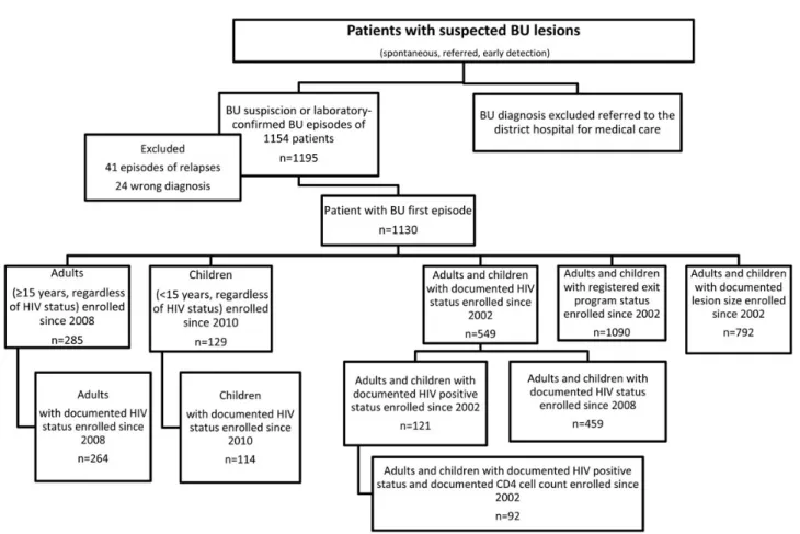
From January 1, 2002 until March 27, 2013, 1195 episodes of BU concerning 1154 patients were registered in the database (Figure2). Two patients had been treated 3 times and 37 pa-tients had been treated twice. Twenty-four papa-tients werefinally considered not to have BU and were excluded from the analyses. Thefinal database included 1130 patients diagnosed with a first BU episode.
HIV Prevalence Among Patients With BU
From January 1, 2008 until March 27, 2013, 93% (264 of 285) of adults (≥15 years old) treated for BU were tested for HIV; 29% were HIV-positive (women, 37%; men, 20%) (Table1). From January 1 2010 to March 27, 2013, 88% of children (114 of 129) treated for BU were tested for HIV and 4% were found to be HIV-positive. The HIV prevalence calculation was performed among PCR-confirmed BU cases and showed similar results. The prevalence of HIV infection in each population was significantly higher than the regional estimated HIV prevalence (Table1). Comparison of BU Baseline Characteristics With and Without HIV Coinfection
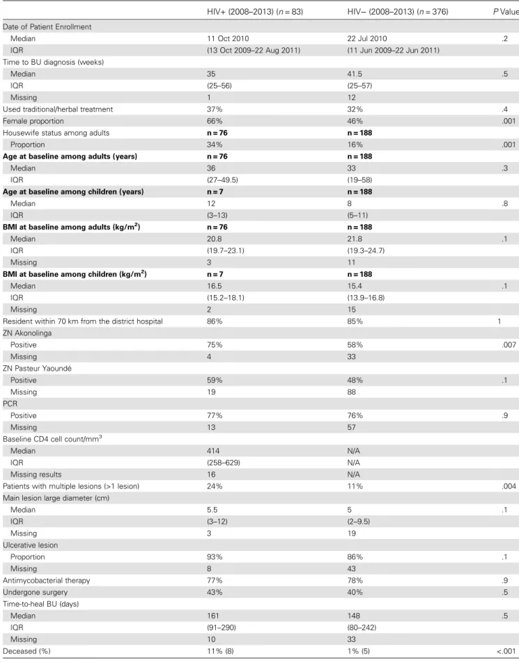
Baseline characteristics of 83 HIV-infected patients (adults and children) included between 2008 and 2013 were compared with 376 HIV-negative patients consecutively admitted to Akonolin-ga District Hospital with a BU diagnosis (Table2). Patients who were HIV positive had significantly more multiple BU lesions at
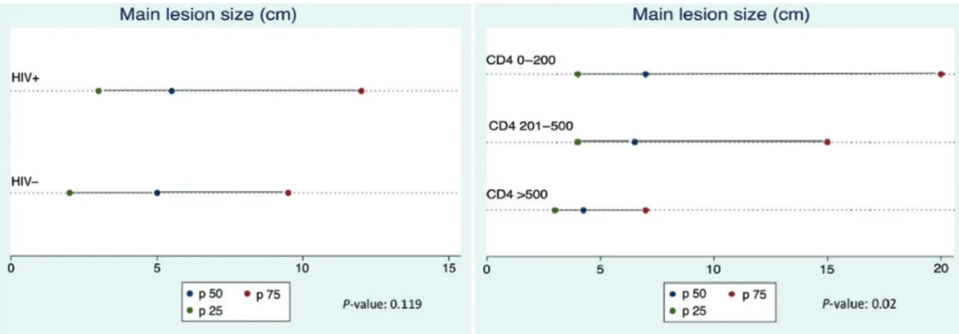
the time of diagnosis compared with individuals who were HIV negative (24% vs 11%, respectively; P = .004). Patients with an HIV infection also tended to have larger BU lesions, defined as the larger diameter of the main lesion (5.5 cm [interquartile range (IQR), 3–12 cm] vs 5 cm [IQR, 2–9.5 cm]; P = .119; Fig-ure3). A higher proportion of HIV-positive patients had ulcer-ated lesions (93% vs 86%, respectively; P = .120) (Figure4), despite a shorter time to BU diagnosis (35 weeks [IQR, 25–56 weeks] vs 42 weeks [IQR, 25–57 weeks]; P = .493) (Table2). Time to BU diagnosis was defined as the duration of the BU ep-isode before enrollment in care.
There were significantly more females among HIV-infected BU patients compared with the HIV-negative group (66% vs 46%; P = .001). Enrollment date, body mass index (BMI), resi-dence, time to BU diagnosis, use of traditional treatment, and the number of PCR-confirmed cases were comparable (Table2).
Baseline HIV Characteristics of BU-HIV Coinfected Patients Only 1 patient was known to be HIV-positive prior to routine testing for HIV and under antiretroviral treatment before BU diagnosis. The median CD4 cell count among 67 HIV-positive patients enrolled since 2008 with CD4 data available (81%) was 414 cell/mm3(IQR, 258–629 cell/mm3) (Table2).
Impact of Immunosuppression in HIV Coinfected Patients From January 1, 2002, a total of 121 patients treated for BU test-ed positive for HIV. Ninety-two (76%) had baseline CD4 data available: 28 patients (30%) had CD4 >500 cell/mm3; 44 (48%) between 201 and 500 cell/mm3; and 20 (22%) below the thresh-old of 201 cell/mm3. The proportion of patients with multiple lesions tended to be lower in the highest CD4 strata, but this did not increase according to immunosuppression status (14%, 30%, and 20%, respectively, from highest to lowest CD4 strata; P = .3). The main lesion size was significantly increased with decreasing CD4 cell counts (4.3 cm [IQR, 3–7 cm]; 6.5 cm [IQR, 4–15 cm]; and 7 cm [IQR, 4–20 cm], respectively; P = .02; Figure3). The proportion of patients with ulcerated le-sions tended to increase with decreasing CD4 cell counts (85%, 95%, and 100%; P = .102; Figure4).
Factors Associated With Time-to-Heal of BU Lesions
We assessed baseline characteristics potentially associated with the time needed to heal BU by univariable and multivariable anal-yses. Human immunodeficiency virus-positive status (n = 547; hazard ratio [HR], 2.35; P = .011; 95% confidence interval [CI], 1.41–3.94), presence of multiple lesions at baseline (n = 1107; HR, 0.42; P < .001; 95% CI, 0.32–0.55), adulthood (n = 1112; HR, 0.64; P < .001; 95% CI, 0.55–0.74), place of residence more than 70 km from the district hospital (n = 1110; HR, 0.76; P = .022; 95% CI, 0.61–0.96), main lesion size more than 5 cm at baseline (n = 785; HR, 0.46; P < .001; 95% CI, 0.39–0.55), use of traditional medicine (n = 1113; HR, 0.71; P < .001; 95% CI, Table 1. HIV Prevalence Among Patients Treated for BU (and
PCR-Confirmed BU) in Akonolinga District Hospital Compared With the Regional Estimated HIV Prevalence
Females With BU≥15 Years (Akonolinga District Hospital) Males With BU ≥15 Years (Akonolinga District Hospital) Children With BU <15 Years (Akonolinga District Hospital) HIV prevalence among patients with BU (95% CI) 37% [29–45]a 20% [13–27]a 4% [2–10]c HIV prevalence among patients with PCR-confirmed BU (95% CI) 39% [30–50] 17% [11–26] 5% [2–13] Regional estimated prevalence 6.9% [5–9]b 5.3% [4 –7]b 0.68% [.36–.98]d BU population compared with reference population P-value <.001 <.001 <.001
Abbreviations: BU, Buruli ulcer; CI, confidence interval; HIV, human immunodeficiency virus; PCR, polymerase chain reaction.
aHIV prevalence among adults has been calculated over a 5-year period (2008– 2013).
bFemale and male (15–49 years) HIV prevalence in Cameroon Central province [37].
c
HIV prevalence among children has been calculated over a 3-year period (2010–2013).
dNational HIV prevalence among 0–14 years children in 2009 (
http://www. unicef.org/sowc2012/pdfs/SOWC-2012-TABLE-4-HIV-AIDS.pdf/http://esa.un. org/unpd/wpp/index.htm).
Table 2. Comparison of Baseline Characteristics, Time-to-Heal BU, Antimycobacterial Therapy, Surgery, and Mortality Among HIV-Pos-itive and -Negative Adults and Children Presenting With BU Lesions Since the Introduction of HIV Systematic Testing in 2008
HIV+ (2008–2013) (n = 83) HIV− (2008–2013) (n = 376) P Value
Date of Patient Enrollment
Median 11 Oct 2010 22 Jul 2010 .2
IQR (13 Oct 2009–22 Aug 2011) (11 Jun 2009–22 Jun 2011) Time to BU diagnosis (weeks)
Median 35 41.5 .5
IQR (25–56) (25–57)
Missing 1 12
Used traditional/herbal treatment 37% 32% .4
Female proportion 66% 46% .001
Housewife status among adults n = 76 n = 188
Proportion 34% 16% .001
Age at baseline among adults (years) n = 76 n = 188
Median 36 33 .3
IQR (27–49.5) (19–58)
Age at baseline among children (years) n = 7 n = 188
Median 12 8 .8
IQR (3–13) (5–11)
BMI at baseline among adults (kg/m2) n = 76 n = 188
Median 20.8 21.8 .1
IQR (19.7–23.1) (19.3–24.7)
Missing 3 11
BMI at baseline among children (kg/m2) n = 7 n = 188
Median 16.5 15.4 .1
IQR (15.2–18.1) (13.9–16.8)
Missing 2 15
Resident within 70 km from the district hospital 86% 85% 1 ZN Akonolinga Positive 75% 58% .007 Missing 4 33 ZN Pasteur Yaoundé Positive 59% 48% .1 Missing 19 88 PCR Positive 77% 76% .9 Missing 13 57
Baseline CD4 cell count/mm3
Median 414 N/A
IQR (258–629) N/A
Missing results 16 N/A
Patients with multiple lesions (>1 lesion) 24% 11% .004 Main lesion large diameter (cm)
Median 5.5 5 .1 IQR (3–12) (2–9.5) Missing 3 19 Ulcerative lesion Proportion 93% 86% .1 Missing 8 43 Antimycobacterial therapy 77% 78% .9 Undergone surgery 43% 40% .5 Time-to-heal BU (days) Median 161 148 .5 IQR (91–290) (80–242) Missing 10 33 Deceased (%) 11% (8) 1% (5) <.001
0.61–0.83), or at least 1 positive laboratory confirmation (ZN or PCR) (n = 1113; HR, 0.80; P = .006; 95% CI, 0.68–0.94) were all significantly associated with a longer healing time of the BU lesion.
By contrast, a hemoglobin level above 11 g/L (n = 344; P = .946), gender (n = 1113; P = .059), a Bacillus Calmette-Guérin scar (n = 947; P = .055), an ulcerative lesion (n = 1024; P = .376), time to BU diagnosis (n = 1076; P = .114), and BMI at baseline (n = 920; P = .061) were not associated with a BU le-sion time-to-heal. In the multivariable analysis, the role of HIV was no longer significantly associated with healing time. How-ever, when CD4 cell strata were entered into the model (n = 92), a CD4 above the threshold of 500 cell/mm3was the only
re-maining significant factor associated with the time needed for BU lesion resolution (HR, 2.39; P = .001; 95% CI, 1.44–3.98). Kaplan-Meier analysis showed that a CD4 above 500 cell/ mm3was significantly associated with halving the time needed
to BU lesion resolution (Figure5).
Factors Associated With Increased BU Lesion Size
Results of risk factor analysis for an increased lesion size are pre-sented in Table3. In the multivariable analysis, an HIV-positive status is significantly and independently associated with an in-creased BU lesion size after adjustment for other influencing fac-tors, such as the hemoglobin level, use of traditional treatment, and place of residence (Table3). When CD4 data were entered into the multivariable analysis, a CD4 above the threshold of 500 cell/mm3was significantly associated with a smaller baseline lesion size (β-coefficient, −0.5; P = .015; 95% CI, −0.91–−0.10) when adjusted for the use of traditional medicine, which was the other remaining factor associated with the baseline lesion.
Mortality Among HIV-Positive and -Negative Patients
During follow-up, the proportion of deceased patients was sig-nificantly higher among HIV-positive than HIV-negative BU patients (11% vs 1%; P < .001) (Table2). The median CD4 cell count among the 8 deceased patients was 228.5 cell/mm3 Figure 3. Main lesion size (median and interquartile range in cm) according to human immunodeficiency virus (HIV) status (left) and immunosuppression level (right).
(IQR, 98–378). None was under antiretroviral therapy (ART). The median duration of time to death was 41.5 days (IQR, 16.5–56.5).
DISCUSSION
We assessed the impact of HIV coinfection on M ulcerans disease in a district hospital in Cameroon. We observed a higher than expected HIV prevalence among patients admitted for BU dis-ease when stratified by gender and age groups. These results are consistent with 2 case-control studies conducted in Benin and Ghana, which showed a higher proportion of HIV-positive patients among BU cases than among controls [9,16] and sup-port the hypothesis of HIV as a risk factor for BU development. Because HIV immunodeficiency is a strong risk factor for devel-oping Mycobacterium tuberculosis disease and ART is strongly associated with a reduction in the incidence of tuberculosis across all CD4 count strata [37], our results suggest a similar pattern of HIV disease in inducing BU overt disease. In addition, we showed an association between HIV-induced immunosuppression (CD4 cell counts) and the clinical severity of BU.
Our data provide further evidence that BU’s clinical manifes-tations are more severe in the presence of HIV-induced immuno-suppression. Indeed, HIV-positive patients frequently present with more multiple and larger lesions compared with HIV-neg-ative individuals. The fact that the proportion of ulcerated lesions is higher among HIV-positive patients across all immunosup-pression strata, together with a shorter time to BU diagnosis, sug-gests an accelerated necrotizing process. Females were overrepresented among HIV-infected patients, reflecting the local HIV epidemiology in Akonolinga, Cameroon [36].
Buruli ulcer treatment consists of surgery and antibiotic treatment. Wound closure is a good marker of cure and
correlates with the follow-up time. Patients who were HIV pos-itive were less likely to have a timely cure of BU, and wound Figure 5. Kaplan-Meier analysis of Buruli ulcer time-to-heal according
to 0–500/>500 CD4 cell count strata. Log rank test: relative ratio, 2.38; P < .0006; 95% confidence interval, 1.43–3.96.
Table 3. Univariable and Multivariable Analyses of BU Main Le-sion Size (Documented Main LeLe-sion of Large Diameter [n = 792])
Main Lesion Large Diameter in cm (log) β-Coefficient P Value 95% CI No. of Observations Univariable Analysis HIV-positive status 0.22 .040 0.01, 0.43 504 Age≥15 years 0.26 .000 0.13, 0.39 791 Hb≥11 g/L −0.29 .010 −0.52, −0.07 330 CD4 >500 −0.58 .004 −0.98, −0.19 88 CD4 0–200/ 201–500/ >500 −0.39 .003 −0.64, −0.13 88 Multiple lesions (>1 lesion) 0.41 .000 0.20, 0.62 789 Enrollment date >2007 −0.18 .012 −0.03, −0.04 791 Used traditional/ herbal treatment 0.56 .000 0.43, 0.69 791 Resident within 70 km of the district hospital −0.38 .000 −0.58, −0.19 791 BU laboratory confirmation (1 or more) 0.09 .235 −0.06, 0.24 791 BMI >19 0.13 .068 −0.01, 0.27 717 Female gender −0.05 .444 −0.18, 0.08 791 BCG scar −0.03 .739 −0.17, 0.12 652 Time to BU diagnosis >12 weeks −0.01 .882 −0.18, 0.15 775
Multivariable Analysis With HIV Status (n = 302) HIV-positive status 0.37 .007 0.10, 0.64 Hb≥11 g/L −0.25 .036 −0.48, −0.02 Used traditional/ herbal treatment 0.47 .000 0.24, 0.70 Resident within 70 km of the district hospital −0.31 .048 −0.62, −0.00
Multivariable Analysis With CD4 Strata 0–500/>500 (n = 88) CD4 0–500/ >500 −0.50 .015 −0.91, −0.10 Used traditional/ herbal treatment 0.40 .038 0.02, 0.77
Abbreviations: BCG, Bacillus Calmette-Guérin; BMI, body mass index; BU, Buruli ulcer; CI, confidence interval; Hb, hemoglobin; HIV, human immunodeficiency virus.
closure time was negatively associated with CD4 cell counts. We also observed a strong association between the level of immuno-deficiency and the baseline size of the main lesion, which might partly explain the longer time-to-heal in patients with low CD4 cell counts. The level of CD4 T-cell counts remained systematically and independently associated with a longer time-to-heal and to main lesion size, despite adjustment for dis-ease severity factors.
The use of traditional medicine, which can consist in local, general, or spiritual interventions [38], is often cited as an ag-gravating factor of the lesion and was also observed in our anal-yses. In our study, the use of traditional medicine had an even a stronger impact on the severity of the BU lesion than the delay of time of access to diagnosis and care. Laboratory-confirmed cases seemed to have a more severe clinical course. One expla-nation may be that the probability of having a positive ZN sam-ple is higher among large and multisam-ple BU lesions with more excision material. Indeed, patients who were HIV positive had a significantly higher proportion of positive ZN compared with individuals who were HIV negative. Socioeconomic status was not assessed in our analyses, although some studies suggested that it was a risk factor for overt BU disease [39].
Our study has some limitations. Observational studies suffer the inherent drawback of uncontrolled bias. Bone involvement, surgical care, antimycobacterial treatment, and ART can all be confounders in the time-to-heal BU analysis. However, we were unable to include these in the analysis due to a lack of data or because they were time- and physician-dependent variables and would have required other statistical models. No conclusion can be drawn on the effect of ART on BU, including possible wors-ening of BU lesions due to its initiation (immune reconstitution inflammatory syndrome). Finally, not all BU cases could be confirmed with a second diagnostic method as recommended by WHO [11]. However, the number of misdiagnosed BU is most probably very low given the high proportion of PCR-confirmed cases and the clear clinical picture of most of our patients.
Overall, our analyses suggest that HIV-associated immuno-suppression affects BU disease. The CD4 level is not only an im-portant determinant of BU baseline severity, but patients with moderate immunosuppression are also at risk for developing se-vere BU disease. In July 2013, WHO issued new recommenda-tions in which all patients with CD4 cell counts below 500 should be offered treatment. We welcome these recommenda-tions, which will allow clinicians to place patients with moderate immunosuppression on antiretroviral drugs. However, 2 ques-tions need further investigation. (1) What is the best timing for ART initiation in eligible patients? Would it be beneficial to offer a rapid introduction of ART within the first weeks of BU (and HIV coinfection) diagnosis? (2) Should patients with CD4 cells over the threshold of 500 be offered ART, similar to patients with tuberculosis?
We conclude that it will be critical to gather prospective data in BU/HIV coinfected patients regarding the impact of ART on the evolution of BU disease, including immune reconstitution inflammatory syndrome risks, and suggest that guidance on how to use and monitor antiretroviral use should be further strengthened. It is becoming increasingly obvious that HIV and its accompanying immunosuppression have an effect on BU incidence and clinical presentation, similar to tuberculosis, another mycobacterial infection. Thus, we recommend testing all BU patients for HIV infection, initiating ART as soon as pos-sible (within 2–4 weeks) in all patients with a CD4 cell count below 500, and to consider ART according to WHO criteria in patients with CD4 cells above this threshold after the comple-tion of antimycobacterial treatment.
Supplementary Data
Supplementary materialare available online at Open Forum Infectious Dis-eases online (http://OpenForumInfectiousDiseases.oxfordjournals.org/).
Notes
Acknowledgments. We thank Ms Rosemary Sudan for careful reading and editing of the original version.
Author contributions. V. C., A. C., and E. C. were responsible for con-cept and design. V. C. and A. C. were responsible for drafting the article. V. C. was responsible for analysis and interpretation of data. The MSF and District hospital team were responsible forfield work. L. R. and A.-B. N. were responsible for data management. V. C. was responsible for patient enrollment, collection of samples, and data assembly. All authors were responsible for critical revision for important intellectual content andfinal approval of article.
Financial support. This work was partially supported by The Human-itarian Committee of the University of Geneva Hospitals, Geneva, Switzerland.
Potential conflicts of interest. All authors: No reported conflicts. All authors have submitted the ICMJE Form for Disclosure of Potential Conflicts of Interest.
References
1. Boulkroun S, Guenin-Mace L, Thoulouze MI, et al. Mycolactone sup-presses T cell responsiveness by altering both early signaling and post-translational events. J Immunol2010; 184:1436–4.
2. Portaels F, Silva MT, Meyers WM. Buruli ulcer. Clin Dermatol2009; 27:291–305.
3. Agbenorku P. Multicenter study of Buruli ulcer disabilities in the head and neck region. Plast Surg Int2011; 2011:647418.
4. Adle-Biassette H, Huerre M, Breton G, et al. [Non tuberculous myco-bacterial diseases]. Ann Pathol2003; 23:216–35.
5. World Health Organization. Buruli ulcer epidemiology. Wkly Epide-miol Rec2009; 84:41–8.
6. Merritt RW, Walker ED, Small PL, et al. Ecology and transmission of Buruli ulcer disease: a systematic review. PLoS Negl Trop Dis2010; 4: e911.
7. Van der Werf TS, Stienstra Y, Johnson RC, et al. Mycobacterium ulcer-ans disease. Bull World Health Org 2005; 83:785–91.
8. Walsh DS, Portaels F, Meyers WM. Buruli ulcer (Mycobacterium ulcer-ans infection). Trulcer-ans R Soc Trop Med Hyg 2008; 102:969–98. 9. Raghunathan PL, Whitney EA, Asamoa K, et al. Risk factors for Buruli
ulcer disease (Mycobacterium ulcerans infection): results from a case-control study in Ghana. Clin Infect Dis2005; 40:1445–53.
10. Herbinger KH, Adjei O, Awua-Boateng NY, et al. Comparative study of the sensitivity of different diagnostic methods for the laboratory diag-nosis of Buruli ulcer disease. Clin Infect Dis2009; 48:1055–64. 11. Portaels F, Johnson P, Meyers WM. Buruli ulcer-Diagnosis of
Mycobac-terium Ulcerans disease. Geneva: WHO; 2001.
12. World Health Organization. Buruli ulcer disease. Geneva: WHO;2007. Available at:http://www.who.int/mediacentre/factsheets/fs199/en/. Ac-cessed April 1, 2014.
13. Nienhuis WA, Stienstra Y, Thompson WA, et al. Antimicrobial treat-ment for early, limited Mycobacterium ulcerans infection: a randomised controlled trial. Lancet2010; 375:664–72.
14. World Health Organization. Provisional guidance on the role of specific antibiotics in the management of Mycobacterium ulcerans disease (Bur-uli ulcer). Geneva: WHO;2011. Available at:http://www.who.int/ buruli/information/antibiotics/en/. Accessed 1 April 2014.
15. World Health Organization. Global tuberculosis report 2013. Geneva: WHO;2013. Available at:http://www.who.int/iris/bitstream/10665/ 91355/1/9789241564656_eng.pdf?ua=1. Accessed April 1, 2014]. 16. Johnson RC, Nackers F, Glynn JR, et al. Association of HIV infection
and Mycobacterium ulcerans disease in Benin. AIDS 2008; 22:901–3. 17. Darie H, Cautoclaud A, Lajaunie C, Millet P. [Dermatological aspects of
AIDS in western Africa. Apropos of 140 cases]. Bull Soc Pathol Exot 1994; 87:176–80.
18. Johnson RC, Ifebe D, Hans-Moevi A, et al. Disseminated Mycobacteri-um ulcerans disease in an HIV-positive patient: a case study. AIDS 2002; 16:1704–5.
19. Kibadi K, Colebunders R, Muyembe-Tamfum JJ, et al. Buruli ulcer lesions in HIV-positive patient. Emerg Infect Dis2010; 16:738–9. 20. Toll A, Gallardo F, Ferran M, et al. Aggressive multifocal Buruli ulcer
with associated osteomyelitis in an HIV-positive patient. Clin Exp Der-matol2005; 30:649–51.
21. Sopoh GE, Dossou AD, Brun LV, et al. Severe multifocal form of Buruli ulcer after streptomycin and rifampin treatment: comments on possible dissemination mechanisms. Am J Trop Med Hyg2010; 83:307–13. 22. Portaels F, Zinsou C, Aguiar J, et al. Les atteintes osseuses dans l’ulcère
de Buruli: A propos de 73 cas. Bull Séanc Acad R Sci Outre-Mer2003; 49:161–90.
23. Ouattara D, Meningaud JP, Saliba F. [Multifocal forms of Buruli ulcer: clinical aspects and management difficulties in 11 cases]. Bull Soc Path-ol Exot2002; 95:287–91.
24. O’Brien DP, Robson ME, Callan PP, et al. “Paradoxical” immune-mediated reactions to Mycobacterium ulcerans during antibiotic treat-ment: a result of treatment success, not failure. Med J Aust2009; 191:564–6.
25. Schutte D, Um-Boock A, Mensah-Quainoo E, et al. Development of highly organized lymphoid structures in Buruli ulcer lesions after
treatment with rifampicin and streptomycin. PLoS Negl Trop Dis 2007; 1:e2.
26. Gooding TM, Johnson PD, Campbell DE, et al. Immune response to infection with Mycobacterium ulcerans. Infect Immun 2001; 69:1704–7.
27. Gooding TM, Johnson PD, Smith M, et al. Cytokine profiles of patients infected with Mycobacterium ulcerans and unaffected household con-tacts. Infect Immun2002; 70:5562–7.
28. Lagarrigue V, Portaels F, Meyers WM, et al. [Buruli ulcer: risk of bone involvement! A propos of 33 cases observed in Benin]. Med Trop2000; 60:262–6.
29. Noeske J, Kuaban C, Rondini S, et al. Buruli ulcer disease in Cameroon rediscovered. Am J Trop Med Hyg2004; 70:520–6.
30. Portaels F, Aguiar J, Debacker M, et al. Mycobacterium bovis BCG vac-cination as prophylaxis against Mycobacterium ulcerans osteomyelitis in Buruli ulcer disease. Infect Immun2004; 72:62–5.
31. Phillips R, Sarfo FS, Guenin-Mace L, et al. Immunosuppressive signa-ture of cutaneous Mycobacterium ulcerans infection in the peripheral blood of patients with buruli ulcer disease. J Infect Dis2009; 200: 1675–84.
32. Prasad R. Pulmonary sarcoidosis and chronic cutaneous atypical myco-bacter ulcer. Aust Fam Physician1993; 22:755–8.
33. Bahebeck J, Bedimo R, Eyenga V, et al. (2004) The management of mus-culoskeletal infection in HIV carriers. Acta Orthop Belg2004; 70: 355–60.
34. Delaporte E, Savage C, Alfandari S, et al. [Buruli ulcer in a Zairian woman with HIV infection]. Ann Dermatol Venereol1994; 121: 557–60.
35. Porten K, Sailor K, Comte E, et al. Prevalence of Buruli ulcer in Akono-linga health district, Cameroon: results of a cross sectional survey. PLoS Negl Trop Dis2009; 3:e466.
36. DHS. Cameroon Demographic and Health Survey and Multiple Indicators Cluster Survey (DHS-MICS).2011. Available at:http:// www.measuredhs.com/pubs/pdf/HF42/HF42.pdf. Accessed 1 April 2014.
37. Suthar AB, Lawn SD, del Amo J, et al. Antiretroviral therapy for preven-tion of tuberculosis in adults with HIV: a systematic review and meta-analysis. PLoS Med2012; 9:e1001270.
38. Laterali M. Ethnographie de la constitution d’un problème de santé publique au Cameroun: l’exemple de l’ulcère de Buruli ou atom dans l’arrondissement d’Ayos [MD Thesis]. Neuchâtel: University of Neu-châtel.2005; 157–62. Available at: http://books.google.ch/books/ about/Ethnographie_de_la_constitution_d_un_pro.html?id=Fivmk QEACAAJ&redir_esc=y.
39. Jacobsen KH, Padgett JJ. Risk factors for Mycobacterium ulcerans infec-tion. Int J Infect Dis2010; 14:e677–81.
![Table 3. Univariable and Multivariable Analyses of BU Main Le- Le-sion Size (Documented Main LeLe-sion of Large Diameter [ n = 792]) Main Lesion Large Diameter in cm (log) β -Coefficient P Value 95% CI No](https://thumb-eu.123doks.com/thumbv2/123doknet/14453676.519157/8.892.86.421.54.292/univariable-multivariable-analyses-documented-diameter-lesion-diameter-coefficient.webp)
