Introduction
Sternoclavicular dislocations are rare. According to the literature, they represent 3% of all injuries of the shoulder girdle [1] or only 10 out of 1,600 trauma cases [2]. Since CT-scan imaging has become more available, the diagno-sis is made earlier and not only post-mortem as often be-fore [3]. The treatment of sternoclavicular lesions remains controversial, and the surgical treatment is associated with many risks.
Case report
A 22-year-old tennis player fell from a scooter onto his right anterolateral shoulder, and was admitted at an outside University hospital with severe pain. After a diagnosis of a posterior sternoclavicular joint dislocation had been estab-lished, he was transferred to our level 1 trauma center.
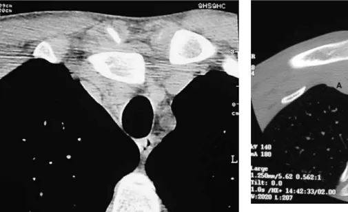
Although the CT-scan showed compression of the right brachiocephalic vein and trunk by the clavicle, the patient showed no signs of any neuro-vascular impairment (Fig. 1). The dislocation was reduced under general anesthesia by traction in abduction of the right arm and direct
percu-taneous grasping of the clavicle with a towel clip. The X-ray control confirmed the reduction. The upper extremity was immobilized in a mitella for a few days.
A week later, the patient presented with an anterior subluxation of the sternoclavicular joint as seen on the CT-scan (Fig. 2) but with no residual symptoms. There-fore, and according to the literature [4,5,6], we decided on no further treatment.
At 6 weeks after the accident, spontaneous reduction was achieved and the patient had almost no residual pain, with normal motion of his right shoulder and no limita-tions in sports.
Review of the literature Anatomy
The sternoclavicular joint is the junction of the shoulder gir-dle to the axial skeleton. A frequent variation is an accessory joint to the first rib. The joint between the clavicle and the sternum is incongruent, with only 50% of the clavicular sur-face being in contact with the sternum. This incongruence is compensated for by a disk. This type of joint design allows a wide range of motion in several directions [3].
Abstract Sternoclavicular disloca-tions represent a rare injury. Based on our clinical experience with a pa-tient showing an anterior subluxation after reduction of a posterior trau-matic dislocation, we review the lit-erature. The emergent reduction of the dislocation is mandatory, always keeping in mind the potentially dev-astating neurovascular complica-tions. If the treatment of a residual anterior instability remains contro-versial, a residual posterior
instabil-ity should be treated by a surgical procedure. The optimal treatment de-pends mainly on each surgeon’s choice and practice.
Keywords Shoulder ·
Sternoclavicular · Dislocation · Instability
S H O U L D E R
Knee Surg Sports Traumatol Arthrosc (2004) 12 : 453–456 DOI 10.1007/s00167-004-0504-x Michael Wettstein Olivier Borens Raffaele Garofalo Cyril Kombot François Chevalley Elyazid Mouhsine
Anterior subluxation
after reduction of a posterior traumatic
sterno-clavicular dislocation:
a case report and a review of the literature
Received: 8 November 2003 Accepted: 27 January 2004 Published online: 3 June 2004 © Springer-Verlag 2004
M. Wettstein · O. Borens · R. Garofalo ·
C. Kombot · F. Chevalley · E. Mouhsine (✉) Service d’Orthopédie et de Traumatologie de l’Appareil Locomoteur,
Centre Hospitalier Universitaire Vaudois, 1011 Lausanne, Switzerland
Tel.: +41-21-3142791, Fax: +41-21-3142800,
The joint is stabilized by the articular capsule, which is reinforced anteriorly by a tiny sternoclavicular ligament and posteriorly by a much stronger sternoclavicular liga-ment (Fig. 3). This may explain why anterior dislocations,
due to indirect stresses, are more frequent than posterior ones [7] in a proportion of approximately 1:20 cases [8]. Cranially, the interclavicular ligament, which unites both clavicles, is a very thin structure. The costo-clavicular lig-ament stabilizes the joint in the caudo-cranial direction and in rotation, due to its configuration with an anterior and a posterior lamina. This ligament also acts as a ful-crum during a postero-lateral blow to the shoulder which results in a medial and backward displacement of the clavicle.
Directly posterior to the right sternoclavicular joint (Fig. 4) pass the right brachiocephalic vein and trunk, as well as the trachea, and directly posterior to the left stern-oclavicular joint run the trachea, the left brachiocephalic vein, the left common carotid artery and the left subcla-vian artery. All these structures may be damaged during an injury to the sternoclavicular joint.
Compression of the brachiocephalic and subclavian veins can lead to swelling and cyanosis of the correspond-ing arm and can also lead to venous thrombosis [9].
Compression of the subclavian artery induces acute ischemia of the arm. Limited compression of the trachea leads to respiratory stridor, but can be fatal due to com-plete obstruction or disruption.
The lower brachial plexus (C5-Th1) lies behind the vessels over the first rib. Thoracic outlet syndrome and brachial plexus compression have been reported in a case of chronic posterior dislocation of the sternoclavicular joint [10]. For these reasons, a careful neurovascular ex-amination of the upper extremities and the exex-amination of the respiratory status are mandatory.
If any arterial injury is suspected, an arteriography should be performed as soon as possible to guide an open
454
Fig. 1 CT-scan showing posterior dislocation of the right medial
clavicle compressing the right brachiocephalic vein and trunk
Fig. 2 CT-scan of the induced anterior subluxation of the right
sternoclavicular joint
Fig. 3 Schematic representation of sternoclavicular environment. A Subclavius muscle, B costoclavicular ligament, C
sternoclavicu-lar ligament, D interclavicusternoclavicu-lar ligament, E interarticusternoclavicu-lar disk
Fig. 4 CT-scan at the medial end-level of both clavicles. A Right
brachiocephalic vein, B brachiocephalic trunk, C trachea, D left common carotid artery, E left brachiocephalic vein, F left sub-clavian artery
reconstruction of the damaged vessel if necessary. Because of the risk of a lesion to one of the above mentioned struc-tures, the reduction of a posterior dislocation should be con-sidered a true emergency. However, because of the possible laceration of a great vessel, it is advisable to proceed to a re-duction only if a vascular surgeon is rapidly available.
Classification
Different classifications can be used to describe a sterno-clavicular dislocation. One is based on the anatomic posi-tion of the dislocaposi-tion, and the other one on the etiology of the lesion [6]. A clinical classification was published by Allman [11] and seems to be a good tool for the treat-ment choice in addition to the direction of dislocation.
Stage 1 corresponds to a contusion or minor distortion with a stable joint and no radiological signs of lesions.
Stage 2 is a subluxation of the joint due to partial rup-ture of the sternoclavicular ligaments, whereas the costo-clavicular ligament remains intact.
Stage 3 is a dislocation of the joint with a complete rupture of all ligaments and the articular disk.
Treatment
Minor sprains with subluxation are best treated function-ally with a protective mitella and non steroidal anti-inflam-matory drugs (NSAI) for a few days.
Anterior dislocations can be treated by closed reduc-tion under general anesthesia. However, these lesions are often unstable and may redislocate [12]. In the case of a recurrent dislocation, no further treatment should be ap-plied if the patient feels no pain or discomfort, which are rare in anterior dislocations [4]. In the case of symptoms, open reduction can be attempted. However, the fixation of the joint by screws is biomechanically unfavorable [6] and the use of Kirschner wires can lead to fatal migrations of the pins [13]. In these cases, the best therapy is proba-bly the excision of the torn disk, suture and plastic rein-forcement of the ligaments as shown in a large series [14]. After reduction or open repair, the shoulder should be held back with a figure-of-eight dressing for 6 weeks to allow ligament healing [6].
Posterior dislocations should first also be treated by means of closed reduction [6]. The reduction is done with the patient in a prone position under general anesthesia, with a sandbag between the shoulder blades. Progressive traction on the abducted arm is applied and, if this is not sufficient for reduction, the clavicle is grasped with a ster-ile towel clip to apply direct anterior traction [6]. Even if other techniques exist [6], this seems to be the best to us. In most cases such a reduction remains stable, and needs only an immobilization with a figure-of-eight dressing for 4–6 weeks.
If a dorsal subluxation persists, some authors [15] have recommended an open repair of the ligaments and fixation by Kirschner wires with the already stated complications. This technique should be definitely abandoned.
As a residual posterior dislocation is not tolerated [16], many techniques for repair have been published. Speed [5] used a fascial loop between the clavicle and the first rib. Allen [17] used a fascia lata flap to reconstruct the stern-oclavicular ligaments. Burrows [18] used the tendon of the subclavius muscle passing through the proximal clav-icle. All these techniques show reliable results, although in each case the series was only small.
If degenerative changes are noted in the joint, several authors [15, 16, 19] have recommended the resection of the medial end of the clavicle. During removal of the ar-ticular part, attention should be paid not to damage the costo-clavicular ligament, or to repair it if already torn.
Arthrodesis of the sternoclavicular joint was reported in the treatment of habitual dislocation of the sternoclav-icular joint [20]. This procedure should not be done any-more because a severe limitation of normal shoulder move-ments can occur.
One recent publication [1] outlined the long immobi-lization which is necessary after ligamentous repairs. The authors proposed a method of plaiting of the sternoclavic-ular joint allowing an early functional recovery.
No patient showed a recurrent dislocation, and the mean functional outcome was good to excellent with this technique. The only critical point of the treatment was the need for removal of the implant.
Conclusion
Sternoclavicular dislocations are rare and they remain a challenging problem. Most cases, both anterior and poste-rior dislocations, should first and immediately be treated by means of closed reduction and immobilization. The potentially disastrous complications of posterior disloca-tions should always be kept in mind during the investiga-tions.
If a residual posterior instability or symptoms occur, a surgical procedure is necessary. Up to now, it is unclear whether a repair of the ligaments with ligamentoplasty or the use of a hook-plate will bring better results, as only small series of patients have been published. The advantage of ligamentoplasty is the need for only one operation, but it carries with it the disadvantage of a long-term immobi-lization. The use of a hook-plate seems an interesting al-ternative, but requires the removal of the plate and can damage the sternoclavicular joint, risking subsequent arthri-tis.
We think that a surgeon should use one single method which he feels most familiar with, so as to obtain the best results.
456
1. Franck WM, Müller O, Hennig F (2000) Die traumatische sternoklaviku-läre Instabilität. Eine therapeutische Alternative. Unfallchirurg 103:834– 838
2. McCulloch P, Henley BM, Linnau KF (2001) Radiographic clues for high energy trauma: three cases of sterno-clavicular dislocation. AJR Am J Roentgenol 176:1534
3. Rajaratnam S, Kerins M, Apthorp L (2002) Posterior dislocation of the sternoclavicular joint: a case report and review of the clinical anatomy of the region. Clin Anat 15:108–111 4. Rockwood CA, Matsen FA (1998)
Dis-orders of the sternoclavicular joint. In: Rockwood CA (ed) The Shoulder, 2nd edn. Saunders, Philadelphia, pp 555– 609
5. Speed K (1942) A textbook of frac-tures and dislocations, 4th edn. Lea and Febiger, Philadelphia, pp 282–290
6. Wirth MA, Rockwood CA Jr (2001) Injuries to the sternoclavicular joint. In: Bucholz RW, Heckman JD (eds) Rockwood and Green’s fractures in adults, 5th edn. Lippincott Williams and Wilkins, Philadelphia, pp 1253– 1255
7. Marker LB, Klareskov B (1996) Posterior sternoclavicular dislocation: an American football injury. Br J Sports Med 30:71–72
8. Nettles JL, Linscheid R (1968) Sterno-clavicular dislocations. J Trauma 8: 158–164
9. Ono K, Iragawa H, Kiyota K, Terada T, Suzuki S, Maekawa K (1998) Poste-rior dislocation of the sternoclavicular joint with obstruction of the innomi-nate vein: a case report. J Trauma In-jury Infect Crit Care 44:381–383 10. Rayan GM (1994) Compression
brachial plexopathy caused by chronic posterior dislocation of the sternoclav-icular joint. J Okla State Med Assoc 87:7–9
11. Allmann FI (1967) Fractures and liga-mentous injuries of the clavicle and its articulation. J Bone Joint Surg Am 49:774–784
12. De Jong KP, Sukul DM (1990) Ante-rior sternoclavicular dislocation: a long-term follow-up study. J Orthop Trauma 4:420–423
13. Kumar P, Godbole R, Rees GM, Sarkar P (2002) Intrathoracic migration of a Kirschner wire. J R Soc Med 95: 198–199
14. Fery A, Sommelet J (1988) Dislocation of the sternoclavicular joint: a review of 49 cases. Int Orthop 12:187–195 15. Bateman JE (1978) The shoulder and
neck, 2nd edn. Saunders, Philadelphia 16. Borrero E (1987) Traumatic posterior displacement of the left clavicular head causing extrinsic compression of the subclavian artery. Physician Sports Med 15:87–89
17. Allen AW (1928) Living suture grafts in the repair of fractures and disloca-tions. Arch Surg 16:1007–1020 18. Burrows HF (1951) Tenodesis of
sub-clavius in the treatment of recurrent dislocation of the sternoclavicular joint. J Bone Joint Surg Br 33:240–243 19. Breitner S, Wirth CJ (1987) Resection
of the acromial and sternal ends of the clavicle. Z Orthop 125:363–368 20. Rice EE (1932) Habitual dislocation of
the sternoclavicular articulation: a case report. J Okla State Med Assoc 25:34– 35