ORIGINAL PAPER
Generation of metabolites by an automated online
metabolism method using human liver microsomes
with subsequent identification by LC-MS(n),
and metabolism of 11 cathinones
Daniel M. Mueller&Katharina M. Rentsch
Received: 7 September 2011 / Revised: 18 November 2011 / Accepted: 18 December 2011 / Published online: 10 January 2012 # Springer-Verlag 2012
Abstract Human liver microsomes (HLMs) are used to simulate human xenobiotic metabolism in vitro. In forensic and clinical toxicology, HLMs are popularly used to study the metabolism of new designer drugs for example. In this work, we present an automated online extraction system we developed for HLM experiments, which was compared to a classical offline approach. Furthermore, we present studies on the metabolism of 11 cathinones; for eight of these, the metabolism has not previously been reported. Metabolites
were identified based on MS2 and MS3 scans. Fifty-three
substances encompassing various classes of drugs were employed to compare the established offline and the new online methods. The metabolism of each of the following 11 cathinones was studied using the new method: 3,4-methy-lenedioxy-N-benzylcathinone, benzedrone, butylone, dime-thylcathinone, ethylone, flephedrone, methedrone, methylone, methylethylcathinone, naphyrone, and pentylone. The agreement between the offline and the online methods was good; a total of 158 metabolites were identified. Using only the offline method, 156 (98.7%) metabolites were iden-tified, while 151 (95.6%) were identified using only the online method. The metabolic pathways identified for the 11 cath-inones included the reduction of the keto group, desalkylation, hydroxylation, and desmethylenation in cathinones containing a methylenedioxy moiety. Our method provides a straightfor-ward approach to identifying metabolites which can then be added to the library utilized by our clinical toxicological screening method. The performance of our method compares
well with that of an established offline HLM procedure, but is as automated as possible.
Keywords LC-MS . Human liver microsomes . Cathinones . Online extraction . Human metabolism
Abbreviations
APCI Atmospheric pressure chemical ionization
AU Arbitrary units
bk Beta-keto
CYP Cytochrome P450
EDTA Ethylenediaminetetraacetic acid
ESI Electrospray ionization
GST Glutathione-S-transferase
G6P Glucose-6-phosphate
G6PD Glucose-6-phosphate dehydrogenase
HPLC High-pressure liquid chromatography
HLMs Human liver microsomes
IU International units
LC-MS Liquid chromatography–mass spectrometry
MDPV Methylenedioxypyrovalerone
NADP+ Nicotinic acid adenine dinucleotide phosphate
(oxidized form)
NADPH Nicotinic acid adenine dinucleotide phosphate
(reduced form)
NAT N-Acetyltransferase
Introduction
Identifying metabolites is a necessary task during the drug development process as well as in the clinical and forensic toxicological laboratory. Whereas the main focus during drug development is to determine the presence of a
D. M. Mueller
:
K. M. Rentsch (*)Institute for Clinical Chemistry, University Hospital Zurich, Raemistrasse 100,
8091 Zurich, Switzerland e-mail: [email protected] DOI 10.1007/s00216-011-5678-8
metabolite, the main focus in the toxicological laboratory is to use the metabolites to prove the intake of a particular drug (e.g., a drug of abuse) when urine samples are analyzed. Therefore, metabolite generation by in vivo and in vitro sys-tems is an important tool in both of these fields of application. Human liver microsomes (HLMs) have become popular tools for simulating human xenobiotic metabolism in vitro, mainly because they are easy to use and store. Phase I metabolism can be studied using HLMs by simply adding NADPH as a cofactor for the main drug-metabolizing
cyto-chrome P450 enzymes (CYP) [1]. However, there are also
some drawbacks of this approach, such as the absence of enzymes such as N-acetyltransferase (NAT) or glutathione-S-transferase (GST). In other words, metabolic pathways
involving those enzymes cannot be studied using HLMs [1].
In the pharmaceutical industry, HLMs are used for me-tabolite identification and to predict the clearance of drug
candidates in vivo, for example [1]. In the field of forensic
and clinical toxicology, HLMs are popularly employed to study, for instance, the metabolism of new designer drugs. The aim of such studies is usually to investigate the metab-olism of a new designer drug in order to make it detectable in toxicological screening procedures, or to quantify it using dedicated methods. Examples include the metabolism of the synthetic cannabimimetic JWH-018, for which Wintermeyer
et al. [2] used HLMs to study its phase I metabolism in vitro.
Meyer et al. [3] studied the metabolism of
methylenedioxy-pyrovalerone (MDPV) using HLMs (amongst other methods),
and Zaitsu et al. [4] identified several metabolites of butylone
and ethylone. The use of mass spectrometry in studies of the metabolism of drugs of abuse and doping agents was recently
reviewed, focusing on clinical and forensic toxicology [5].
The process used in HLM experiments can be automated, and this is an especially common approach in the pharma-ceutical industry, where a high throughput of experiments is needed. Several methodologies with fully integrated
work-flows have recently been presented. Drexler et al. [6]
devel-oped a sophisticated automated approach to study the metabolic stabilities of drug candidates. This approach in-volved incubation using HLMs, which was carried out in microplates using an automated liquid handling robot. Similar
approaches were presented by Xu et al. [7] and by Jenkins et
al. [8], which also used a liquid handling robot to carry out the
HLM experiments. Lai et al. [9] described a system where the
autosampler was modified to include a heated agitator and the samples were subsequently injected into the online extraction system. The system was used to screen the stability of a
prodrug in development. Kool et al. [10] developed an online
system using rat liver microsomes to study the affinity of ligands for CYP enzymes.
Regarding the throughput required, assays using, for example, specialized liquid-handling robots are usually over-kill for metabolite-generating experiments in the clinical
toxicological laboratory, and the high throughput achievable is also not needed.
One possible method for achieving a certain degree of lower-level automation and miniaturization was presented
by Nicoli et al. [11] and Curcio et al. [12], which used a
capillary as the reaction vessel for HLM experiments. This method included automated metabolite generation, but re-quired manual sample preparation of the incubation mixture. The main aim of the work presented in this paper was to create an automated method with adequate throughput for our toxicological laboratory to generate phase I metabolites which could then be added to the library utilized by our clinical
toxicological screening method [13]. Our approach used a
modified, heated autosampler loop for incubation, and the samples were then transferred onto an online extraction LC-MS system. After loading the components for the incubation onto the autosampler, no additional manual steps were needed. A second aim of this work was to study the metabolism of 11 different cathinones using HLMs; for eight of these, the metabolism has not previously been reported, to our knowledge. Cathinones are a new class of designer drugs that have appeared in the drugs-of-abuse market in recent years. They are reported to inhibit plasma membrane amine transporters, thereby increasing the amount of
mono-amine neurotransmitter in the synaptic cleft [14], which
leads to the pharmacological effects of these drugs.
Materials and methods Chemicals and reagents
Acetonitrile was purchased from Romil (Cambridge, UK), acetone from Merck (Darmstadt, Germany), and methanol and 2-propanol from Seelze (Seelze, Germany); all were of HPLC gradient grade purity. Purified water was produced in-house using a central water purification installation (Burkhalter AG, Worblaufen, Switzerland).
HPLC-grade ammonium acetate and ammonium carbon-ate were obtained from Scharlau (Taegerig, Switzerland), while analytical grade formic acid and ammonia were from Merck. Analytical grade EDTA, potassium dihydrogen phosphate, potassium hydrogen phosphate, tris base and glacial acetic acid were obtained from Sigma–Aldrich (Buchs, Switzerland). Glycerol Ph. Eur. was purchased from Haenseler (Herisau, Switzerland).
Human liver microsomes (pooled, male and female), β-nicotinamide adenine dinucleotide phosphate hydrate
(NADP+), D-glucose-6-phosphate dipotassium hydrate
(G6P), and glucose-6-phosphate dehydrogenase (G6PD) from S. cerevisiae were purchased from Sigma–Aldrich. Amitriptyline, chlorpromazine, chlorprothixene, duloxe-tine, ethylone, fenfluramine, fluoxeduloxe-tine, fluphenazine,
ketamine, methylone, methylphenidate, mianserine, naphyr-one, oxycodnaphyr-one, and phencyclidine were purchased from Cerilliant (Round Rock, TX, USA). Bromazepam, butylone, flephedrone, methaqualone, methylfentanyl, psilocin, tilidine, and zolpidem were obtained from Lipomed (Arlesheim, Swit-zerland). Amlodipine, bisacodyl, desipramine, flupentixol, maprotiline, perphenazine, promazine, propafenone, sertindole, strychnine, and thioridazine were purchased from Sigma– Aldrich. Amisulpride, diclofenac, opipramol, pipamperone, and reboxetine were obtained from TRC (North York, Canada). 3,4-Methylenedioxy-N-benzylcathinone, benzedrone, dime-thylcathinone, methedrone, methyledime-thylcathinone, and penty-lone were purchased from LGC Standards (Wesel, Germany). Aripiprazol, clotiapine, cinnarizine, cyclizine, dextropropoxy-phene, doxylamine, fluvoxamine, meclozine, paroxetine, pen-fluridol, pethidine, pentazocine, remifentanil, sildenafil, thiethylperazine, thioproperazine, tramadol, and zaleplone were provided by their respective manufacturers.
LC-MS analysis
The turbulent flow online extraction HPLC system con-sisted of an HTC PAL autosampler, one Allegro pump for the online extraction, and one Allegro pump for the analytical chromatography, all controlled by Aria 1.6.3 software (all from Thermo Fisher Scientific, Basel, Switzer-land). The autosampler was equipped with a Hamilton
100μl syringe for CTC autosamplers (Hamilton, Bonaduz,
Switzerland).
The detailed conditions for the online extraction and
LC-MS analysis are described in our previous publications [13,
15]. In brief, samples were extracted online using
chroma-tography based on turbulent flow under alkaline conditions using ammonium carbonate buffer 10 mM, pH 8.0. A Cy-clone and a C18 XL column (both from Thermo Fisher Scientific) were connected in series to achieve sufficient extraction for substances over a wide polarity range. Ana-lytical chromatography was performed under acidic
condi-tions on a Betasil phenyl/hexyl column (100×3 mm, 3μm
particles, Thermo Fisher Scientific) using ammonium ace-tate 5 mM in water plus 0.1% formic acid and ammonium acetate 5 mM in methanol plus 0.5% formic acid as mobile phases. A mixture of acetonitrile/acetone/2-propanol 1/1/1 (v/v/v) was used as a wash phase. The combination of the two extraction columns and the analytical column could be connected inline and divided again via two six-port switching valves controlled by Aria software.
A LXQ linear ion trap mass spectrometer was used for detection, as controlled by XCalibur 2.0.7 SP1 software (Thermo Fisher Scientific). Ionization was performed under atmospheric pressure chemical ionization (APCI) conditions because of its better performance in terms of ion suppression susceptibility as compared with electrospray ionization
(ESI). The vaporizer temperature was fixed at 450 °C, the capillary temperature at 275 °C, the sheath gas flow at 30 arbitrary units (AU), the auxiliary gas flow at 5 AU, and the
discharge current was set to 5μA. Both positive and
nega-tive ionization were used, with the polarity constantly switching. Chromatograms were recorded in untargeted data-dependent acquisition mode. In positive ionization
mode, MS2spectra were recorded for the four most abundant
ions, and in negative ionization mode they were recorded for
the two most abundant ions. MS3spectra were recorded for
the most abundant ion from each MS2spectrum. The
thresh-old for the acquisition of fragment spectra was set to 100 counts per second. The isolation width was set to 1 amu, while the normalized collision energy for the fragmentation was set to 35%.
For the online metabolism experiment, the injection loop
was modified to a 200μl injection loop placed in a column
oven (Thermo Fisher Scientific) that kept the temperature constant at 37 °C.
Metabolism studies
Identification of metabolites
Metabolites were identified using their MS2and MS3mass
spectra. Methods applied were neutral-loss scans and com-parisons of their fragmentation patterns to the parent sub-stances. Raw files were examined both manually and assisted by MassFrontier 7 (Thermo Fisher Scientific). Solutions
G6P was prepared as a 0.1 M solution in water and NADP+
as a 10 mg/ml solution in water; both were stored as aliquots
at−20 °C until use. The G6PD was dissolved in a mixture
consisting of 20% 40 mM Tris-acetate buffer containing 1 mM EDTA and 80% glycerol to achieve an activity of
103IU/ml at a protein concentration of 1 mg/ml. Aliquots
were stored at−20 °C until use. The HLM suspensions were
stored as aliquots at−80 °C until usage.
Stock solutions of the test substances were prepared at 1 mg/ml in water or methanol, depending on the solubility
of the substance, and stored at−20 °C until use. Dilutions to
10 mg/l were freshly prepared in phosphate buffer 0.1 M, pH 7.4. The remaining amount of organic solvent was always below 1% (v/v).
Offline method
The method was adapted from that of Sohl et al. [16]. To
generate the NADPH needed for the reactions, a NADPH-generating system was used. It consisted of 50 parts 0.1 M
concentration of 1 mg/ml and an activity of 103IU/ml; all solutions were prepared as described above. The mixture was pre-incubated for 1 min before adding it to the incubation mixture to start the reaction.
Five hundred picomoles of total CYP were used in the incubation mixture per incubation. The substances were added diluted in phosphate buffer 0.1 M, pH 7.4 at a concentration of 10 mg/l. The total volume of the incubation
mixture was 112μl.
The mixture was incubated in a water bath at 37 °C for 3 h under occasional gently mixing. After 3 h, the reaction
was stopped by adding 110μl of ice-cold acetonitrile to the
incubation mixture. After vigorous vortexing and centrifu-gation for 5 min at 11,700×g and 10 °C, the supernatant was transferred into autosampler vials and stored at 10 °C until injection. One hundred microliters were analyzed as described above.
Online method
The same components (NADPH-generating system, HLM suspension, and substances in phosphate buffer) were used as described above and used for the offline method. The
autosampler was modified with a 200 μl injection loop
which could be heated to 37 °C using a column oven. The heated autosampler loop served as the incubation vessel. The HLM suspension, the NADPH-generating system, and the phosphate buffer 0.1 M, pH 7.4 containing the substances at a concentration of 10 mg/l were stored separately in the cooled autosampler stack at 10 °C until usage.
To combine the incubation mixture, the autosampler was programmed using the standard options of the Aria software
to first pipette the HLM suspension (15 μl), then the
NADPH-generating system (10μl), and finally the phosphate
buffer containing the substances (75μl) into the heated loop.
Afterwards, using the installed 100μl syringe of the
autosam-pler, the whole incubation mixture was slowly mixed twice by aspiration followed by immediate ejection. To ensure that the
100μl incubation mixture was in the heated region of the loop
inside the column oven, a 30μl air gap was pushed behind the
incubation mixture.
The mixture was incubated in the loop for 50 min, and then the injection valve was switched to the active position, which caused the incubation mixture to be transferred onto the online extraction columns. By transferring the incubation mixture onto the online extraction columns, the reaction was stopped by separating the drug-metabolizing enzymes from the small organic molecules.
Intermediate precision of the online method
To check the stability of the HLM suspension and the NADPH-generating system in the cooled autosampler stack,
venlafaxine was chosen as a test compound and repeatedly injected over a 6 h period. Stability was evaluated by calcu-lating the ratio of the peak area of O-desmethylvenlafaxine to the peak area of venlafaxine.
To check the between-day intermediate precision of the method, including different lots of HLM suspensions, clo-tiapine was injected on four different days. We checked whether all of the metabolites could be identified on all of the days.
Method comparison
Fifty-three compounds from various drug classes were me-tabolized with the offline method as well as the online method in order to compare the efficiency of metabolite generation by the new online procedure.
HLM metabolism of 11 cathinones
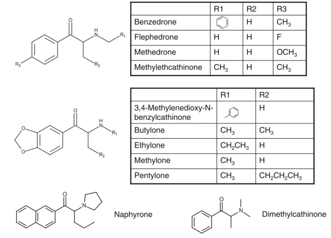
To study the phase I metabolism of the 11 cathinones (3,4-methylenedioxy-N-benzylcathinone, benzedrone, butylone, dimethylcathinone, ethylone, flephedrone, methedrone, meth-ylone, methylethylcathinone, naphyrone, and pentylone), they were metabolized with the new online metabolite generation system, and the resulting chromatograms were analyzed in order to identify metabolites. The structures of the different
cathinones studied are shown in Fig.1.
Results Online method
Using the online method, manual workup was reduced signif-icantly. The only manual steps needed were the loading of the HLM suspension, the substances in phosphate buffer, and the NADPH-generating system onto the autosampler.
During method development, the poor dispensability of the highly viscous HLM suspension was identified as a problem. This was solved by setting the drawing speed of
the autosampler syringe to a low value (5μl/s) and adjusting
the waiting time before the syringe was lifted out of the autosampler vial from 0.1 s to 1 s in order to allow enough time for the viscous suspension to flow into the autosampler syringe.
The remaining volume of HLM suspension that could not
be dispensed was reduced to <5μl by adjusting the
pene-tration level of the autosampler syringe.
The incubation duration of 50 min was a compromise between the speed and efficiency of metabolite generation; both shorter and longer incubation times are possible with this system. The chromatographic run after the 50 min incubation took about 33 min. However, a new incubation
could be started 3 min after the chromatography of the previous incubation was initiated. When a series of different drugs were tested, the time needed for single substances could therefore be reduced to only 50 min (because the previous incubation was analyzed during the incubation of the next sample) instead of 83 min, which represents the sum of the incubation time plus the chromatographic time. Intermediate precision of the online method
The autosampler stability experiment using the demethylation of venlafaxine to its main metabolite, O-desmethylvenlafax-ine, showed that the HLM suspension and the NADPH-generating system were stable enough to be used over a time period of at least 6 h when kept in the cooled autosampler stack at 10 °C; the O-desmethylvenlafaxine to venlafaxine peak ratio was still 50% of its initial value after 6 h. This is acceptable considering the goal of the method, which was to generate metabolites which would then be added to the library of our toxicological screening method.
In the interday experiment using the metabolites of clo-tiapine and different lots of HLM suspensions, the same six metabolites (desmethyl-, MI, MII, MIII, MIV, and MV, as
shown in Table1) were identified on all four days,
demon-strating that the method achieves reproducible results.
Method comparison
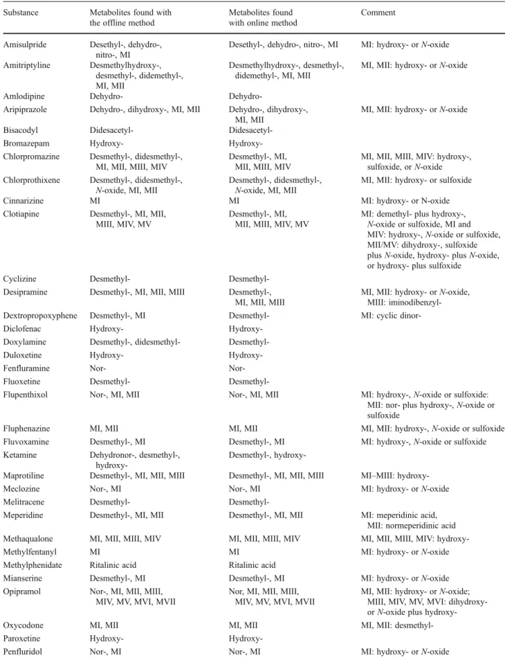
The phase I metabolites of the 53 test compounds identified
with the offline and the online methods are shown in Table1.
A chromatogram for pipamperone and its metabolites as well as the corresponding mass spectra of the metabolites
can be seen in Fig.2.
Using the offline method, a total of 156 metabolites were identified. Using the online method, a total of 151 metabolites were identified, among which two metabolites were identified that were not identified by the offline method, and seven metabolites identified by the offline method were not identi-fied using the online method. Therefore, a total of 158 metab-olites were identified using both methods, among which 156 (98.7%) were identified by the offline method and 151 (95.6%) by the online method. The seven metabolites that were not identified using the online method were dehydronor-ketamine, didesmethyltramadol, didesmethyldoxylamine, didesmethylchlorpromazine, cyclic dinordextropropoxy-phene, and two oxidation products of thioridazine. The two metabolites identified solely by the online method were dehy-drosertindole and an oxidation product of thioproperazine.
Not all of the metabolites could be identified unambigu-ously using only LC-MS. For example, the differentiation of N-oxide- and hydroxy metabolites was not always possible
Dimethylcathinone
Naphyrone
R1
R2
R3
Benzedrone
H
CH
3Flephedrone
H
H
F
Methedrone
H
H
OCH
3Methylethcathinone
CH
3H
CH
3R1
R2
3,4-Methylenedioxy-N-benzylcathinone
H
Butylone
CH
3CH
3Ethylone
CH
2CH
3H
Methylone
CH
3H
Pentylone
CH
3CH
2CH
2CH
3Table 1 Comparison of the offline and online methods. MI, MII, etc. refer to metabolite I, metabolite II, etc. Different chemical structures are possible for those metabolites based on the acquired MS spectra, as listed in the final column
Substance Metabolites found with the offline method
Metabolites found with online method
Comment
Amisulpride Desethyl-, dehydro-, nitro-, MI
Desethyl-, dehydro-, nitro-, MI MI: hydroxy- or N-oxide Amitriptyline Desmethylhydroxy-,
desmethyl-, didemethyl-, MI, MII
Desmethylhydroxy-, desmethyl-, didemethyl-, MI, MII
MI, MII: hydroxy- or N-oxide
Amlodipine Dehydro-
Dehydro-Aripiprazole Dehydro-, dihydroxy-, MI, MII Dehydro-, dihydroxy-, MI, MII
MI, MII: hydroxy- or N-oxide Bisacodyl Didesacetyl-
Didesacetyl-Bromazepam Hydroxy- Hydroxy-Chlorpromazine Desmethyl-, didesmethyl-,
MI, MII, MIII, MIV
Desmethyl-, MI, MII, MIII, MIV
MI, MII, MIII, MIV: hydroxy-, sulfoxide, or N-oxide Chlorprothixene Desmethyl-, didesmethyl-,
N-oxide, MI, MII
Desmethyl-, didesmethyl-, N-oxide, MI, MII
MI, MII: hydroxy- or sulfoxide Cinnarizine MI MI MI: hydroxy- or N-oxide Clotiapine Desmethyl-, MI, MII,
MIII, MIV, MV
Desmethyl-, MI, MII, MIII, MIV, MV
MI: demethyl- plus hydroxy-, N-oxide or sulfoxide, MI and MIV: hydroxy-, N-oxide or sulfoxide, MII/MV: dihydroxy-, sulfoxide plus N-oxide, hydroxy- plus N-oxide, or hydroxy- plus sulfoxide
Cyclizine Desmethyl- Desmethyl-Desipramine Desmethyl-, MI, MII, MIII Desmethyl-,
MI, MII, MIII
MI, MII: hydroxy- or N-oxide, MIII: iminodibenzyl-Dextropropoxyphene Desmethyl-, MI Desmethyl- MI: cyclic dinor-Diclofenac Hydroxy-
Hydroxy-Doxylamine Desmethyl-, didesmethyl- Desmethyl-Duloxetine Hydroxy- Hydroxy-Fenfluramine Nor- Nor-Fluoxetine Desmethyl-
Desmethyl-Flupenthixol Nor-, MI, MII Nor-, MI, MII MI: hydroxy-, N-oxide or sulfoxide: MII: nor- plus hydroxy-, N-oxide or sulfoxide
Fluphenazine MI, MII MI, MII MI, MII: hydroxy-, N-oxide or sulfoxide Fluvoxamine Desmethyl-, MI Desmethyl-, MI MI: hydroxy-, N-oxide or sulfoxide Ketamine Dehydronor-, desmethyl-,
Desmethyl-,
hydroxy-Maprotiline Desmethyl-, MI, MII, MIII Desmethyl-, MI, MII, MIII MI–MIII: hydroxy-Meclozine Nor-, MI Nor-, MI MI: hydroxy- or N-oxide Melitracene Desmethyl-
Desmethyl-Meperidine Desmethyl-, MI, MII Desmethyl-, MI, MII MI: meperidinic acid, MII: normeperidinic acid Methaqualone MI, MII, MIII, MIV MI, MII, MIII, MIV MI, MII, MIII, MIV: hydroxy-Methylfentanyl MI MI MI: hydroxy- or N-oxide Methylphenidate Ritalinic acid Ritalinic acid
Mianserine Desmethyl-, MI Desmethyl-, MI MI: hydroxy- or N-oxide Opipramol Nor-, MI, MII, MIII,
MIV, MV, MVI, MVII
Nor, MI, MII, MIII, MIV, MV, MVI, MVII
MI, MII: hydroxy- or N-oxide; MIII, MIV, MV, MVI: dior N-oxide plus hydroxy-Oxycodone MI, MII MI, MII MI, MII: desmethyl-Paroxetine Hydroxy-
using the acquired MS spectra only. In Table 1, those metabolites are denoted using the naming scheme of M plus a Roman numeral, and the different chemical structures are mentioned in the final column. To elucidate the exact struc-ture of each metabolite, complementary techniques need to be applied.
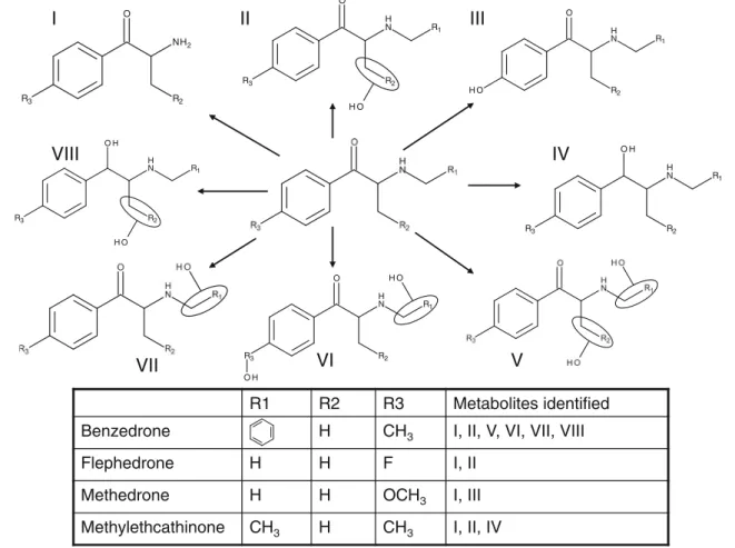
HLM phase I metabolism of 11 cathinones
The proposed structures of the identified phase I metabolites
are shown in Figs. 3, 4, and 5. The general metabolism
pathways seen in several substances include desalkylation
of the amine group (cf. Fig.1). This metabolite was seen in
all cathinones tested apart from naphyrone, which contains a tertiary amine included in a 5-ring instead of the secondary amine present in all of the other tested substances. Other metabolites observed for more than one substance include those resulting from the reduction of the keto group and hydroxylation products. In cathinones containing a methyl-enedioxy moiety, this moiety can be desmethylenated to the respective diol, a metabolite which exhibited quite a high intensity in our experiments. Combinations of the men-tioned phase I metabolites were also observed for some substances, indicating subsequent metabolism steps.
Table 1 (continued)
Substance Metabolites found with the offline method
Metabolites found with online method
Comment
Pentazocine MI, MII, MIII MI, MII, MIII MI, MII, MIII: hydroxy-Perphenazine Nor-, MI, MII, MIII Nor-, MI, MII, MIII MI, MII: hydroxy-, N-oxide or
sulfoxide; MIII: nor- plus hydroxy-, N-oxide or sulfoxide
Phencyclidine MI, MII MI, MII MI, MII: hydroxy- or N-oxide Pipamperone Dehydro-, dihydro-, MI Dehydro-, dihydro-, MI MI: hydroxy- or N-oxide
Promazine Desmethyl-, MI, MII Desmethyl-, MI, MII MI, MII: hydroxy-, N-oxide or sulfoxide Propafenone Hydroxy-, Nor- Hydroxy-,
Nor-Psilocine HIAA HIAA HIAA: hydroxyindoleacetic acid Reboxetine Desethyl-, hydroxy-, MI Desethyl-, hydroxy-, MI MI: oxidation of morpholino ring Remifentanil MI, MII MI, MII MI: GI-94219; MII: GI-90291 Sertindole Hydroxy- Dehydro-,
hydroxy-Sildenafil Desmethyl-, MI, MII, MIII
Desmethyl-, MI, MII, MIII
MI: hydroxy- or N-oxide, MII: opening of piperazine ring, MIII: cleavage of piperazine moiety Strychnine MI, MII, MIII MI, MII, MIII MI, MII: hydroxy- or N-oxide, MIII:
dihydroxy- or hydroxy- plus N-oxide Thiethylperazine Desmethyl-, MI, MII,
MIII, MIV, MV, MVI, MVII, MVIII
Desmethyl-, MI, MII, MIII, MIV, MV, MVI, MVII, MVIII
MI, MII: hydroxy-, N-oxide or sulfoxide; MIII, MIV, MV: dihydroxy- or combinations of N-oxide, sulfoxide and hydroxy-; MVI: opening of piperazine ring; MVII, MVIII: desmethyl-plus hydroxy-, N-oxide or sulfoxide Thioproperazine Desmethyl-I, desmethyl-II,
didesmethyl-I, didesmethyl-II, MI, MII, MIII, MIV, MV
Desmethyl-I, desmethyl-II, didesmethyl-I, didesmethyl-II, MI, MII, MIII, MIV, MV
MI, MII: hydroxy- or N-oxide or sulphoxide; MIII: didesmethyl- plus hydroxy-, N-oxide or sulfoxide; MIV, MV: desmethyl- plus hydroxy-, N-oxide or sulfoxide
Thioridazine MI, MII, MIII, MIV, MV, MVI, MVII, MVIII, MIX
MI, MII, MIII, MIV, MV, MVI; MIX
MI–MIII: hydroxy-, N-oxide or sulfoxide, MIV–MVI: dihydroxy- or combinations of hydroxy-, N-oxide or sulfoxide, MVII, MVIII: trihydroxy- or combinations of hydroxy-, N-oxide or sulfoxide Tilidine Desmethyl-, didesmethyl Desmethyl-, didesmethyl
Tramadol Desmethyl-, desmethyl-II,
didesmethyl-Desmethyl-, desmethyl-II Zaleplone Desethyl-, oxo-, oxodesethyl- Desethyl-, oxo-,
oxodesethyl-Zolpidem Hydroxy-I, hydroxy-II, MI Hydroxy-I, hydroxy-II, MI MI: oxidation of methyl group to carboxylic acid
Discussion
The majority of the metabolites identified using the offline method (95.6% of them) could be identified using the online method. A possible issue for the other seven metabolites that could not be identified using the online method may be the difference in incubation times. In the offline method, sub-stances were incubated for 3 h, but this was reduced to 50 min in the online method to speed up the process.
As sertindole is light sensitive, the lack of identification of dehydrosertindole by the offline method could be due to the instability of the parent compound. In the opaque loop used for online incubation, the incubation mixture was
always protected from the light, which was not the case in the Eppendorf vials that were used for the offline method.
The online method minimizes the manual preparation steps required compared to the offline method. This helped to speed up the whole process and to minimize errors. The autosampler modifications required were minimal and could be done in a few minutes. The system can therefore be used sequentially for clinical toxicological analysis and for me-tabolism experiments.
In 2005, a method using similar instrumentation was
published. Lai et al. [9] used an automated online extraction
system to study pro-drug stability. The autosampler was mod-ified and included a heated agitator for sample incubation.
2 4 6 8 10 12 14 16 18 20 22 24 Time (min) 0 20 40 60 80 1000 20 40 60 80 1000 20 40 60 80 100 R el ative A bu nda nc e 0 20 40 60 80 100 RT: 9.5 min m/z376 RT: 11.4 min m/z374 RT: 8.7 min m/z378 RT: 9.1 min m/z392 5 10 15 20 25 Time Relative Abundance 50 100 50 100 50 100 50 100
Pipamperone
Dehydro-M
Dihydro-M
MI
a)
100 120 140 160 180 200 220 240 260 280 300 320 340 360 380 m/z 0 5 10 15 20 25 30 35 40 45 50 55 60 65 70 75 80 85 90 95 100 R ela tive A bund ance 291.31 331.52 165.18 262.26 165 262 291 331 25 50 75 100 Relative Abundance 100 200 300 400 m/zb)
100 120 140 160 180 200 220 240 260 280 300 320 340 360 380 400 m /z 0 5 10 15 20 25 30 35 40 45 50 55 60 65 70 75 80 85 90 95 100 Re la tiv e A bu nda nce 291.31 347.40 165.27 262.29 25 50 75 100 Relative Abundance 100 200 300 400 m/z 165 262 291 347e)
100 120 140 160 180 200 220 240 260 280 300 320 340 360 380 m /z 0 5 10 15 20 25 30 35 40 45 50 55 60 65 70 75 80 85 90 95 100 Re la tiv e A bu nda nce 289.32 165.17 291.28 210.32 123.10 25 50 75 100 Relative Abundance 100 200 300 400 m/zc)
123 165 210 289 291 200 300 400 m/z 100 0 5 10 15 20 25 30 35 40 45 50 55 60 65 70 75 80 85 90 95 100 Re la ti ve A bu nda nce 360.47 25 50 75 100 Relative Abundance 165 263 275 293 360d)
333Fig. 2a–e Chromatogram and mass spectra for pipamperone and its metabolites. a Example of a chromatogram of pipamperone and its metabolites. Extracted ion chromatograms obtained in positive mode are shown. The corresponding retention times (RTs) as well as the mass-to-charge ratios (m/z) are indicat-ed. b Mass spectrum of pipam-perone. c Mass spectrum of the dehydro metabolite of pipamperone. d Mass spectrum of the dihydro metabolite of pipamperone. e Mass spectrum of metabolite I (MI) of pipam-perone. As indicated in Table1, the hydroxy metabolite or the N-oxide metabolite cannot be distinguished
Incubation vials containing the biological matrix (e.g., S9 fraction) diluted with buffer were stored in the cooled auto-sampler stack. The autoauto-sampler was programmed to add the cofactors (e.g., the NADPH-generating system) to this vial and subsequently transport the whole vial to the heated agita-tor. After a pre-incubation, the test substance was added. At programmed time points, the autosampler withdrew an aliquot for analysis, which was subsequently extracted online using turbulent flow chromatography. Compared with our method, the autosampler needed more modifications (e.g., the heated agitator). In our approach, only the loop needed to be changed in daily operation. Also, fewer manual steps were needed, since the autosampler adds not only cofactors and the test substance to the incubation mixture but also the HLM sus-pension. However, our method was slower and did not allow sampling at different time points, since the aim of the method was a different one. We were not interested in kinetic experi-ments, only the end points.
Compared to the method of Nicoli et al. [11] and Curcio
et al. [12], where a capillary was used as reaction vessel for
the HLM experiments, our method coupled the use of a modified injection loop online to an online extraction system. No manual sample pretreatment was necessary.
Compared to fully integrated and automated solutions, as
presented by Drexler et al. [6], Xu et al. [7], and Jenkins et
al. [8] for instance, our method had a much lower throughput.
The aim of this work was not a fully integrated high-throughput method but a method that is as automated and makes it as easy as possible to generate metabolites that can be added to the library of our toxicological screening method, and to study the metabolism of new designer drugs.
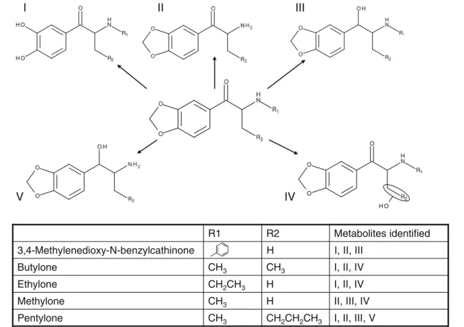
To our knowledge, among the 11 cathinones studied, only butylone, ethylone, and methylone have had their
metabolisms published already [4,17,18], and these have
always been based on the use of HLMs. For butylone and
ethylone, Zaitsu et al. [4] identified the normetabolite,
pro-duced by the reduction of the keto group to the corresponding alcohol and demethylenation with subsequent methylation by
the catechol-O-methyltransferase. Meyer et al. [18] were also
able to detect those metabolites for butylone using their stan-dard toxicological GC-MS screening procedure. In our experi-ments, we also detected the normetabolites for butylone and ethylone. Since we did not add the co-substrate needed for the methylation (S-adenosylmethionine; SAM) to the incubation mixture, we could not detect the methylated derivatives of both cathinones, only the demethylenyl-diol-metabolites.
R1 R2 R3 Metabolites identified
Benzedrone H CH3 I, II, V, VI, VII, VIII
Flephedrone H H F I, II
Methedrone H H OCH3 I, III
Methylethcathinone CH3 H CH3 I, II, IV O H N H O R2 R3 R1
I
I
I
I
I
I
VI
V
IV
R3 R2 O NH2 R2 R1 O H N H O R2 R3 R1 N H O H O H N H O HO R3 R1 R2 N H O H O H R3 R1 R2VII
VIII
Interestingly, we did not observe the products of keto-group reduction, but we were able to identify an additional hydroxy metabolite for both butylone and ethylone.
The metabolism of methylone was studied by Kamata
et al. [17]. They identified the normetabolite and the
demethylenation products, which were methylated by
the catechol-O-methyltransferase. Meyer et al. [18]
identi-fied an additional metabolite, the demethylenated normetabo-lite, which was again methylated by the catechol-O-methyltransferase. We also detected the normetabolite, but
I
I
I
I
I
I
V
I
V
R1 R2 Metabolites identified3,4-Methylenedioxy-N-benzylcathinone H I, II, III
H C e n o l y t u B 3 CH3 I, II, IV H C e n o l y h t E 2CH3 H I, II, IV H C e n o l y h t e M 3 H II, III, IV H C e n o l y t n e P 3 CH2CH2CH3 I, II, III, V R1 R2 N H O O H O H O H N H O O O R2 R1
Fig. 4 Structures of the proposed metabolites for 3,4-methylenedioxy-N-benzylcathinone, butylone, ethylone, methylone, and pentylone
a)
N O
b)
no demethylenation products. Instead, we detected a metabo-lite due to the reduction of the keto group plus a hydroxy metabolite.
No information on the metabolism of the other cathinones could be found in the literature. However, the metabolic path-ways described for butylone, ethylone, and methylone were also observed in most other cathinones.
Conclusions
The method presented in this paper provides an easy way to identify metabolites (which can then be added to the library of our clinical toxicological screening method) at an ade-quate throughput. The performance of the method compared well with that of an established offline HLM procedure, but it requires less manual work.
Acknowledgements The authors would like to thank Thermo Fisher Scientific for providing the LC-MS system and the online extraction instrument.
References
1. Asha S, Vidyavathi M (2010) Role of human liver microsomes in in vitro metabolism of drugs—a review. Appl Biochem Biotechnol 160:1699–1722
2. Wintermeyer A, Moller I, Thevis M, Jubner M, Beike J, Rothschild MA, Bender K (2010) In vitro phase I metabolism of the synthetic cannabimimetic JWH-018. Anal Bioanal Chem 398:2141–2153 3. Meyer MR, Du P, Schuster F, Maurer HH (2010) Studies on the
metabolism of the alpha-pyrrolidinophenone designer drug methylenedioxy-pyrovalerone (MDPV) in rat and human urine and human liver microsomes using GC-MS and LC-high-resolution MS and its detectability in urine by GC-MS. J Mass Spectrom 45:1426– 1442
4. Zaitsu K, Katagi M, Kamata HT, Kamata T, Shima N, Miki A, Tsuchihashi H, Mori Y (2009) Determination of the metabolites of the new designer drugs bk-MBDB and bk-MDEA in human urine. Forensic Sci Int 188:131–139
5. Meyer MR, Maurer HH (2011) Current status of hyphenated mass spectrometry in studies of the metabolism of drugs of abuse, including doping agents. Anal Bioanal Chem
6. Drexler DM, Belcastro JV, Dickinson KE, Edinger KJ, Hnatyshyn SY, Josephs JL, Langish RA, McNaney CA, Santone KS, Shipkova PA, Tymiak AA, Zvyaga TA, Sanders M (2007) An automated high throughput liquid chromatography–mass spectrometry process to assess the metabolic stability of drug candidates. Assay Drug Dev Technol 5:247–264
7. Xu R, Manuel M, Cramlett J, Kassel DB (2010) A high throughput metabolic stability screening workflow with automated assessment of data quality in pharmaceutical industry. J Chromatogr A 1217:1616–1625
8. Jenkins KM, Angeles R, Quintos MT, Xu R, Kassel DB, Rourick RA (2004) Automated high throughput ADME assays for meta-bolic stability and cytochrome P450 inhibition profiling of combi-natorial libraries. J Pharm Biomed Anal 34:989–1004
9. Lai F, Khojasteh-Bakht SC (2005) Automated online liquid chro-matographic/mass spectrometric metabolic study for prodrug sta-bility. J Chromatogr B 814:225–232
10. Kool J, van Liempd SM, van Rossum H, van Elswijk DA, Irth H, Commandeur JN, Vermeulen NP (2007) Development of three parallel cytochrome P450 enzyme affinity detection systems coupled on-line to gradient high-performance liquid chromatography. Drug Metab Dispos 35:640–648
11. Nicoli R, Curcio R, Rudaz S, Veuthey JL (2009) Development of an in-capillary approach to nanoscale automated in vitro cytochromes p450 assays. J Med Chem 52:2192–2195
12. Curcio R, Nicoli R, Rudaz S, Veuthey JL (2010) Evaluation of an in-capillary approach for performing quantitative cytochrome P450 activity studies. Anal Bioanal Chem 398:2163–2171 13. Mueller DM, Duretz B, Espourteille FA, Rentsch KM (2011)
Development of a fully automated toxicological LC-MS(n) screening system in urine using online extraction with turbulent flow chroma-tography. Anal Bioanal Chem 400:89–100
14. Cozzi NV, Sievert MK, Shulgin AT, Jacob P 3rd, Ruoho AE (1999) Inhibition of plasma membrane monoamine transporters by beta-ketoamphetamines. Eur J Pharmacol 381:63–69
15. Mueller DM, Rentsch KM (2011) Online extraction toxicological MS(n) screening system for serum and heparinized plasma and comparison of screening results between plasma and urine in the context of clinical data. J Chromatogr B
16. Sohl CD, Cheng Q, Guengerich FP (2009) Chromatographic assays of drug oxidation by human cytochrome P450 3A4. Nat Protoc 4:1252–1257
17. Kamata HT, Shima N, Zaitsu K, Kamata T, Miki A, Nishikawa M, Katagi M, Tsuchihashi H (2006) Metabolism of the recently encoun-tered designer drug, methylone, in humans and rats. Xenobiotica 36:709–723
18. Meyer MR, Wilhelm J, Peters FT, Maurer HH (2010) Beta-keto amphetamines: studies on the metabolism of the designer drug mephedrone and toxicological detection of mephedrone, butylone, and methylone in urine using gas chromatography–mass spectrome-try. Anal Bioanal Chem 397:1225–1233