HAL Id: hal-02346635
https://hal.archives-ouvertes.fr/hal-02346635
Submitted on 8 May 2020
HAL is a multi-disciplinary open access archive for the deposit and dissemination of sci-entific research documents, whether they are pub-lished or not. The documents may come from teaching and research institutions in France or abroad, or from public or private research centers.
L’archive ouverte pluridisciplinaire HAL, est destinée au dépôt et à la diffusion de documents scientifiques de niveau recherche, publiés ou non, émanant des établissements d’enseignement et de recherche français ou étrangers, des laboratoires publics ou privés.
Characterization of Kcnk3 -Mutated Rat, a Novel Model
of Pulmonary Hypertension
Mélanie Lambert, Véronique Capuano, Angele Boet, Laurent Tesson, Thomas
Bertero, Morad Nakhleh, Séverine Remy, Ignacio Anegon, Christine Péchoux,
Aurélie Hautefort, et al.
To cite this version:
Mélanie Lambert, Véronique Capuano, Angele Boet, Laurent Tesson, Thomas Bertero, et al.. Charac-terization of Kcnk3 -Mutated Rat, a Novel Model of Pulmonary Hypertension. Circulation Research, American Heart Association, 2019, 125 (7), pp.678-695. �10.1161/CIRCRESAHA.119.314793�. �hal-02346635�
Circulation Research is available at www.ahajournals.org/journal/res
Correspondence to: Fabrice Antigny, INSERM UMR_S 999, Hôpital Marie Lannelongue, 133, Ave de la Résistance, F-92350 Le Plessis Robinson, France. Email fabrice.antigny@u-psud.fr
The online-only Data Supplement is available with this article at https://www.ahajournals.org/doi/suppl/10.1161/CIRCRESAHA.119.314793. For Sources of Funding and Disclosures, see page 694.
© 2019 American Heart Association, Inc.
ORIGINAL RESEARCH
Characterization of Kcnk3-Mutated Rat, a Novel
Model of Pulmonary Hypertension
Mélanie Lambert, Véronique Capuano, Angèle Boet, Laurent Tesson, Thomas Bertero, Morad K. Nakhleh, Séverine Remy, Ignacio Anegon, Christine Pechoux, Aurélie Hautefort, Catherine Rucker-Martin, Boris Manoury, Valérie Domergue, Olaf Mercier, Barbara Girerd, David Montani, Frédéric Perros, Marc Humbert, Fabrice Antigny
RATIONALE: Pulmonary arterial hypertension is a severe lethal cardiopulmonary disease. Loss of function mutations in KCNK3 (potassium channel subfamily K member 3) gene, which encodes an outward rectifier K+ channel, have been identified in
pulmonary arterial hypertension patients.
OBJECTIVE: We have demonstrated that KCNK3 dysfunction is common to heritable and nonheritable pulmonary arterial hypertension and to experimental pulmonary hypertension (PH). Finally, KCNK3 is not functional in mouse pulmonary vasculature.
METHODS AND RESULTS: Using CRISPR/Cas9 technology, we generated a 94 bp out of frame deletion in exon 1 of Kcnk3 gene and characterized these rats at the electrophysiological, echocardiographic, hemodynamic, morphological, cellular, and molecular levels to decipher the cellular mechanisms associated with loss of KCNK3. Using patch-clamp technique, we validated our transgenic strategy by demonstrating the absence of KCNK3 current in freshly isolated pulmonary arterial smooth muscle cells from Kcnk3-mutated rats. At 4 months of age, echocardiographic parameters revealed shortening of the pulmonary artery acceleration time associated with elevation of the right ventricular systolic pressure. Kcnk3-mutated rats developed more severe PH than wild-type rats after monocrotaline exposure or chronic hypoxia exposure. Kcnk3-mutation induced a lung distal neomuscularization and perivascular extracellular matrix activation. Lungs of Kcnk3-mutated rats were characterized by overactivation of ERK1/2 (extracellular signal–regulated kinase1-/2), AKT (protein kinase B), SRC, and overexpression of HIF1-α (hypoxia-inducible factor-1 α), survivin, and VWF (Von Willebrand factor). Linked with plasma membrane depolarization, reduced endothelial-NOS expression and desensitization of endothelial-derived hyperpolarizing factor, Kcnk3-mutated rats presented predisposition to vasoconstriction of pulmonary arteries and a severe loss of sildenafil-induced pulmonary arteries relaxation. Moreover, we showed strong alteration of right ventricular cardiomyocyte excitability. Finally, Kcnk3-mutated rats developed age-dependent PH associated with low serum-albumin concentration.
CONCLUSIONS: We established the first Kcnk3-mutated rat model of PH. Our results confirm that KCNK3 loss of function is a key event in pulmonary arterial hypertension pathogenesis. This model presents new opportunities for understanding the initiating mechanisms of PH and testing biologically relevant therapeutic molecules in the context of PH.
VISUAL OVERVIEW: An online visual overview is available for this article.
Key Words: echocardiography ◼ exon ◼ extracellular matrix ◼ hypoxia ◼ rats
Editorial, see p 696 | In This Issue, see p 659 | Meet the First Author, see p 660
P
ulmonary arterial hypertension (PAH) is an uncom-mon, progressive, and severe disease with an esti-mated prevalence of 15 to 50 per million.1 PAHhas been hemodynamically defined by an elevation of the mean pulmonary artery pressure >20 mm Hg and
pulmonary vascular resistance >3 Wood units at rest.2
PAH results from increased pulmonary vascular resis-tance because of remodeling of the small distal pulmo-nary arteries (PAs) and arterioles (diameter <500 µm), causing adaptive right ventricular (RV) hypertrophy and
ORIG
IN
AL R
ESE
ARCH
Lambert et al Characterization of Kcnk3-Mutated Rats
when unchecked, right heart failure.3 In 2013,
whole-exome sequencing revealed 6 types of heterozygous mutations in KCNK3 (potassium channel subfamily K member 3), representing the first channelopathy identi-fied in PAH.4 In 2016, Navas et al5 identified 2 additional
KCNK3 mutations in PAH patients and described the
first PAH patient with a homozygous KCNK3 mutation associated with an aggressive form of PAH and early disease development.4 Finally, in 2017, 2 new mutations
were identified in an Asian and an American cohort of PAH patients.6,7 KCNK3 mutations carriers’ patients are
younger at diagnosis than idiopathic PAH patients and have a higher mean pulmonary artery pressure.8
KCNK3 encodes an outward-K+ channel member of the
2 pore K+ channel family, characterized by 4
transmem-brane domains and 2 pore domains per subunit.4 KCNK3
channel, also known as TASK-1 (Twik-related-acid-sensitive-K+ channel) or K2P3.19 participates in
regu-lating resting membrane potential in several cell types including human pulmonary arterial smooth muscle cells
Nonstandard Abbreviations and Acronyms
α-SMA α-smooth muscle actinCH chronic hypoxia
ECM extracellular matrix
EDHF endothelium-derived hyperpolarizing factor
HIF1-α hypoxia-inducible factor-1 α
MCT monocrotaline NOS nitric oxide synthase
PA pulmonary artery
PAH pulmonary arterial hypertension
PASMC pulmonary arterial smooth muscle cell
PH pulmonary hypertension
RVSP right ventricular systolic pressure
TPR total pulmonary resistance
VWF Von Willebrand factor
WT wild type
Novelty and Significance
What Is Known?
• Loss of function mutations in the KCNK3 (potassium channel subfamily K member 3) gene have been described in pulmonary arterial hypertension (PAH) patients and those who are carriers of KCNK3 muta-tions are younger as compared with idiopathic PAH patients.
• Reduced KCNK3 expression/function is common to heritable and nonheritable PAH and to experimental pulmonary hypertension.
• KCNK3 is not functional in mouse pulmonary arte-rial smooth muscle cell limiting previous mechanistic exploration.
What New Information Does This Article
Contribute?
• By CRISPR/Cas9 technology, we generate Kcnk3-mutated rats to inactivate the KCNK3 channel. • At 4 months old, Kcnk3-mutated rats develop
sponta-neous elevation of right ventricular systolic pressure, are sensitized to induced pulmonary hypertension (monocrotaline or chronic hypoxia), and develop age-dependent pulmonary hypertension at 12 months. • Kcnk3 dysfunction leads to distal pulmonary vessels
neomuscularization and perivascular collagen deposi-tion associated with overactivadeposi-tion of proproliferative pathways (ERK1/2 [extracellular signal–regulated kinase1-/2] and HIF-1α [hypoxia-inducible factor-1 α]) and endothelial phenotype switch.
• We identify that an increase of HIF1-α and ERK1/2 are the primary molecular events linked to
Kcnk3-dysfunction.
• Pulmonary arteries from Kcnk3-mutated rats are sen-sitized to constriction and are less responsive to vaso-relaxing stimuli.
Pulmonary arterial hypertension (PAH) is a progressive and severe disease. Since 2013, several loss of function mutations in KCNK3 gene have been detected in select PAH patients. KCNK3 encodes an outward-K+ channel
member of the 2-pore K+ channel family. Patients with the
KCNK3 mutation are younger at diagnosis than idiopathic
PAH patients. Loss of function and expression is a hallmark of PAH at pulmonary vascular and right ventricular levels. However, the mechanisms linking KCNK3 dysfunction with PAH were unknown. Using unique Kcnk3-mutated rats and human pulmonary arterial smooth muscle cells we aimed to decipher the cellular mechanisms associ-ated with KCNK3-dysfunction. Our results demonstrate that KCNK3 loss of function is a key event in PAH patho-genesis. Using a combination of hemodynamic mea-surements, molecular biology, histological analyses, and electrophysiology approaches, we show that KCNK3-dysfunction leads to plasma membrane depolarization, pulmonary arterial smooth muscle cells overproliferation, ERK1/2 and HIF1-α over-activation, mitochondrial depo-larization, pulmonary artery vasoconstriction, pulmonary vessels remodeling, endothelial cell phenotype switch, and reduction of RV cardiomyocytes excitability. These results highlight that KCNK3 loss of function is consis-tent with human PAH pathobiology and demonstrate that KCNK3-dysfunction is a key events in PAH. This model presents new opportunities for understanding the initiat-ing mechanisms of pulmonary hypertension.
ORIG IN AL R ESE ARCH
(PASMCs).8 Electrophysiological recording demonstrated
a loss of function for all KCNK3 mutations identified.4 We
recently determined that KCNK3 dysfunction contributes to the development of heritable PAH (BMPR2 mutated patients) and idiopathic PAH, indicating that KCNK3 dys-function is a hallmark of PAH at pulmonary vascular and RV levels.10,11 However, the mechanisms linking KCNK3
dysfunction with PAH are still unknown. Contrary to rats where KCNK3 is functionally expressed in pulmonary vasculature,10 mice are not a suitable models to study
the role of KCNK3 in pulmonary hypertension (PH) as KCNK3 does not form a functional channel in mouse PASMCs12 and is replaced by KCNK6 channel.13
There-fore, we developed the first Kcnk3-mutated rat line to decipher the role of KCNK3 dysfunction in the context of PAH pathobiology, representing an interesting model to study the time-dependent cellular and molecular mecha-nisms involved in PH development.
Using a combination of hemodynamic measurements, molecular biology, histological analyses, and electrophys-iology approaches, we characterized the first Kcnk3-mutated rat model, providing tools for developing specific therapeutic targets for PH.
METHODS
The authors declare that all supporting data are available within the article and its Online Data Supplement.
Because of space limitations, a detailed description of the Materials and Methods is presented in the Online Data Supplement.
RESULTS
KCNK3 Current Is Absent in Freshly isolated
PASMCs From Kcnk3-Mutated Rats
Kcnk3-mutated Sprague-Dawley rats were generated
using CRISPR-Cas9 system with a specific sgRNA-rKCNK3 and Cas9 mRNA,14 targeting the first exon of
Kcnk3 gene. In one of the newborn rats, a deletion of 94
bp (Δ94ex1) was found, which originated an out of frame shift in the open reading frame that leads to a premature stop codon and the generation of a completely different aa (amino acid) sequence (Figure 1A). Although pre-mature stop codon may generate a degradation of the mRNA,15 we showed the presence of a mutated mRNA
deleted of 94 bp but not its absence (Figure 1B) sug-gesting an absence of nonsense mRNA degradation pathway as already mentioned elsewhere.15
Neverthe-less, a putative translation of the truncated mRNA could produce a truncated 90-aa protein instead of the 411 aa of the wild-type (WT) protein (Figure 1A) and share only the first 14 aa in common with the WT protein. The sequencing of the Kcnk3 mRNA from Kcnk3+/+ and
Kcnk3Δ94ex1/Δ94ex1 rats confirmed the deletion of the 94
bp and an aberrant potential protein sequence with 8 potential premature stop codons (Online Figure I).
The founder animal with the Δ94ex1 mutation was crossed with a WT partner, and the mutation was transmit-ted to the offspring as shown by genotype DNA analysis, demonstrating that the rats were either Kcnk3+/+,
het-erozygous Kcnk3Δ94ex1/+, or homozygous Kcnk3Δ94ex1/Δ94ex1
(Figure 1C). Unfortunately, there is no available specific antibodies for KCNK3,16,17 thus avoiding any measurement
of the protein expression of KCNK3 in Kcnk3-mutated rats. We showed that all commercially anti-KCNK3 antibodies tested were unusable because when tested in Kcnk3-knockout mice, they were either nonreactive or gave the same signal as in the WT mice (Online Figure II and IIbis). The same unspecific staining occurred in Kcnk3-mutated rats. Thus, we could not assert the absence of KCNK3 protein in Kcnk3-mutated rats. Nevertheless, whole cell patch-clamp measurement on freshly isolated PASMCs from WT and Kcnk3-mutated rats clearly showed a defect in KCNK3 current in mutated rats. Indeed, the global-K+
current was significantly reduced: 50% in Kcnk3Δ94ex1/+
rats and 60% in Kcnk3Δ94ex1/Δ94ex1rats compared with the
WT (Figure 1D and 1E). We then specifically measured the KCNK3 current (IKCNK3) after applying a specific KCNK3 channel inhibitor (A293 at 200 nmol/L)10 and
defined it as the A293-sensitive K+-current. I
KCNK3 was
strongly decreased (60%) in freshly isolated PASMCs from Kcnk3Δ94ex1/+ rats and was abolished (95%) in
Kcnk3Δ94ex1/Δ94ex1 rats (Figure 1F and 1G). Consistent
with the role of KCNK3 in resting membrane potential, PASMCs isolated from Kcnk3-mutated rats were signifi-cantly depolarized compared with PASMCs from WT rats (Figure 1H). We performed RT-qPCR (quantitative reverse transcription polymerase chain reaction) experiments to evaluate the consequences of Kcnk3-dysfunction on other lung K+ channels (Online Figure III). No expression
of KCNK4, KCNK9, KV1.4, or KCa4 channels was detected. The mRNA expression levels of KCNK2, KCNK6, KV1.5, KV2.1, KV9.3, KCa1.1, KCa2.1, KCa2.2, KCa2.3, and KCa4.2 was unchanged in Kcnk3-mutated rats compared with WT. However, we observed a significant increase in the mRNA level of KV1.2 and KCa3.1 in Kcnk3Δ94ex1/Δ94ex1 rat lungs. Yet,
the A293-insensitive K+-current was similar in PASMCs
isolated from WT and Kcnk3-mutated rats (not shown), suggesting that Kcnk3-dysfunction is not compensated by other K+ currents.
Kcnk3-Dysfunction Leads to an Increase in
Right Ventricular Systolic Pressure in Males and
Female
To determine whether Kcnk3-dysfunction leads to spon-taneous PH, we performed closed chest right-heart catheterization at 4 months of age. We observed a slight but significant increase in right ventricular systolic pres-sure (RVSP) in Kcnk3Δ94ex1/Δ94ex1 rats compared with WT
ORIG
IN
AL R
ESE
ARCH
Lambert et al Characterization of Kcnk3-Mutated Rats
Figure 1. KCNK3 (potassium channel subfamily K member 3) is no longer functional in pulmonary arterial smooth muscle cells (PASMCs) from Kcnk3-mutated rats.
A, Schematic overview of the Kcnk3 gene with wild-type (WT) and Kcnk3-mutant alleles. We generated Kcnk3-mutated rats with 94 bp
deletion in exon 1 of the Kcnk3 (Δ94ex1). Below, Kcnk3 DNA and Δ94 bp DNA (strikethrough letter) and protein sequences in WT and Δ94ex1 rats. Gray: aberrant/potential protein sequence. *STOP codon. B, mRNA levels of Kcnk3 analyzed by RT-PCR (reverse transcription polymerase chain reaction) in Kcnk3Δ94ex1/Δ9e4x1 rats, Kcnk3Δ94ex1/+ rats, and Kcnk3+/+ rats, using 2 oligo combinations rKcnk3-17F/rKcnk3-1146R
(left; WT: 1148 bp/Δ94ex1: 1054 bp) and rKcnk3-17F/rKcnk3-303R (right; WT: 304 bp/Δ94ex1: 210 bp). C, Validation of the deletion by PCR, showing genomic DNA amplicons from rats carrying the Kcnk3+/+allele (400 bp) and Δ94ex1deletion allele (306 bp). D,
Current-voltage relationship of global-K+-currents, in freshly isolated PASMCs from Kcnk3+/+, Kcnk3Δ94ex1/+ and Kcnk3Δ94ex1/Δ94ex1 rats, corresponding to
the depolarizing step protocol (+60 mV to −100 mV, holding potential −40 mV). E, The current was normalized by the capacitance of the cell and is represented in pA/pF (pico ampere/pico farad; WT=16 cells, 4 rats, Kcnk3Δ94Ex1/+=21 cells, 4 rats, Kcnk3Δ94Ex1/Δ94Ex1=20 cells, 4 rats).
After recording, the global-K+-current, KCNK3 current was isolated by applying a specific inhibitor (200 nmol/L, A293). I
KCNK3 (A293
sensitive-K+-current) was then measured and presented in (F, WT=12 cells, 4 rats, Kcnk3Δ94Ex1/+=10 cells, 4 rats, Kcnk3Δ94Ex1/Δ94Ex1=13 cells, 4 rats). G, Current density of IKCNK3 measured at +60 mV. H, Using a current clamp, we measured the resting membrane potential of freshly isolated PASMCs (WT=34 cells, 4 rats, Kcnk3Δ94Ex1/+=28 cells, 4 rats, Kcnk3Δ94Ex1/Δ94Ex1=35 cells, 4 rats). *P<0.05, **P<0.01, ***P<0.001 vs Kcnk3+/+.
Experiments were analyzed using 1-way ANOVA followed by Tukey post hoc test for multiple comparisons. ns indicates nonsignificant.
ORIG IN AL R ESE ARCH
in males (Figure 2A) and in females (Online Figure IVA). However, increased RVSP was not accompanied by RV hypertrophy (Fulton index) nor by a change in cardiac output or mean carotid artery pressures in males (Fig-ure 2A) and in females (Online Fig(Fig-ure IVA). Total pul-monary resistances (TPR) were significantly increased in Kcnk3Δ94ex1/Δ94ex1 rats compared with WT males
(Fig-ure 2A) and females (Online Fig(Fig-ure IVA).
Using echocardiography, we examined the conse-quences of Kcnk3-dysfunction at 4 months old (males) on cardiac function. Pulmonary artery acceleration time was significantly reduced in Kcnk3-mutated rats (Figure 2B), indicating a higher resistive PA profile. In addition, heart rate was significantly increased in Kcnk3Δ94ex1/Δ94ex1 rats.
We did not observe modification in the other parameters measured in Kcnk3-mutated rats (Online Table IV).
In mice, Kcnk3 deletion causes primary hyper-aldosteronism.18 Conversely, in 4-month-old male
Kcnk3Δ94ex1/Δ94ex1, serum aldosterone concentration
signif-icantly decreased, whereas no variations were observed in female (Online Figure V).
Kcnk3-Mutated Rats Were More Susceptible
to Monocrotaline (MCT)-or Chronic Hypoxia
(CH)–Induced PH
As shown in Figure 2A, 4-month-old Kcnk3-mutated rats displayed a modest but significant increase in RVSP. Therefore, we exposed 4-month-old male and female rats to MCT (60 mg/kg). Kcnk3-dysfunction increased the severity of MCT-induced PH as demonstrated by higher RVSP in both males and females Kcnk3-mutated group, associated with higher TPR (Figure 2C; Online Figure IVB) with no modification of mean carotid artery pressure (not shown). These results demonstrate that
Kcnk3-dysfunction sensitizes rats to develop PH under
MCT-exposure.
In addition, male rats were exposed to CH (10% O2 for 3 weeks). As shown in Figure 2D, Kcnk3-dys-function increased the severity of CH-induced PH, as demonstrated by higher RVSP and TPR values in Kcnk3-mutated rats, compared with WT rats.
Kcnk3-Dysfunction Leads to Distal Pulmonary
Vessel Neomuscularization and Increases
Collagen Crosslinking
We analyzed muscularization of small pulmonary ves-sels (<30 µm) by immunostaining against α-SMA (α-smooth muscle actin, (α-smooth-muscle marker) and VWF (Von Willebrand factor, endothelium marker; Figure 3A).
Kcnk3-mutated rats presented a significantly decreased
percentage of nonmuscularized vessels along with sig-nificantly increased numbers of muscularized vessels (Figure 3B). Moreover, the level of fibrillar collagen
assembly was abnormally increased in pulmonary ves-sels from Kcnk3-mutated rats (Figure 3C).
The increase in RVSP in Kcnk3-mutated rats could also be explained by the modification of pulmonary microvessel density. By counting the number of CD31+
(cluster of differentiation-31) pulmonary vessels, we did not detect modification of microvessel density in Kcnk3-mutated rats compared with WT (Figure 3D). However, Western blot analysis showed significant decreases of CD31 and significant increases of VWF expression in lungs from Kcnk3Δ94ex1/Δ94ex1 rats (Figure 3E). Both
mol-ecules are critical to maintaining endothelial integrity, suggesting that Kcnk3-dysfunction may result in an alteration of the endothelial functions.
Proliferation Pathways Were Activated in
Kcnk3-Mutated Rat Lungs
We analyzed the expression and activation of some proproliferative or antiapoptotic proteins known to contribute to PAH pathobiology. Compared with WT, the phosphorylation state of ERK 1/2, and SRC sig-nificantly increased in lung from Kcnk3-mutated rats, without modification of P38 (Figure 4A). We found that HIF1-α (hypoxia-inducible factor-1 α) expression was also significantly increased in lungs from Kcnk3-mutated rats (Figure 4A), without alteration of HIF1-α
mRNA expression (not shown). We also measured an increase in the survivin protein in Kcnk3Δ94ex1/Δ94ex1 rats
(Figure 4A). AKT (protein kinase B) phosphorylation at Thr308 significantly increased in Kcnk3Δ94ex1/Δ94ex1 rats
(Figure 4A). ERK1/2 and AKT are targets for serine– threonine PP2A (protein phosphatase 2A). As pre-sented in Online Figure VIA, the expression of PP2A-A, PP2A-B, and PP2A-C proteins was unchanged. How-ever, we showed that PP2A phosphatase activity was strongly reduced (50%) in PASMCs from Kcnk3Δ94ex1/ Δ94ex1 compared with PASMCs from WT rats, while PP2B
(or calcineurin) activity was unchanged (Figure 4B). We also measured several other molecules known to be involved in PAH pathogenesis. We observed an increase in the expression of the endothelial-to-mes-enchymal (endoMT) transcription factor TWIST1 (twist family bHLH transcription factor 1) and a decrease in the phosphorylation of CREB (cAMP response element–binding protein) in Kcnk3Δ94ex1/Δ94ex1rats
(Fig-ure 4C), which are altered in PAH patients.19,20 We
measured an increase in the mRNA of Nfatc1, Notch3,
Mmp2, and Stat3 in Kcnk3Δ94ex1/ Δ94ex1 rats (Online Figure
VIB). No variation for BMPRII, phospho-SMAD1/5/8, phospho-SMAD2/3, and Caveolin-1 protein expression was observed in lungs from Kcnk3-mutated rats (Online Figure VII).
The mRNA expression of IL-6, IL-6 receptor (GP130),
IL-17Ra and Timp1 was unchanged in the lung from Kcnk3-mutated rats at 4 months old (Online Figure VIIIA).
ORIG
IN
AL R
ESE
ARCH
Lambert et al Characterization of Kcnk3-Mutated Rats
Figure 2. Kcnk3 (potassium channel subfamily K member 3) dysfunction leads to a significant increase in right ventricular systolic pressure (RVSP) in males at 4 months of age.
A, Quantification of right ventricular systolic pressure (RVSP), Fulton index (weight of right ventricle [RV]/left ventricle [LV]+septum [S]), cardiac
output (CO), mean carotid pressure (mCP), and total pulmonary resistance (TPR evaluated by RVSP/CO; wild type [WT]=8 Kcnk3Δ94Ex1/+=9, Kcnk3Δ94Ex1/Δ94Ex1=9 rats). B, Representative pulmonary artery (PA) flow through the pulmonary valve in Kcnk3+/+and Kcnk3-mutated rats and
quantification of the PA acceleration time (PAAT; WT=26, Kcnk3Δ94Ex1/+=29, Kcnk3Δ94Ex1/Δ94Ex1=17rats). C, At 4-month old, male Kcnk3+/+and
Kcnk3-mutated rats were exposed to 60 mg/kg of monocrotaline (MCT) or saline (SHAM). Three weeks later, we measured RVSP, CO, and TPR (SHAM; WT=9, Kcnk3Δ94Ex1/+=12, Kcnk3Δ94Ex1/Δ94Ex1=10, MCT; WT=12, Kcnk3Δ94Ex1/+=17, Kcnk3Δ94Ex1/Δ94Ex1=7 rats). D, Four-month-old male Kcnk3+/+and Kcnk3-mutated rats were exposed 3 weeks to chronic hypoxia (CH, 10% O2) or normoxia (NX). Three weeks later, we measured RVSP, CO, and TPR (NX; WT=8, Kcnk3Δ94Ex1/+=16, Kcnk3Δ94Ex1/Δ94Ex1=11, CH; WT=18, Kcnk3Δ94Ex1/+=36, Kcnk3Δ94Ex1/Δ94Ex1=14 rats). *P<0.05, **P<0.01,
***P<0.001. Experiments were analyzed using 1-way ANOVA followed by Tukey post hoc test and experiments with 2 different categorical independent variables were analyzed with 2-way ANOVA completed by Tukey post hoc test for multiple comparisons. ns indicates nonsignificant.
ORIG IN AL R ESE ARCH
Figure 3. Kcnk3 (potassium channel subfamily K member 3)-dysfunction leads to distal pulmonary vessel neomuscularization and increased collagen crosslinking.
A, Immunolabeling and confocal imaging of α-SMA (α-smooth muscle actin) in green, VWF (Von Willebrand factor) in red, and DAPI in blue
on rat lung sections. Scale bar: 20 µm. B, Percentage of nonmuscularized vessels and muscularized vessels (100 vessels counted per rat, wild type [WT]=9, Kcnk3Δ94Ex1/+=13, Kcnk3Δ94Ex1/Δ94Ex1 =14 rats). C, Upper, Picrosirius Red staining and below quantification of total collagen
signal, collagen crosslinked, and crosslinked/collagen total ratio in small (<50 µm) and medium (50–200 µm) pulmonary artery from rats at 4 months old (WT=17, Kcnk3Δ94Ex1/+=13, Kcnk3Δ94Ex1/Δ94Ex1=13 rats). Scale bar: 100 µm. D, Pulmonary vessel density quantification based on
CD31 (endothelium marker) labeling in paraffin-embedded lung sections, expressed as the percentage of small (<20 µm) or large (>20 µm) CD31+ vessels measured in 20 fields of Kcnk3+/+and Kcnk3-mutated rats at 4 months old (WT=6, Kcnk3Δ94Ex1/+=7, Kcnk3Δ94Ex1/Δ94Ex1=4 rats). E, Representative Western blots and quantification of CD31 and VWF expression in lungs from Kcnk3+/+and Kcnk3-mutated rats (WT=8, Kcnk3Δ94Ex1/+=8, Kcnk3Δ94Ex1/Δ94Ex1=8 rats). β-actin was used as the loading control. . *P<0.05, **P<0.01, ***P<0.001 vs Kcnk3+/+. Experiments
were analyzed using 1-way ANOVA followed by Tukey post hoc test for multiple comparisons. ns indicates nonsignificant.
ORIG
IN
AL R
ESE
ARCH
Lambert et al Characterization of Kcnk3-Mutated Rats
Figure 4. In vivo and in vitro Kcnk3 (potassium channel subfamily K member 3)-dysfunction leads to activation of several proproliferative and antiapoptotic pathways.
A, Representative Western blots and quantification of p-ERK1/2, p-P38, p-SRC, Survivin, HIF1-α (hypoxia-inducible factor-1 α), and p-308
AKT (protein kinase B) expression in lungs from wild-type (WT) and Kcnk3-mutated rats (WT=7, Kcnk3Δ94Ex1/+=8, Kcnk3Δ94Ex1/Δ94Ex1=6 rats). B, Quantification of PP2A and PP2B activity in pulmonary arterial smooth muscle cells (PASMCs) from WT and Kcnk3Δ94ex1/Δ9e4x1 (WT=3, Kcnk3Δ94Ex1/Δ94Ex1=3rats). C, Representative Western blots and quantification of TWIST1 (twist family bHLH transcription factor 1) and p-CREB
expression in lungs from Kcnk3+/+and Kcnk3-mutated rats (WT=8, Kcnk3Δ94Ex1/+=8, Kcnk3Δ94Ex1/Δ94Ex1 =7 rats). D, Upper: left, KCNK3 mRNA level
in human control PASMCs transfected 72 h with a si-RNA control (si-Control) or against KCNK3 (si-KCNK3; n=8patients), normalized by 18S mRNA expression. Right, percentage of proliferation of human PASMCs (hPASMCs) transfected with si-KCNK3 compared with Si-Control (n=6patients). D, Below: representative Western blots and quantifications of HIF1α, Survivin, p-ERK 1/2, ERK 1/2, p-AKT308, p-SRC, and p-P38 in hPASMCs, 72 h after si-RNA transfection. E, Upper, KCNK3 mRNA level in human control PASMCs, 9 days after cell transfection with si-RNA (n=4 patients), normalized by 18S mRNA expression. Below, percentage of si-KCNK3 hPASMC proliferation compared with si-Control (n=4patients). F, confocal imaging of mitochondrial membrane potential (Δφm) using TMRE (Tetramethylrhodamine, ethyl ester) on hPASMCs treated with si-RNA. Right, quantification of TMRE intensity and mitochondria perimeter. ns: nonsignificant. *P<0.05, **P<0.01 vs Kcnk3+/+or
si-Control. Experiments were analyzed using 2-tailed unpaired Student test. ERK1/2 indicates extracellular signal–regulated kinase1-/2.
ORIG IN AL R ESE ARCH
Likewise, the lung expression of CD45 (pan-leukocyte marker) was also unchanged (Online Figure VIIIB).
In Vitro KCNK3 Knockdown or Pharmacological
KCNK3 Inhibition Increases Human PASMCs
Proliferation
To evaluate the role of KCNK3 in human PASMCs (hPASMCs) and human pulmonary arterial endothe-lial cells proliferation, we abolished KCNK3 function using either si-RNA-mediated or specific pharmaco-logical inhibition. Seventy two hours after transfection,
KCNK3 mRNA decreased in hPASMCs transfected with
si-KCNK3 (by 85%). KCNK3 knockdown enhanced hPASMCs proliferation by 40% as measured by BrdU incorporation (Figure 4D). Pharmacological inhibition of KCNK3 by 24-hour exposure to A293 at 1 µmol/L enhances the proliferation of control PASMCs from humans and rats (Online Figure IXA).
In human pulmonary arterial endothelial cells exposed to the KCNK3 inhibitor, we observed significantly decreased proliferation rate compared with the vehi-cle condition (Online Figure IXC). Using iCelligenceTM,
we observed that specific KCNK3 inhibition enhances human pulmonary arterial endothelial cells adhesion without alteration of endothelial barrier opening by TNFα (tumor necrosis factor α) stimulation (5 ng/mL; Online Figure IXD and IXE).
Consistent to an enhancement of PASMCs prolifera-tion by the knockdown of KCNK3, we found a significant increase in the expression of HIF1-α, an over-phos-phorylation of ERK1/2, and a nonsignificant increase of survivin, without modification of P38, SRC, and AKT phosphorylation (Figure 4D). Nine days after si-RNA transfection, KCNK3 expression returned to the nor-mal, hPASMCs proliferation was normalized (Figure 4E) as well as HIF1-α expression and ERK1/2 activation (Online Figure IXB), demonstrating that loss of KCNK3 enhances PASMCs proliferation. In addition to HIF1-α and ERK1/2 overactivation in si-KCNK3-treated hPASMCs, we found that mitochondria membrane were depolarized (TMRE fluorescence intensity, tetramethyl-rhodamine, ethyl ester) and fragmented compared with mitochondria from si-Control hPASMCs (Figure 4F), without modification of the total expression of the reg-ulating mitochondrial fission protein DRP1 (dynamin related protein-1; Online Figure XA). Likewise, we found that the mitochondria of PASMCs from Kcnk3Δ94ex1/ Δ94ex1rats were also significantly depolarized and
frag-mented compared with WT. This strongly suggests that KCNK3 is involved in mitochondria integrity in human and rat PASMC (Online Figure XB and XC). We found that plasma membrane depolarization (induced by138 mM KCl) strongly depolarized mitochondria membrane without modification of mitochondrial web (Online Fig-ure XD and XE).
Kcnk3-Dysfunction Predisposes PA to
Constriction
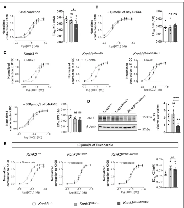
We investigated the consequences of Kcnk3-dysfunction on PA contractility. Using isolated PA from 4-month-old WT and Kcnk3-mutated rats, we first assessed the conse-quence of Kcnk3-dysfunction on basal PA tone, measured as the extracellular Ca2+-dependent tone. The basal tone
was unaltered in Kcnk3-mutated rats (Online Figure XIA). The contractile dose-response to increasing doses of KCl was unchanged in PA from Kcnk3Δ94ex1/+ compared with PA
from WT rats whereas the dose-response curve of PA from
Kcnk3Δ94ex1/Δ94ex1 rats was significantly shifted to the left as
shown by the significant decrease in EC50 (Figure 5A). When PA were pretreated with Bay K 8644, a drug that potentiates voltage-sensitivity of L-Type-Ca2+
chan-nels, the contractile response was shifted to the left com-pared with the condition without Bay K 8644 (Figure 5B). Importantly, L-Type Ca2+ channel potentiation abolished
the previously observed differences in Figure 5A, consis-tent with the PASMCs depolarization (Figure 1H).
To rule out the contribution of endothelial-dependent PA relaxation via nitric oxide synthase (NOS) activi-ties, we pretreated PA with L-NAME (L-NG-nitroarginine
methyl ester; NOS inhibitor). With L-NAME, PA contracted at a lower concentration of KCl than in the absence of L-NAME, demonstrating the contribution of NOS activi-ties and validating the efficacy of L-NAME treatment (Fig-ure 5C). Interestingly, with L-NAME, in Kcnk3Δ94ex1/Δ94ex1 rats,
the difference in the KCl contractile dose-response was lost (Figure 5C). Western blot analyses showed that endo-thelial-NOS expression was significantly reduced in lungs from Kcnk3Δ94ex1/Δ94ex1 rats (Figure 5D), whereas
inducible-NOS expression was unchanged (Online Figure XIB). Pulmonary vascular endothelium also releases EDHF (endothelium-derived hyperpolarizing factor). EDHF rep-resents a family of endothelial factors as an arachidonic acid metabolite produced by cytochrome P450-2C9 that hyperpolarize and relax smooth muscles. To investigate the contribution of EDHF in PA tone, we used flucon-azole, a cytochrome P450 epoxygenase inhibitor. With fluconazole, WT PA contracted at a lower KCl concentra-tion than in the basal condiconcentra-tion (Figure 5E, left), whereas fluconazole had no impact on PA contraction in Kcnk3-mutated rats (Figure 5E, middle and right). These data suggested that EDHF production is strongly reduced or inefficient in Kcnk3-mutated rats. All these parame-ters (PASMCs depolarization, reduced endothelial-NOS expression, reduced EDHF sensitivity) may contribute to enhanced PA vasoconstriction in Kcnk3Δ94ex1/ Δ94ex1 rats.
Kcnk3-Dysfunction in Rats Reduced PA
Relaxation
After inducing contraction with a thromboxane-A2 mimetic (U46619), relaxation was stimulated using
ORIG
IN
AL R
ESE
ARCH
Lambert et al Characterization of Kcnk3-Mutated Rats
Figure 5. Kcnk3 (potassium channel subfamily K member 3)-dysfunction in rats predisposes pulmonary arteries to constriction. A, Normalized dose-response curve (normalized to K100) established by applying an increasing concentrations of KCl on isolated pulmonary
artery (PA) from Kcnk3+/+and Kcnk3-mutated rats and quantification of EC50 (WT=6, Kcnk3Δ94Ex1/+=7, Kcnk3Δ94Ex1/Δ94Ex1=10 rats). B, In
presence of 1 µmol/L of Bay K 8644, normalized dose-response curve established by applying increasing concentrations of KCl on PA isolated from Kcnk3+/+and Kcnk3-mutated rats and EC50 quantification (WT=7, Kcnk3Δ94Ex1/+=6, Kcnk3Δ94Ex1/Δ94Ex1=5 rats). C, Comparison of PA
contraction, in response to increasing dose of KCl, between PA pretreated with 300 µmol/L of L-NAME (L-NG-nitroarginine methyl ester) and
PA nontreated (WT=5, Kcnk3Δ94Ex1/+=7, Kcnk3Δ94Ex1/Δ94Ex1=5 rats). Bottom, normalized dose-response curve established by applying increasing
concentrations of KCl in the presence of 300 µmol/L of L-NAME and EC50 quantification (WT=5, Kcnk3Δ94Ex1/+=7, Kcnk3Δ94Ex1/Δ94Ex1=5 rats). D,
Representative Western blots and quantification of eNOS expression in lungs from Kcnk3+/+and Kcnk3-mutated rats (WT=8, Kcnk3Δ94Ex1/+=8, Kcnk3Δ94Ex1/Δ94Ex1=7 rats). β-actin was used as the loading control. E, Normalized dose-response curve established by applying increasing
concentrations of KCl on isolated PA from Kcnk3+/+and Kcnk3-mutated rats in presence of 10 µmol/L fluconazole and EC50 quantification
(WT=8, Kcnk3Δ94Ex1/+=6, Kcnk3Δ94Ex1/Δ94Ex1=7 rats) . *P<0.05, **P<0.01 vs Kcnk3+/+. Experiments were analyzed using 1-way ANOVA followed
by Tukey post hoc test for multiple comparisons. ns indicates nonsignificant.
ORIG IN AL R ESE ARCH
acetylcholine to induce endothelium-dependent relax-ation, an NO donor (sodium nitroprusside [SNP]) to induce endothelium-independent relaxation and a PDE5 inhibitor (sildenafil; Figure 6). PA relaxation induced by acetylcholine or SNP was similar between WT and
Kcnk3Δ94ex1/+ rats, whereas significantly reduced in Kcnk
3Δ94ex1/Δ94ex1 rats (Figure 6A and 6B). Compared with WT,
sildenafil-induced PA relaxation was reduced in Kcnk3-mutated rats, with a higher degree in Kcnk3Δ94ex1/Δ94ex1 rats
(Figure 6C), suggesting that KCNK3 is critical for PDE5 inhibition-mediated PA relaxation. To explore this hypoth-esis, we preincubated PA from WT rats with a KCNK3 inhibitor (A293, 1 µmol/L) and measured the sildenafil-induced relaxation. Sildenafil-mediated relaxation was reduced compared with vehicle condition (Figure 6D). The amount of PDE5A (Online Figure XIC) and its local-ization was similar in Kcnk3-mutated rats (not shown). These results suggest that KCNK3 is involved in the PDE5 inhibition-induced PA relaxation.
In addition, we found a gain of mRNA expression of various genes known to be involved in SMC phenotype switch, including Hexim1, MYL6, and PLN in isolated PA from Kcnk3Δ94ex1/Δ94ex1 rats and decrease expression in
Hrpt1 in isolated PA from Kcnk3-mutated rats (Online
Figure XID).
Kcnk3-Dysfunction Did Not Alter Both
Constriction and Relaxation of Aorta or
Pulmonary Veins
Interestingly, the contractile dose-response to KCl and the SNP-mediated relaxation were unchanged in aorta from Kcnk3-mutated rats (Online Figure XIIA and XIIB). Importantly, in isolated aorta, Kcnk3-dysfunction was not associated to an overactivation of MAP-kinase and antiapoptotic pathways (Online Figure XIIC). Finally, the contractile dose-response to KCl and the sildenafil-mediated relaxation in isolated pulmonary veins were unchanged in Kcnk3-mutated rats compared with WT (not shown), demonstrating specificity of the KCNK3 channel contribution in PAs. In line with normal aorta tone in Kcnk3-mutated rats, LV weights were similar in each group (Online Figure XIID and XIIE).
Kcnk3-Dysfunction Leads to Loss of Plasma
Membrane Caveolae
Using electron microscopy imaging of PA, we found in
Kcnk3Δ94ex1/Δ94ex1 a decrease in the number of caveolae
per micrometer localized into the luminal ECs surface compared with WT, while the total number of caveo-lae was unchanged (Figure 6E and 6F). Morphometric analysis of the depth of these caveolae invaginations in
Kcnk3-mutated rats revealed that they were shallower
than caveolae from WT pulmonary arterial endothelial cells (Figure 6F).
Kcnk3-Dysfunction Induces Action Potential
Prolongation in RV Cardiomyocytes
In RV cardiomyocytes from Kcnk3-mutated rats, we recorded significant depolarization of resting mem-brane potential compared with WT (Figure 7A). More-over, the action potentials were significantly prolonged in RV cardiomyocytes from Kcnk3Δ94ex1/ Δ94ex1 rats at −60
mV (Figure 7B). Kcnk3-mutated rats also showed some early after depolarization events (data not shown). Along with increased action potential duration, we measured a significant decrease in the outward-K+-transient current
(Ito; Figure 7C) and in an outward-K+-sustained current
(Isus; Figure 7E). Ito was reduced without modification of inactivation time constants (data not shown). At mRNA level, we observed a significant decrease in the sion of Kv4.3 channels, while Kv4.2, Kv1.5, Kv1.2 expres-sions remained unchanged (Figure 7D and 7F). The background-inward-K+-rectifier current (I
K1) was also
strongly reduced in isolated RV cardiomyocytes from
Kcnk3-mutated rats (Figure 7G), while KCNJ12 mRNA
expression remained unchanged (Figure 7H). Together, these results demonstrate that KCNK3 contributes to RV cardiomyocyte excitability. The expression of Atp2a2,
Myh6, and Myh7 were also unchanged (Online Figure
XIIIA). About RV inflammation in Kcnk3-mutated rats, we measured a significant increase of Gp130 (IL-6 receptor component) and Timp-1 mRNA in RV from
Kcnk3Δ94ex1/Δ94ex1 rats (Online Figure XIIIB).
Both Male and Female Kcnk3-Mutated Rats
Developed an Age-Related Increase in RVSP
(12 Months)
To determine whether PH development could be age-dependent, we performed right-heart catheterization in WT and Kcnk3-mutated rats at 12 months old (Fig-ure 8A and Online Fig(Fig-ure XIVA). PH is defined by an elevation of RVSP superior to 40 mm Hg. In males, RVSP was superior to 40 mmHg in 25% of Kcnk3Δ94ex1/+ rats
and in 45% of Kcnk3Δ94ex1/Δ94ex1 rats associated with a
sig-nificant increase in TPR (Figure 8A). In females, RVSP was superior to 40 mmHg in 20% of Kcnk3Δ94ex1/+ rats
and in 36% of Kcnk3Δ94ex1/Δ94ex1 rats, associated with a
significant increase in TPR (Online Figure XIVA).
In males and females, the elevation of RVSP was not associated with the development of RV hypertrophy (Ful-ton index) or a reduction in cardiac output (Figure 8A; Online Figures XIV and XV) nor alteration of mean carotid artery pressure values (Figure 8A; Online Figure XIVA).
In addition, we observed an increase in the number of muscularized pulmonary vessels and a decrease in nonmuscularized pulmonary vessels in 12-month-old male Kcnk3-mutated rats (Figure 8B) associated with HIF1-α overexpression in lung from Kcnk3Δ94ex1/Δ94ex1 rats
(Online Figure XIVB). Significant higher level of fibrillary
ORIG
IN
AL R
ESE
ARCH
Lambert et al Characterization of Kcnk3-Mutated Rats
Figure 6. Kcnk3 (potassium channel subfamily K member 3)-dysfunction in rats altered the relaxation of pulmonary artery (PA). A, Concentration-relaxation response curves of isolated segment of PA established by applying increasing concentrations of (A) acetylcholine
(Ach; 1 nmol/L to 10 µmol/L), (B) sodium nitroprusside (SNP; 10 nmol/L to 10 µmol/L) and (C) sildenafil (1 nmol/L to 5 µmol/L). Right, represent graph of the percentage of relaxation (WT=5, Kcnk3Δ94Ex1/+=9, Kcnk3Δ94Ex1/Δ94Ex1=7 rats). D, Concentration-relaxation response curves
established by applying increasing concentrations of sildenafil (1 nmol/L to 10 µmol/L) to PA segments isolated from Kcnk3+/+rats, treated with
or without KCNK3 inhibitor (A293 1 µmol/L), and percentage of relaxation to 10−6 M of sildenafil (WT=6, Kcnk3Δ94Ex1/+=9, Kcnk3Δ94Ex1/Δ94Ex1=6
rats). E, Analysis of the ultrastructure of PA by electron microscopy in 4-month-old rats. Endothelial cells (ECs) were based on the basal lamina (BL). F, Caveolae density was quantified as the number of caveolae by micrometer square of total EC surface (Left; WT=34 cells, 2 rats,
Kcnk3Δ94Ex1/+=29 cells, 2 rats, Kcnk3Δ94Ex1/Δ94Ex1=73 cells, 3 rats). Caveolae were counted as omega-shaped membrane profiles open at the
lumen surface (L) and normalized per micrometer; WT=34 cells, 2 rats, Kcnk3Δ94Ex1/+=29 cells, 2 rats, Kcnk3Δ94Ex1/Δ94Ex1=73 cells, 3 rats; middle).
Depths of the lumen caveolae (invagination depths) were quantified in right (WT=86 caveolae, 2 rats, Kcnk3Δ94Ex1/+=131 caveolae, 2 rats, Kcnk 3Δ94Ex1/Δ94Ex1=234 caveolae, 3 rats). Bars= 400 nm. ns: nonsignificant. *P<0.05 **P<0.01, ***P<0.001 vs Kcnk3+/+. Experiments were analyzed
using 1-way ANOVA followed by Tukey post hoc test for multiple comparisons.
ORIG IN AL R ESE ARCH
collagen assembly was observed in small (<50 µm) and medium (50–200 µm) PA in Kcnk3-mutated rats (Fig-ure 8C), while total collagen deposition was similar (Online Figure XIVC).
At 12-month-old rats (males), echocardiography analyses (Online Table V) revealed a reduction in pulmo-nary artery acceleration time in Kcnk3-mutated rats. LV diastolic and systolic diameters (normalized by weight) significantly increased in Kcnk3-mutated rats along with reduction in RV and LV thickness, demonstrating devel-opment of LV dilatation in Kcnk3Δ94ex1/Δ94ex1. LV dilatation
seems to be compensated by higher RV and LV contrac-tility as evaluated by RV and LV fractional shortening. At 12 months of age, Kcnk3-mutated rats showed chronic echography signs of left and right heart dilatation.
As low serum-albumin concentrations was associated with high mortality in PAH patients, serum-albumin was proposed as a marker of disease severity.21 Interestingly,
the serum-albumin level was unchanged in 4-month-old Kcnk3-mutated rats (Online Figure XIVD) but was significantly reduced in 12-month Kcnk3-mutated rats (Figure 8D), supporting the notion of age-dependent PH development in these animals.
DISCUSSION
In this study, we performed electrophysiological, hemo-dynamic, histological, cellular, and molecular character-ization of Kcnk3-mutated rat line (Δ94ex1). First, we succeeded in generating a model of Kcnk3-inactiva-tion with a decrease in IKCNK3 in PASMCs from Kcnk3-mutated rats. Second, at 4 months of age, RVSP are increased in Kcnk3-mutated rats and they are sensitized to PH induce by MCT-exposure or CH-exposure. Third,
Kcnk3-dysfunction leads to increased pulmonary vessel
muscularization and perivascular fibrillar collagen deposi-tion. At the molecular level, Kcnk3-mutated rats are char-acterized by overactivation of proproliferative pathways, with alteration of endothelial marker expression. Fourth, in human PASMCs, KCNK3 knockdown increase the amount of HIF1-α and overactivate ERK1/2 pathway as primary molecular events linked to Kcnk3-dysfunc-tion. Fifth, PAs from Kcnk3-mutated rats are sensitized to constriction and are less responsive to vasorelaxing stimuli. Finally, Kcnk3-mutated rats develop spontaneous PH in an age-dependent manner with penetrance rang-ing between 30% and 60%. The variety of pulmonary vascular alterations observed in Kcnk3-mutated rats are summarized in Figure 8E is consistent with recent find-ings related to human PAH pathobiology.3
KCNK3 channels are commonly accepted as con-tributors to background-K+-currents that stabilize
rest-ing membrane potential.8 In excitable cells including
PASMCs, plasma membrane depolarization causes opening of L-type Ca2+ channels leading to influx of Ca2+,
PASMCs contraction, and proliferation via activation of
Ca2+ sensitive pathways.22 KCNK3 knockdown in mouse
neuroblastoma cells increases proliferation rates by >25%.23 In vivo chronic inhibition of KCNK3 induces
aberrant pulmonary vascular cell proliferation.10 We
pres-ent evidence that pharmacological or genetic inactivation of KCNK3 promotes PASMCs proliferation, confirming that KCNK3 downregulation is an initial trigger for PAH pathobiology possibly via HIF1-α and ERK1/2 activation. In mice, hif1-α-dysfunction reduces the development of
PH after CH-exposure.24 In PAH, there is normoxic
acti-vation of HIF1-α inducing a pseudo-hypoxic cell sta-tus leading to aberrant cell proliferation,25 which might
be mediated by an activation of specific kinases (like P38 or ERK1/2)26,27 or phosphatases inactivation.26 We
found that Kcnk3-dysfunction leads to overactivation of ERK1/2, which could partly enhance HIF1-α expres-sion. Mitochondrial membrane depolarization induced by the mitochondrial uncoupler (FCCP [carbonyl cya-nide-4(trifluoromethoxy)phenylhydrazone]) leads to the activation of ERK1/2.28 Moreover, the resting plasma
membrane potential and ΔΨm are intimately linked.29
We found that extracellular high-K+ concentration as
well as si-KCNK3 induces mitochondrial membrane depolarization, which could also partly explain HIF1-α overexpression. Furthermore, mitochondrial dysfunction has been investigated in PAH, demonstrating that ΔΨm from PAH PASMCs are more hyperpolarized compared with mitochondria from healthy PASMCs because of an alteration of SOD2 expression.30 However, SOD2
pro-tein expression was unchanged in Kcnk3-mutated rats (not shown). Finally, previous reports demonstrated that mitochondrial network is fragmented in iPAH PASMCs19
in association with the development of PAH. Here, we also found that mitochondria network is fragmented in the context of Kcnk3-dysfunction.
Our results suggest that Kcnk3-dysfunction enhances the phosphorylation of pro-mitogen proteins ERK1/2, 308AKT, and SRC and decreases PP2A activity. Indeed, we recently showed in iPAH PASMCs that T-type-Ca2+
channels signaling contributes to pathological hyperprolif-eration/cell survival and apoptosis resistance via phos-phorylation of ERK1/2, 308AKT, and decreased PP2A activity.31
Higher levels of lung VWF have been associated with poor survival in PAH patients.32 Mojiri et al33
dem-onstrated that CH-exposure induced elevated lung VWF expression. We found that VWF expression is also severely increased in lung of Kcnk3-mutated rats, sug-gesting that Kcnk3-dysfunction alter also pulmonary endothelial function. CD31 is highly expressed at endo-thelial cell-cell junctions.34 Here, we showed a decrease
in CD31 expression in lungs from Kcnk3-mutated rats. Migrating endothelial cells lose specific endothelial markers such as CD31.35 Increasing evidence
dem-onstrates that EndMT contributes to PAH develop-ment.20 EndMT is a process by which endothelial cells
ORIG
IN
AL R
ESE
ARCH
Lambert et al Characterization of Kcnk3-Mutated Rats
acquire a mesenchymal phenotype in association with expression of SMC genes or loss of endothelial marker expression.36 We showed an overexpression of TWIST1,
mainly defined as a master controller of EndMT and
epithelial-mesenchymal-transition,20 suggesting
Kcnk3-dysfunction contributes to EndMT pathway.
As caveolae are demonstrated to mediate albumin tran-scytosis in human ECs,37 the loss of plasma-membrane
Figure 7. Kcnk3 (potassium channel subfamily K member 3)-dysfunction leads to action potential prolongation in adult right ventricular (RV) cardiomyocytes.
A, Average resting membrane potential in RV cardiomyocytes isolated from Kcnk3+/+and Kcnk3-mutated rats (wild-type [WT]=11 cells, 3 rats, Kcnk3Δ94Ex1/+=11 cells, 3 rats, Kcnk3Δ94Ex1/Δ94Ex1=11 cells, 3 rats). B, Representative membrane potential of RV cardiomyocytes from Kcnk3+/+and Kcnk3-mutated rats and analysis of RV action potential duration (APD) at 0, −20, and −60 mV of membrane repolarization (WT=20 cells, 4 rats, Kcnk3Δ94Ex1/+=11 cells, 3 rats, Kcnk3Δ94Ex1/Δ94Ex1=11 cells, 3 rats). C, We analyzed the current-voltage relationship of the outward-K+
-transient current (Ito) or 4-AP-sensitive-K+ current (WT=8 cells, 3 rats, Kcnk3Δ94Ex1/+=8 cells, 3 rats, Kcnk3Δ94Ex1/Δ94Ex1=9 cells, 3 rats). D, mRNA
expression of Kv4.2 and Kv4.3 in RV tissues (WT=11, Kcnk3Δ94Ex1/+=11, Kcnk3Δ94Ex1/Δ94Ex1=11 rats). E, Outward-sustained-K+-current (I sus)
current (WT=11 cells, 3 rats, Kcnk3Δ94Ex1/+=11 cells, 3 rats, Kcnk3Δ94Ex1/Δ94Ex1=11 cells, 3 rats). F, mRNA expression of Kv1.5 and Kv2.1 in RV
tissues from Kcnk3+/+and Kcnk3-mutated rats (WT=5, Kcnk3Δ94Ex1/+=5, Kcnk3Δ94Ex1/Δ94Ex1=5 rats). G, Inward-K+-rectifier-current (I
K1; WT=8 cells,
3 rats, Kcnk3Δ94Ex1/+=11 cells, 3 rats, Kcnk3Δ94Ex1/Δ94Ex1=9 cells, 3 rats). H, mRNA expression of KCNJ12 in RV tissues from Kcnk3+/+and
Kcnk3-mutated rats (WT=11, Kcnk3Δ94Ex1/+=11, Kcnk3Δ94Ex1/Δ94Ex1=11 rats). *P<0.05 vs Kcnk3+/+. Experiments were analyzed using 1-way ANOVA
followed by Tukey post hoc test for multiple comparisons. ns indicates nonsignificant.
ORIG IN AL R ESE ARCH
Figure 8. Kcnk3 (potassium channel subfamily K member 3)-mutated rats develop age-related pulmonary hypertension (12 mo). A, Right ventricular systolic pressure (RVSP), cardiac output (CO), total pulmonary resistance (TPR), and mean carotid pressure (mCP)
were assessed at 12 months old (wild type [WT]=13, Kcnk3Δ94Ex1/+=41, Kcnk3Δ94Ex1/Δ94Ex1=11 rats). B, Percentage of nonmuscularized and
muscularized vessels (100 vessels count per rats; WT=6, Kcnk3Δ94Ex1/+=11, Kcnk3Δ94Ex1/Δ94Ex1=6 rats). C, Collagen crosslinked to total collagen
ratio in small (<50 µm) and medium (50–200 µm) pulmonary artery (PA) from Kcnk3+/+and Kcnk3-mutated rats at 12 months old (WT=11, Kcnk3Δ94Ex1/+=7, Kcnk3Δ94Ex1/Δ94Ex1=9 rats). D, Serum-albumin concentration in plasma from Kcnk3+/+and Kcnk3-mutated rats at 12 months old
(WT=5, Kcnk3Δ94Ex1/+=5, Kcnk3Δ94Ex1/Δ94Ex1=6 rats). *P<0.05, **P<0.01 vs Kcnk3+/+. Experiments were analyzed using 1-way ANOVA followed
by Tukey post hoc test. E, Proposed events arising from Kcnk3-dysfunction. Loss of KCNK3 results in plasma membrane depolarization, mitochondrial membrane depolarization, endothelial phenotype switch, reduces RV cardiomyocytes excitability. All the events leads to pulmonary arterial vasoconstriction, pulmonary vascular remodeling, and pulmonary arterial smooth muscle cells (PASMCs) proliferation (partly explained by ERK1/2 increased phosphorylation and overexpression of HIF1-α [hypoxia-inducible factor-1 α]). Altogether, these events predispose for pulmonary vascular remodeling and pulmonary arterial hypertension (PAH). ns indicates nonsignificant.
ORIG
IN
AL R
ESE
ARCH
Lambert et al Characterization of Kcnk3-Mutated Rats
caveolae in pulmonary arterial endothelial cells lacking KCNK3 could partly explain abnormal serum-albumin production in Kcnk3-mutated rats.
Moreover, increasing evidences suggest an impor-tant role of ECM (extracellular matrix) deposition in PAH. Numerous experimental PH models exhibit pulmonary vascular matrix deposition.38 Interestingly at 4 months
old, perivascular collagen cross-linking is markedly pro-nounced in heterozygous Kcnk3-mutated rats, which may contribute to abnormal RVSP elevation because perivas-cular collagen cross-linking is associated to PA stiffness.3
Bertero et al39 demonstrated in hPASMCs that the
knock-down of KCNK3 promotes expression of the collagen crosslinking enzyme (lysyl oxidase) and promotes expres-sion of miR130/301 that coordinate ECM remodeling.
In addition to PA remodeling and fibrosis, PAH patients have aberrant PA vasoconstricition mainly explained by the disruption of the NO pathway.3 In PAH,
endothelial-derived NO activity is decreased, promoting vasocon-striction and thrombosis.40 As caveolae are shown to
regulate endothelial-NOS activity,41 the abnormalities
observed in caveolae shape from pulmonary arterial endothelial cell Kcnk3-mutated rats could contribute to the decrease in NOS-mediated relaxation. Although EDHF is important to maintain normal vascular tone, the exact role of EDHF in PH pathogenesis is unclear. Alter-ation of EDHF-mediated responses has been reported with aging, hypertension, atherosclerosis, heart failure, and diabetes mellitus.42 We showed that
Kcnk3-dys-function predisposes to PA vasoconstriction associated with reduced endothelial-NOS expression and alteration of EDHF pathway. Ouyang et al43 have suggested that
K+-channels regulate NO secretion in human pulmonary
arterial endothelial cells.43 Our results strongly suggest a
crosslink between KCNK3 and NO or EDHF in PA. We found that PA relaxation, induced by PDE5 inhibi-tion, is reduced when KCNK3 is not functional. PDE5 inhibition increase intracellular levels of cGMP, thus increasing PKG activity and consequently relaxing smooth muscles.44 KCNK3 contains multiple PKG
phos-phorylation sites. As PKG regulates KCNK3 activity,45 we
propose that KCNK3 channel is the end point of PDE5/ GMP/PKG dependent signaling in PASMCs.
At cardiac level, we confirmed that KCNK3 contributes to RV cardiomyocyte excitability and action potential repo-larization. Moreover, we recently demonstrated that phar-macological inhibition of KCNK3 (by A293) increased the action potential duration in adult RV cardiomyocytes from rats.11 Previous reports showed similar results in
kcnk3-knockout mice.46 In human iPSC (induced
pluripo-tent stem cells)-derived cardiomyocytes, KCNK3 knock-down was found to prolong action potential duration.47
Kcnk3-dysfunction should increase the susceptibility for
early after-depolarizations and ventricular arrhythmias. Collectively, our findings, summarized in Figure 8E, underscore the concept that genetic Kcnk3-inactivation
in rats leads to pulmonary vascular alteration, thus facili-tating PH development in an age-dependent manner. Importantly, our results highlight that loss of KCNK3 function acts on PA compliance resulting in increased RVSP via 2 different ways. Heterozygous-Kcnk3Δ94ex1/+
rats were characterized by PA remodeling and perivas-cular collagen crosslinking whereas PA remodeling and PA vasoconstriction (Online Figure XVI) characterize homozygous-Kcnk3Δ94ex1/Δ94ex1 rats. These results confirm
the importance of PA tone and mechanical cues at the early stages of PH development. This model provides new opportunities for understanding the initiating mech-anisms of PH and represents a relevant tool for testing therapeutic targets against PH.
Limitations
Our Kcnk3-mutated rats cannot be considered as knockout animals. Using CRISPR/Cas9 approach, we induced a deletion of 94 bp in the exon 1 of Kcnk3 gene, shifting the open reading frame. This shift in the coding sequence results in an aberrant aa sequence and leads to a premature stop codon (8 potential premature stop codons; Online Figure I. Unfortunately, commercially available KCNK3 antibodies are not specific to KCNK3 preventing any evaluation of the expression of WT or truncated KCNK3 proteins. The putative translation of the truncated mRNA should produce a 90-aa protein (instead of a 411-aa for the WT protein) with only 14-aa in common with the WT protein, which could partly affect a signaling cascade induced by Kcnk3-mutation. How-ever, we show that most of KCNK3-dependent signaling observed in Kcnk3-mutated rats were also detected in hPASMC in which KCNK3 is targeted by siRNA strat-egy. These similar results (ex vivo versus in vivo) indicate that the truncated protein negligibly affected the Kcnk3-mutated phenotype.
ARTICLE INFORMATION
Received February 1, 2019; revision received July 17, 2019; accepted July 24, 2019.
Affiliations
From the University Paris–Sud, Faculté de Médecine, Université Paris-Saclay, Le Kremlin Bicêtre, France (M.L., V.C., A.B., M.K.N., A.H., C.R.-M., O.M., B.G., D.M., F.P., M. H., F.A.); Assistance Publique Hôpitaux de Paris, Service de Pneumologie, Centre de Référence de l’Hypertension Pulmonaire, Hôpital Bicêtre, Le Kremlin Bicêtre, France (M.L., V.C., A.B., M.K.N., A.H., C.R.-M., O.M., B.G., D.M., F.P., M.H., F.A.); Inserm UMR_S 999, Hôpital Marie Lannelongue, Le Plessis Robinson, France (M.L., V.C., A.B., M.K.N., A.H., C.R.-M., O.M., B.G., D.M., F.P., M.H., F.A.); Université Côte d’Azur, CNRS, IPMC, Valbonne, France (T.B.); Centre de Recherche en Transplantation et Immunologie UMR 1064, INSERM, Université de Nantes, France (L.T., S.R., I.A.); PTransgenic Rat ImmunoPhenomic (TRIP) facility Nantes, Nantes, France (L.T., S.R., I.A.); GABI, INRA, AgroParisTech, Université Paris-Saclay, 78350 Jouy-en-Josas, France (C.P.); Signalisation et Physiopathologie Cardiovasculaire - UMR_S 1180, Univ. Paris-Sud, INSERM, Université Paris-Saclay, Châtenay-Malabry, France (B.M.); Animal Facility, Institut Paris Saclay d’Innovation Thérapeutique (UMS IPSIT), Université Paris-Sud, Université Paris-Saclay, Châtenay-Malabry, France (V.D.); and Centre de Recherche de l’Institut Universitaire de Cardiologie et de Pneumologie de Québec, Laval University, Canada (F.P.).