HAL Id: hal-00015165
https://hal.archives-ouvertes.fr/hal-00015165
Submitted on 24 Feb 2006
HAL is a multi-disciplinary open access
archive for the deposit and dissemination of
sci-entific research documents, whether they are
pub-lished or not. The documents may come from
teaching and research institutions in France or
abroad, or from public or private research centers.
L’archive ouverte pluridisciplinaire HAL, est
destinée au dépôt et à la diffusion de documents
scientifiques de niveau recherche, publiés ou non,
émanant des établissements d’enseignement et de
recherche français ou étrangers, des laboratoires
publics ou privés.
Structure cristalline de la
L-glycyl-L-leucyl-L-phénylalanine hémihydraté
Jean Delettré, Jean Berthou, Alain Lifchitz, Pierre Jollès
To cite this version:
Jean Delettré, Jean Berthou, Alain Lifchitz, Pierre Jollès. Structure cristalline de la
L-glycyl-L-leucyl-L-phénylalanine hémihydraté. Acta Crystallographica Section C Structural Chemistry, International
Union of Crystallography, 1988, 44, pp.902-904. �10.1107/S0108270188000952�. �hal-00015165�
902 SULFAPYRIDINE
C C
A
Fig. 2. Packing diagram of SP IV, showing the hydrogen bonds. For clarity only N and O are labeled, and only the hydrogens on N(1) and N(3) have been included.
may be formed, thus leading to the proliferation of polymorphic structures. The hydrogen-bonding motifs of polymorphs II, III and V have been discussed earlier in considerable detail. One of the features which distinguish the current structure is a pair of hydrogen bonds of the type N ( 3 ) - H ( N 3 ) . . . N ( 1 ) forming a cyclic system about a center of symmetry (at ½, ½, ½ in Fig. 2). This type of hydrogen bonding was found previously only in form V, but in that case the cyclic system is based on the two crystallographically independent molecules in the asymmetric unit. In forms II and III there is an interaction involving parallel plane-to-plane contact of neighboring molecules. For form II the pyridine rings exhibit this mode, while in form III the aniline rings are in this arrangement. No such inter- action exists in form IV reported here.
The hydrogen bonding of the anilino hydrogens also differs considerably among the four forms. In both forms II and V one of these H atoms is hydrogen bonded to 0(2) of the sulfone, in both cases with
symmetry-related molecules. In form III the anilino hydrogen interacts with N(1) rather than an oxygen. In the present structure one of the anilino hydrogens forms a hydrogen bond with 0(2) of the sulfone, leading to the formation of chains along the c axis. These chains are in turn linked by the cyclic hydrogen-bonding pattern noted above. The second anilino H atom forms a hydrogen bond with O(1) of a second sulfone moiety (Table 2).
We thank Mr A1 Swanson of Cyanamid for providing a sample of sulfapyridine. This work was supported in part by the United States-Israel Bi- national Science Foundation, Jerusalem.
References
BAR, I. & BERNSTEIN, J. (1985). J. Pharm. Sci. 74, 255-263. BASAK, A. K., CHAUDHURI, S. & MAZUMDAR, S. K. (1984). Acta
Cryst. C40, 1848-1851.
BERNSTEIN, J. & HAGLER, A. T. (1978). J. Am. Chem. Soc. 100, 673-681.
CROMER, D. T. & WASER, J. T. (1974). In International Tables for X-ray Crystallography, Vol. IV, edited by J. A. IBERS & W. C. HAMILTON. Birmingham: Kynoch Press. (Present distributor D. Reidel, Dordrecht.)
HALEBLIAN, J. K. & MCCRONE, W. C. (1969). J. Pharm. Sci. 58, 911-929.
JOHNSON, C. K. (1976). ORTEPII. Report ORNL-5138. Oak Ridge National Laboratory, Tennessee, USA.
KUHNERT-BRANDSTATTER, M. (1971). Thermomicroscopy in the Analysis of Pharmaceuticals, Oxford: Pergamon Press.
SHELDRICK, G. M. (1976). SHELX76. Program for crystal structure determination. Univ. of Cambridge, England.
YANG, S. S. & GUILLORY, J. K. (1972). J. Pharm. Sci. 61, 26-40.
Acta Cryst. (1988). C44, 902-904
Structure Cristalline de la L-GlycyI-L-leucyl-L-phenylalanine H~mihydrat~
PAR JEAN DELETTRI~, JEAN BERTHOU ET ALAIN LIFCHITZ
Laboratoire de Mindralogie et Cristallographie (LA C N R S n ° 09), Universitd Pierre et Marie Curie, 75231 Paris C E D E X 05, France
ET PIERRE JOLLI~S
Laboratoire des Protdines (UA C N R S n ° 1188), Universitd Paris V, 45 rue des Saints P~res, 75270 Paris C E D E X 06, France
(Regu le 23 septembre 1987, acceptd le 20janvier 1988)
Abstract. C I7H25N304.½H20, M r - - 344.4, monoclinic, B2, a = 18.299 (6), b = 18.781 (6), c = 5.917 (3)/~,, y = 110.65 (3) °, V = 1902.9 (10)A 3, Z = 4, C u K a , 2 = 1.54178/~, a = 0.73 mm -~, F(000) = 740, D x = 0108-2701/88/050902-03 $03.00
1.202 Mg m -3, room temperature, final R = 0.048 and
wR = 0.048 for all (1470) reflections. The molecules are stacked in layers along the a axis. There are five
intermolecular hydrogen bonds.
J. DELETTRE, J. BERTHOU, A. L I F C H I T Z ET P. JOLLES 903
Introduction. De nombreux peptides se sont r6v~l&s comme de puissants agents immunomodulateurs. Cette d~couverte a suscit6 un inter& consid6rable quant/~ leur utilisation en th6rapeutique humaine. Pour une revue g6n~rale, voir Werner, Floe'h, Migliore-Samour & Joll&s (1986). Certains sont d'origine exog~ne, soit microbienne comme le muramyl dipeptide (MDP), soit fongique comme la cyclosporine. A partir de ces substances on a pu synth&iser des d6riv~s ou des analogues, tous actifs, comme des lipopeptides (Migliore-Samour, Bouehaudon, Floe'h, Zerial, Ninet, Werner & Joll~s, 1980) ou des d6riv~s du MDP (Adam & Lederer, 1984).
I1 existe des peptides d'origine endog~ne poss6dant les m~mes propri&~s comme les hormones thymiques (Bach, 1984) et la tuftsine (Najjar & Fridkin, 1983). I1 a ~t6 montr~ (Joll~s, Parker, Floc'h, Migliore, Alliel, Zerial & Werner, 1982) que des fragments de la cas~ine humaine, fraction prot~ique principale du lait maternel, poss6daient, eux-aussi, des propri&~s stimulantes. De cette cas~ine, de courts peptides actifs, ont pu &re isol~s et caract~ris~s comme l'hexapeptide Val-Glu-Pro-Ile- Pro-Tyr (Parker et al., 1984). Mieux encore, des tripeptides de m~me origine poss~dent cette propri&~ (Berthou, Migliore-Samour, Lifchitz, Delettr~, Floc'h & Joll~s, 1987).
C'est dans ce contexte que nous proposons l'6tude du tripeptide naturel L-glycyl-leueyl-phenylalanine (GLP) afin de rechereher une ~ventuelle influence conformation- nelle sur son activit6.
Partie exp6rimentale. Le cristal, 1,5 x 0,3 x 0,1 mm, a &6 obtenu sous forme d'un h6mihydrate par 6vaporation lente (six semaines) d'une solution m&hanol-eau (1:3). Mesures: diffractom~tre Philips PW 1100, monochromateur graphite, temp6rature am- biante; mesure des intensit6s par 'flying stepscan'; 1470 r6flexions mesur~es; r6flexions de contr61e 2 i l , l l i , 220, variations maximales de 0,4% autour de la valeur moyenne; - 2 0 < h < 1 8 , - 2 0 < k < 9 , 0 < 1 < 6 ; valeurs moyenn6es pour I kl > 10; param&res cristal- lins obtenus it partir de 25 r6flexions 5 < O < 20 °.
Corrections de Lorentz-polarisation, pas de correc- tion d'absorption. [(sing)/2]max = 0,549 A-~; r~solution par m&hodes directes: programme MULTAN (Ger- main, Main & Woolfson, 1971), facteurs de diffusion atomique (Cromer & Mann, 1968). Affinement en matrice compl&e: programme AFFINE (Delettr+, Mornon & Lepicard, 1980) converge jusqu'/~ R = 0,048, wR = 0.048; minimisation de ~ w ( I F o l - IFcl) 2 avec w = 1/o2; atomes H plac+s sur section de Fourier-difference et affin6s; coefficients d'agitation thermique anisotrope pour t o u s l e s atomes sauf H, r6sidu de densit6 6lectronique sur sections de Fourier- diff+rence finales 0,46 e/~-3;
(A/O.)max
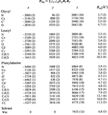
pour les x, y, z des C, O: 0,6.Tableau 1. Coordonndes atomiques fractionnaires
(X10 4)
a v e c leurs dcarts-type entre parentheses etfacteurs d'agitation thermique dquivalents
x y z Glycyl N -3060 (2) 46 (2) Co -3146 (2) 800 (2) C' -3040 (2) 1224 (2) O -2816 (2) 1010 (2) Leucyl N -3218 (2) 1869 (2) Cr~ -3109 (2) 2371 (2) C' -3728 (2) 2090 (2) O -3561 (2) 2275 (2) Cfl -3089 (2) 3155 (2) Cy -2391 (3) 3584 (2) C3(1) -2540 (4) 4223 (3) C3(2) - 1643 (3) 3929 (4) Phenylalanine N -4446 (2) 1684 (2) Cn -5126 (2) 1417 (2) C' -5837 (2) 904 (2) O' -5734 (2) 822 (2) O" -6463 (1) 636 (2) Cfl -5307 (2) 2073 (3) C), -5292 (2) 2703 (2) C~(I) -5829 (4) 2589 (3) C6(2) -4734 (3) 3410 (3) Cr.(I) - 5 8 1 1 ( 6 ) 3141 (4) Cr,(2) -4700 (4) 3992 (3) C~ -5257 (5) 3816 (4) Solvant Wat 0 0 Oeq(A 2) 2081 (24) 3,7 (2) 1710 (10) 3,9 (2) 3948 (10) 3,5 (2) 5663 (8) 4.7 (1) 3809 (8) 3.5 (1) 5725 (10) 3.4 (2) 7543 (9) 3.4 (2) 9520 (8) 4.8 ( I ) 4882 (10) 4.0 (2) 3394 (12) 5.2 (2) 2100 (14) 8.1 (3) 4823 (19) 10.5 (4) 6864 (8) 3.9 ( I ) 8325 (10) 3.8 (2) 6943 (10) 3.8 (2) 4873 (8) 4.5 ( 1 ) 7970 (8) 4.7 (1) 9492 (10) 4.7 (2) 7913 (12) 4.8 (2) 6196 (15) 8.9 (4) 8066 (17) 7.9 (3) 4639 (18) 13.1 (6) 6431 (21) 10.9 (5) 4775 (19) 11.3 (5) 7433 (12) 6.8 (3)
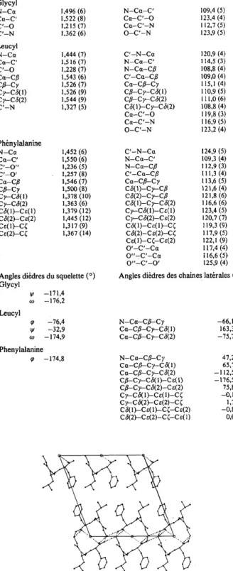
Discussion. Les coordonn~es atomiques sont report~es dans le Tableau 1,* les longueurs de liaison, angles de valence et angles de torsion dans le Tableau 2. La Fig. 1 montre la projection selon raxe c.
Nous sommes en pr6sence d'un zwitterion tout h fait caract~ristique. Notons la s6gr6gation des groupes hydrophobes des chaines lat6rales de la ph6nylalanine et de la leucine qui d~limitent un canal parall61e it l'axe a de diam&re environ 4/~. Un tel regroupement a d~jh &6 observ~ (Prang~ & Pascard, 1979). Il en r~sulte une grande agitation de ces chalnes. Ce ph6nom6ne est courant dans de tels compos~s (Pr6cigoux, Geoffre & Ouvrard, 1986). Notons les longueurs de liaison Ce(1)-C~, 1,32 et C6(2)-Ce(2), 1,45/~, dont rinterpr&ation ne semble pas imm6diate. Les liaisons hydrog6ne rencontr~es sont tout ~ fait classique, remarquons toutefois l'absence de telles liaisons sur O (Gly) et N (Phe), voir Tableau 3.
Les liaisons peptidiques sont bien en conformation
trans, les groupes peptidiques restent plans (09 = 176 et 175°). Les angles ~0 et ~' sont tout h fait normaux mais l'extr6mit6 ph+nylalanine est en conformation +tendue
(tp= -174,9°).
* Les listes des facteurs de structure, des param&res d'agitation thermique anisotrope, des angles de valence, et des coordonn&es des atomes d'hydrog6ne, ont 6t~ d~pos6es au d~p6t d'archives de la British Library Document Supply Centre (Supplementary Pub- lication No. SUP 44711: 14 pp.). On peut en obtenir des copies en s'adressant /l: The Executive Secretary, International Union of Crystallography, 5 Abbey Square, Chester CH 1 2HU, Angleterrc.
904 C
~7H2sNaO4.½H20
Tableau 2. Longueurs de liaison (A), angles de valence(o) et angles de torsion (o), les ddviations standards dtant donndes entre parentheses
Angles de torsion en accord avec les normes de la I U P A C - I U B Commission on Biochemical Nomenclature (1970). Glycyl N-Ca 1,496 (6) Ca-C' 1,522 (8) C'-O 1,215 (7) C'-N 1,362 (6) Leueyl N-Ca 1,444 (7) Ctt-C' 1,516 (7) C'-O 1,228 (7) Ca-Eft 1,543 (6) Cfl--Cy 1,526 (7) Cy-C6(l) 1,526 (9) Cy--C~(2) 1,544 (9) C'-N 1,327 (5) Ph6nylalanine N-Cfl 1,452 (6) Ctt-C' 1,550 (6) C'-O" 1,236 (5) C'-O' 1,257 (8) Ctt-Cfl 1,546 (7) c#-c;, 1,50o (8) c),-c6(i) 1,378 (I0) Cy--C6(2) 1,363 (6) C6(1)-Ce(1) 1,379 (12) C6(2)--Ce(2) 1,445 (12) Ct:(1)--C¢ 1,317 (9) Ce(2)-C/~ 1,367 (14)
Angles di&lres du squelette (o) Glyeyl ~v -171,4 -176,2 Leueyl '76,4 -32,9 to -174,9 Phenylalanine -174,8 N-Cct-C' 109,4 (5) C~t-C'-O 123,4 (4) Ctt-C'-N 112,7 (5) O-C'-N 123,9 (5) C'-N-Ctt 120,9 (4) N-Cct-C' 114,5 (3) N-Ca-C~' 108,8 (4) C'--Cct-Cfl 109,0 (4) Ca-Cfl---Cy 115,1 (4) C~-Cy-C6(l) 110,9 (5) Cfl-Cy-CtJ(2) 111,0 (6) C6(I)-Cy-C6(2) 108,8 (4) Cct-C'-O 119,8 (3) Cct-C'-N 116,9 (5) O-C'-N 123,2 (4) C'-N-Ca 124,9 (5) N-Cct-C' 109,3 (4) N--Ctt-Cfl 112,9 (3) C'-Cu-Cfl 111,3 (4) Cct-C/~-Cy 113,6 (5) C6(1)--Cy--Cfl 121,6 (4) C6(2)-C),-Cfl 121,8 (6) C6(I)-Cy--C6(2) 116,6 (6) Cy--C6(1)-Ce(l) 123,4 (5) Cy--C6(2)-Ce(2) 120,7 (7) C6(l)-C~:(l)-C~ 119,3 (9) C6(2)-Ce(2)-CC 117,9 (5) Ce(1)-C~-Ce(2) 122,1 (9) O'-C'-Ctt 117,4 (4) O"-C'-Cct 116,6 (5) O"-C'-O' 125,9 (4) Angles di6dres des chaines lat&ales (o)
N-Ca-Cfl---C y -66,1 Ca-Cfl-Cy---C 6(1) 163,3 Cct-Cfl---Cy-C6(2) -75,7 N-Ca-Cfl---Cy 47,2 Ctt-Cfl--Cy---C 6(1) 65,7 Cct-Cfl--Cy---C 6(2) - 112,5 Cfl---Cy---C 6( 1 )---C ~( 1 ) -176,5 Cfl--Cy--C ~(2)-Ce(2) 75,8 Cy---C~(1)-Ce(1)-C~ -O,l Cy-C6(2)-Ce(2)-C~ 1,7 c,~(1)-c ~(1)-c~-c ~(2) -0,8 C6(2)--Ce(2)-c~r-ce(1) 0,0 %" ~ " "" ¢ 1
Fig. 1. Projection de rarrangement mol6culaire selon l'axe c. Les liaisons hydrog~ne sont repr6sent6es en pointill6s.
Tableau 3.
Liaisons hydrogkne
(A)N(Gly)(i)H...O'(Phe)(v) 2,779 N(GIy)(i)H...O"(Phe)(iii) 2,786 N (Gly)(i)H-..O"(Phe)(vi) 2,74, N(Leu)(i)H-..O(Leu)(ii) 2,78~ Wat(i)H.--O'(Phe)(iv) 2,78 t
Les op&ations de sym&rie sont: (i) x, y, z; (ii)x, y, 1 + z; (iii) ½+ x, y, ½+ z; (iv) ½+x,y, -½+z; (v) - 1 - x , - y , z; (vi) - 1 - - x , - y , I +z.
..--o Fig. 2. Repr&entation des ellipso'ides d'agitation thermique/l 50%
de probabilit&
Les r6gions hydrophiles, COO- et NH~, sont f6d6r6es par une mol6cule d'eau. Le r6seau de liaisons hydrog~ne est donn6 par le Tableau 3; il est constitu~ de cinq liaisons voisines de 2,8 A.
La Fig. 2 repr6sente les eUipso'/des d'agitation thermique.
R~f~renees
ADAM, A. & LEDERER" E. (1984). Med. Res. Rev. 4, 111-152. BACH, J. F. (1984). Dans Thymic Factor Therapy, Tome 16, pp.
21-29. New York: Raven Press.
BERTHOU, J., MIGLIORE-SAMOUR" D., Ln~CHITZ, A., DELETTRt~, J., FLOC'H, F. & JOLLES, P. (1987). FEBS Lett. 2i8, 55-58. CROMER, D. T. & MANN, J. B. (1968). Acta Cryst. A24, 321-324. DELETTP~, J., MORNON, J. P. & LEPICARD, G. (1980). Acta Cryst.
B36, 1430-1435.
GERMAIN, G., MAIN, P. & WOOLFSON, M. M. (1971). Acta Cryst. A27, 368-376.
IUPAC-IUB COMMXSSION ON BIOCHEMICAL NOMENCLATURE ( 1970). Biochemistry, 9, 3471-3479.
JOLL~.S, P., PARKER, F., FLOC'H, D., MIGLIORE, D., ALLIEL, P., ZERIAL, A. & WERNER, G. H. (1982). J. Immunopharmacol. 3, 363-369.
MIGLIORE-SAMOUR, D., BOUCHAUDON, J., FLOC'H, F., ZERIAL, A., NINET, L., WER~ER, G. H. & JOLL~S, P. (1980). Life Sci. 26, 883-888.
NA.IJAR, V. & FRIDKIN, M. (1983). Ann. NYAcad. Sci. 419, 1-273. PARKER, F., MIGLIORE-SAMOUR, D., FLOC'H, F., ZERIAL, A.,
WERNER" G. H., JOLLES, J., CASARETTO, M., ZAHN, H. & JOLLES, P. (1984). Eur. J. Biochem. 145, 677-682.
PRANGI~, T. & PASCARD, C. (1979). Acta Cryst. B35, 1812-1819. PRECIGOUX, G., GEOFFRE, S. & OUVRARD, E. (1986). Acta Cryst.
C42, 721-724.
WERNER, G. H., FLOC'H, F., MIGLIORE-SAMOUR, D. & JOLLIES, P. ( 19 86). Experientia, 42, 5 21-5 30.