HAL Id: hal-01595704
https://hal.archives-ouvertes.fr/hal-01595704
Submitted on 26 Sep 2017
HAL is a multi-disciplinary open access
archive for the deposit and dissemination of
sci-entific research documents, whether they are
pub-lished or not. The documents may come from
teaching and research institutions in France or
abroad, or from public or private research centers.
L’archive ouverte pluridisciplinaire HAL, est
destinée au dépôt et à la diffusion de documents
scientifiques de niveau recherche, publiés ou non,
émanant des établissements d’enseignement et de
recherche français ou étrangers, des laboratoires
publics ou privés.
of Dendritic Cells byGardnerella vaginalis: The
“Invisible Man” of Bacterial Vaginosis?
Thomas Bertran, Patrick Brachet, Marjolaine Vareille, Julie Falenta, Annie
Dosgilbert, Marie-Paule Vasson, Christiane Forestier, Arlette Tridon,
Bertrand Evrard
To cite this version:
Thomas Bertran, Patrick Brachet, Marjolaine Vareille, Julie Falenta, Annie Dosgilbert, et al.. Slight
Pro-Inflammatory Immunomodulation Properties of Dendritic Cells byGardnerella vaginalis: The
“In-visible Man” of Bacterial Vaginosis?. Journal of Immunology Research, Hindawi Publishing
Corpora-tion, 2016, pp.1-13. �10.1155/2016/9747480�. �hal-01595704�
Research Article
Slight Pro-Inflammatory Immunomodulation Properties of
Dendritic Cells by
Gardnerella vaginalis: The
(Invisible Man)
of Bacterial Vaginosis?
Thomas Bertran,
1Patrick Brachet,
2Marjolaine Vareille-Delarbre,
1Julie Falenta,
1Annie Dosgilbert,
3Marie-Paule Vasson,
4Christiane Forestier,
5Arlette Tridon,
1,3and Bertrand Evrard
1,31UFR M´edecine-Pharmacie, Clermont Universit´e, Universit´e d’Auvergne, UMR 1019 UNH, ECREIN,
Laboratoire d’Immunologie, 63001 Clermont-Ferrand, France
2INRA de Theix, UMR 1019 UNH, ECREIN, 63122 Saint-Gen`es-Champanelle, France 3Service d’Immunologie, CHU Clermont-Ferrand, 63001 Clermont-Ferrand, France
4UFR Pharmacie, Clermont Universit´e, Universit´e d’Auvergne, UMR 1019 UNH, ECREIN, Laboratoire de Biochimie,
63001 Clermont-Ferrand, France
5UFR Pharmacie, Clermont Universit´e, Universit´e d’Auvergne, UMR CNRS 6023 LMGE, Laboratoire de Bact´eriologie,
63001 Clermont-Ferrand, France
Correspondence should be addressed to Bertrand Evrard; bevrard@chu-clermontferrand.fr Received 22 October 2015; Revised 4 January 2016; Accepted 10 January 2016
Academic Editor: Silvia Beatriz Boscardin
Copyright © 2016 Thomas Bertran et al. This is an open access article distributed under the Creative Commons Attribution License, which permits unrestricted use, distribution, and reproduction in any medium, provided the original work is properly cited. Bacterial vaginosis (BV), the most common genital infection in reproductive-aged women, is associated with increased risk of sexually transmitted infections. Its etiology remains unclear, especially the role of Gardnerella (G.) vaginalis, an anaerobic bacterium characteristic of the BV-alteration of the vaginal ecosystem. In the genital mucosa, dendritic cells (DCs) sense bacteria of the microenvironment via receptors and then orchestrate the immune response by induction of different T cell subtypes. We investigated the interactions between G. vaginalis and human monocyte-derived DCs using a wide range of bacterial concentrations (multiplicity of infection from 0.01 to 100), and the effects of this pathogen on PHA-induced lymphocyte proliferation. As observed by electron microscopy and cytometry, G. vaginalis reduced the internalization ability of DCs by forming extracellular clusters and induced neither DC maturation, nor DC secretion of cytokines, except at the highest dose with a very early DC maturation state. The same profile was observed on lymphocytes with significant increases of proliferation and cytokine secretion only at the highest bacterial concentration. Our findings indicate that G. vaginalis possesses slight immune-stimulating activities against DCs and T cells, reflecting thus a defective inflammatory response and giving rise to the atypical, non- or low-grade, inflammatory clinical disease profile.
1. Introduction
Bacterial vaginosis (BV) is the most common low genital infection among reproductive-aged women, with a preva-lence of 29% among 14- to 49-year-old US women and almost 40% in individuals at high risk for sexually trans-mitted infections (STIs) [1]. BV is associated with serious medical complications, including adverse pregnancy out-comes, endometritis, and pelvic inflammatory disease such
as endometriosis [2, 3]. BV also increases women’s risk of acquiring STIs, particularly HIV infections [4, 5].
Clinically, one-half of BV-positive women are asymp-tomatic while the others suffer only from mild symptoms, such as homogeneous white vaginal discharge and amine (fishy) odor [6]. These signs are associated with a vaginal pH > 4.5 and the presence of characteristic “clue cells” on micro-scopic examination. These four manifestations constitute Amsel’s clinical criteria [6]. The microbiological diagnosis
Volume 2016, Article ID 9747480, 13 pages http://dx.doi.org/10.1155/2016/9747480
of BV is usually based on Nugent’s score, which includes assessment of lactobacilli by Gram’s stain of vaginal fluid samples. BV is associated with an alteration of the vaginal ecosystem, characterized by a decrease in hydrogen peroxide-producing Lactobacillus (L.) species such as L. crispatus and L. jensenii and a concomitant increase in polymicrobial anaerobic bacteria like Gardnerella (G.) vaginalis [7, 8].
The microbial etiology of BV is unclear and a matter of debate [9]. Two opposing hypotheses exist [10]. In the monomicrobial hypothesis, historically the first one, G.
vaginalis is the single, specific etiologic agent of BV [11].
In the polymicrobial hypothesis, which has gained general acceptance in the last 20 years, G. vaginalis acts synergically with other anaerobes to unbalance the vaginal flora and trigger the disease [12]. Vaginal inoculation experiments in the monkey show thus that the co-occurrence of anaerobes and G. vaginalis is required to induce BV [13]. Moreover,
G. vaginalis is frequently isolated in healthy women without
BV [14]. Nevertheless, recent works have relaunched the debate by confirming its importance in the pathophysiology of the disease. G. vaginalis predominates in vaginal BV-associated biofilms, which are implicated in persistent BV, thus constituting a major factor of resistance to standard treatment [15].
Dendritic cells (DCs) are professional antigen present-ing cells (APCs) which, by inducpresent-ing both tolerance and immunity, are critical for the orchestration of the adaptive immune response [16]. Immature DCs reside in peripheral mucosa, where they sense the microenvironment via pat-tern recognition receptors (PRRs) that recognize pathogen-associated molecular patterns (PAMPs). PRRs include toll-like receptors (TLRs) and C-type lectin receptors (CLRs) [17]. PRR stimulation triggers a DC maturation process with up-regulation or down-regulation of membrane molecules (CD83, CD86 and HLA-DR, and DC-SIGN and Mannose Receptor, resp.) and cytokine production. DC activation by several PAMPs, via distinct PRRs with antagonistic or synergistic effects, modulates their differentiation, which secondarily determines the polarization of the effector T cell responses, that is, the balance between Th1, Th2, Th17, and T regulatory (Treg) subsets [18]. Cytokine production by DCs is an important factor in this process. IL-12 production drives polarization towards Th1 cells, whereas synthesis of IL-1𝛽, IL-6, TGF𝛽 and IL-23, and IL-10 promotes induction of Th17 or Treg cells, respectively [19]. These different stages of the immune response have been recently described in the human genital mucosa. Notably, in both the upper and lower tracts, several DC subsets exist and express specific TLRs, such as TLR-6, TLR-7, and TLR-8, and CLRs, such as langerin and DC-SIGN, and are able to induce different T cell subpopulations [20–22].
The effects of mucosal fluids from women with BV or healthy flora, without analysis of implicated bacterium species, were examined on DC function [23, 24]. BV samples induced IL-12 and IL-23 production, as well as expression of maturation markers (HLA-DR, CD40, and CD83) by monocyte-derived DCs (moDCs). Concerning T cells, there has been yet no investigation on the impact of BV on the polarization of the different lymphocyte subpopulations.
Only one study reported effects of BV on the percentage of Treg cells in peripheral blood mononuclear cells (PBMCs). However, this study did not test the possibility of a specific impact of G. vaginalis and it did not objectify differences in the distribution of Treg in BV+ versus BV− HIV-negative women, decreased Treg being solely observed in BV+/HIV+ women compared to BV−/HIV+ women [25]. Many studies have attempted to measure cervicovaginal production of cytokines in BV, but disparate results were obtained. Most articles reported an elevation in IL-1𝛽, and less consistently in IL-6 and IL-8, in BV-affected women [26–29]. Additionally, BV mucosal fluid was found to increase proliferation of T cells in allogeneic mixed-leukocyte reaction (MLR) [23]. Finally, the specific effects of G. vaginalis on DC and T cells have never been evaluated yet.
Unlike conventional vaginitis that is characterized by burning, dysuria, dyspareunia, and frequent pruritus, BV causes scant inflammatory signs without primary pain or pruritus in affected women [14]. Likewise, a relative paucity of inflammatory cells and a near normal number of vaginal neutrophils are characteristic of BV status. In view of the literature data, we hypothesized that BV corresponds to a unique local immunological environment, with a low-grade inflammation, potentially mediated by so far unknown immunomodulatory mechanisms of action of G. vaginalis on the vaginal immune system, particularly on the DCs and T cells. In the present study, we investigated this hypothesis in in vitro models by monitoring (i) the internalization, maturation, and cytokine secretion of moDCs; (ii) lympho-cyte proliferation and subset cytokine production, after cell exposure to G. vaginalis or to commensal or pathogenic microorganisms potentially found in the vaginal mucosa.
2. Material and Methods
2.1. Bacterial Strains and Culture Conditions. G. vaginalis
ATCC14018 was grown in brain heart infusion (Biom´erieux) supplemented with maltose (0.1%), glucose (0.1%), yeast extract (1%), and horse serum (10%) in 5% CO2at 37∘C for 72 h. L. reuteri ATCC23272 was grown in De Man, Rogosa, Sharpe (MRS) medium (BD Difco™) at 37∘C overnight.
Candida albicans ATCC10231 was grown in Sabouraud broth
at 37∘C overnight. Microbial cells were harvested by centrifu-gation (11,000×g for 10 min), and the pellet was washed twice and then resuspended in RPMI 1640 (Cambrex Bio Science). Optical density (OD) measurements were performed at 620 nm to adjust the final concentration of the microbial suspension, and the exact number of colony forming units (CFU) was determined by plating serial dilutions of the inocula onto adapted agar plates (Columbia 5% Sheep Blood Agar, MRS, or Sabouraud). Before being added to the cell samples, the microbial cells were inactivated by exposure to UV for 1 h. The effectiveness of the inactivation was evaluated by plating 20𝜇L of the irradiated inocula on adapted agar plates.
2.2. Ethics Statement. The human cells used in this study were
from the local French blood agency (Etablissement Franc¸ais du Sang, EFS, Saint-Etienne). It is a statutory requirement that blood donors be given full necessary information (arti-cle R.1221-5 of the Public Health Code, 12/01/2009 and 11/06/2006 decrees). Written informed consent was obtained by the EFS from all volunteers involved in our study.
2.3. In Vitro Differentiation of Monocyte-Derived Dendritic Cells. DCs were generated from PBMCs. Briefly, PBMCs
were isolated from the buffy-coats of healthy volunteers by Ficoll-Histopaque (Sigma) density gradient centrifugation. PBMCs were washed twice in RPMI 1640 and resuspended at a final concentration of 5× 10⁷cells per mL of phosphate buffered saline (PBS) supplemented with 2% fetal calf serum (FCS, Biowest-Abcys) and 1 mM EDTA. Monocytes were purified by negative selection using the EasySepÝ Human Monocyte Enrichment Kit, as recommended by the manu-facturer (StemCell Technologies). They were then cultured for 5 days in RPMI 1640 supplemented with 1% L-glutamine (Sigma), 10% FCS, and 0.5% penicillin-streptomycin (Sigma), in the presence of 500 U/mL IL4 (R&D systems) and 800 U/mL granulocyte-macrophage colony-stimulating fac-tor (GM-CSF, R&D systems). After 3 days of incubation, one-half volume of fresh culture medium containing 2x concentrations of IL4 and GM-CSF was added to each well.
2.4. Electron Microscope Observations. DCs obtained as
pre-viously described were plated in a sterile 12-well plate at a concentration of 1 × 10⁶ cells/mL. G. vaginalis was added to wells at a multiplicity of infection (MOI) of 10 for 1 h (Scanning Electron Microscopy, SEM) and at a MOI of 0.01, 1, or 100 for 3 h (Transmission Electron Microscopy, TEM). Cells were harvested, centrifuged (400×g for 10 min), rinsed with Natrium Cacodylate (0.2 M pH 7.4) for 10 min, and then fixed at 4∘C overnight with glutaraldehyde 1.6% in Natrium Cacodylate buffer. The samples were then rinsed, postfixed with 1% Osmium Tetroxide (1 h, room temperature), rinsed again, and dehydrated with graded series of ethanol (70 to 100%) and eventually with 100% hexamethyldisilazane. Finally, after overnight drying, samples were placed on a Jeol SEM filter and metallised with carbon (40 s). For TEM, dried samples were embedded in a polymerized 2 mm thick Epon coating, and ultrathin sections were picked up with Formvar-coated copper grids (300 mesh). Sections were counterstained with 4% aqueous uranyl acetate. For negative staining, bacteria were grown overnight in M63B1-0.4% Glu medium and negatively stained with 2% phosphotungstic acid on Formvar-coated copper grids (300 mesh). Images were captured at the Centre d’Imagerie Cellulaire Sant´e (CICS) of the Universit´e d’Auvergne with a Jeol JSM-6060LV (SEM) and a Hitachi H-7650 (TEM).
2.5. Flow Cytometry Analysis of DC Maturation and Via-bility. On day 6, the immature DCs from each well were
harvested, pooled, centrifuged, and reseeded at 1 × 10⁵ cells/mL. UV-killed bacteria were then added at 10𝜇L of suspension per well to reach a final concentration ranging from 103 to 107CFU/mL, that is, a MOI between 0.01
and 100. Lipopolysaccharide (LPS) from Escherichia coli (Sigma) at a final concentration of 100 ng/mL was used as positive control. Immature DCs without addition of LPS or bacteria were used as negative control. After 48 h of maturation at 37∘C in a 5% CO2atmosphere, DCs were col-lected, centrifuged, and resuspended in PBS with 1% bovine serum albumin (BSA, Sigma). Cell surfaces were stained with the appropriate fluorescence-labeled murine antibod-ies: APC-Cy7-conjugated anti-CD14 (LPS coreceptor specific to monocytes), PE-conjugated anti-CD86 (costimulatory molecule, activation marker), V450-conjugated anti-HLA-DR, PerCP-Cy5.5-conjugated anti-DC-SIGN (DC-specific ICAM-3 grabbing-nonintegrin or CD209, member of the CLR family, specific marker of immature DCs), Alexa FluorÝ 488-conjugated anti-MR (Mannose Receptor or CD206, member of the CLR family, specific marker of immature DCs and macrophages), and streptavidin APC-conjugated anti-TLR4 (biotin antibody, an LPS receptor with activation functions). Antibodies were obtained from BD Biosciences, except anti-MR (Biolegend). Corresponding murine isotype-matched and non-labeled antibodies (BD Biosciences or Biolegend) were used as controls. The cells were analyzed by a BD-LSRII flow cytometer with FACSDiva Software (BD Biosciences) at the CICS. Fluorescence compensation adjustments were performed. Gates were set on living DCs based on their forward/side scatter (FSC/SSC) properties. The analysis was halted at a count of 3,000 DCs. The level of staining was expressed as the mean fluorescence intensity (MFI). Culture supernatants were collected and stored at −20∘C until cytokine analysis. To determine cell viability, dye LIVE/DEADÝbeads (Fixable Blue Dead Cell Stain Kit, for UV excitation Life technologies) were added to cells and used as markers of dead cells. DC mortality was assessed by gating together dead cells and live DCs on SSC/FSC diagram. Two other gates were created from this gate to separate the two populations. From the dead cell gate, cells that expressed LIVE/DEAD marker were considered as effectively dead. This dead cell number was expressed as a ratio of the first created overall gate to obtain a dead DC percentage.
2.6. Lymphocyte Proliferation Assays. The mitogenic response
to plant lectins, as phytohemagglutinin A (PHA), is conven-tionally used to measure cell-mediated immunity in mam-mals in general and especially in humans [30]. These tests are named lymphocyte proliferation assays or lymphocyte transformation test (LTT). They were performed here to study the functional properties of G. vaginalis and especially its capabilities to modulate PHA-induced T cell proliferation. On day 1, PBMCs collected from a buffy-coat as previously described were adjusted to a concentration of 1× 10⁶cells per mL of complete medium, that is, RPMI 1640 supplemented with 1% L-glutamine and 10% FCS. The cell suspension was deposited in a sterile 96-well plate (100𝜇L per well). Each measurement was done in triplicate. Polyclonal proliferation of lymphocytes was induced with 2𝜇g/mL of PHA (Sigma). Cells without PHA or bacteria were used as a negative control and cells with PHA without bacteria as a positive one. G. vaginalis was added to other PHA-treated wells
to obtain concentrations ranging from 103 to 107CFU/mL. After a 72 h incubation at 37∘C under 5% CO2, 1𝜇Ci of tritiated thymidine (Perkin Elmer) was added to each well and the cells were further incubated for 4 h. Labeling was stopped by cooling the plate to 4∘C. The cells were then collected under vacuum onto a Whatman filter paper and incorporation of tritiated thymidine was measured using a𝛽 counter (Tri-Carb 2300TR, Canberra-Packard). Proliferation results were expressed as mean cpm (counts per min) values of triplicate measurements. Identical incubations, however, without addition of tritiated thymidine, were carried out in parallel to collect supernatants for cytokine quantification.
2.7. Cytokine Quantification. For DC maturation
experi-ments, the cytokines IL-10, TNF-𝛼, IFN-𝛾, and IL-12p70 were assayed in culture supernatants with Biolegend enzyme-linked immunosorbent assay (ELISA) kits according to the manufacturer’s instructions. For lymphocyte proliferation assays, the cytokines IFN-𝛾, IL-4, IL-17A, IL-10, IL-12p70, and TNF-𝛼 were quantified in supernatants of PBMC cultures using Pro Human Cytokine Group 1 6-Plex 1× 96 kit (Bio-Rad) on a Bio PlexÝ200 (Bio-Rad).
2.8. Statistical Analysis. All data were expressed as means
+ SD. After variance dispersion test, three different statisti-cal tests were performed. Two-way ANOVA with post hoc Bonferroni test, Friedman’s test with Nemenyi’s group, and Kruskal-Wallis test with Dunn test were used to analyze the significant effect of bacteria on DCs or PBMCs with XLStat 7.5.2 software (Addinsoft, Paris, France).𝑝 values lower than 0.05 were considered statistically significant.
3. Results
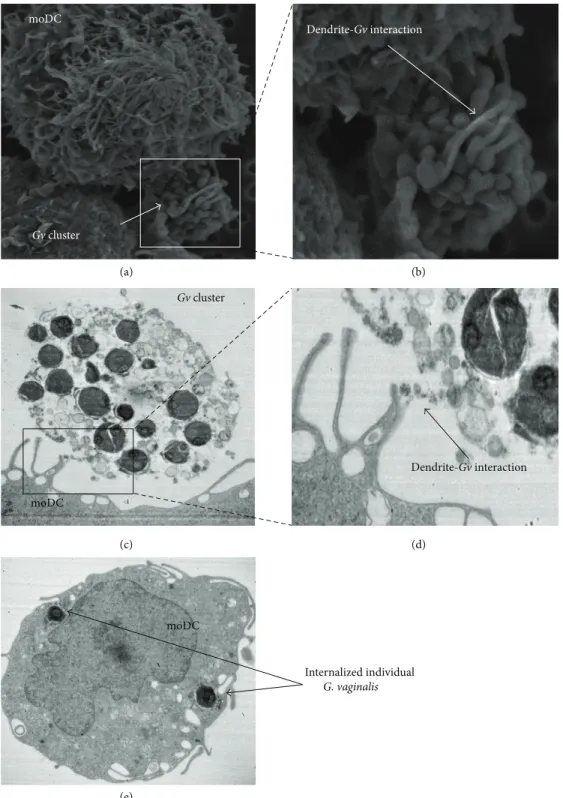
3.1. Electron Microscopy Observation of G. vaginalis-DC Inter-action. In vivo, the first step of the immune response in
the vaginal mucosa corresponds to the interactions between bacteria and immature DCs, which lead to the internalization of bacteria. To mimic this initial phenomenon in vitro, we put in contact DCs with G. vaginalis during 1 to 3 h and took SEM and TEM pictures. SEM produced 3D images of cell and bacteria surfaces after 1 h of contact (Figures 1(a) and 1(b)). G. vaginalis bacteria were rarely found on isolated cell forms (in contrast with what we observed with
Lactobacillus, data not shown) and were in general organized
in clusters (Figure 1(a)). However, DC dendrites interacted with this cluster of bacteria and sometimes surrounded it (Figure 1(b)). TEM realized at 3 h, to allow the cells to have time to internalize bacteria, confirmed interactions of DC dendrites with clusters of G. vaginalis (Figures 1(c) and 1(d)). Figure 1(c) shows additionally that the cluster is composed of bacteria enrobed in an extracellular matrix which may interact with DCs. TEM also shows internalized intracellular
G. vaginalis bacteria, but only in their isolated form, without
intracellular clusters (Figure 1(e)). At a MOI of 0.01 no DCs with internalized G. vaginalis were found, very few at a MOI of 1, and only 35% at a MOI of 100 (count on 100 DCs). For each DC with internalized G. vaginalis, the
number of bacteria ranged from 1 to 9 bacteria per cell. This experiment was performed in parallel with L. reuteri at the same concentrations. Internalization of Lactobacillus, unlike
G. vaginalis, could be observed on few cells at a MOI of 0.01, a
majority of cells at a MOI of 1, and up to 82% of cells at a MOI of 100 (count on over 160 DCs). The number of Lactobacillus internalized in those cells was higher than with that of G.
vaginalis since it ranged from 6 to 18 bacteria per cell (data
not shown). To conclude on this part, we showed that DCs were able to interact with G. vaginalis and to internalize it, but less effectively than lactobacilli, probably due to G. vaginalis ability to form extracellular clusters.
3.2. Flow Cytometric Analysis of Microbial Effects on DC Phe-notype. During the immune response, bacterium
internal-ization can then induce DC activation and maturation, result-ing in modifications detectable by cytometry of numerous membrane markers and permitting to distinguish immature DCs from mature ones. In our study, DC activation and maturation were assessed by changes affecting an extensive phenotype of the cell membrane. As expected, immature DCs were characterized by low levels of CD86, HLA-DR, and CD14 expression (the latter was compared to its initial level in monocytes before IL-4 and GM-CSF treatment, data not shown), together with high levels of DC-SIGN and MR expression (Figures 2 and 3). Comparatively, LPS-induced DCs expressed a phenotype characteristic of fully mature DCs with increased levels of CD86 and HLA-DR (Figures 3(a) and 3(b)) associated with decreased levels of DC-SIGN, MR, TLR4, and CD14 (Figures 3(c), 3(d), 3(e), and 3(f)).
Incubation with low or medium doses of G.
vagi-nalis (103–106CFU/mL, i.e., MOI from 0.01 to 10) did not alter immature DC cell surface phenotype, as shown by the absence of significant change in either membrane marker (Figures 2 and 3). At the highest concentration (107CFU/mL, i.e., MOI 100), G. vaginalis caused a very slight increase in HLA-DR expression (statistically non-significant, Figure 3(b)) combined with moderate decreases in MR and CD14 expressions (both non-significant, Figures 3(e) and 3(f)), which reflect early signs of maturation. However, the lack of increase in CD86 expression and decrease in DC-SIGN and TLR4 expressions indicated that this maturation process was incomplete.
Taken together, our overall cytometric results indicate that G. vaginalis induced no maturation of DCs or an incomplete DC maturation process at high doses.
Unlike G. vaginalis, L. reuteri and C. albicans induced a clear dose-dependent higher expression of CD86 and HLA-DR upon maturation of DCs (Figures 3(a) and 3(b)). Combined with decreased expressions of DC-SIGN, MR, CD14, and TLR4 (Figures 3(c), 3(d), 3(e), and 3(f)), these results show that the last two microorganisms induced fully mature DCs, similarly to LPS extract.
3.3. Flow Cytometric Analysis of G. vaginalis Effects on DC Mortality. Given the very low level of maturation of DC by G. vaginalis observed in our experiments, we hypothesized
(d) moDC Gv cluster Dendrite-Gv interaction Gv cluster moDC Dendrite-Gv interaction moDC Internalized individual G. vaginalis (e) (a) (b) (c)
Figure 1: Pictures of DCs after exposure to G. vaginalis. (a) SEM (×5500), after a 1 h exposure, DC dendrites surrounding clusters of G.
vaginalis. (b) Zoom (×13000) on the contact zone between DCs and bacteria. (c) TEM (×12000), after a 3 h exposure, a cluster of G. vaginalis
in contact with DC dendrites. (d) Enlargement of DC dendrite-G. vaginalis interaction zone. (e) TEM cutting, internalized G. vaginalis in one DC. moDC: monocyte-derived dendritic cell, Gv: Gardnerella vaginalis.
L. reuteri (CFU/mL) L. reuteri (CFU/mL)
L. reuteri (CFU/mL) L. reuteri (CFU/mL) L. reuteri (CFU/mL)
G. vaginalis (CFU/mL) G. vaginalis (CFU/mL)
G. vaginalis (CFU/mL) G. vaginalis (CFU/mL) G. vaginalis (CFU/mL)
CD86 (MFI, a.u.) CD86 (MFI, a.u.) CD86 (MFI, a.u.) CD86 (MFI, a.u.) CD86 (MFI, a.u.)
CD86 (MFI, a.u.) CD86 (MFI, a.u.)
CD86 (MFI, a.u.) CD86 (MFI, a.u.)
CD86 (MFI, a.u.) CD86 (MFI, a.u.) CD86 (MFI, a.u.)
Immature DCs Fully mature DCs 102 103 104 105 D C-S
IGN (MFI, a.u
.) 103 102 104 105 Imm-DCs 103 102 104 105 LPS-DCs 102 103 104 105 D C-S
IGN (MFI, a.u
.) 103 102 104 105 103 103 102 104 105 104 102 103 104 105 102 103 104 105 102 103 104 105 102 103 104 105 103 102 104 105 107 103 102 104 105 106 103 102 104 105 105 102 103 104 105 102 103 104 105 103 102 104 105 104 102 103 104 105 102 103 104 105 103 102 104 105 103 102 103 104 105 D C-S
IGN (MFI, a.u
.) 102 103 104 105 103 102 104 105 105 103 102 104 105 106 103 102 104 105 107
Figure 2: Surface phenotype of human DCs after exposure to a range of G. vaginalis or L. reuteri concentrations. The dot plots and histograms show MFI values on gated DCs. DC-SIGN/CD86 dot plots gated on human DCs. Data from a representative experiment comparing G.
vaginalis-induced DC process of maturation with that induced by L. reuteri.
this assertion, a viability marker was added during flow cytometric experiments. Immature DCs alone had almost no mortality. In comparison, exposure of DCs to LPS extract induced a slight, but significant, increase in mortality (Figure 4). Three concentrations of G. vaginalis were tested to analyze their impact on DC survival. Results showed that, from 103 to 107CFU/mL (MOI 0.01 to 100), G. vaginalis induced no increase in dead DCs.
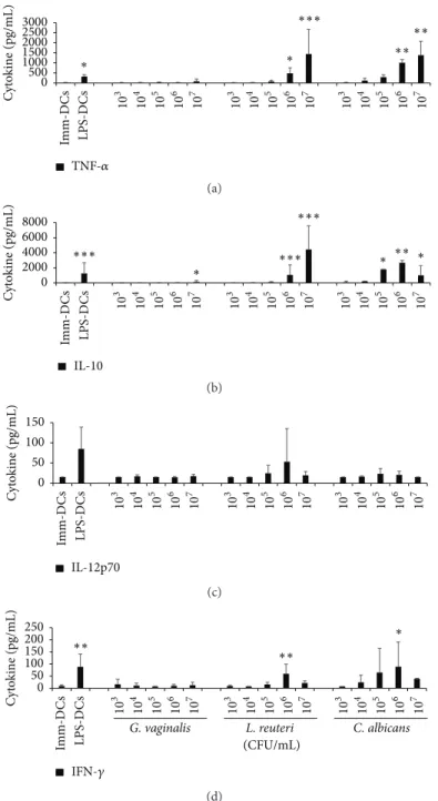
3.4. Cytokine Secretion by Microbial-Matured DCs. In vivo,
mature DCs migrate to secondary lymphoid organs and present the antigen to T cells. During this interaction, DCs deliver three signals, notably a cytokinic signal polarizing the differentiation of lymphocytes into several subpopulations. To decipher the preferential pathway of T cell polarization induced by G. vaginalis, four cytokines assumed to be of particular interest were selected for ELISA measurements. Compared to untreated immature DCs, DCs incubated with
G. vaginalis did not significantly increase the production of
TNF-𝛼 or IL-10, except at the upper dose of 107CFU/mL
(Figures 5(a) and 5(b)). Additionally, the production of IL-12p70 and IFN-𝛾 was not or barely detectable in G. vaginalis-treated or immature DCs (Figures 5(c) and 5(d)). Conversely, a strong dose-dependent increase in the production of TNF-𝛼 and IL-10 was induced by L. reuteri and C. albicans and, to a lesser extent, of IL-12p70 by L. reuteri and of IFN-𝛾 by C.
albicans (Figures 5(a), 5(b), 5(c), and 5(d)). The fold changes
determined by comparing the level of cytokines produced by DCs exposed to 107CFU/mL G. vaginalis (i.e., a MOI of 100) to that produced by immature DCs were equal to 8.9 for IL-10, 6.5 for TNF-𝛼, and only 1.1 and 1.3 for IL-12p70 and IFN-𝛾, respectively. By comparison, the fold changes induced by the same dose of L. reuteri or C. albicans ranged from 70 to 307 for IL-10 and TNF-𝛼 production. Whatever the concentration of G. vaginalis, the level of DC-produced cytokines attained only 10 to 28% of that generated by LPS-treated DCs. Overall, these results show that G. vaginalis barely induces cytokinic secretion, in accordance with our findings on DC maturation (Figure 3), thus indicating absence, or slight induction, of DC activation by G. vaginalis.
CD86 0 5000 10000 10 3 10 4 10 5 10 6 10 7 10 3 10 4 10 5 10 6 10 7 10 3 10 4 10 5 10 6 10 7 Imm-D Cs LPS-D Cs ∗ ∗ ∗ ∗ (a) 0 5000 10000 HLA-DR 10 3 10 4 10 5 10 6 10 7 10 3 10 4 10 5 10 6 10 7 10 3 10 4 10 5 10 6 10 7 ∗ ∗ ∗ Imm-D Cs LPS-D Cs (b) DC-SIGN ∗ ∗ ∗ 0 5000 10000 10 3 10 4 10 5 10 6 10 7 10 3 10 4 10 5 10 6 10 7 10 3 10 4 10 5 10 6 10 7 Imm-D Cs LPS-D Cs (c) TLR4 ∗ 0 500 10 3 10 4 10 5 10 6 10 7 10 3 10 4 10 5 10 6 10 7 10 3 10 4 10 5 10 6 10 7 Imm-D Cs LPS-D Cs (d) CD14 ∗ ∗ ∗ ∗ 0 5000 10000 10 3 10 4 10 5 10 6 10 7 10 3 10 4 10 5 10 6 10 7 10 3 10 4 10 5 10 6 10 7 Imm-D Cs LPS-D Cs (e) MR CD206 ∗ ∗ ∗ ∗∗ ∗ 0 5000 10000 (CFU/mL) L. reuteri C. albicans G. vaginalis 10 3 10 4 10 5 10 6 10 7 10 3 10 4 10 5 10 6 10 7 10 3 10 4 10 5 10 6 10 7 Imm-D Cs LPS-D Cs (f)
Figure 3: Human DC membrane marker expression after exposure to a range of G. vaginalis, L. reuteri, or C. albicans concentrations. (a, b) Differential expression of CD86 and HLA-DR, two membrane markers typically increasing during DC maturation. (c, d, e, f) Differential expression of DC-SIGN, TLR4, CD14, and MR CD206, four membrane markers typically decreasing during DC maturation. Data are means (+ SD). For every marker, the isotypic control values were subtracted from the MFI values. Imm-DCs: immature DCs, LPS-DCs: DCs matured by a 48 h exposure to 100 ng/mL E. coli LPS; G. vaginalis, L. reuteri, and C. albicans: DCs matured after a 48 h incubation at different
concentrations (103to 107CFU/mL, i.e., MOI = 0.01 to 100) of G. vaginalis (𝑛 = 5), L. reuteri (𝑛 = 3), and C. albicans (𝑛 = 2). Kruskal-Wallis
IMM-DCs LPS-DCs G. vaginalis 0 5 10 15 20 25 D ead cell (%) 103 105 107 ∗
Figure 4: DC viability after 48 h of culture with G. vaginalis. Histogram shows the dead cell percentage, calculated from SSC/FSC diagram and live/dead marker. LPS induced a significant increase in DC mortality compared to immature DCs alone in the medium. Low and high concentrations of G. vaginalis did not induce any increase in DC mortality, even at a MOI of 100. Data are means (+ SD) from 6 individual experiments. Imm-DCs: immature DCs, LPS-DCs: DCs matured by a 48 h exposure to 100 ng/mL E. coli LPS; G. vaginalis.
DCs matured after a 48 h incubation at different concentrations (103
to 107CFU/mL, i.e., MOI = 0.01 to 100) of G. vaginalis. Friedman’s
test with Nemenyi’s comparison∗𝑝 < 0.05.
3.5. Slight Increase in PHA-Stimulated Lymphocyte Prolifer-ation by G. vaginalis. To investigate lymphocyte activProlifer-ation,
the next classical stage of the immune response, we carried out functional tests of bacterium-induced modulation of lymphocytic proliferation, using a model of PHA-induced T cell proliferation. Addition of the strain to the medium caused a slight dose-dependent increase in PHA-stimulated lympho-cyte proliferation in comparison to lympholympho-cyte control assays without bacteria (Figure 6). The increase in proliferation was only significant at a dose of 107CFU/mL (𝑝 < 0.001) and attained an upper average value of 20% compared to that of PHA-stimulated control cells. Lower concentrations of G.
vaginalis did not cause any significant modulation of
PHA-stimulated lymphocyte proliferation. Similar experiments we performed in parallel showed that L. reuteri and C. albicans did not induce a similar pattern (data not shown).
3.6. G. vaginalis-Dependent Increase in Cytokine Secretion by PHA-Stimulated Leukocytes. To depict the type of immune
response involved during the G. vaginalis-dependent increase in PHA-stimulated lymphocyte proliferation, secretion of cytokines by the four main subpopulations of T cells (IFN-𝛾, IL-4, IL-17A, and IL-10 corresponding to Th1, Th2, Th17, or Treg, resp.) or by APCs (IL-12p70 and TNF-𝛼) was measured in extracellular media of lymphocyte proliferation assays. PHA (2𝜇g/mL) alone induced a strong secretion of IFN-𝛾, IL-17A, IL-10, IL-12p70, and TNF-𝛼 from PBMCs (Figure 7). A clear dose-dependent increase in IFN-𝛾 and IL-17A production was observed in PHA-stimulated PBMCs exposed to varying concentrations of G. vaginalis (Figures 7(c) and 7(d)). As shown previously for PHA-stimulated
proliferation, only the highest dose of G. vaginalis tested induced a significant augmentation in the production of these two cytokines, compared to the PHA control without bacteria. For TNF-𝛼, IL-12p70, and IL-10, the increases were non-significant, even at high doses (Figures 7(a), 7(b), and 7(e)). Compared to the control conditions without bacteria, exposure to 107CFU/mL G. vaginalis induced fold increases of 4.9, 4.0, 3.0, 2.2, and 2.2 for IFN-𝛾, 10, TNF-𝛼, IL-17A, and IL-12p70 production, respectively. Contrastingly, the secretion level of IL-4 was barely measurable or undetectable at any dose of G. vaginalis (not shown). Furthermore, at their highest concentration, L. reuteri and C. albicans caused an increase in cytokine secretion that, depending on the cytokine, was 4- to 5-fold higher than that caused by G.
vaginalis (data not shown). Overall, these data show that G. vaginalis can induce a slight dose-dependent secretion of
cytokines on both the inflammatory (IFN-𝛾, TNF-𝛼, IL-17A, and IL-12p70) and anti-inflammatory (IL-10) sides.
4. Discussion
DCs, the main sentinels of the immune system, are abundant in the human vaginal mucosa, both in the epithelium and in the lamina propria [21]. In this mucosal area, DCs can interact with luminal microorganisms, either indirectly via epithelial transport mechanisms or directly via dendrites extended across epithelial cells that take up bacteria from the vaginal lumen [31, 32]. As BV is characterized by an imbalance of the normal H2O2-producing Lactobacillus flora toward a polymorphic anaerobic flora with predominant
G. vaginalis, it is likely that the PRRs of vaginal DCs are
subsequently affected by this alteration. To gain insight into the pathophysiological role of G. vaginalis in BV, we decided to characterize the interactions between this bacterium and DCs.
We first carried out an electron microscope study of bacteria-DC interactions, the first stage in the immune response, to determine whether G. vaginalis can internalize DCs. This was confirmed by using TEM, but we observed that the bacteria were very sparsely represented in free form in culture medium and rather formed clusters coated in an extracellular matrix. As compared to a Lactobacillus species which remained in free form, G. vaginalis was internalized by DCs much less efficiently. It can reasonably be assumed that this difference is related to the propensity of G. vaginalis to form clusters. In addition, G. vaginalis forms, in vivo, biofilms in the vagina, a process that is involved in BV pathogen-esis [33]. These clusters might be the beginning of biofilm formation. Biofilms reduce the host-immune response by decreasing bacteria internalization owing to their large size and to the fact that their extracellular matrix can prevent antigen recognition by APCs [34]. This conformation might thus allow bacteria like G. vaginalis to be less internalized by DCs comparatively to strains like Lactobacillus that are unable to form biofilms.
We then studied the impact of internalization of G.
vaginalis on DC maturation status. The maturation state of
10 3 10 4 10 5 10 6 10 7 10 3 10 4 10 5 10 6 10 7 10 3 10 4 10 5 10 6 10 7 Imm-D Cs LPS-D Cs 0 500 1000 1500 2000 2500 3000 C yt o kine (pg/mL) ∗ ∗ ∗∗ ∗∗ ∗∗∗ TNF-𝛼 (a) IL-10 10 3 10 4 10 5 10 6 10 7 10 3 10 4 10 5 10 6 10 7 10 3 10 4 10 5 10 6 10 7 Imm-D Cs LPS-D Cs 0 2000 4000 6000 8000 C yt o kine (pg/mL) ∗ ∗ ∗ ∗∗ ∗∗∗ ∗∗∗ ∗∗∗ (b) IL-12p70 10 3 10 4 10 5 10 6 10 7 10 3 10 4 10 5 10 6 10 7 10 3 10 4 10 5 10 6 10 7 Imm-D Cs LPS-D Cs 0 50 100 150 C yt o kine (pg/mL) (c) ∗ ∗∗ ∗∗ (CFU/mL) L. reuteri C. albicans G. vaginalis 10 3 10 4 10 5 10 6 10 7 10 3 10 4 10 5 10 6 10 7 10 3 10 4 10 5 10 6 10 7 Imm-D Cs LPS-D Cs 0 50 100 150 200 250 C yt o kine (pg/mL) IFN-𝛾 (d)
Figure 5: Cytokine production by human DCs exposed to a range of G. vaginalis, L. reuteri, or C. albicans concentrations. (a, b) TNF-𝛼 and IL-10 cytokine production. G. vaginalis induceda slight increase in IL-10 secretion at high doses but no significant production of TNF-𝛼. (c, d) IL-12p70 and IFN-𝛾 cytokine production. G. vaginalis induced no significant production of the 2 cytokines, even at the highest bacteria concentrations. Data are means (+ SD) of measurements from 6 independent experiments, except for C. albicans (3 experiments). Imm-DCs: immature DCs; LPS-DCs: DCs matured by a 48 h exposure to 100 ng/mL E. coli LPS; G. vaginalis, L. reuteri, and C. albicans: DCs matured
after a 48 h incubation at different concentrations (103to 107CFU/mL, i.e., MOI = 0.01 to 100) of G. vaginalis, L. reuteri, and C. albicans,
respectively. Friedman’s test with Nemenyi’s comparison∗𝑝 < 0.05,∗∗𝑝 < 0.01, and∗∗∗𝑝 < 0.001 as compared to the Imm-DCs.
tolerogenic and immunogenic abilities [35]. Immature or semi-mature DCs generally promote tolerogenic responses whereas mature DCs promote immunogenic responses [36]. Flow cytometry analysis showed that, whatever bacterial concentrations, G. vaginalis elicited minimal changes in the DC membrane phenotype, thus inducing a very incom-plete maturation of human DCs. Concurrently, cytokine
production remained very low compared to that from LPS-induced fully mature DCs or L. reuteri- or C. albicans-matured DCs, even at high doses of microorganisms. Taken together, our cytometric and cytokinic data characterize a non-inflammatory DC response at low G. vaginalis doses and a very slight pro-inflammatory DC response at the highest concentration. G. vaginalis concentrations interacting
PBMC G. vaginalis (CFU/mL) A B, C B, C B, C B C, D D 0 5000 10000 15000 20000 25000 30000 P ro lif era ti o n (CPM) 103 104 105 106 107 ∗∗∗ + PHA2 𝜇g/mL PHA 2 𝜇g/mL
Figure 6: Modulation of PHA-induced lymphocyte proliferation after exposure to a range of G. vaginalis concentrations.As expected,
2𝜇g/mL PHA induced a strong proliferation of the lymphocyte
control cells, reaching on average about 20 000 cpm. Compared to the PHA-stimulated control cells, a significant slight increase in lymphocyte proliferation was observed with the G. vaginalis-treated cells at a high dose. Data are means (+ SD) of measures from 13 independent experiments. PBMC: PBMCs cultured for 72 h without
any effector; PHA control: PBMCs only exposed to PHA (2𝜇g/mL)
for 72 h; G. vaginalis: PBMCs exposed for 72 h to PHA (2𝜇g/mL) in
the presence of 103to 107CFU/mL of G. vaginalis. Two-way ANOVA
with post hoc Bonferroni test (∗∗∗𝑝 < 0.001). A, B, C, and D: means
with different superscript letters are significantly different from each other.
in vivo with mucosal DCs in the human genital tract have
not been widely assessed. However, in women with BV, the number of bacteria present can be equal or superior to 108 per mL of vaginal fluid, including about 107CFU/mL of G.
vaginalis [37, 38]. Although unknown, the actual number
of bacteria in direct contact with vaginal DCs within the mucosa is certainly much lower than this number. Thus, the concentrations of G. vaginalis interacting with vaginal DCs in the mucosa of women with BV probably correspond to the low or intermediate MOIs used in our model and causing no or very little DC maturation. The results obtained with G.
vaginalis were compared to those obtained with two other
microorganisms potentially present in the vaginal mucosa, a commensal bacterium (L. reuteri) and a pathogenic yeast responsible for mycotic vaginitis (C. albicans). Each induced a clear-cut maturation of the DCs, similar to that we previously observed with other pathogens and probiotic strains [39, 40] but in strong contrast to the G. vaginalis DC response. In our model, G. vaginalis did not induce DC mortality, unlike the two other microorganisms (data not shown). Overall, our findings show that G. vaginalis is slightly pro-inflammatory, but less than the Lactobacillus strain we used as control.
The effects of G. vaginalis on DCs observed in our study were slighter than those reported in other studies, which showed activation and maturation of human moDCs when exposed to the mucosal fluid of women with BV [23, 24]. In these previous studies, DCs were exposed to a mixture of numerous bacterial products secreted by the characteristic polymorphic BV flora and to molecules produced by the
mucosal immune system of women with BV. By contrast, in our model, DCs were placed in the presence of G. vaginalis alone to find new evidence of its role as a putative BV etiolog-ical agent. Thus, the absence of or the very slight G. vaginalis-induced DC maturation by in vitro direct contact is not inconsistent with a DC maturation induced by substantially secreted products of the overall BV flora, like, for example, the LPS of Prevotella bivia. In the vaginal mucosa, G. vaginalis could have local specific modulatory effects on immune cells, independently of an overall effect on the maturation of BV flora.
We next performed functional tests of lymphocytic proliferation using a model of PHA-stimulated PBMCs to investigate the activation of lymphocytes, the following stage in the classical immune response. We observed no significant variation in lymphocyte proliferation at all doses of G.
vaginalis that we used, except the highest one where a
sig-nificant increase was measured. These findings are consistent with the variations observed in DC status and confirm that
G. vaginalis induces very few immunologic effects at low
doses and a slight pro-inflammatory response at the highest concentrations, which are unlikely to be encountered in vivo. This profile of the immunological response to G. vaginalis could explain the characteristic lack of external inflammatory signs during BV, in contrast with bacterial or mycotic vagini-tis, despite increased pro-inflammatory TLR2 and TLR4 signaling and IL-1𝛽 secretion reported elsewhere [41–43]. In light of our results, we hypothesize that, depending on its actual amount in contact with the mucosal immune cells,
G. vaginalis could either go unnoticed or induce low-grade
inflammation in vivo.
To explore the mechanism of the slight G. vaginalis-induced increase in lymphocyte proliferation, a large panel of cytokines was measured in the cell supernatants including molecules secreted by APCs (IL-10, IL-12p70, and TNF-𝛼) and/or by T cells (IFN-𝛾, IL-4, IL-17A, and IL-10). We evidenced a similar G. vaginalis dose-dependent profile of secretion for all these cytokines, except for IL-4, which remained undetectable. The higher the dose of the pathogen was, the stronger the PHA-stimulation of cytokine secretion was. Thus, this cytokine secretion profile is clearly related to that of the proliferation of PHA-stimulated PBMCs, irrespec-tive of the anti- or pro-inflammatory nature of the cytokines. With regard to the response to G. vaginalis of the four main subpopulations of T cells, cytokine secretion suggests a clear dose-dependent induction of Th1 (IFN-𝛾, IL-12p70) and Th17 (IL-17A) and Tregs (IL-10), but not of Th2 (IL-4). Thus, Th1, Th17, and Tregs could be involved in the immune response to high doses of G. vaginalis. This topic deserves further investigation in future studies of T cell polarization.
Taken together, our findings show that G. vaginalis, by forming clusters and reducing the internalizing ability of DC, induces a slight immunological host response includ-ing maturation of DCs, lymphocyte proliferation, and pro-Th1, pro-Th17, and pro-Tregs cytokine production. These immunomodulatory properties are consistent with the atyp-ical clinatyp-ical profile of BV, which is characterized by a low-grade inflammatory process, as we observed in our in vitro model at the highest dose of the bacterium. Our results
PBMC TNF-𝛼 A A, B A, B A, B B B B 103 104 105 106 107 PHA2 𝜇g/mL 0 2000 4000 6000 8000 10000 12000 C yt o kine (pg/mL) G. vaginalis (CFU/mL) + PHA2 𝜇g/mL (a) IL-12p70 A A A A A A A PBMC PHA2 𝜇g/mL 103 104 105 106 107 0 1 2 3 4 5 C yt o kine (pg/mL) G. vaginalis (CFU/mL) + PHA2 𝜇g/mL (b) IFN-𝛾 A A, B A, B, C A, B, C B, C B, C C PBMC PHA2 𝜇g/mL 103 104 105 106 107 0 2000 4000 6000 8000 10000 12000 C yt o kine (pg/mL) G. vaginalis (CFU/mL) + PHA2 𝜇g/mL (c) IL-17A A A, B A, B, C A, B, C B, C B, C C PBMC PHA2 𝜇g/mL 103 104 105 106 107 0 200 400 600 800 1000 1200 1400 C yt o kine (pg/mL) G. vaginalis (CFU/mL) + PHA2 𝜇g/mL (d) IL-10 A A, B A, B A, B B B B PBMC PHA2 𝜇g/mL 103 104 105 106 107 0 100 200 300 400 500 600 700 C yt o kine (pg/mL) G. vaginalis (CFU/mL) + PHA2 𝜇g/mL (e)
Figure 7: Modulation of PHA-induced cytokine secretion after exposure to a range of G. vaginalis concentrations. (a) TNF-𝛼 secretion (𝑛 = 6). (b) IL-12p70 secretion (𝑛 = 8). (c) IFN-𝛾 secretion (𝑛 = 7). (d) IL-17a secretion (𝑛 = 8). (e) IL-10 secretion (𝑛 = 8). Compared to the PHA-stimulated control cells, a significant and dose-dependent increase in cytokine secretion was observed for IL-17a and IFN-𝛾 with the G. vaginalis-treated cells. Other cytokines showed no significant increase in secretion. Data are means (+ SD). PBMC: PBMCs cultured
for 72 h without any effector; PHA control: PBMCs only exposed to PHA (2𝜇g/mL) for 72 h; G. vaginalis: PBMCs exposed for 72 h to PHA
(2𝜇g/mL) in the presence of 103to 107CFU/mL of G. vaginalis. Friedman’s test with Nemenyi’s comparison (𝑝 < 0.05). A, B, and C: means
with different superscript letters are significantly different from each other.
show the potential immunological effects of the bacteria of the vaginal flora and suggest the existence of a mechanism whereby BV and its associated changes in flora composition could affect host vaginal immunity. Finally, these results lend weight to the putative role of G. vaginalis in the pathophysiology of BV and open up broader prospects, in particular for the understanding of the contribution of local immunological alterations to the increased risk of STIs in women with BV.
Conflict of Interests
The authors declare that there is no conflict of interests regarding the publication of this paper.
Acknowledgments
The authors thank the CICS of Clermont-Ferrand and are very grateful to Christelle Blavignac, Claire Szczepaniak, and
Lorraine Novais Gameiro for their generous cooperation. This work was partially supported by a grant of the regional council of Auvergne.
References
[1] J. E. Allsworth and J. F. Peipert, “Prevalence of bacterial vagi-nosis: 2001–2004 National Health and Nutrition Examination Survey data,” Obstetrics and Gynecology, vol. 109, no. 1, pp. 114– 120, 2007.
[2] H. Leitich, B. Bodner-Adler, M. Brunbauer, A. Kaider, C. Egarter, and P. Husslein, “Bacterial vaginosis as a risk factor for preterm delivery: a meta-analysis,” American Journal of
Obstetrics and Gynecology, vol. 189, no. 1, pp. 139–147, 2003.
[3] S. L. Hillier, R. P. Nugent, D. A. Eschenbach et al., “Association between bacterial vaginosis and preterm delivery of a low-birth-weight infant,” The New England Journal of Medicine, vol. 333, no. 26, pp. 1737–1742, 1995.
[4] J. Atashili, C. Poole, P. M. Ndumbe, A. A. Adimora, and J. S. Smith, “Bacterial vaginosis and HIV acquisition: a meta-analysis of published studies,” AIDS, vol. 22, no. 12, pp. 1493– 1501, 2008.
[5] N. Sewankambo, R. H. Gray, M. J. Wawer et al., “HIV-1 infection associated with abnormal vaginal flora morphology and bacterial vaginosis,” The Lancet, vol. 350, no. 9077, pp. 546– 550, 1997.
[6] E. H. Koumans, L. E. Markowitz, and V. Hogan, “Indications for therapy and treatment recommendations for bacterial vaginosis in nonpregnant and pregnant women: a synthesis of data,”
Clinical Infectious Diseases, vol. 35, no. 2, pp. S152–S172, 2002.
[7] M. A. D. Antonio, S. E. Hawes, and S. L. Hillier, “The identi-fication of vaginal Lactobacillus species and the demographic and microbiologic characteristics of women colonized by these species,” The Journal of Infectious Diseases, vol. 180, no. 6, pp. 1950–1956, 1999.
[8] D. N. Fredricks, T. L. Fiedler, and J. M. Marrazzo, “Molecular identification of bacteria associated with bacterial vaginosis,”
The New England Journal of Medicine, vol. 353, no. 18, pp. 1899–
1911, 2005.
[9] U. Forsum, E. Holst, P. G. Larsson, A. Vasquez, T. Jakobsson, and I. Mattsby-Baltzer, “Bacterial vaginosis—a microbiological and immunological enigma,” APMIS, vol. 113, no. 2, pp. 81–90, 2005.
[10] J. R. Schwebke, “New concepts in the etiology of bacterial vaginosis,” Current Infectious Disease Reports, vol. 11, no. 2, pp. 143–147, 2009.
[11] H. L. Gardner and C. D. Dukes, “New etiologic agent in nonspecific bacterial vaginitis,” Science, vol. 120, no. 3125, p. 853, 1954.
[12] W. E. Josey and J. R. Schwebke, “The polymicrobial hypothesis of bacterial vaginosis causation: a reassessment,” International
Journal of STD and AIDS, vol. 19, no. 3, pp. 152–154, 2008.
[13] P. A. M˚ardh, E. Holst, and B. R. Møller, “The grivet monkey as a model for study of vaginitis. Challenge with anaerobic curved rods and Gardnerella vaginalis,” Scandinavian Journal of
Urology and Nephrology Supplement, vol. 86, pp. 201–205, 1984.
[14] J. D. Sobel, “Vaginitis,” The New England Journal of Medicine, vol. 337, no. 26, pp. 1896–1903, 1997.
[15] A. Swidsinski, W. Mendling, V. Loening-Baucke et al., “An adherent Gardnerella vaginalis biofilm persists on the vaginal epithelium after standard therapy with oral metronidazole,”
American Journal of Obstetrics and Gynecology, vol. 198, no. 1,
pp. 97.e1–97.e6, 2008.
[16] N. Cools, P. Ponsaerts, V. F. I. Van Tendeloo, and Z. N. Berneman, “Balancing between immunity and tolerance: an interplay between dendritic cells, regulatory T cells, and effector T cells,” Journal of Leukocyte Biology, vol. 82, no. 6, pp. 1365– 1374, 2007.
[17] J. Sabatt´e, J. Maggini, K. Nahmod et al., “Interplay of pathogens, cytokines and other stress signals in the regulation of dendritic cell function,” Cytokine and Growth Factor Reviews, vol. 18, no. 1-2, pp. 5–17, 2007.
[18] A. Mazzoni and D. M. Segal, “Controlling the Toll road to dendritic cell polarization,” Journal of Leukocyte Biology, vol. 75, no. 5, pp. 721–730, 2004.
[19] J. Zhu and W. E. Paul, “Heterogeneity and plasticity of T helper cells,” Cell Research, vol. 20, no. 1, pp. 4–12, 2010.
[20] T. Kaldensj¨o, P. Petersson, A. Tolf, G. Morgan, K. Broliden, and T. Hirbod, “Detection of intraepithelial and stromal langerin and CCR5 positive cells in the human endometrium: potential targets for HIV infection,” PLoS ONE, vol. 6, no. 6, Article ID e21344, 2011.
[21] D. Duluc, J. Gannevat, E. Anguiano et al., “Functional diversity of human vaginal APC subsets in directing T-cell responses,”
Mucosal Immunology, vol. 6, no. 3, pp. 626–638, 2013.
[22] L. de Witte, A. Nabatov, and T. B. H. Geijtenbeek, “Distinct roles
for DC-SIGN+-dendritic cells and Langerhans cells in HIV-1
transmission,” Trends in Molecular Medicine, vol. 14, no. 1, pp. 12–19, 2008.
[23] E. P. St John, J. Martinson, J. A. Simoes, A. L. Landay, and G. T. Spear, “Dendritic cell activation and maturation induced by mucosal fluid from women with bacterial vaginosis,” Clinical
Immunology, vol. 125, no. 1, pp. 95–102, 2007.
[24] E. P. St John, M. R. Zariffard, J. A. Martinson, J. A. Simoes, A. L. Landay, and G. T. Spear, “Effect of mucosal fluid from women with bacterial vaginosis on HIV trans-infection mediated by dendritic cells,” Virology, vol. 385, no. 1, pp. 22–27, 2009. [25] J. J. Schellenberg, C. M. Card, T. B. Ball et al., “Bacterial
vaginosis, HIV serostatus and T-cell subset distribution in a cohort of East African commercial sex workers: retrospective analysis,” AIDS, vol. 26, no. 3, pp. 387–393, 2012.
[26] G. Anton, J. Rid, I. Mylonas, K. Friese, and E.-R. Weissenbacher, “Evidence of a TH1-shift of local vaginal inflammatory response during bacterial vaginosis,” Infection, vol. 36, no. 2, pp. 147–152, 2008.
[27] S. Cauci, S. Guaschino, D. De Aloysio et al., “Interrelationships of interleukin-8 with interleukin-1𝛽 and neutrophils in vaginal fluid of healthy and bacterial vaginosis positive women,”
Molec-ular Human Reproduction, vol. 9, no. 1, pp. 53–58, 2003.
[28] S. Cauci, J. F. Culhane, M. Di Santolo, and K. McCol-lum, “Among pregnant women with bacterial vaginosis, the hydrolytic enzymes sialidase and prolidase are positively asso-ciated with interleukin-1𝛽,” American Journal of Obstetrics and
Gynecology, vol. 198, no. 1, pp. 132.e1–132.e7, 2008.
[29] C. Mitchell and J. Marrazzo, “Bacterial vaginosis and the cervi-covaginal immune response,” American Journal of Reproductive
Immunology, vol. 71, no. 6, pp. 555–563, 2014.
[30] C.-Y. Li, H.-C. Lin, C.-H. Lai, J. J.-Y. Lu, F. Wu, and S.-H. Fang, “Immunomodulatory effects of Lactobacillus and
Bifidobacterium on both murine and human mitogen-activated
T cells,” International Archives of Allergy and Immunology, vol. 156, no. 2, pp. 128–136, 2011.
[31] M. Rescigno, M. Urbano, B. Valzasina et al., “Dendritic cells express tight junction proteins and penetrate gut epithelial monolayers to sample bacteria,” Nature Immunology, vol. 2, no. 4, pp. 361–367, 2001.
[32] D. M. LeBlanc, M. M. Barousse, and P. L. Fidel Jr., “Role for dendritic cells in immunoregulation during experimental vaginal candidiasis,” Infection and Immunity, vol. 74, no. 6, pp. 3213–3221, 2006.
[33] A. Swidsinski, W. Mendling, V. Loening-Baucke et al., “Adher-ent biofilms in bacterial vaginosis,” Obstetrics & Gynecology, vol. 106, no. 5, part 1, pp. 1013–1023, 2005.
[34] J. W. Costerton, P. S. Stewart, and E. P. Greenberg, “Bacterial biofilms: a common cause of persistent infections,” Science, vol. 284, no. 5418, pp. 1318–1322, 1999.
[35] S. Manicassamy and B. Pulendran, “Dendritic cell control of tolerogenic responses,” Immunological Reviews, vol. 241, no. 1, pp. 206–227, 2011.
[36] M. B. Lutz and G. Schuler, “Immature, semi-mature and fully mature dendritic cells: which signals induce tolerance or immunity?” Trends in Immunology, vol. 23, no. 9, pp. 445–449, 2002.
[37] A. N. Masfari, B. I. Duerden, and G. R. Kinghorn, “Quantitative studies of vaginal bacteria,” Genitourinary Medicine, vol. 62, no. 4, pp. 256–263, 1986.
[38] C. A. Spiegel, R. Amsel, D. Eschenbach, F. Schoenknecht, and K. K. Holmes, “Anaerobic bacteria in nonspecific vaginitis,” The
New England Journal of Medicine, vol. 303, no. 11, pp. 601–607,
1980.
[39] B. Evrard, D. Balestrino, A. Dosgilbert et al., “Roles of capsule and lipopolysaccharide O antigen in interactions of human monocyte-derived dendritic cells and Klebsiella pneumoniae,”
Infection and Immunity, vol. 78, no. 1, pp. 210–219, 2010.
[40] B. Evrard, S. Coudeyras, A. Dosgilbert et al., “Dose-dependent immunomodulation of human dendritic cells by the probiotic
Lactobacillus rhamnosus Lcr35,” PLoS ONE, vol. 6, no. 4, Article
ID e18735, 2011.
[41] A. R. Goepfert, M. Varner, K. Ward et al., “Differences in inflammatory cytokine and Toll-like receptor genes and bacte-rial vaginosis in pregnancy,” American Journal of Obstetrics &
Gynecology, vol. 193, no. 4, pp. 1478–1485, 2005.
[42] M. R. Zariffard, R. M. Novak, N. Lurain, B. E. Sha, P. Graham, and G. T. Spear, “Induction of tumor necrosis factor-𝛼 secretion and toll-like receptor 2 and 4 mRNA expression by genital mucosal fluids from women with bacterial vaginosis,” Journal
of Infectious Diseases, vol. 191, no. 11, pp. 1913–1921, 2005.
[43] S. Cauci, “Vaginal immunity in bacterial vaginosis,” Current